Rapid communication
Calcium waves driven by
“sensitization” wave-fronts
Markus Keller
a, Joseph P.Y. Kao
b, Marcel Egger
a, Ernst Niggli
a,⁎
aDepartment of Physiology, University of Bern, Bühlplatz 5, CH-3012 Bern, Switzerland
b
Medical Biotechnology Center, University of Maryland Biotechnology Institute and Department of Physiology, University of Maryland School of Medicine, Baltimore, USA
Received 11 January 2007; received in revised form 30 January 2007; accepted 6 February 2007 Time for primary review 11 days
Available online 12 February 2007
Abstract
Objective: Cellular Ca2+waves are understood as reaction–diffusion systems sustained by Ca2+-induced Ca2+release (CICR) from Ca2+stores. Given the recently discovered sensitization of Ca2+release channels (ryanodine receptors; RyRs) of the sarcoplasmic reticulum (SR) by luminal SR Ca2+, waves could also be driven by RyR sensitization, mediated by SR overloading via Ca2+pump (SERCA), acting in tandem with CICR. Methods: Confocal imaging of the Ca2+indicator fluo-3 was combined with UV-flash photolysis of caged compounds and the whole-cell configuration of the patch clamp technique to carry out these experiments in isolated guinea pig ventricular cardiomyocytes.
Results: Upon sudden slowing of the SERCA in cardiomyocytes with a photoreleased inhibitor, waves indeed decelerated immediately. No secondary changes of Ca2+ signaling or SR Ca2+ content due to SERCA inhibition were observed in the short time-frame of these experiments.
Conclusions: Our findings are consistent with Ca2+ loading resulting in a zone of RyR ‘sensitization’ traveling within the SR, but inconsistent with CICR as the predominant mechanism driving the Ca2+waves. This alternative mode of RyR activation is essential to fully conceptualize cardiac arrhythmias triggered by spontaneous Ca2+release.
© 2007 European Society of Cardiology. Published by Elsevier B.V. All rights reserved.
Keywords: Calcium; SR; Ca2+pump; Myocytes; Ec-coupling
1. Introduction
Intracellular Ca2+signals are encoded in various ways, such as their amplitude, frequency and subcellular spatial localiza-tion, which allows the targeting of specific cellular reactions with the universal second messenger Ca2+. In cardiac muscle, the amplitude of cellular Ca2+signals is locally controlled by Ca2+influx via L-type Ca2+channels and subsequent ampli-fication by CICR from the SR. Since this ampliampli-fication system exhibits positive feedback, it has an inherent tendency to oscillate, particularly under pathological conditions such as SR Ca2+overload. These oscillations are clinically important as they are known to trigger arrhythmias[1]. On the cellular level,
Ca2+ oscillations often manifest themselves as Ca2+ waves
[2,3]. Propagation of Ca2+ waves in both excitable and unexcitable cells is thought to be sustained by CICR[4–6]. However, in cardiac muscle this paradigm faces the conceptual complexity of having to reconcile the low Ca2+sensitivity[7]
of the Ca2+-release channels (∼50 to 100 μM for the ryanodine receptor (RyR))[8,9]with the fact that the cytosolic Ca2+concentration ([Ca2+]i) at the wave-front rarely exceeds
1μM[2,10]. However, recent observations on the termination of Ca2+release suggest a prominent role for the luminal intra-store Ca2+ concentration ([Ca2+]SR) in sensitizing the RyRs
towards Ca2+triggers from the cytosolic side of the channel
[11]. Such a mechanism suggests that Ca2+waves could, in principle, be mainly driven by a region of RyR sensitization traveling inside the sarcoplasmic reticulum at the wave-front, with the sensitization being mediated by local SR Ca2+(over) loading via the SERCA.
⁎ Corresponding author. Tel.: +41 31 631 8730; fax: +41 631 4611. E-mail address:[email protected](E. Niggli).
0008-6363/$ - see front matter © 2007 European Society of Cardiology. Published by Elsevier B.V. All rights reserved. doi:10.1016/j.cardiores.2007.02.006
2. Methods 2.1. Cell isolation
Experiments were carried out according to the Swiss Animal Protection Law and conform with the Guide for the Care and Use of Laboratory Animals published by the US National Institutes of Health (NIH Publication No. 85-23, revised 1996). Ventricular myocytes were isolated from male guinea-pig hearts using established enzymatic methods based on collagenase and protease digestion
[12]. Hearts were rapidly excised after cervical dislocation
and mounted on a Langendorff apparatus for retrograde perfusion with nominally Ca2+ free solution followed by enzyme containing solution for 5 min each.
2.2. Current recordings and solutions
Cardiomyocytes were voltage clamped in the whole-cell configuration of the patch-clamp technique. Internal solution contained (mM): Cs-Asp 120, HEPES 20, TEA-Cl 20, K-ATP 5, NaCl 8, O-[o-nitromandelyloxycarbonyl]-2,5-di (tert-butyl)hydroquinone (Nmoc-DBHQ) 1, fluo-3-K50.05,
Fig. 1. Photorelease of the SERCA inhibitor DBHQ affects Ca2+wave propagation velocity. (A) Cell image and example of a confocal line-scan recording with
photorelease of DBHQ during spontaneous Ca2+waves (provoked by 6 mM [Ca2+]
o). Angled gray lines mark the wave-fronts (91μm/s before, 80 μm/s and 69 μm/s
after flash). The trace was extracted from the line-scan image (a) by averaging after aligning the lines. Red lines show monoexponential fits to the wave decay. (B) Statistical analysis in control and after photorelease of DBHQ (maximal effect observed within 1 s): wave velocity 95.8 ± 5.9μm/s was slowed to 69.3±1.8 μm/s. No alterations in wave amplitude (0.99 ± 0.02 of control) or in resting [Ca2+]
i(0.99 ± 0.01 of control) were observed after the UV-flash, confirming absence of photolytic
side-effects. Consistent with the inhibition of the SERCA, theτ of wave-decay was longer after a UV-flash (2.39±0.78 of control). (⁎: pb0.05; ⁎⁎: pb0.01, n=5 cells).
pH 7.2 (adjusted with CsOH). External solution (mM): NaCl 140, KCl 5, glucose 10, CaCl21.8 (or 6), CsCl 1, BaCl20.5,
HEPES 10, pH 7.4 (adjusted with NaOH). Puffs of this solution containing 10 mM caffeine were applied to estimate SR Ca2+content where indicated. Inhibition of the SERCA by photoreleased DBHQ was evaluated by analyzing the
decay of the [Ca2+]i-transient during long lasting (1500 ms)
depolarizations (to minimize Ca2+-efflux via Na+/Ca2+ exchanger). Membrane currents were recorded with an Axopatch 200 amplifier (Axon Instruments, Foster City, CA), data were acquired using custom written software developed under LabView (National Instruments, Austin,
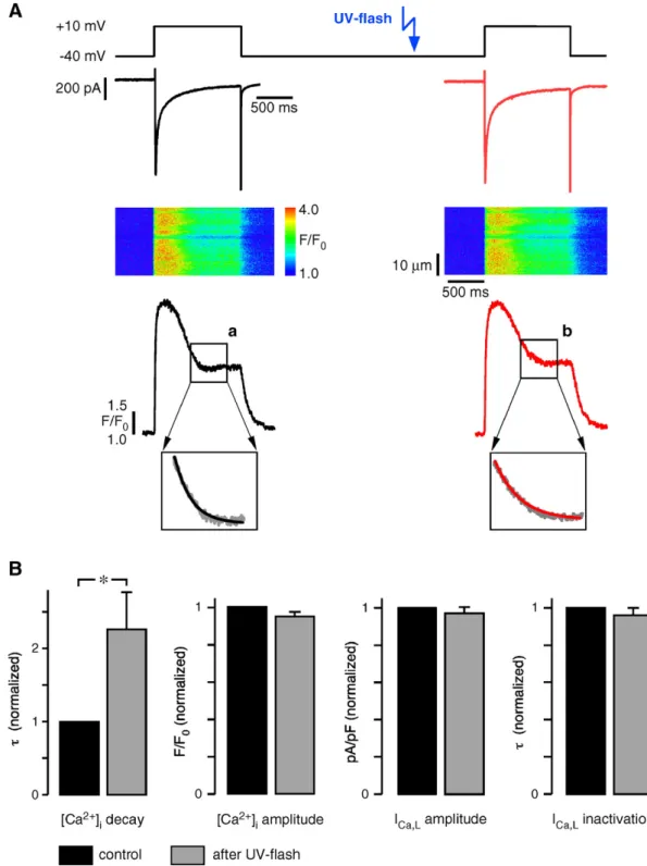
Fig. 2. During Ca2+transients elicited by ICa,Lonly the Ca 2+
removal is slowed by photorelease of DBHQ, ICa,Land CICR were unaffected. (A) ICa,Lbefore (left)
and after UV-flash (right) and the corresponding Ca2+transients in 1.8 mM extracellular [Ca2+]. Monoexponential function fitted to decay of Ca2+signals (after inactivation of ICa,L):τ=146 ms before (a), 240 ms after flash (b). (B) Statistical analysis (from left to right): τ of Ca
2+
-transient decay (2.26 ± 0.51 of control),Ca2+ -transient amplitude (0.95 ± 0.02 of control), peak ICa,L(0.97 ± 0.03 of control) andτ of ICa,Linactivation (0.96 ± 0.04 of control). (⁎: pb0.05, n=8 cells).
TX). Data analysis was carried out with IgorPro software (WaveMetrics, Lake Oswego, OR).
2.3. Confocal calcium imaging
Fluo-3 was excited at 488 nm line of an argon laser (50μW) and fluorescence (N515 nm) was collected with a PMT on an MRC-100 confocal microscope (Bio-Rad).
Image analysis was carried out with NIH-Image-SXM. Fluorescence signals are expressed as normalized fluores-cence F/F0. For details of the image acquisition and flash
photolysis see [12]. Amplitude information was extracted from line-scan images after aligning the scanned lines to correct for the signal distortion due to the slow wave prop-agation. Statistical analysis of the Ca2+signals: ⁎: pb0.05; ⁎⁎: pb0.01, one-way ANOVA followed by Dunnett's test, mean ± SE.
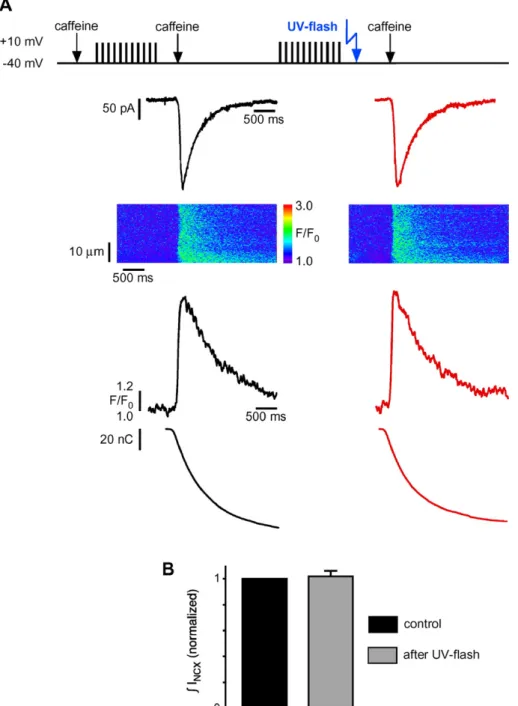
Fig. 3. The SR Ca2+content is not reduced within seconds after photorelease of DBHQ. (A) Example recording of Na–Ca exchange current (INCX) during rapid
caffeine application (10 mM for 2 s) before (left) and after DBHQ photorelease (right) in 1.8 mM extracellular [Ca2+]. From top to bottom: experimental protocol, INCX, line-scan image, fluorescence changes, integrated INCX, representing amount of Ca2+released from the SR. (B) Statistical analysis of normalized SR Ca2+
content after UV-flash was 1.04 ± 0.02 of control (n = 4 cells).
2.4. UV-flash photolysis
For photolysis of Nmoc-DBHQ UV-flashes from a Xenon short-arc flashlamp (wavelength 340–390 nm, flash duration 400 μs) were coupled into the microscope via an optical light-guide[12]. Spontaneous Ca2+waves were induced by increasing the extracellular Ca2+concentration to 6 mM.
3. Results
To assess whether Ca2+ waves in cardiomyocytes are predominantly driven by CICR directly or by a sensitization wave inside the SR, we used UV-flash photolytic liberation of the SERCA inhibitor DBHQ from a “caged” precursor, Nmoc-DBHQ [13]. Spontaneous Ca2+ waves were evoked by elevating the extracellular Ca2+concentration to 6 mM. Photolysis of Nmoc-DBHQ during Ca2+waves rapidly (i.e. within 1 s) slowed the propagation from around 95μm/s to 69μm/s, as revealed by laser-scanning confocal microscopic Ca2+imaging (Fig. 1A). To determine the extent of SERCA block that can be achieved with one photolytic flash we assessed the slowing of Ca2+transient decays in 100μM of the SERCA inhibitor TBQ applied extracellularly, a con-centration which blocks the Ca2+pump completely. By com-parison with the slowing observed after a single UV-flash we estimated the degree of SERCA inhibition by photoreleased DBHQ to be around 31%. Such an immediate deceleration of Ca2+wave-propagation is consistent with Ca2+uptake via the SERCA mediating a wave-front of RyR sensitization traveling inside the SR. Unbound luminal Ca2+ diffuses rapidly inside the longitudinal SR to the dyads of the next
sarcomere, as it has been reported very recently [14]. This leads to RyR sensitization ahead of the cytosolic Ca2+wave, which travels more slowly and mainly by diffusion of Ca2+ bound to mobile Ca2+ buffers[15]. A consequence of this sensitization is that a less pronounced elevation of [Ca2+]iis
required at the next sarcomere, such that RyRs will open earlier and even with the moderate [Ca2+]ipresent at the Ca2+
wave-front [2,10]. The opposite experimental result – an acceleration of the propagation– would be expected after the UV-flash for Ca2+waves mainly driven by CICR since, after SERCA inhibition, less Ca2+is removed from the wave-front and more remains in the cytosol to trigger CICR. An alternative mechanism could result from sensitization of RyRs after SERCA mediated SR Ca2+uptake taking place in the same site where the release occurs, since SERCAs are also present in the junctional SR. This mechanism would not require fast diffusion of SR Ca2+. But it seems somewhat less likely because the SR Ca2+pumps have low turnover rates (5–7 s− 1[16]), which would not allow for sufficient time to
sensitize the RyRs before they are exposed to cytosolic trigger Ca2+.
Previous studies of Ca2+wave propagation using conven-tional pharmacological inhibition of the SERCA have provided contradictory results. Inhibiting the SERCAwith the irreversible blocker thapsigargin led to an acceleration of the waves[17], while application of the SERCA blocker DBHQ (a.k.a. TBQ) resulted in a slowing of wave propagation[18]. While the exact reason for this discrepancy is still elusive, several confounding factors need to be considered. The prolonged SERCA inhibi-tion, used in these pharmacological experiments, is known to cause secondary, long-term changes in cell physiology, including elevations of [Ca2+]i, loss of SR Ca2+ content, Fig. 4. Possible mechanisms for Ca2+wave propagation acting in tandem. 1: Ca2+waves driven by CICR; the elevated [Ca2+]
iin the wave-front induces Ca2+
release from the SR via RyRs. 2: Wave of RyR sensitization mediated by Ca2+pumping into the SR by the SERCA. Diffusion of free Ca2+inside the longitudinal
SR results in slight elevations of [Ca2+]
SRahead of the cytosolic wave, leading to RyR sensitization for cytosolic Ca2+triggers. Rapid Ca2+diffusion inside the
impaired SR Ca2+ release and changes of the L-type Ca2+ current. However, with the rapid flash photolytic liberation of a SERCA inhibitor used in the present study, these secondary changes are expected to be minimal in the short time-frame of our experiments. Indeed, none of the parameters known to change during prolonged experiments were significantly affected in our study using a flash photolytic approach, except when applying series of flashes over more prolonged times. As expected, however, the time-course of the Ca2+decay trailing the wave was immediately slowed. To exclude changes of the SR Ca2+release mechanism and the Ca2+ current by photo-released DBHQ we recorded Ca2+transients triggered by L-type Ca2+ currents (Fig. 2). Both parameters remained unchanged, only the late phase of the Ca2+ transient decay during the voltage-clamp depolarization was significantly slowed. While the early phase of the Ca2+transient contains contributions from the inactivating Ca2+current, the late phase is governed by SR Ca2+uptake via the SERCA, and therefore this immediate slowing was expected. Also the SR Ca2+content did not change noticeably, as confirmed by recording Ca2+ transients and Na–Ca exchange currents induced by puffs of caffeine before and after the flash (Fig. 3). Taken together, the observed changes of the Ca2+decline were precisely what we had anticipated, since they reflect immediate consequences of SERCA inhibition. Furthermore, these experiments provide evidence that neither DBHQ nor other photolytic by-products of the caged compound interfered with L-type Ca2+ current, CICR, or SR Ca2+content.
4. Discussion
The observation that an SR luminal mechanism can sustain Ca2+wave propagation is not entirely unexpected. First of all, it may be anticipated based on several recent reports linking CICR termination to functional SR Ca2+depletion[12,19,20]. In cardiac muscle, CICR must terminate after each heartbeat to ensure muscle relaxation. Among several proposed mechan-isms for CICR termination, functional SR Ca2+depletion has recently gained strong experimental support. It appears that Ca2+unbinding from the intra-SR Ca2+buffer calsequestrin makes the RyRs less sensitive to cytosolic Ca2+[21], via a retrograde signaling pathway from calsequestrin to the RyRs, presumably mediated by the small accessory SR proteins triadin and junctin[22]. Thus, overloading the SR with Ca2+ would be expected to lead to a“sensitization” of the RyRs. In fact, it is well established that elevations of SR Ca2+content increase fractional SR Ca2+release in a way that is steeply Ca2+-dependent[23]. This implies that even a small increase in [Ca2+]SR in the region of the Ca2+ wave-front would be
sufficient to sensitize the RyRs locally, while on the level of the entire cell this would not change the [Ca2+]SRnoticeably. In
contrast, in myocytes with a normal SR Ca2+ load the local Ca2+signals are known not to propagate as waves, indicating that Ca2+overload is required[24].
Ca2+ waves are known to occur in many cell types. Interestingly, when comparing different varieties of Ca2+
waves in many systems and over a broad range of conditions, the waves in cardiomyocytes have consistently been found to exhibit extraordinarily fast propagation [6]. Based on this observation it had been suspected that a luminal propagation mechanism may contribute to wave propagation, acting in tandem with CICR (seeFig. 4).
Taken together, the findings reported here are contrary to predictions derived from the classical view of Ca2+ wave propagation by CICR, but entirely consistent with the concept that SR Ca2+loading via SERCA leads to sensitization of the RyRs, thereby creating a traveling wave of high Ca2+sensitivity initiating Ca2+-release. Thus, in cardiac muscle, and presum-ably in many other cell types, Ca2+wave propagation appears to be driven by a traveling wave-front of CICR“sensitization” rather than by CICR alone. This fundamental mechanism of RyR activation is most likely also crucial for our understanding of life threatening cardiac arrhythmias triggered by spontane-ous Ca2+ releases, since it can render the RyRs abnormally sensitive for [Ca2+]i and thus lowers the threshold for
regenerative Ca2+release events[25]. This type of arrhythmia is linked to RyR hyperphosphorylation[26], or to the recently discovered mutations of the RyR[1], or of the Ca2+buffering protein inside the SR (calsequestrin[27]).
Acknowledgements
We thank the grant support by the Swiss National Science Foundation (to E.N. and M.E.), by the NIH (to J.P.Y.K), the Swiss Cardiovascular Research and Training Network (SCRTN) and the Swiss State Secretariat for Education and Research (SER). We are grateful to Natalia Shirokova, Stephan Rohr and Jakob Ogrodnik for the helpful discussions and to Daniel Lüthi for the excellent technical assistance.
References
[1] Lehnart SE, Wehrens XH, Laitinen PJ, Reiken SR, Deng SX, Cheng Z, et al. Sudden death in familial polymorphic ventricular tachycardia associated with calcium release channel (ryanodine receptor) leak. Circulation 2004;109:3208–14.
[2] Lipp P, Niggli E. Microscopic spiral waves reveal positive feedback in subcellular calcium signaling. Biophys J 1993;65:2272–6.
[3] Capogrossi MC, Suarez-Isla BA, Lakatta EG. The interaction of electrically stimulated twitches and spontaneous contractile waves in single cardiac myocytes. J Gen Physiol 1986;88:615–33.
[4] Fabiato A. Time and calcium dependence of activation and inactivation of calcium-induced release of calcium from the sarcoplasmic reticulum of a skinned canine cardiac Purkinje cell. J Gen Physiol 1985;85: 247–89.
[5] Sneyd J, Keizer J, Sanderson MJ. Mechanisms of calcium oscillations and waves: a quantitative analysis. FASEB J 1995;9:1463–72. [6] Jaffe LF. The path of calcium in cytosolic calcium oscillations: a
unifying hypothesis. Proc Natl Acad Sci U S A 1991;88:9883–7. [7] Niggli E, Lederer WJ. Voltage-independent calcium release in heart
muscle. Science 1990;250:565–8.
[8] Meissner G, Henderson JS. Rapid calcium release from cardiac sarcoplasmic reticulum vesicles is dependent on Ca2+and is modulated
by Mg2+, adenine nucleotide, and calmodulin. J Biol Chem 1987;262:3065–73.
[9] Cannell MB, Soeller C. Numerical analysis of ryanodine receptor activation by L-type channel activity in the cardiac muscle diad. Biophys J 1997;73:112–22.
[10] Wier WG, Cannell MB, Berlin JR, Marban E, Lederer WJ. Cellular and subcellular heterogeneity of [Ca2+]iin single heart cells revealed by
fura-2. Science 1987;235:325–8.
[11] Gyorke S, Gyorke I, Terentyev D, Viatchenko-Karpinski S, Williams SC. Modulation of sarcoplasmic reticulum calcium release by calsequestrin in cardiac myocytes. Biol Res 2004;37:603–7. [12] DelPrincipe F, Egger M, Niggli E. Calcium signalling in cardiac muscle:
refractoriness revealed by coherent activation. Nat Cell Biol 1999;1:323–9. [13] Rossi FM, Kao JP. Nmoc-DBHQ, a new caged molecule for modulating sarcoplasmic/endoplasmic reticulum Ca2+ATPase activity
with light flashes. J Biol Chem 1997;272:3266–71.
[14] Wu X, Bers DM. Sarcoplasmic reticulum and nuclear envelope are one highly interconnected Ca2+store throughout cardiac myocyte. Circ Res 2006;99:283–91.
[15] Michailova A, DelPrincipe F, Egger M, Niggli E. Spatiotemporal features of Ca2+buffering and diffusion in atrial cardiac myocytes with inhibited sarcoplasmic reticulum. Biophys J 2002;83:3134–51. [16] Levitsky DO, Benevolensky DS, Levchenko TS, Smirnov VN, Chazov
EI. Calcium-binding rate and capacity of cardiac sarcoplasmic reticulum. J Mol Cell Cardiol 1981;13:785–96.
[17] Lukyanenko V, Subramanian S, Gyorke I, Wiesner TF, Gyorke S. The role of luminal Ca2+in the generation of Ca2+waves in rat ventricular
myocytes. J Physiol 1999;518:173–86.
[18] O'Neill SC, Miller L, Hinch R, Eisner DA. Interplay between SERCA and sarcolemmal Ca2+efflux pathways controls spontaneous release of
Ca2+from the sarcoplasmic reticulum in rat ventricular myocytes. J Physiol 2004;559:121–8.
[19] Terentyev D, Viatchenko-Karpinski S, Valdivia HH, Escobar AL, Gyorke S. Luminal Ca2+controls termination and refractory behavior of Ca2+-induced Ca2+release in cardiac myocytes. Circ Res 2002;91: 414–20.
[20] Sobie EA, Dilly KW, dos Santos Cruz J, Lederer WJ, Jafri MS. Termination of cardiac Ca2+sparks: an investigative mathematical
model of calcium-induced calcium release. Biophys J 2002;83:59–78. [21] Szentesi P, Pignier C, Egger M, Kranias EG, Niggli E. Sarcoplasmic reticulum Ca2+ refilling controls recovery from Ca2+-induced Ca2+
release refractoriness in heart muscle. Circ Res 2004;95:807–13. [22] Gyorke I, Hester N, Jones LR, Gyorke S. The role of calsequestrin,
triadin, and junctin in conferring cardiac ryanodine receptor respon-siveness to luminal calcium. Biophys J 2004;86:2121–8.
[23] Shannon TR, Ginsburg KS, Bers DM. Potentiation of fractional sarcoplasmic reticulum calcium release by total and free intra-sarco-plasmic reticulum calcium concentration. Biophys J 2000;78:334–43. [24] O'Neill SC, Mill JG, Eisner DA. Local activation of contraction in
isolated rat ventricular myocytes. Am J Physiol 1990;258:C1165–8. [25] Wakayama Y, Miura M, Stuyvers BD, Boyden PA, ter Keurs HEDJ.
Spatial nonuniformity of excitation–contraction coupling causes arrhyth-mogenic Ca2+waves in rat cardiac muscle. Circ Res 2005;96:1266–73. [26] Marks AR. Cardiac intracellular calcium release channels: role in heart
failure. Circ Res 2000;87:8–11.
[27] Viatchenko-Karpinski S, Terentyev D, Gyorke I, Terentyeva R, Volpe P, Priori SG, et al. Abnormal calcium signaling and sudden cardiac death associated with mutation of calsequestrin. Circ Res 2004;94:471–7.

![Fig. 4. Possible mechanisms for Ca 2+ wave propagation acting in tandem. 1: Ca 2+ waves driven by CICR; the elevated [Ca 2+ ] i in the wave-front induces Ca 2+](https://thumb-eu.123doks.com/thumbv2/123doknet/14885593.646659/5.892.201.693.100.441/possible-mechanisms-propagation-acting-tandem-driven-elevated-induces.webp)
