The neo-adjuvant, surgical and adjuvant treatment of
gastric adenocarcinoma. Current expert opinion
derived from the Seventh World Congress on
Gastrointestinal Cancer, Barcelona, 2005
E. Van Cutsem
1*, M. Dicato
2, N. Arber
3, A. Benson
4, D. Cunningham
5, E. Diaz-Rubio
6,
B. Glimelius
7, R. Goldberg
8, D. Haller
9, K. Haustermans
1, Y. Koo-Kang
10, R. Labianca
11,
I. Lang
12, B. Minsky
13, B. Nordlinger
14, A. Roth
15, P. Rougier
14, H.-J. Schmoll
16, A. Sobrero
17,
J. Tabernero
18, A. Szawlowski
19& C. van de Velde
201
University Hospital Gasthuisberg, Leuven, Belgium;2
Luxembourg Medical Center, Luxembourg;3
Ichilov Hospital, Tel Aviv, Israel;4
Northwestern University Chicago, Illinois, United States;5
The Royal Marsden Hospital, Surrey, United Kingdom;6
San Carlos Hospital Clinic, Madrid, Spain;7
University of Uppsala, Uppsala, and Karolinska Institutet, Sweden;8
University of North Carolina at Chapel Hill, Chapel Hill, North Carolina, United States;9
University of Pennsylvania Cancer Center, Philadelphia, Pennsylvania, United States;10
Asan Medical Center, University of Ulsan, Seoul, Korea;11
Ospedali Riuniti, Bergamo, Italy;12
National Institute of Oncology, Budapest, Hungary;13
Memorial Sloan-Kettering Cancer Center, New York, New York, United States;14
Hoˆpital Ambroise Pare´, Boulogne, France;15
Geneva University Hospital, Geneva, Switzerland;16
Martin Luther Universita¨t, Halle, Germany;17
Ospedale S. Martino, Genova, Italy;18
Vall d’Hebron University Hospital, Barcelona, Spain;19
Marie Sklodowska-Curie Memorial Cancer Center, Warsaw, Poland;20
Leiden University Medical Center, Leiden, The Netherlands
Key words:adjuvant treatment, chemotherapy, gastric cancer, radiotherapy
Gastric cancer is an important problem and is worldwide one of the most frequently-diagnosed cancers [1, 2]. Although the incidence of gastric cancer has been declining in the last decades, gastric cancer still accounts for approximately 10% of all cancers and is responsible for approximately 12% of all cancer deaths [3]. The incidence of gastric cancer varies substantially among racial and ethnic groups and is the highest in Japan, Korea, Eastern Asia, Eastern Europe, and parts of Latin America. Western Europe, the USA and other industrialised nations have relatively low incidences.
Surgical resection remains the primary curative treatment option in gastric cancer, with 5-year survival rates of 58%–78% and 34% reported for stage I and II disease, respectively [4]. Despite this, the overall 5 year survival rate for all patients remains poor and ranges between 15 and 38%. Recurrences are frequent after surgery. Recurrence rate and subsequent survival is dependent on the stage at diagnosis. Treatment decisions are usually made in reference to the staging systems of the American Joint Committee on Cancer (AJCC) and the international Union Against Cancer (UICC) [5, 6].
Recently new data focussing on the extent of gastric resection and on the additional treatment pre-, post- or perioperatively have become available and give hope for improved outcome for patients with gastric cancer.
This article reports on an expert discussion on the (neo-) adjuvant treatment of gastric cancer. The expert discussion was organised during the seventh World Congress on
Gastrointestinal Cancer in June 2005 in Barcelona, Spain. Well known opinion leaders and experts from different nationalities participated in the discussion. In preparation of this expert discussion a detailed survey and questionnaire was sent to all experts and the questions, answers and conclusions were rediscussed during the meeting.
The article reports on the evidence based conclusions and advises on the (neo-)adjuvant treatment of gastric cancer as proposed by these experts.
staging algorithm
Most patients with gastric cancer have at an early stage mild or no symptoms. The main reason for late diagnosis is that patients typically present with vague and non-specific symptoms.
Upper gastrointestinal endoscopy with biopsies for histologic confirmation is the procedure of choice for the diagnosis of gastric cancer. Although the accuracy of the histologic diagnosis increases with the number of biopsies, histologic confirmation may sometimes be difficult in some cases of diffuse carcinoma, as the intramucosal component may be small in comparison with an extensive and mural involvement [1].
The primary staging of gastric cancer includes:
• A thorough physical examination with the search for
pathological lymph nodes. A special attention to the supraclavicular lymph nodes (Virchow’s lymph node) is required.
• Biochemical tests. They do not contribute to the diagnosis of
gastric cancer, although blood counts, liver and renal function are usually evaluated. Tumour markers (CEA, CA 19.9) have
symposium
article
*Correspondence to: Prof. E. Van Cutsem, Digestive Oncology Unit, University Hospital Gasthuisberg Leuven, Herestraat 49, 3000 Leuven, Belgium. E-mail: Eric.Vancutsem@uz.kuleuven.ac.be
not been found to be useful in the diagnosis of gastric cancer, but CEA and CA 19.9, in particular, are elevated in 30–40% of patients with primary gastric cancer. Significantly higher levels of CEA and CA 19.9 have been found in patients with more advanced disease. Although there are no data that prove the use of CEA and CA 19.9 in the surveillance of gastric cancer, the determination of these markers may be more useful in the monitoring of gastric cancer than in the primary diagnosis.
• Endoscopic ultrasound is useful for the local staging of
tumours at the gastro-oesophageal junction and for small tumours and is most reliable for T-staging [7].
• A good quality CT scan of the abdomen (including pelvis) and
chest are required for the evaluation of locoregional extension and of metastases.
For the secondary staging specific procedures can be required.
• There is no evidence for routinely performing a FDG-PET
scan in all patients. FDG-PET scanning can, however, contribute to the detection and/or diagnosis of metastases. The sensitivity of FDG-PET in gastric cancer is lower than in the tumours localised at the gastro-oesophageal junction (GE-junction) [8]. In GE-junction tumours FDG-PET scan can give additional information and may be more conclusive compared to good quality conventional imaging [9].
• Barium X-ray has only a limited role and does not contribute
in the staging, but may suggest a diagnosis of gastric cancer in case of diffuse gastric cancers in which mucosal biopsies do not confirm the suspicion of malignancy. Barium X-ray is more useful in the preoperative evaluation and planning of GE-junction tumours than in more distally located tumours.
• MRI imaging may replace CT scan only in selected patients
and should not be performed routinely in the staging of gastric cancer.
• Bone scintigraphy and bone marrow aspiration should not
be performed routinely in the staging of gastric cancer.
• A diagnostic laparoscopy can be recommended if a
neo-adjuvant treatment is considered, especially in the case of a T3 or T4 and subdiaphragmatic tumour. In addition to the diagnostic laparoscopy a peritoneal lavage can be useful in staging. A peritoneal lavage containing malignant cells worsens the prognosis of patients with gastric cancer. A positive peritoneal lavage, however, does not exclude surgical resection of the gastric cancer, but implies that an extensive lymph-adenectomy is not useful.
treatment of gastric cancer
Important data have recently been published that led to increased knowledge in the treatment of gastric cancer, esp. in relation to the adjuvant and neo-adjuvant treatment of gastric cancer. Guidelines for treatment are therefore adjusted according to results of these important trials. Further research remains, however, important because gastric cancer has still a high mortality rate and less than 50% of the patients can be treated with a R0 resection. The prognosis of metastatic gastric cancer is poor, a median survival of 3 to 4 months for untreated patients and 6 to 9 months for patients treated with
chemotherapy. The only potentially curative treatment for gastric cancer is surgery. However, relapse after surgical resection is common and accounts for the high mortality rate. The poor survival after surgery for patients who had lymph node metastases suggests that surgery alone is not adequate and that an additional therapy is needed. Strategies of peri-operative chemotherapy and of postoperative chemoradiotherapy have shown to improve the overall survival of patients with gastric cancer.
A multidisciplinary discussion and treatment planning of all patients with gastric cancer is mandatory to guarantee optimal quality of care.
surgery
Surgical resection of the primary tumour and regional lymph nodes is the treatment of choice for gastric cancer. Surgery should be performed by well trained and experienced surgeons in centres of excellence to optimise the treatment planning and the surgical treatment. Quality control of surgery and of pathology is also very important.
For tumours located in the proximal and middle third of the stomach or when a diffuse type gastric cancer is found a total gastrectomy is recommended [1, 2, 10]. For tumours localised in the distal (antral) stomach, most surgeons recommend today a distal gastrectomy, since randomised trials did not show any advantage of total gastrectomy over subtotal gastrectomy in this setting [11–13]. For tumours involving the GE junction a partial oesophagectomy should also be performed.
In very small and superficial cancers an endoscopic mucosal resection can be performed by experienced physicians [14]. A limited gastric resection in combination with a sentinel node examination gives adequate locoregional control and good chances of survival for T1 tumours in the hands of very experienced surgeons.
Guidelines for the standardisation of surgical treatment and pathologic evaluation have been made [10, 15]. According to these guidelines 16 different lymph node compartments are identified surrounding the stomach. The perigastric lymph node stations along the lesser and greater curvature are group N1, whereas the nodes along the left gastric, hepatic, celiac and splenic arteries are group N2. Other lymph node stations are described as N3 and N4 and include nodes at the posterior aspect of the pancreas head, nodes at the root of the
mesenterium, nodes in the mesocolon of the transverse colon and para-aortic nodes.
A D1 dissection entails removal of the involved part of the stomach including greater and lesser omentum. For a D2 dissection, the omental bursa is removed with the front leave of the transverse mesocolon and the mentioned vascular pedicles of the stomach are cleared completely [10, 15]. The experts agreed that at least a D1 resection should be performed and that it is mandatory that at least 15 lymph nodes are removed and recovered. The resection margins should be free of tumour. A splenectomy and pancreas tail resection should not be performed routinely unless there is tumour invasion (Table 1).
The experts base their recommendations on data from different publications. In Japan complete removal of the N1 and N2 nodes is considered standard practice for curative resection based on evidence from large retrospective studies [16].
Available trials in Western countries comparing D2 and D1 lymphadenectomies have failed to support extended lymph node dissection. Several prospective randomized trials have evaluated the role of D1 or D2 resection in the management of gastric cancer and they did not show any advantage in terms of overall survival in favor of D2 lymphadenectomy [12, 17–20]. In the British trial the 5-year survival rates were 35% for D1 and 33% for D2 dissections [18]. Postoperative complications were significantly higher in the D2 group [18]. The DGC trial in the Netherlands randomized 996 patients between D1 and D2 lymph node dissection (380 with D1 and 331 with D2; 285 required palliative treatment). D2 resection had a higher postoperative mortality and significantly more complications leading to prolonged hospitalisation. At the medium follow-up of 11 years the survival rate for D1 was 30% and D2 35% (NS). The risk for relapse was not significantly different [10, 19]. If hospital deaths are excluded, survival rates are 32% for D1 and 39% for D2 (NS). The relapse risk of these patients tends to be in favor of D2 resection (P = 0.07) [10, 19]. In a recently reported relatively small Chinese trial patients were randomised between D1 and D3 surgery. The overall 5-year survival was significantly higher in patients assigned to D3 surgery than in those assigned to D1 surgery (59.5% versus 53.6%; log-rank P = 0.041) [20].
(neo-)adjuvant treatment
In view of the high frequency of recurrences after surgery, clinicians should consider in multidisciplinary discussions the options of a neo-adjuvant or adjuvant therapy. A neo-adjuvant or adjuvant treatment does not replace adequate surgery, which remains the cornerstone of a therapeutic strategy with curative intentions. In view of the decision of a (neo-) adjuvant therapy adequate staging is important, the TNM classification (version 2002) should be used (Table 2). Identification of the risk of
recurrence and risk-benefit analysis of a (neo-) adjuvant treatment should always be done.
Patterns of relapse are important when considering adjuvant therapy. It has been shown that more than half of the patients who undergo a resection with curative intention will have locoregional recurrence [2]. This highlights the fact that surgery alone was unable to eradicate all locoregional disease and supports again the need for optimal surgery and also for the evaluations of complementary strategies aiming at decreasing local relapse as well as distant metastases.
Although the treatment delivered determines a patient’s prognosis to a large extent, other factors, such as patient age and gender, the stage of disease at presentation, tumour localisation and morphology play a substantial role. Current staging modalities, which solely focus on the extent of tumour invasion and the presence of lymph node disease, do not take these factors into account. Therefore nomograms have been developed to address this problem. They are predictive tools for the individual patient based on known prognostic variables including the extent of surgical treatment. Nomograms help with patient counselling, follow-up scheduling and clinical trial determination and have been developed for several tumours. Recently a statistical model developed for gastric carcinoma was able to predict an individual patient’s probability for 5–9 year disease specific survival after R0 resection for gastric cancer in a single institution US population involving more than 1000 patients [21]. This nomogram has been validated in a multicentre study of 459 patients from the Dutch Gastric Cancer trial. The nomogram provided predictions that discriminated better than the AJCC staging system, regardless of the extent of lymph node dissection [22, 23].
adjuvant chemotherapy
Most of the individual trials studying the effect of postoperative adjuvant chemotherapy do not show a survival advantage compared to surgery alone. These studies often randomised a low number of patients and are clearly underpowered. The trials studied also predominantly older chemotherapy regimens, although the most recently reported trials have used adequate cisplatin-containing regimens [24]. Further, the patient populations studied were heterogeneous, including patient populations with both high and low risk of recurrence.
Five meta-analyses (or combined analyses) of adjuvant chemotherapy have been published [25–29]. Most of the analyses show a small benefit in survival for patients treated with postoperative adjuvant chemotherapy. When analysed, this small gain is only seen in Asian, and not in Western world, studies. Because of the nature of the data adjuvant
chemotherapy is not generally advised to patients who undergo a complete surgical resection of gastric cancer.
postoperative chemoradiotherapy
A very important study is the trial of the US GI-Intergroup study, which randomised 556 patients with resected
adenocarcinoma of the stomach or gastro-oesophageal junction to surgery plus postoperative chemoradiotherapy or surgery alone [30]. The adjuvant treatment consisted of 425 mg/m2 5-FU (bolus infusion) per day plus 20 mg/m2of leucovorin (LV)
Table 1. Surgery in gastric cancer
• Should be performed by
– well trained and experienced surgeons – in centres of excellence
• Quality control of surgery is important
• Quality control of pathology is important
• Multidisciplinary approach and discussion is warranted
• Type of surgery
– Total gastrectomy for proximal tumours – Subtotal gastrectomy for distal tumours
– Subtotal or distal oesophagectomy for distal oesophageal tumours and GE junction tumour – Siewert type 1
– Endoscopic mucosal resection only for very superficial lesions – Limited resection + sentinel node resection for T1 tumours:
adequate locoregional control and survival in experienced hands
• At least D1 resection
• Mandatory: at least 15 lymph nodes
• No routine sentinel lymph node analysis
• Free margins
• No splenectomy unless tumour extends to spleen.
World Congress on Gastrointestinal Cancer: Barcelona 2005 expert discussion.
for five days, followed by 45 Gy in 25 fractions of 1.8 Gy over 5 weeks with bolus 5-FU and LV during the first and last week of the radiotherapy. This was followed 4 weeks later by two cycles of bolus 5-FU/LV. The median overall survival in the surgery only arm was 27 months, compared to 36 months in the chemoradiotherapy group (P < 0.05). The survival at 3 years was 50 versus 40% in favour of patients treated with postoperative chemoradiotherapy. After a median follow-up of 5 years, compared with surgery alone, the 5 year overall survival was improved by 11.6% (28.4% versus 40% respectively; P < 0.001) and the relapse-free survival was increased from 25 to 31% in favour of patients treated with postoperative
chemoradiotherapy. Patients treated with postoperative chemoradiotherapy had significantly fewer locoregional recurrences. Three patients died from toxic effects of
chemoradiotherapy. Grade 3 toxic effects occurred in 41% and grade 4 in 32% of the patients treated with chemoradiotherapy [30]. Most patients did not undergo an extensive surgical resection although the protocol recommended a D2 resection. Fifty-four percent of the patients did not even have a D1 resection [24]. It has therefore been suggested that the postoperative chemoradiotherapy was simply making up for inadequate surgery. However, a large observational study from Korea, while confirming the results of the US Intergroup study, suggested also a benefit of postoperative chemoradiotherapy after D2 resection [31]. It is, however, also possible that the effects could have been even larger if surgery had been more adequate leaving fewer cancer cells to be killed by the additional therapy. The chemotherapy used in this study was never considered to be a highly effective combination for stomach cancer. Therefore better chemotherapeutic options should be investigated in this setting: actually infused regimens of 5-FU are recommended in this combination regimen because of better tolerability. In gastric cancer, there are no trials having compared bolus with infused 5-FU. The addition of new cytotoxic agents is under investigation [32].
The experts at the World Congress on Gastrointestinal Cancer recommend, however, to use a more optimised Intergroup schedule. There was an agreement that infused 5-FU instead of bolus 5-FU regimens before, during and after radiotherapy should be recommended. There was an agreement that cisplatin or irinotecan based chemotherapy before, during and after the radiotherapy is reserved for evaluation in protocols.
The experts recommend irradiating similar volumes as those irradiated in the Intergroup protocol. A dose of 45 Gy in fractions of 1.8 Gy is recommended. Postoperative radiotherapy in gastric cancer should be performed by experienced radiation oncologists.
The target volume should be delineated on a CT scan in the treatment position taking into account the surgery and pathology reports. Up to 30% deviations from the treatment protocol were noticed in the Intergroup study so careful planning by experienced radiation oncologists is a prerequisite [30, 33].
perioperative chemotherapy
The Magic trial compared a strategy of surgery alone with the administration of three cycles of preoperative chemotherapy followed by surgery followed by three cycles of postoperative chemotherapy (perioperative chemotherapy) in 504 patients with gastric and GE junction adenocarcinoma. The ECF regimen was selected as chemotherapy regimen: epirubicin, cisplatin and protracted 5-FU every 3 weeks. The patients treated with perioperative chemotherapy had a significantly better survival: 50 versus 41% were alive at 2 years and 36 versus 23% at 5 years compared to patients who were treated with surgery only. The median survival was significantly longer for patients treated with perioperative chemotherapy: 24 versus 20 months (P = 0.009). The progression free survival was also significantly improved for patients treated with perioperative chemotherapy: HR 0.66 (95% CI 0.53 – 0.81; P = 0.0001) [34]. Approximately 40 % of the patients enrolled in the experimental
Table 2. TNM classification [6]
Primary tumour (T)
TX: primary tumour cannot be assessed
T0: no evidence of primary tumour
Tis: carcinoma in situ: intraepithelial tumour without invasion of the lamina propria T1: tumour invades lamina propria or submucosa
T2: tumour invades muscularis propria or subserosa T2a: tumour invades muscularis propria
T2b: tumour invades subserosa
T3: tumour invades the serosa (visceral peritoneum) without invasion of adjacent structures T4: tumour directly invades adjacent structures
Regional lymph nodes (N)
NX: regional lymph node(s) cannot be assessed N0: no regional lymph node metastasis
N1: metastasis in one to six regional lymph nodes N2: metastasis in 7–15 regional lymph nodes N3: metastasis in more than 15 regional lymph nodes Distant metastasis (M)
MX: presence of distant metastasis cannot be assessed
MO: no distant metastasis
arm of the MAGIC trial did not receive the planned adjuvant part of systemic therapy. This is in accordance with the observed decreased patient tolerance to chemotherapy observed early after gastrectomy which is probably related to poor food intake capacity.
specific issues
Although phase II studies with preoperative chemoradiotherapy for resectable tumours are appealing, this approach is at the moment investigational. In T4 or ‘difficult’ tumours (especially GE junction tumours) there is an emerging use of preoperative chemoradiotherapy, based on the studies in oesophageal cancer and on the phase II studies in gastric cancer.
The experts recognise the lack of specific studies in patients with GE junction adenocarcinomas and recommend, because most of the studies with gastric cancer include GE junction adenocarcinomas as well, a treatment algorithm for patients with GE junction tumours similar to patients with gastric cancer.
In patients who underwent a partial gastrectomy and in whom Helicobacter pylori was found, Helicobacter pylori eradication in accordance with standard regimens, containing a combination of a proton pomp inhibitor and a double of antibiotics, is recommended, after recovery from surgery.
Intramuscular vitamin B12 injections should be administered at regular intervals after gastrectomy.
general recommendations for treatment
The experts recognise the growing evidence that (neo-)adjuvant treatment increases the outcome of selected patients. As general strategy there was an agreement to recommend postoperative chemoradiotherapy in patients who underwent inadequate surgery or less than D1 resection. The strategy of either postoperative chemoradiotherapy or perioperative
chemotherapy can be recommended as strategies in centres after multidisciplinary team discussions. For both strategies the evidence is based on data from a large well performed
randomized trial (level 2 evidence). The evidence supporting postoperative chemotherapy is not so strong and is based on several combined analyses (level 3 evidence or possibly level 1 evidence for no sufficient positive benefit of therapy). The experts agree to recommend a perioperative chemotherapy or postoperative chemoradiotherapy for patients who have stage T3, T4 or N+ M0 gastric cancer. There was no general agreement whether patients with stage T2bN0 should be offered an adjuvant treatment. In this setting other factors should be taken into consideration (e.g. factors presented in Nomograms).
A neo-adjuvant or adjuvant treatment should be offered only to fit patients without important comorbidities. Crucial in good tolerance, especially for the postoperative chemoradiotherapy, is the ability of the patient to have an adequate calorie intake during the treatment. Adequate measures are therefore necessary, with eventually the administration of enteral nutrition (Table 3).
surveillance of gastric cancer
There is no clear evidence in the literature that systematic follow-up is useful. A detection of recurrence usually does not lead to curative therapeutic interventions. The purpose of follow-up visits is for general symptom care. The experts therefore recommend limiting technical examinations to a minimum if the patient is asymptomatic. The frequency of a surveillance visit is usually every 3–6 months in the first three years and becomes less intensive after 3 years.
future research
There are still many open questions for future research:
• The evaluation of diagnostic modalities.
• The evaluation of prognostic and predictive markers. • The evaluation of molecular characteristics.
• The evaluation of treatment strategies. • The evaluation of new agents.
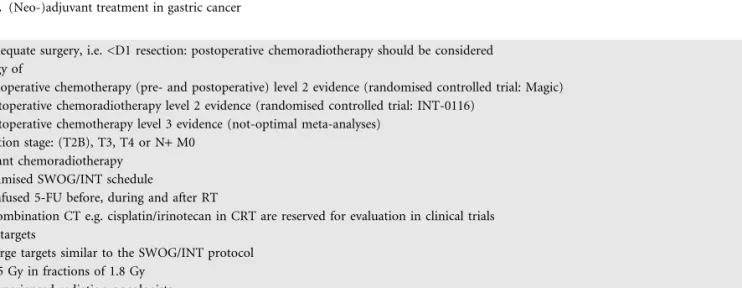
Table 3. (Neo-)adjuvant treatment in gastric cancer
• If inadequate surgery, i.e. <D1 resection: postoperative chemoradiotherapy should be considered
• Strategy of
– perioperative chemotherapy (pre- and postoperative) level 2 evidence (randomised controlled trial: Magic) – postoperative chemoradiotherapy level 2 evidence (randomised controlled trial: INT-0116)
– postoperative chemotherapy level 3 evidence (not-optimal meta-analyses)
• Indication stage: (T2B), T3, T4 or N+ M0
• Adjuvant chemoradiotherapy – optimised SWOG/INT schedule
• infused 5-FU before, during and after RT
• combination CT e.g. cisplatin/irinotecan in CRT are reserved for evaluation in clinical trials – RT targets
• large targets similar to the SWOG/INT protocol
• 45 Gy in fractions of 1.8 Gy
• experienced radiation oncologists
• Perioperative chemotherapy – ECF regimen
– 5-FU/cisplatin-based
conclusions
Consensus cannot be reached on all aspects of the management of gastric cancer, but expert advice can be given based on clinical data and on clinical experience. The knowledge on gastric cancer treatment is increasing and the evidence is growing that ‘optimal’ care improves the outcome of patients with gastric cancer.
A multidisciplinary approach in experienced centres is mandatory for adequate staging, for optimal surgery and for the selection of an adequate (neo-)adjuvant strategy.
Relapses after gastric cancer are frequent. Recent data on the (neo-)adjuvant therapy have changed clinical practice in patients with gastric cancer at risk of recurrence after complete gastric cancer resection. There is actually level 2 evidence for the strategy of postoperative chemoradiotherapy and for the strategy of perioperative, i.e. pre- and postoperative
chemotherapy and level 3 evidence for postoperative adjuvant chemotherapy. The strategy of postoperative
chemoradiotherapy and of perioperative chemotherapy decreases the risk of recurrence and improves the outcome for patients fit to undergo these treatments.
Those involved in the treatment of patients with gastric cancer should be encouraged to participate in well-designed clinical trials, in order to increase the evidence-based knowledge and to make further progress.
references
1. Catalano V, Labianca R, Beretta G et al. Gastric cancer. Crit Rev Oncol Hematol 2005; 54: 209–241.
2. Lim L, Michael M, Mann GB, Leong T. Adjuvant therapy in gastric cancer. J Clin Oncol 2005; 23(25): 6220–6232.
3. Parkin DM, Pisani P, Ferlay J. Global cancer statistics. CA Cancer J Clin 1999; 49(1): 33–64.
4. Hundahl SA, Phillips JL, Menck HR. The National Cancer Data Base Report on poor survival of U.S. gastric carcinoma patients treated with gastrectomy: Fifth edition American Joint Committee on cancer staging, proximal disease, and the ‘different disease’ hypothesis. Cancer 2000; 88(4): 921–932.
5. Green FL, Page DL, Fleming ID et al. AJCC cancer staging manual. 6th ed. New York: Springer; 2002.
6. UICC (International Union Against Cancer). In: Sobin LH, Wittekind Ch. (eds). TNM classification of malignant tumours. 6th edn. New York. Chichester.weinheim, Brisbane, Singapore, Toronto: Wiley-Liss; 2002.
7. Polkowski M, Palucki J, Wronska E et al. Endosonography versus helical computed tomography for locoregional staging of gastric cancer. Endoscopy 2004 Jul; 36(7): 617–623.
8. De Potter T, Flamen P, Van Cutsem E et al. Whole-body PET with FDG for the diagnosis of recurrent gastric cancer. Eur J Nucl Med Mol Imaging 2002; 29(4):525–529.
9. Flamen P, Lerut A, Van Cutsem E et al. Utility of positron emission tomography for the staging of patients with potentially operable esophageal carcinoma. J Clin Oncol 2000; 18(18): 3202–3210.
10. Jansen EP, Boot H, Verheij M, van de Velde CJ. Optimal locoregional treatment in gastric cancer. J Clin Oncol 2005; 23(20): 4509–4517.
11. Gouzi JL, Huguier M, Fagniez PL et al. Total versus subtotal gastrectomy for adenocarcinoma of the gastric antrum. A French prospective controlled study. Ann Surg 1989; 209: 162–166.
12. Robertson CS, Chung SC, Woods SD et al. A prospective randomized trial comparing RI subtotal gastrectomy with R3 total gastrectomy for antral cancer. Ann Surg 1994; 220: 176–182.
13. Bozzetti F, Marubini E, Bonfanti G et al. Total versus subtotal gastrectomy: surgical morbidity and mortality rates in a multicenter Italian randomized trial. The Italian Gastrointestinal Tumor Study Group. Ann Surg 1997; 226: 613–620.
14. Wang YP, Bennett C, Pan T. Endoscopic mucosal resection for early gastric cancer. Cochrane Database Syst Rev 2006 Jan 25; (1): CD004276 15. Kajitani T. Japanese Research Society for the Study of Gastric Cancer: The
general rules for gastric cancer study in surgery and pathology. Jpn J Surg 1981; 11: 127–145.
16. Maruyama K, Sasako M, Kinoshita T et al. Surgical treatment for gastric cancer: the Japanese approach. Semin Oncol 1996; 23: 360–368.
17. Dent DM, Madden MV, Price SK. Randomized comparison of RI and R2 gastrectomy for gastric carcinoma. Br J Surg 1988; 75: 110–112. 18. Cuschieri A, Weeden S, Fielding J et al. Patient survival after DI and D2
resections for gastric cancer: long-term results of the MRC randomized surgical trial. Surgical Co-operative Group. Br J Cancer 1999; 79: 1522–1530. 19. Bonenkamp JJ, Hermans J, Sasako M, van de Velde CJ. Extended lymph-node
dissection for gastric cancer. Dutch Gastric Cancer Group. N Engl J Med 1999; 340: 908–914.
20. Wu CW, Hsiung CA, Lo SS et al. Nodal dissection for patients with gastric cancer: a randomised controlled trial. Lancet Oncol 2006; 7(4): 309–315.
21. Kattan MW, Karpeh MS, Mazumdar M, Brennan MF. Postoperative nomogram for disease-specific survival after an R0 resection for gastric carcinoma. J Clin Oncol 2003; 21(19): 3647–3650.
22. Peeters KC, Kattan MW, Hartgrink HH et al. Validation of a nomogram for predicting disease-specific survival after an R0 resection for gastric carcinoma. Cancer 2005: 103(4): 703–707.
23. Peeters KC, Hundahl SA, Kranenbarg EK et al. Low Maruyama index surgery for gastric cancer: blinded reanalysis of the Dutch D1-D2 trial. World J Surg 2005; 29(12): 1576–1584.
24. Bouche O, Ychou M, Burtin P et al. Adjuvant chemotherapy with 5-fluorouracil and cisplatin compared with surgery alone for gastric cancer: 7-year results of the FFCD randomized phase III trial (8801). Ann Oncol 2005; 16(9): 1488–1497
25. Hermans J, Bonenkamp JJ, Boon MC et al. Adjuvant therapy after curative resection for gastric cancer: a meta-analysis of randomized trials. J Clin Oncol 1993; 11: 1441–1447.
26. Earle CC, Maroun JA. Adjuvant chemotherapy after curative resection for gastric cancer in non-Asian patients: revisiting a meta-analysis of randomised trials. Eur J Cancer 1999; 35(7): 1059–1064.
27. Mari E, Floriani I, Tinazzi A et al. Efficacy of adjuvant chemotherapy after curative resection for gastric cancer: a meta-analysis of published randomised trials. A study of the GISCAD. Ann Oncol 2000; 11(7): 837–843.
28. Gianni L, Panzini I, Tassinari D et al. Meta-analyses of randomized trials of adjuvant chemotherapy in gastric cancer. Ann Oncol 2001; 12(8): 1178–1180. 29. Janunger KG, Hafstro¨m L, Nygren P et al. A systematic overview of chemotherapy
effects in gastric cancer. Acta Oncol 2001; 40(2–3): 309–326.
30. Macdonald JS, Smalley SR, Benedetti J et al. Chemoradiotherapy after surgery compared with surgery alone for adenocarcinoma of the stomach or gastroesophageal junction. N Engl J Med 2001; 345(10): 725–730. 31. Kim S, Lim do H, Lee J, Kang WK et al. An observational study suggesting clinical
benefit for adjuvant postoperative chemoradiation in a population of over 500 cases after gastric resection with D2 nodal dissection for adenocarcinoma of the stomach. Int J Radiat Oncol Biol Phys 2005; 63(5): 1279–1285.
32. Kollmannsberger C, Budach W, Stahl M et al. Adjuvant chemoradiation using 5-fluorouracil/folinic acid/cisplatin with or without paclitaxel and radiation in patients with completely resected high-risk gastric cancer: two cooperative phase II studies of the AIO/ARO/ACO. Ann Oncol 2005; 16(8): 1326–1333. 33. Macdonald J. Role of post-operative chemoradiation in resected gastric cancer.
J Surg Oncol. 2005; 90(3): 166–170.
34. Cunningham D, Allum W, Stenning S, Weeden S. Perioperative chemotherapy in operable gastric and lower oesophageal cancer: final results of a randomised, controlled trial (the MAGIC trial, ISRCTN 93793971). Proc ASCO 2005; 23 (16S): 308s, abstr no. 4001.
![Table 2. TNM classification [6]](https://thumb-eu.123doks.com/thumbv2/123doknet/14888254.647975/4.1026.131.861.173.509/table-tnm-classification.webp)