ORIGINAL ARTICLE
Biomaterial-induced sarcomagenesis is not associated
with microsatellite instability
Achim Weber&Annette Strehl&Erik Springer&
Torsten Hansen&Arno Schad&C. James Kirkpatrick
Received: 18 June 2008 / Revised: 4 October 2008 / Accepted: 13 November 2008 / Published online: 19 December 2008 # Springer-Verlag 2008
Abstract Sarcomagenesis, in contrast to carcinogenesis, is poorly understood. Microsatellite instability has been impli-cated in the development of many cancers, in particular those associated with chronic inflammatory conditions. In an experimental animal model, rats developed not only a peri-implantational chronic inflammatory reaction, but also malignant mesenchymal tumors in response to different biomaterials. Therefore, it was the aim of our study to test if the development of biomaterial-induced sarcomas is charac-terized by a mutator phenotype. A multiplex-PCR approach was designed to screen biomaterial-induced sarcomas for the presence of microsatellite instability. Seven different micro-satellite loci were tested in ten tumors for micromicro-satellite instability using a fluorochrome-labelled multiplex-PCR and subsequent fragment analysis. All tumors provided a microsatellite-stable phenotype at all loci tested. Our data suggest that microsatellite instability is rarely or not at all a feature of malignant transformation of biomaterial-induced soft tissue tumors. Thus, there is no evidence that a mutator phenotype is a hallmark of biomaterial-induced sarcomagenesis.
Keywords Biomaterials . Sarcoma . Microsatellite instability . Mutator phenotype
Introduction
Tumorigenesis is now widely accepted as resulting from a stepwise accumulation of genetic instability [1]. The biological and molecular mechanisms involved in the development of carcinomas in humans and mammals have been extensively investigated. In contrast, the molecular steps in the development of sarcomas and other soft tissue tumors are less well understood. A progression from a benign precursor lesion to malignant tumor, characterisitic of many epithelial tumors, is exceptional for soft tissue tumors. However, a biological continuum of benign and malignant soft tissue tumors has been postulated for example in the case of lipomatous [2] and nerve sheath tumors [3,4].
In a previous study, we had established an animal model for the tumorigenesis of soft tissue tumors [5]. Malignant mesenchymal tumors were induced around subcutaneously implanted biomaterial disks. In addition to fully developed tumors, implantation sites showed lymphocyte and macro-phage infiltrates reflecting an inflammatory reaction, as a characteristic response to the implantation of foreign materials [6, 7] as well as hyper-proliferative lesions, reflecting precursor lesions. This animal model allowed a detailed investigation of sarcoma tumorigenesis, especially because a spectrum of preneoplastic lesions could be identified.
Several mechanisms are known to result in genetic instability and consequent malignant transformation of cells. One pathway is the development of chromosomal instability with numerical and structural chromosomal
A. Weber
:
A. Strehl:
E. Springer:
T. Hansen:
A. Schad:
C. J. KirkpatrickInstitute of Pathology, Johannes Gutenberg University of Mainz, Langenbeckstr. 1,
55101 Mainz, Germany Present address: A. Weber (*)
Department of Pathology, Institute of Surgical Pathology, University Hospital of Zurich,
Schmelzbergstr. 12, 8091 Zurich, Switzerland e-mail: [email protected]
changes and aneuploidy of tumor cells. Another pathway is the development of a“mutator phenotype” with changes at the nucleotide level. These alterations may result in the accumulation of multiple mutations in genes relevant to cell growth and homeostasis, eventually leading to growth advantage and transformation of cells. Multiple short tandem repetitive DNA-sequences, also known as micro-satellites, are present throughout the genome of humans and other mammals [8]. A mutator phenotype is frequently reflected by widespread sequence length alterations of these microsatellites, designated as microsatellite instability [9,
10]. Besides inherited deficiencies of mismatch repair (MMR) genes and inactivation by methylation, an under-lying impairment of repair enzymes in association with chronic inflammation [11,12] has been demonstrated, most likely due to oxidative stress [13].
While there is a clear association of specific or typical chromosomal translocations or translocation of gene fusion products with many soft tissue tumors, microsatellite instability has also been reported for some entities [14– 17]. Since a chronic inflammatory condition is associated with microstallite-associated carcinogenesis, we sought to investigate if the tumorigenesis of biomaterial-induced sarcomas is based on the development of a mutator pheno-type. Using the above-mentioned rat model, biomaterial-induced sarcomas were screened for microsatellite instability.
Materials and methods Animal model
Ten formalin-fixed, paraffin-embedded tumor samples (Table 1) were retrieved from the previously published rat sarcoma collective [5]. Briefly, in this study a total of 490 Fischer rats had been implanted with nine different standard medical grade biomaterials. In about one quarter of implan-tations, animals developed (mostly malignant) mesenchymal tumors of different histological type, including fibrosarcomas,
leimyosarcomas, and rhabdomyosarcomas, around the implantation sites.
Laser capture microdissection
Sections of normal and transformed tissues (either tumors or precursor lesions) from the above mentioned paraffin blocks were prepared on membrane mounted-slides (PALM MembraneSlides, P.A.L.M., Bernried, Germany) and de-paraffinized. Microdissection was performed using a PALM MicroBeam system with a pulsed UV-A nitrogen laser (337 nm). The selected areas were marked, cut out, and pulsed into a 0.5-ml microfuge tube with adhesive filling (PALM AdhesiveCaps, P.A.L.M.), taking care to keep the normal and tumor tissues separate. “Normal tissue” was prepared so distant from the implantation site that it was morphologically not affected by the implants and corresponding tissue reactions.
Multiplex PCR and fragment analysis
Since (in contrast to human and mouse) no well-established and characterized panels of markers for the analysis of microsatellite instability was described in rat, sequence information of rat microsatellite loci was taken from several studies [18–20]. In order to compensate for a potentially low sensitivity of these markers in detecting MSI, seven markers instead of the more common panel of five markers was used (Table 2). DNA was isolated by proteinase K digestion, followed by phenol-chloroform extraction and ethanol/salt precipitation using standard protocols [21]. Two multiplex PCR-reactions were performed using two different primer mixtures with fluorochrome-labelled oligonucleotide primers. In each case, either the forward or the reverse primer was fluorochrome-labelled. Primermix A contained oligonucleotide primers of the loci GH, NGFR, SECR, and UCP; primermix B contained oligonucleotide primers of the loci ANF, PPY, and SMST (Table2). Non-fluorescent oligonucleotide primers were purchased from
Table 1 Biomaterial-induced soft tissue tumors
The study included a total of ten different biomaterial-induced soft tissue tumors. Every rat was given an identi-fication code describing the implanted material. The result-ing tumor was histologically diagnosed
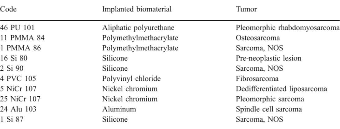
Code Implanted biomaterial Tumor
46 PU 101 Aliphatic polyurethane Pleomorphic rhabdomyosarcoma 11 PMMA 84 Polymethylmethacrylate Osteosarcoma
1 PMMA 86 Polymethylmethacrylate Sarcoma, NOS
16 Si 80 Silicone Pre-neoplastic lesion
2 Si 90 Silicone Sarcoma, NOS
4 PVC 105 Polyvinyl chloride Fibrosarcoma
5 NiCr 107 Nickel chromium Dedifferentiated liposarcoma
25 NiCr 107 Nickel chromium Pleomorphic sarcoma
24 Alu 103 Aluminum Spindle cell sarcoma
biomers.net, Ulm, Germany and the fluorescent oligonu-cleotide primers from Sigma-Aldrich, Germany.
The final 25-μl multiplex reaction mix consisted of the following: 12.5 μl Qiagen-Multiplex-Mastermix (Qiagen, Hilden, Germany), 2.5μl 10× primer-mix, 7.5 μl H2O, and
2.5 μl template. The 10× primer-mix was composed by mixing 50 μM fluorescent primers and 100 μM non-fluorescent primers at a concentration of 2μM each in H2O.
PCR was performed using a GeneAmp 9700 thermocycler (Applied Biosystems, Foster City, CA, USA) under the following conditions: initial denaturation at 95° for 15 min, followed by 45 cycles of: 30 s at 94°, 90 s at 54°, and 1 min at 72° with a final 30-min extension period at 60°. The resulting PCR-product was then analyzed by gel-electrophoresis on a 2% agarose gel and subsequent fragment analysis of the fluorescently labelled PCR products was performed using a Beckman Coulter CEQ 8000 Sequencer (Beckman Coulter, Krefeld, Germany) with the corresponding CEQ System software. The detected fragments of normal and malignant tissue were compared to determine the stability for each locus.
Results
Tissue selection and isolation of tumor and normal DNA
Histology of the samples was re-evaluated in order to define suitable areas of tumor and non-neoplastic tissue, respectively. In all cases studied, a clear identification of
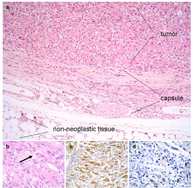
tumors and non-tumorous areas was possible, providing the basis for the laser-capture analysis and tissue preparation (Fig. 1). Following laser-capture (P.A.L.M.)-based tissue preparation and standard nucleic acid extraction, a suffi-cient amount and quality of DNA could be isolated from all samples analyzed.
Development of a multiplex-PCR assay for microsatellite instability testing
PCR conditions were first optimized, and all samples were initially tested in a single PCR setting. In the case of the GH and PPY locus, respectively, significantly lower amplification sizes were observed when compared to information from either the gene bank (see Table 2) and/ or the literature [18]. In the case of the PPY locus, higher amplification sizes were observed. The SECR locus revealed rather a single peak instead of the characteristic stuttering pattern in all samples tested. However, all loci displayed a constant size comparing different animals (due to the same genetic background of animals).
After successfully testing each pair of oligonucleotide primers in a single-PCR, a multiplex-PCR-assay was designed by dividing the seven different primers into two groups of three or four primer pairs, respectively. Using this multiplex-PCR approach, all loci test were successfully amplified and analyzed by fluorescence-based fragment analysis. No difference between single PCR fragment analysis patterns and the multiplex approach was observed,
Table 2 Microsatellite loci and oligonuceotide primer sets
Locus Acc. No. Oligo nucleotide sequence Sizea Dye Ref.
Mix A GH X12967 F 5′-ATGGGAGGGAACAAGTCTTC-3′ 135 D4 [18,19] R 5′-GAGGGAGAGAGAAAGAGAGACAG-3′ NGFR X05137 F 5′-ACCCCACAATCCAACACTATAC-3′ 126 D3 [18,19] R 5′-GCAGGATCTAGTCTCAGCCC-3′ SECR M64033 F 5′-ACCATGGAGCCTCTACTGC-3′ 194 D4 [18,20] R 5′-GTCCCGTCCGAGTGTCTT-3′ UCP X12925 F 5′-TGCCCGTCTCTGTTACTCAT-3′ 111 D4 [18,19] R 5′-CAAGAACCCTGAGGCAATAA-3′ Mix B SMST K02248 F 5′-TTTCCAGGTGCCAAATGTAG-3′ 154 D4 [18,20] R 5′-TATATTGTGACAAAAGAAAGGCAC-3′ ANF K02062 F 5′-TCCACAACCTTGATCTTTCG-3′ 160 D2 [18,20] R 5′-GTTGAGGGCCATAGTGTGAC-3′ PPY M27450 F 5′-CACACTGACCAGGCCTAGG-3′ 153 D3 [18,20] R 5′-TGTCAGCTCAGCTGCTTTG-3′ Microsatellite loci and oligonucletide primer sequences information
GH growth hormone, NGFR nerve growth factor receptor, SECR secretin, UCP uncoupling protein, SMST somatostatin, ANF atrial natriuretic factor, PPY pancreatic polypeptide
aSizes are indicated as expected according to NCBI database information and differed significantly from the fragment sizes observed in this study
indicating that the multiplex assay used was a valid method for the simultaneous testing of several microsatellite loci (Fig.2).
All microsatellite loci showed a stable phenotype
A total of 140 microsatellites was analyzed, with 70 microsatellites displaying microsatellites of neoplastic or preneoplastic tissue, and the other 70 reflecting the corre-sponding normal tissue, respectively. Nearly all micro-satellites were informative with the exception of three cases (PND locus of animals: one PMMA 86, two Si90, and four
PVC 105). In these cases, the fragment analysis revealed signals which were too low or unspecific to be readable, so that a definite statement about the stability of the micro-satellite loci for these particular samples could not be made. For all other microsatellite loci tested, no size or frame shifts were observed on comparing normal and the tumor/ precursor lesion tissue (see examples in Fig.3). Thus, even assuming that the PND locus in case of the three tumors, in which this particular locus was not amplified, had shown a change in microstatellite pattern, the overall diagnostic criteria for microsatellite instability would not have been fulfilled. Therefore, all loci provided stable microsatellites.
Fig. 1 Examples of a biomaterial-induced sarcoma (pleomorphic rhabdomyosarcoma from animal 46 PU 101). a Histology revealed a pleomorphic tumor with rhabdomyoblastic differentiation (upper left) and a rather well-defined border surrounded by a fibrous connective tissue capsule with proliferative lesions, possibly pre-neoplastic. (Tissue for DNA isolation was prepared by laser-microdissection
from tumors, capsule tissue, and surrounding non-neoplastic tissue, respectively). b Higher magnification of the tumor shows atypical tumor cells with vacuoles similar to lipoblasts and a mitotic figure (arrow). c Tumor cells show a widespread positive cytoplasmic immunoreactivity for desmin, d as well as a strong nuclear positivity for myogenin
Discussion
The etiology of most benign and malignant soft tissue tumors is unknown. The vast majority of soft tissue tumors seem to arise without an apparent causative factor. Among suspected or proven causative factors are genetic and environmental factors, viral infections, immune deficien-cies, local environmental factors like scars or implants as well as chronic inflammatory conditions. In contrast to many epithelial tumors, a clear multistage tumorigenesis or a clear precursor-sarcoma-sequence so far has rarely been identified in soft tissue tumors. However, the demonstration of common genetic alterations in the case of benign and malignant lipomatous tumors [2,22] as well as benign and
malignant nerve sheath tumors [3, 4] makes it reasonable that a stepwise development in soft tissues tumors is relevant for at least some entities.
In a previous study, an experimental animal model has been presented in which a high incidence of malignant soft tissue tumors arose around implanted biomaterials [5]. Sarcoma induction by biomaterials required direct contact with the respective substances. Remarkably, a broad spectrum of soft tissue tumors developed, obviously independent of the specific kind of agent implanted and lacking any consistent correlation between biomaterial groups and specific histological type of tumor. This observation suggested that it is not a specific genetic damage, interfering with a defined pathway, which is
Fig. 2 Comparison of micro-satellite profiles in single versus multiplex amplification show no changes in microsatellite pat-terns. In each pair, the micro-satellite pattern from the single PCR amplification is shown in the upper row and the profile of the multiplex amplification is shown in the lower row. (In case of NGRF and PPY, respectively, only one locus is shown while the fluorochromes of further loci are not displayed). Amplificates result from tumor DNA of ani-mal 5 NiCr 107 showing loci UCP and GH and NGFR loci (primer mix A) and locus PPY (primer mix B)
induced. In this case, one would expect a less heterogenous spectrum of histologic entities, as are described for several specific carcinogenic substances resulting in defined tumor entities, e.g. mesotheliomas following asbestos exposure [23]. The heterogenous spectrum of lesions observed in this study rather argues for a (or several) more or less random insult(s). What might be the nature of this insult?
A characteristic hallmark of tumors developing in this model was the reaction of the peri-tumorous tissue, characterized by a chronic inflammation and hyperpro-liferative changes at peri-implantational sites, known as a typical response to foreign materials for a long time [6]. Chronic inflammation, characterized by free radical stress, can be a significant contributor to carcinogenesis [13,24]. One could easily imagine that the microenvironment around the implants, probably through inflammation-mediated factors, might interfere with replication repair enzymes in this proliferation. Such a defect is expected to be reflected by microsatellite instability. There are several observations providing evidence that chronic inflammation indeed can result in an impaired mismatch-repair capacity, even in the absence of any genetic inactivation of the mismatch repair system [11–13]. Although rare,
micro-satellite instability has been observed in sarcomas of different histogenetic type [14, 16]. This prompted us to search for microsatellite instability in biomaterial-induced sarcomas. However, in the present study we have not observed a single microsatellite instability, neither in fully developed tumors nor in preneoplastic lesions. Based on this observation, we found no evidence that a replication-repair-associated defect is crucial for biomaterial-induced sarcomagenesis.
If this is not the case, what could be the responsible factor? As stated above, with respect to the heterogenous spectrum of tumor entities, it seems unlikely that a specific genetic insult underlies biomaterial-induced sarcomagenesis. Although the diversity of different physical compounds able to induce soft tissue tumors argues against a direct toxic effect of substances released, there is obviously the possi-bility of a genotoxic effect with subsequent genetic or epigenetic changes. Alternatively, sarcomagenesis is proba-bly associated with genetic instabilty on the chromosomal (cytogenetic) level, for example induced by interference with cell check points. Carcinogen-induced sarcomagenesis in Trp53+/− mice suggests that loss of p53 function is, at least in part, an important mechanism in the development of
Fig. 3 No difference in microsatellite profiles comparing tumor and normal tissue in all tumor and all seven loci tested. In each pair, the microsatellite pattern from the normal tissue (N) is shown in the upper
row and the pattern of the tumor (T) is shown in the lower row. Loci UCP, GH, NGFR, SECR, and PPY are shown from animal 11 PMMA 84, loci SMST, and ANF from animal 25 NiCr 107
sarcomas at the implantation site [24, 25]. Since the hypothesis that a mutator phenotype could be responsible for the development of biomaterial-induced sarcomas is not supported by our data, further investigation should test the latter possibilities.
Acknowledgments We thank Mrs U. Hildebrand for her expert technical assistance and the state of Rhineland-Palatinate for financial support.
Conflict of interest The authors declare that they have no conflict of interest.
References
1. Hanahan D, Weinberg RA (2000) The hallmarks of cancer. Cell 100(1):57–70
2. DeiTos A, Doglioni C, Piccinin S et al (2000) Coordinated expression and amplification of the MDM2, CDK4, and HMGI-C genes in atypical lipomatous tumours. J Pathol 190(5):523–525 3. Woodruff JM, Selig AM, Crowley K et al (1994) Schwannoma
(neurilemoma) with malignant transformation. A rare, distinctive peripheral nerve tumor. Am J Surg Pathol 18(9):882–895 4. Kindblom L, Ahlden M, Meis-Kindblom J et al (1995)
Immuno-histochemical and molecular analysis of p53, MDM2, proliferating cell nuclear antigen and Ki67 in benign and malignant peripheral nerve sheath tumours. Virchows Arch 427:19–26
5. Kirkpatrick CJ, Alves A, Kohler H et al (2000) Biomaterial-induced sarcoma: a novel model to study preneoplastic change. Am J Pathol 156(4):1455–1467
6. Brand KG, Buoen LC, Johnson KH et al (1975) Etiological factors, stages, and the role of the foreign body in foreign body tumorigenesis: a review. Cancer Res 35(2):279–286
7. Donaldson K, Tran CL (2002) Inflammation caused by particles and fibers. Inhal Toxicol 14(1):5–27
8. Ellegren H (2004) Microsatellites: simple sequences with complex evolution. Nat Rev Genet 5(6):435–445
9. Ionov Y, Peinado MA, Malkhosyan S et al (1993) Ubiquitous somatic mutations in simple repeated sequences reveal a new mechanism for colonic carcinogenesis. Nature 363(6429):558–561 10. Thibodeau SN, Bren G, Schaid D (1993) Microsatellite instability
in cancer of the proximal colon. Science 260(5109):816–819
11. Hofseth LJ, Khan MA, Ambrose M et al (2003) The adaptive imbalance in base excision-repair enzymes generates microsatellite instability in chronic inflammation. J Clin Invest 112(12):1887– 1894
12. Lee SH, Chang DK, Goel A et al (2003) Microsatellite instability and suppressed DNA repair enzyme expression in rheumatoid arthritis. J Immunol 170(4):2214–2220
13. Chang CL, Marra G, Chauhan DP et al (2002) Oxidative stress inactivates the human DNA mismatch repair system. Am J Physiol Cell Physiol 283(1):C148–C154
14. Ottini L, Esposito DL, Richetta A et al (1995) Alterations of microsatellites in neurofibromas of von Recklinghausen’s disease. Cancer Res 55(23):5677–5680
15. Martin SS, Hurt WG, Hedges LK et al (1998) Microsatellite instability in sarcomas. Ann Surg Oncol 5(4):356–360
16. Sourvinos G, Parissis J, Sotsiou F et al (1999) Detection of microsatellite instability in sporadic cardiac myxomas. Cardiovasc Res 42(3):728–732
17. Kawaguchi K, Oda Y, Takahira T et al (2005) Microsatellite instability and hMLH1 and hMSH2 expression analysis in soft tissue sarcomas. Oncol Rep 13(2):241–246
18. Serikawa T, Kuramoto T, Hilbert P et al (1992) Rat gene mapping using PCR-analyzed microsatellites. Genetics 131(3):701–721 19. Mironov NM, Aguelon AM, Hollams E et al (1995) Microsatellite
alterations in human and rat esophageal tumors at selective loci. Mol Carcinog 13(1):1–5
20. Walchle C, Diwan BA, Shiao YH et al (1999) Microsatellite instability is infrequent in azoxymethane-induced rat intestinal tumors: an assessment by capillary electrophoresis. Toxicol Appl Pharmacol 157(1):9–15
21. Sambrook J, Fritsch E, Maniatis T (1989) Molecular cloning: a laboratory manual. Cold Spring Harbor, New York
22. Mentzel T (2000) Biological continuum of benign, atypical, and malignant mesenchymal neoplasms—does it exist? J Pathol 190 (5):531–536
23. Spirtas R, Heineman EF, Bernstein L et al (1994) Malignant mesothelioma: attributable risk of asbestos exposure. Occup Environ Med 51(12):804–811
24. Tazawa H, Tatemichi M, Sawa T et al (2007) Oxidative and nitrative stress caused by subcutaneous implantation of a foreign body accelerates sarcoma development in Trp53+/− mice. Carcinogenesis 28(1):191–198
25. French JE, Lacks GD, Trempus C et al (2001) Loss of heterozygosity frequency at the Trp53 locus in p53-deficient (+/−) mouse tumors is carcinogen-and tissue-dependent. Carcinogenesis 22(1):99–106