ORIGINAL ARTICLE
Effect of transnasal insufflation on sleep disordered
breathing in acute stroke: a preliminary study
José Haba-Rubio&Daniela Andries&Vincianne Rey&
Patrik Michel&Mehdi Tafti&Raphael Heinzer
Received: 30 March 2011 / Revised: 14 July 2011 / Accepted: 22 July 2011 / Published online: 19 August 2011 # Springer-Verlag 2011
Abstract
Background and Purpose Sleep disordered breathing (SDB) is frequent in acute stroke patients and is associated with early neurologic worsening and poor outcome. Although continuous positive airway pressure (CPAP) effectively treats SDB, compliance is low. The objective of the present study was to assess the tolerance and the efficacy of a continuous high-flow-rate air administered through an open nasal cannula (transnasal insufflation, TNI), a less-intrusive method, to treat SDB in acute stroke patients.
Methods Ten patients (age, 56.8 ± 10.7 years), with SDB ranging from moderate to severe (apnea–hypopnea index, AHI, >15/h of sleep) and on a standard sleep study at a mean of 4.8 ±3.7 days after ischemic stroke (range, 1–15 days), were selected. The night after, they underwent a second sleep study while receiving TNI (18 L/min).
Results TNI was well tolerated by all patients. For the entire group, TNI decreased the AHI from 40.4±25.7 to 30.8± 25.7/h (p=0.001) and the oxygen desaturation index >3% from 40.7±28.4 to 31±22.5/h (p=0.02). All participants except one showed a decrease in AHI. The percentage of slow-wave sleep significantly increased with TNI from 16.7± 8.2% to 22.3±7.4% (p=0.01). There was also a trend toward a reduction in markers of sleep disruption (number of awakenings, arousal index).
Conclusions TNI improves SDB indices, and possibly sleep parameters, in stroke patients. Although these changes are modest, our findings suggest that TNI is a viable treatment alternative to CPAP in patients with SDB in the acute phase of ischemic stroke.
Keywords Transnasal insufflation . Stroke . Sleep disordered breathing
Abbreviations
SDB Sleep disordered breathing
CPAP Continuous positive airway pressure TNI Transnasal insufflation
AHI Apnea–hypopnea index CBFV Cerebral blood flow velocity PSG Polysomnography
BL Baseline diagnostic night
RERAs Respiratory effort-related arousals RDI Respiratory disturbance index ODI Oxygen desaturation indexes
NIHSS National Institutes of Health Stroke Scale OAI Obstructive Apnea Index
CAI Central Apnea Index MAI Mixed Apnea Index HI Hypopnea Index
Trial registration number ACTRN12611000150943
J. Haba-Rubio
:
D. Andries:
V. Rey:
M. Tafti:
R. Heinzer (*)Center for Investigation and Research in Sleep, Centre Hospitalier Universitaire Vaudois and Université de Lausanne,
Rue du Bugnon 46, 1011 Lausanne, Switzerland e-mail: [email protected]
V. Rey
:
P. MichelNeurology Service, Centre Hospitalier Universitaire Vaudois and Université de Lausanne,
Lausanne, Switzerland M. Tafti
Center for Integrative Genomics, Centre Hospitalier Universitaire Vaudois and Université de Lausanne, Lausanne, Switzerland
Background
A recent meta-analysis found that sleep disordered breath-ing (SDB) is present in up to 72% of stroke patients [1]. SDB was primarily obstructive in nature, only 7% of patients having primarily central apneas. SDB is known to increase the risk of stroke and stroke risk factors (as hypertension) [2]. In the acute phase of stroke, when ischemic penumbra is still present [3], the consequences of recurrent apneas may have the most deleterious effect on critically ischemic, but viable, brain tissue. It has been shown that, in the acute stroke phase, sleep apnea is associated with early neurologic worsening [4]. The presence of SDB in stroke patients is also associated with worse functional outcomes, daily living dependence, a longer period of hospitalization and rehabilitation [5], and more psychiatric and behavioral symptoms [6]. Despite the absence of scientific proof, it is reasonable to assume that treatment of poststroke SDB may favorably influence stroke outcome [7]. Furthermore, it may be reasonably assumed that early treatment may be more effective than late intervention.
Continuous positive airway pressure (CPAP) is the most effective and widely used therapy for SDB. However, 25– 50% of patients with SDB will refuse or not tolerate the use of CPAP therapy [8,9]. The adherence to CPAP treatment seems even lower in stroke patients with SDB. With the exception of some studies performed in the rehabilitation poststroke setting and in selected patients showing higher adherence rates [10–12], reported CPAP compliance in the acute [13–16] and sub-acute stroke phase [12, 17–19] is around 25–50%. Most patients had stroke-related problems that caused non-compliance, including facial palsy inducing CPAP mask leaks [20]. Reasons for discontinuation treatment also include mask discomfort, difficulty tolerating pressure, claustrophobia, skin breakdown, inability to apply headgear, mask, and hose, and to readjust equipment. In addition, little is known on the effects of CPAP on cerebral hemodynamics. Data about the effects of CPAP upon cerebral perfusion are conflicting [21–23], but studies, performed in healthy volunteers [24] and in acute stroke patients [16], have shown that CPAP could lead to a decrease in cerebral blood flow velocity (CBFV). Thus, there is a clear need for alternative treatments in these patients.
Transnasal insufflation (TNI) is a less-invasive method to treat apneas/hypopneas. It delivers warm and humidified air at a continuous high flow rate through an open nasal cannula. Treatment with TNI has been shown, in previous studies, to significantly reduce the apnea–hypopnea index (AHI) in adults [25, 26] and children [27]. The primary mechanism of action of TNI appears to be related to small increases in end-expiratory pharyngeal pressure. The
present study was designed to determine if TNI is well tolerated in acute stroke patients and whether treatment with TNI alleviates SDB in these patients.
Materials and methods
Recruitment and demographic characteristics
Participants were recruited from patients admitted for acute ischemic stroke at the stroke unit of the CHUV (Lausanne, Switzerland). The study received institutional review board approval, and after receiving a detailed explanation of the protocol, each subject signed a written informed consent to take part in the study. Patients unable to consent due to the severity of the stroke were excluded. We calculated that we needed to include ten subjects to have an 80% power to detect a 5±5-event-per-hour difference on AHI using a paired t test with an alpha of 0.05.
Study protocol
Patients underwent two consecutive polysomnographies (PSG) as soon as circumstances allowed it. Night 1 was used to diagnose the presence of SDB (baseline diagnostic night: BL). PSG was performed in the hospital room with titanium recorders and Somnologica biosignal analysis software (Embla, Broomfield, CO). Signals included electroencephalograms (F3-A2, F4-A1, C3-A2, C4-A1), left and right electrooculograms, submental electromyo-gram, electrocardioelectromyo-gram, oxyhemoglobin saturation, body position, nasal pressure, and thoracic and abdominal respiratory movements.
If AHI was >15/h, a second polysomnography with TNI was performed on the following night (TNI treatment night: TNI). As previously described [25], TNI consists of an air compressor (Seleon, Freiburg, Germany) delivering a constant airflow up to 20 L/min through a nasal cannula. We chose to apply a flow of 18 L/min for comfort reasons. A heater and humidifier included in the TNI device regulated the temperature and humidity. A heated wire was incorporated into the lumen of the nasal cannula tubing to achieve a temperature of 30–33°C and relative humidity of approximately 80% at the nasal outlet.
Analysis
Standard PSG scoring techniques were used to stage sleep and arousals [28]. Respiratory events were scored accord-ing to the“Chicago criteria” [29]. An apnea was defined as complete cessation of airflow for more than 10 s. We differentiate apneas into obstructive, mixed, or central, according to the presence or absence of discernable
excursions of either the chest or abdomen. A hypopnea was defined as a decrease >50% of airflow from baseline or a clear amplitude reduction (>30% but <50%) associated with either an oxygen desaturation of >3% or an arousal. Respiratory effort-related arousals (RERAs) were identified as a series of flow-limited breaths that was terminated by an arousal. An AHI for each individual was calculated, as well as a respiratory disturbance index (RDI), consisting of the number of apnea + hypopnea + RERA per hour of sleep. We also determined an AHI in REM and NREM sleep.
We calculated oxygen desaturation indexes (ODI): ODI3 was defined as the number of 3% desaturation per hour of sleep, and the ODI4 was defined as the number of 4% desaturation per hour of sleep. Sleep studies were scored by a unique, experienced, and registered polysomnographic technician (DA) and independently reviewed by one of the authors (JHR).
Statistical analysis
Data are shown as mean and standard deviation, if not otherwise specified. A paired Student's t test was performed to compare differences in polysomnographic indices between BL diagnostic and TNI treatment night. A p value <0.05 was considered statistically significant. Statistical analyses were performed with a software program (IBM SPSS Statistics 18, SPSS Inc., Chicago, IL).
Results
Subject demographics
For this study, we approached 15 patients admitted for acute ischemic stroke. Five were excluded for analysis after the BL night: one for technical problems with the PSG recording, two refused the second PSG, and two had an AHI <15/h at BL. The remaining ten patients who completed the study were all men, with a mean age of 56.8 ± 10.7 years. Their median score on the National Institutes of Health Stroke Scale at admission was 4±4.3 (range 1–13). According to the Oxford Community Stroke Project classification [30], 3 had partial anterior circulation infarct, 2 had posterior circulation infarct, and 5 had lacunar infarct. PSG was performed 4.8±3.7 days after the stroke symptom onset (range 1–15 days). No patient included in the study was previously diagnosed for sleep apnea syndrome, and none of them had ever been treated by CPAP.
Effect of TNI on SDB indices and on sleep variables
TNI was well tolerated by patients with acute stroke: there were no reports of significant discomfort or side
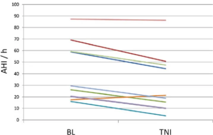
effects after a full night of treatment with TNI. Overall, TNI led to a 23.8% reduction in the AHI (40.4 ± 25.7 to 30.8 ± 25.7/h, p = 0.001) (Fig. 1). The RDI was also significantly decreased (41 ± 25.4 vs. 31.4 ± 25.4/h, p = 0.001) (Table 1). TNI decreased the AHI both in REM (39.8 ± 16.8 vs. 28.7 ±21.4/h, p = 0.048) and NREM sleep (40.5 ± 28.9 vs. 31 ± 28.9/h, p = 0.003).
The ODI3 and the ODI4 both significantly improved with TNI: 40.7±28.4 vs. 31±22.5/h, p=0.02, and 32.1± 27.7 vs. 22.8±19/h, p=0.04, respectively. No significant change was observed in the mean oxygen saturation (92.8± 1.6% vs. 93.1±1.8%, p=0.1).
TNI induced heterogeneous response rates among participants. Noticeable reductions in the AHI were observed for eight patients, in particular in patients with moderate SDB. In one of them, the AHI normalized (<5/h). In one patient with severe SDB, the AHI fell only minimally from 87.3 to 86.2/h. In one subject, the AHI was greater with TNI (17.5 vs. 21.4/h).
Sleep structure also improved with TNI. The percentage of slow-wave sleep (SWS) significantly increased with TNI (16.7±8.2% vs. 22.3±7.4% of total sleep time, p=0.01). No changes were noted in the distribution of other sleep stages. TNI showed a tendency to decrease markers of sleep disruption, with fewer awakenings (24.5±9.3 vs. 17.9±8.3, p=0.05), a decreased arousal index (28.9±24.1 vs. 22.4± 22.4/h, p=0.06), and a decreased respiratory arousal index (23.9 ± 27.6 vs. 17.3 ± 24.5/h, p = 0.06). The sleep time spent in the supine position was similar in both nights (336.4 ± 41.8 vs. 326.6 ± 89 min, p = 0.7).
Discussion
This study was designed to examine the tolerability and the effect of treatment with TNI on SDB in a series of patients with acute stroke. TNI was well accepted, and no significant side effects were reported. Globally, TNI
significantly lowered the AHI and the ODI and improved sleep stage distribution with an increase in slow-wave sleep, but the magnitude of the improvement was rather modest.
In a previous study, McGinley et al. assessed the efficacy of TNI in 11 selected subjects with mild to severe obstructive apnea–hypopnea syndrome [25]. TNI led to a mean reduction of 63.2% in the overall AHI (28±5 to 10± 3/h), and some improvement of the AHI was observed in each subject. Recently, in a larger study, Nilius et al. studied 56 patients with a wide spectrum of disease severity [26]. TNI decreased the RDI from 22.6±15.6 to 17.2±13.2 events/h, thus a 31% mean reduction of the RDI, which is closer to our results. Although TNI decreased RDI in the majority of patients, some demonstrated a marked increase in the RDI. In our study, we obtained a mean global reduction of 23.8% of the AHI. We also found heterogeneous response rates between patients. In one subject with moderate SDB, the AHI even increased in the night with TNI (17.5 vs. 21.4/h), and in one patient
with severe SDB, the AHI fell only minimally. Com-pared with the previous populations studied, our patients were older (57.1 ± 11.3 years) and, overall, had a more severe SDB (AHI 44.5 ± 25.3/h). We used a slightly lower flow rate of 18 L/min (vs. 20 L/min in previous studies) to guarantee a maximum of comfort.
We observed that the effect of TNI on respiratory events was modest in most patients. Nasal CPAP is very likely to be more efficacious in reducing the AHI. However, CPAP is often not well tolerated, in particular in the acute phase of stroke. In addition, CPAP is known to increase intrathoracic pressure and therefore potentially could increase central venous and intracranial pressure and reduce cardiac output [31–33]. The anxiety induced by the sensation of breathing against a positive pressure could induce hyperventilation and hypocapnia and, in turn, increase cerebrovascular resistance. Moreover, studies performed in healthy volunteers [24] and in acute stroke patients [16] have shown that CPAP could lead to a decrease in CBFV, as measured by transcranial doppler. The negative conse-quences of a fall in CBFV may outweigh the therapeutic benefits of CPAP in the poststroke settings.
We thus believe that TNI offers a simplified nasal interface for delivering relatively low levels of pharyngeal pressure. Additionally, TNI does not require titration. TNI could be an alternative treatment of SDB when the use of CPAP is not feasible. The main question is whether the relatively modest AHI reduction with TNI will have a clinically significant effect on short- and long-term out-comes. In some clinical circumstances, even small decreases in SDB severity have had a profound clinical impact: Redline et al., analyzing data from a large prospective cohort of middle-aged and older adults, recently confirmed that OSA increases risk of first-time ischemic stroke, in particular in men [34]. In this population, the risk of stroke increased by 6% with every unit increase in baseline AHI from 5 to 25/h.
Moreover, the impact of treating SDB in the acute stroke phase, when cerebral tissue perfusion is seriously compro-mised, could be particularly important: apneas in the acute stroke phase have been associated with early neurologic worsening [4], and it has been shown that the presence of SDB in stroke patients is associated with worse functional outcomes [5, 6, 12]. Two studies have demonstrated reductions in cerebral tissue hemoglobin saturation levels with apneas [35,36]. The severity of tissue deoxygenation correlated with the length of the respiratory disturbance events and the degree of related desaturation. Respiratory events also induce marked hemodynamic changes. At the onset of apnea, there is hypotension and progressive bradycardia, followed by abrupt tachycardia on resumption of breathing [37]. These changes are associated with large fluctuations in CBFV [38,39]. The repeated hemodynamic
Table 1 Indices of SDB and sleep characteristics BL diagnostic night TNI treatment night p value TST, min 419.4±43 410.3±95.1 0.77
TST in supine position, min 336.4±41.8 326.6±86.8 0.76
Sleep efficiency, % 81±8 77±13 0.31 Stage 1, % 19.6±12.1 14.4±4.8 0.13 Stage 2, % 43.8±6.2 42.3±7.7 0.63 SWS, % 16.7±8.2 22.3±7.4 0.01 REM, % 19.7±6 20.8±5.4 0.71 WASO, min 100.3±43.1 118.9±61.4 0.33 Awakenings, nr 24.5±9.3 17.9±8.3 0.05 Arousal index, nr/h 28.9±24.1 22.4±22.4 0.06 Respiratory arousal index, nr/h 23.9±27.1 17.3±24.5 0.06 AHI, nr/h 40.4±25.7 30.8±25.7 0.001 OAI, nr/h 8.6±8.6 4.9±4.9 0.08 CAI, nr/h 2±3.1 3.3±4 0.16 MAI, nr/h 8.2±13 3.5±5.7 0.1 HI, nr/h 23.8±16.2 19.2±18.2 0.26 RDI, nr/h 41±25.4 31.4±25.4 0.001 AHI NREM, nr/h 40.5±28.9 31±28.9 0.003 AHI REM, nr/h 39.8±16.8 28.7±21.4 0.04 Mean SpO2, % 92.8±1.6 93.1±1.8 0.16 ODI >3%, nr/h 40.7±28.4 31±22.5 0.02 ODI >4%, nr/h 32.1±27.7 22.8±19 0.04
TST total sleep time, SWS slow-wave sleep, REM rapid eye movement, WASO wake after sleep onset, AHI apnea/hypopnea index, OAI obstructive apnea index, CAI central apnea index, MAI mixed apnea
index, HI hypopnea index, RDI respiratory disturbance index, SpO2
oscillations that accompany both occlusion of the upper airway and the post-apneic phase may have a deleterious effect in the penumbra zone. Reducing the AHI, even mildly, may have a positive effect on tissue survival after stroke.
There are several limitations to the current study. First of all, only a limited number of patients were included in this preliminary study and makes its generalizability to all stroke patients questionable. Also, only male patients were recruited. Because of the small number of patients studied, it is not possible to determine demographic or polysomno-graphic predictors for treatment responses. TNI was used for only one night, and further studies of TNI administered over several nights would be required to examine its long-term effects. We used a fixed relatively low flow rate of 18 L/min, determined empirically (no titration) to guarantee a maximum of comfort of TNI. We could speculate that a higher flow rate could yield better results. Since the order of baseline and TNI nights were not randomized, one could argue that the observed therapeutic effects of TNI may be attributed to the expected spontaneous decrease of apneas as stroke recovery progresses. The response rate to TNI was almost twofold as high as the expected night-to-night variability in sleep apnea severity of 13% [40]; however, randomizing TNI nights was beyond the scope of the current study. As patients were studied in the acute phase of stroke, they spent most of the night in the supine position (around 80% of the total sleep time, TST). This could be another factor responsible for the modest effects of the treatment: the supine position increases the severity of respiratory events, counteracting the expected improvement of treatment, if TNI does not act as a mechanical splint (as CPAP or oral devices) but by means of a functional increase of the expiratory pharyngeal pressure.
In summary, this preliminary study provides evidence that TNI is a viable alternative treatment, when CPAP is not tolerated or not indicated, in patients with SDB in the acute phase of ischemic stroke and induces a significant reduction in AHI in these patients. Further studies will be required to confirm these findings and to determine the effect of TNI in long-term outcome of stroke patients.
Acknowledgments The authors thank Pr Hartmut Schneider for his
helpful comments.
Disclosure statement This is not an industry-supported study. The
authors have no financial conflicts of interest.
References
1. Johnson KG, Johnson DC (2010) Frequency of sleep apnea in stroke and TIA patients: a meta-analysis. J Clin Sleep Med 6:131–137
2. Bradley TD, Floras JS (2009) Obstructive sleep apnoea and its
cardiovascular consequences. Lancet 373:82–93
3. Fisher M, Garcia JH (1996) Evolving stroke and the ischemic
penumbra. Neurology 47:884–888
4. Iranzo A, Santamaria J, Berenguer J, Sanchez M, Chamorro A (2002) Prevalence and clinical importance of sleep apnea in the first night after cerebral infarction. Neurology 58:911–916 5. Kaneko Y, Hajek VE, Zivanovic V, Raboud J, Bradley TD (2003)
Relationship of sleep apnea to functional capacity and length of
hospitalization following stroke. Sleep 26:293–297
6. Sandberg O, Franklin KA, Bucht G, Gustafson Y (2001) Sleep apnea, delirium, depressed mood, cognition, and ADL ability after
stroke. J Am Geriatr Soc 49:391–397
7. Hermann DM, Bassetti CL (2009) Sleep-related breathing and
sleep-wake disturbances in ischemic stroke. Neurology 73:1313–1322
8. Dinges DF, Kribbs NB, Schwartz AR, Smith PL, Pack AI (1994) Objective measurement of nasal continuous positive airway pressure use: ethical considerations. Am J Respir Crit Care Med
149:291–292
9. McArdle N, Devereux G, Heidarnejad H, Engleman HM, Mackay TW, Douglas NJ (1999) Long-term use of CPAP therapy for sleep apnea/hypopnea syndrome. Am J Respir Crit Care Med 159:1108–1114
10. Wessendorf TE, Wang YM, Thilmann AF, Sorgenfrei U, Konietzko N, Teschler H (2001) Treatment of obstructive sleep apnoea with nasal continuous positive airway pressure in stroke.
Eur Respir J 18:623–629
11. Disler P, Hansford A, Skelton J, Wright P, Kerr J, O'Reilly J, Hepworth J, Middleton S, Sullivan C (2002) Diagnosis and treatment of obstructive sleep apnea in a stroke rehabilitation unit:
a feasibility study. Am J Phys Med Rehabil 81:622–625
12. Sandberg O, Franklin KA, Bucht G, Eriksson S, Gustafson Y (2001) Nasal continuous positive airway pressure in stroke patients with sleep apnoea: a randomized treatment study. Eur
Respir J 18:630–634
13. Hui DS, Choy DK, Wong LK, Ko FW, Li TS, Woo J, Kay R (2002) Prevalence of sleep-disordered breathing and continuous positive airway pressure compliance: results in Chinese patients
with first-ever ischemic stroke. Chest 122:852–860
14. Broadley SA, Jorgensen L, Cheek A, Salonikis S, Taylor J, Thompson PD, Antic R (2007) Early investigation and treatment of obstructive sleep apnoea after acute stroke. J Clin Neurosci 14:328–333
15. Palombini L, Guilleminault C (2006) Stroke and treatment with
nasal CPAP. Eur J Neurol 13:198–200
16. Scala R, Turkington PM, Wanklyn P, Bamford J, Elliott MW (2009) Acceptance, effectiveness and safety of continuous positive airway pressure in acute stroke: a pilot study. Respir
Med 103:59–66
17. Martinez Garcia MA, Galiano Blancart R, Cabero Salt L, Soler Cataluna JJ, Escamilla T, Roman Sanchez P (2004) Prevalence of sleep-disordered breathing in patients with acute ischemic stroke: influence of onset time of stroke. Arch
Bronconeumol 40:196–202
18. Hsu CY, Vennelle M, Li HY, Engleman HM, Dennis MS, Douglas NJ (2006) Sleep-disordered breathing after stroke: a randomised controlled trial of continuous positive airway pressure. J Neurol Neurosurg Psychiatry 77:1143–1149
19. Martinez-Garcia MA, Soler-Cataluna JJ, Ejarque-Martinez L, Soriano Y, Roman-Sanchez P, Illa FB, Canal JM, Duran-Cantolla J (2009) Continuous positive airway pressure treatment reduces mortality in patients with ischemic stroke and obstructive sleep apnea: a 5-year follow-up study. Am J Respir Crit Care Med
180:36–41
20. Teschler H, Stampa J, Ragette R, Konietzko N, Berthon-Jones M (1999) Effect of mouth leak on effectiveness of nasal bilevel
ventilatory assistance and sleep architecture. Eur Respir J
14:1251–1257
21. Haring HP, Hormann C, Schalow S, Benzer A (1994) Continuous positive airway pressure breathing increases cerebral blood flow velocity in humans. Anesth Analg 79:883–885
22. Droste DW, Ludemann P, Anders F, Kemeny V, Thomas M, Krauss JK, Ringelstein EB (1999) Middle cerebral artery blood flow velocity, end-tidal pCO2 and blood pressure in patients with obstructive sleep apnea and in healthy subjects during continuous positive airway pressure breathing. Neurol
Res 21:737–741
23. Bowie RA, O'Connor PJ, Hardman JG, Mahajan RP (2001) The effect of continuous positive airway pressure on cerebral blood
flow velocity in awake volunteers. Anesth Analg 92:415–417
24. Scala R, Turkington PM, Wanklyn P, Bamford J, Elliott MW (2003) Effects of incremental levels of continuous positive airway pressure on cerebral blood flow velocity in healthy adult humans.
Clin Sci (Lond) 104:633–639
25. McGinley BM, Patil SP, Kirkness JP, Smith PL, Schwartz AR, Schneider H (2007) A nasal cannula can be used to treat
obstructive sleep apnea. Am J Respir Crit Care Med 176:194–200
26. Nilius G, Wessendorf T, Maurer J, Stoohs R, Patil SP, Schubert N, Schneider H (2010) Predictors for treating obstructive sleep apnea with an open nasal cannula system (transnasal insufflation). Chest 137:521–528
27. McGinley B, Halbower A, Schwartz AR, Smith PL, Patil SP, Schneider H (2009) Effect of a high-flow open nasal cannula system on obstructive sleep apnea in children. Pediatrics
124:179–188
28. Iber C, Ancoli-Israel S, Chesson A, Qaun SF (2007) The AASM manual for the scoring of sleep and associated events: rules, terminology and technical specifications, 1st edn. American Academy of Sleep Medicine, Westchester
29. No authors listed (1999) Sleep-related breathing disorders in adults: recommendations for syndrome definition and measure-ment techniques in clinical research. The report of an American
Academy of Sleep Medicine Task Force Sleep 22:667–689
30. Bamford J, Sandercock P, Dennis M, Burn J, Warlow C (1991) Classification and natural history of clinically identifiable
subtypes of cerebral infarction. Lancet 337:1521–1526
31. Werner C, Kochs E, Dietz R, Schulte am Esch J (1990) The effect of positive end expiratory pressure on the blood flow velocity in the basal cerebral arteries during general anesthesia. Anasth Intensivther Notfallmed 25:331–334
32. Hormann C, Mohsenipour I, Gottardis M, Benzer A (1994) Response of cerebrospinal fluid pressure to continuous positive
airway pressure in volunteers. Anesth Analg 78:54–57
33. Feldman Z, Robertson CS, Contant CF, Gopinath SP, Grossman RG (1997) Positive end expiratory pressure reduces intracranial
compliance in the rabbit. J Neurosurg Anesthesiol 9:175–179
34. Redline S, Yenokyan G, Gottlieb DJ, Shahar E, O'Connor GT, Resnick HE, Diener-West M, Sanders MH, Wolf PA, Geraghty EM, Ali T, Lebowitz M, Punjabi NM (2010) Obstructive sleep apnea-hypopnea and incident stroke: the sleep heart health study.
Am J Respir Crit Care Med 182:269–277
35. Hayakawa T, Terashima M, Kayukawa Y, Ohta T, Okada T (1996) Changes in cerebral oxygenation and hemodynamics during
obstructive sleep apneas. Chest 109:916–921
36. Valipour A, McGown AD, Makker H, O'Sullivan C, Spiro SG (2002) Some factors affecting cerebral tissue saturation during obstructive sleep apnoea. Eur Respir J 20:444–450
37. Guilleminault C, Connolly S, Winkle R, Melvin K, Tilkian A (1984) Cyclical variation of the heart rate in sleep apnoea syndrome. Mechanisms, and usefulness of 24 h electrocardiography
as a screening technique. Lancet 1:126–131
38. Balfors EM, Franklin KA (1994) Impairment of cerebral perfusion during obstructive sleep apneas. Am J Respir Crit Care Med
150:1587–1591
39. Klingelhofer J, Hajak G, Sander D, Schulz-Varszegi M, Ruther E, Conrad B (1992) Assessment of intracranial hemodynamics in
sleep apnea syndrome. Stroke 23:1427–1433
40. Stepnowsky CJ Jr, Orr WC, Davidson TM (2004) Nightly variability of sleep-disordered breathing measured over 3 nights.