Differentiation between systolic and diastolic dysfunction
M. FEDERMANN AND 0. M. HESS
Cardiology, Department of Internal Medicine, University Hospital of Zurich, Switzerland
KEY WORDS: Systolic dysfunction, diastolic dysfunction, left ventricular relaxation, left ventricular filling, passive-elastic properties, mitral filling patterns, medical treatment.
Left ventricular (LV) failure can be divided into systolic and diastolic dysfunction. The former is characterized by a reduced ejection fraction and an enlarged LV chamber, the latter by an increased resistance to filling with increased filling pressures. Systolic dysfunction is clinically associated with left ventricular failure in the presence of marked cardiomegaly,
while diastolic dysfunction is accompanied by pulmonary congestion together with a normal or only slightly enlarged ven-tricle. Echocardiography is currently the most relevant technique for non-invasive differentiation of the two forms.
Systolic dysfunction is easily assessable by estimation of global ejection fraction and regional wall motion. Diastolic dysfunction can be diagnosed indirectly by means of a normal or nearly normal ejection fraction and changes of the mitral filling pattern in the context of LV failure. For an exact determination ofdiastolic dysfunction LV catheterization is required. Systolic dysfunction treatment is well defined, consisting of ACE inhibitors, followed by diuretics and digitalis. Calcium channel blockers are usually contraindicated. Diastolic dysfunction therapy is more dependent on the underlying disease. Calcium channel blockers, ACE inhibitors or beta-blockers are first line drugs in most instances: diuretics can be added with increasing symptoms. Digitalis should be avoided, except in atrial fibrillation, to control heart rate.
Introduction
Chronic heart failure (CHF) is generally associated with a poor prognosis, with the annual mortality rate ranging between 20 and 50% depending on the severity of cardiac dysfunction'1'. Heart failure has been subdivided into
dif-ferent forms such as forward and backward failure, high-and low-output failure, systolic or diastolic dysfunction etc. All these entities show considerable variations in regard to clinical course and prognosis During the last two decades, the role of diastolic dysfunction in the development of heart failure has been increasingly recognized. The differ-ent pathophysiology of systolic and diastolic dysfunction affects prognosis and therapeutic management.
Definition and aetiology
Systolic dysfunction is determined by an impaired pump function with reduced ejection fraction and an enlarged end-diastolic chamber volume'21. Diastolic dysfunction is
characterized by an increased resistance to filling with increased filling pressures'2'. Isolated systolic dysfunction is
rare; most patients with CHF have predominating systolic dysfunction with some degree of diastolic impairment. The main aetiology of systolic dysfunction is coronary artery disease followed by hypertensive heart disease'3'4'. Other,
but less frequent causes are dilated cardiomyopathy and myocarditis Left ventricular (LV) failure is caused by iso-lated diastolic dysfunction in up to 40% of all cases'51,
mainly due to long-standing hypertension!3'4'. Coronary
artery disease is secondary, hypertrophic cardiomyo-pathy, restrictive cardiomyopathies and aortic valve
Correspondence: Prof. Dr O. M. Hess, Cardiology, Department of Internal Medicine, University Hospital, 8091 Zurich, Switzerland.
diseases are less frequent. Age also has a bearing on physiological diastolic dysfunction'6!.
Pathophysiology
The pathophysiology of systolic dysfunction begins with a reduction in systolic pump function following a loss of muscle cells, a decrease in myocardial contractility, and/or structural changes of the myocardium with an increase in interstitial fibrosis (= myocardial remodelling). The con-secutive fall in cardiac output is counteracted by two major compensatory mechanisms, the Frank-Starling mechanism and activation of the sympathetic nervous system'71. The
Frank-Starling mechanism leads to an enlargement of the
Table 1 The three major diastolic functional abnormalities and their main aetiology
Slowed or incomplete relaxation hypertrophy
asynchrony ischaemic Reduced LV filling
early diastolic filling: delayed relaxation asynchrony late diastolic filling:
restrictive filling pattern chamber dilatation
Accumulation of collagen—altered collagen architecture (increased myocardial stiffness)
hypertrophy hypertension infarction chamber dilatation
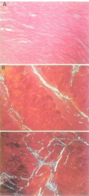
Figure 1 Photomicrographs (magnification X 300 and
repro-duced here at 80%) of different patterns of collagen accumulation and altered collagen architecture. Differently orientated collagen fibres are represented by white or blue in the polarized light micro-scope. (A) Normal myocardium with few, tiny collagen fibres. (B) Biopsy sample from a patient with aortic valve disease showing an increase in interstitial collagen fibres, which are mostly orientated longitudinally to muscle fibres. (C) Biopsy sample from another patient with aortic valve disease. The collagen concentration is increased with evidence of extensive malalignment of collagen fibres (= cross-hatching).
chamber and an increase in end-diastolic pressure. Sympathetic activation results in an increase in the heart's inotropic and chronotropic state. The adverse effects of these compensatory mechanisms—higher wall stress and higher oxygen requirements—are reduced by subsequent myocardial hypertrophy, activation of the baroreceptor reflex and release of atrial natriuretic peptide. The barore-ceptor reflex inhibits sympathetic activation and atrial natriuretic peptide exerts diuretic and vasodilating effects.
During the development of chronic heart failure, these pri-mary and secondary compensatory mechanisms lose their beneficial influences, for instance by a down-regulation of the beta-receptors and dysfunction of the baroreceptor reflex. A vicious circle evolves, which is enhanced by increasing activation of the renin-angiotensin system and by the release of vasopressin, with subsequent vasocon-striction and water retention.
The pathophysiology of diastolic dysfunction is gener-ally more diverse, depending on the underlying disease. Factors extrinsic to the ventricular chamber, such as peri-cardial constriction or ventricular interdependence must be distinguished from those intrinsic to the chamber181.
There are three major diastolic functional abnormalities, namely slowed or incomplete relaxation, reduced LV fill-ing, and altered passive elastic properties, especially due to accumulation of collagen or altered collagen architecture (Table I)'9'. On the cellular level, an increase in myocyte
calcium content, a decrease in ATP levels and an activation of angiotensin II seem to be responsible for impaired relax-ation!101. Alterations of the passive elastic properties are
probably the result of enhanced interstitial fibrosis and changes in the collagen structure. A change in the collagen architecture with an abnormal alignment (cross-hatching) and/or endomyocardial fibrosis appears to be responsible for an increase in myocardial stiffness, whereas the sole increase in collagen leaves passive diastolic function unaltered (Figs 1 and 2)'ll>.
LV diastolic dysfunction results in an elevation of ven-tricular and atrial filling pressures, with consecutive pul-monary congestion. In this situation, atrial contraction becomes more important'121. Chronic atrial enlargement
favours the onset of atrial fibrillation. This is poorly toler-ated in patients with diastolic dysfunction and results often in acute CHF181. An accompanying tachycardia contributes
0 4 8 12 Total interstitial collagen volume fraction (%)
Figure 2 Plot of the constant of myocardial stiffness (b) vs the
total collagen volume fraction in normals and patients with aortic valve disease and different amounts of myocardial collagen and/or architecture. The relationship is exponential and increases only slightly from normal (O) to enhanced myocardial collagen volume fraction ( • ) , but increases significantly in the presence of cross-hatching and/or endomyocardial fibrosis (x). Likewise, isolated cross-hatching and/or endomyocardial fibrosis ( • left upper point) is associated with a significant increase of myocardial stiff-ness Reproduced with permission from the American College of Cardiology!"].
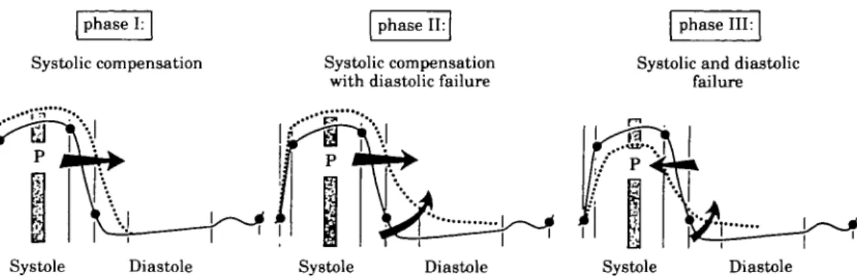
phase I: phase II: phase III:
Systolic compensation Systolic compensation
with diastolic failure Systolic and diastolicfailure
Systole Diastole Systole Diastole Systole Diastole
Figure 3 Pressure (P)-time relationship of the left ventricle in successive phases during the pathophysiological evolution
of pressure or volume overloading, or both (modified from Brutsaert et ai'131, reproduced with permission). There are three
different phases. (1) Systolic compensation: prolonged contraction leads to retarded (but not prolonged) relaxation with shortening of diastole. (2) Systolic compensation with diastolic failure; impaired relaxation and increased myocardial stiff-ness occur resulting in elevated left ventricular diastolic pressures. (3) Systolic and diastolic failure: in a third phase, systolic failure develops.
to diastolic failure by shortening of diastolic filling time. During the clinical course, chronic mechanical overload and subendocardial hypoperfusion induce further hyper-trophy and interstitial fibrosis (= myocardial remodelling) and finally systolic dysfunction may occur. According to Brutsaert and co-workers!131 three phases can be
distin-guished in the evolution of diastolic dysfunction in patients with chronic pressure and volume overload (Fig. 3). Initially a prolongation of systolic contraction is associated with retarded relaxation, which shortens diastolic filling time. In a second phase, impairment of myocardial relax-ation occurs, which (along with increased stiffness and ele-vated heart rate) may result in enhanced diastolic filling pressures. In a third phase, concomitant systolic dysfunc-tion develops.
Clinical symptoms and diagnostic approach
In clinical practice, systolic dysfunction is characterized by the occurrence of CHF in the presence of marked car-diomegaly. Diastolic dysfunction is associated with pul-monary congestion and a normal or only slightly enlarged ventricle.
Echocardiography is currently the most relevant non-invasive method for differentiation of systolic and diastolic dysfunction. Systolic performance of the ventricle can be quantified by regional and global function parameters such as LV-ejection fraction'1*1 and regional wall motion
analy-sis. In contrast, diastolic function cannot be estimated directly because the measurable filling parameters are a function of multiple determinants. Diastolic dysfunction is often associated with typical, but non-specific, mitral filling patterns (Fig. 4), such as the pattern of delayed relaxation with increased isovolumic relaxation time and a low E/A ratio, and the restrictive pattern with shortened isovolumic relaxation time and a high E/A ratio'151.
During the course of progressive diastolic dysfunction, mitral filling may shift from the first to the second pattern with a 'pseudonormalization' in between. Additional infor-mation for interpretation of mitral filling pressures may be gained from the pulmonary venous flow velocity pattern
and the colour Doppler M-mode display of mitral inflow1151.
Radionuclide angiography, another non-invasive diagnos-tic method, has similar advantages and limitations in the assessment of diastolic dysfunction.
Only LV catheterization allows reliable assessment of LV diastolic function and differentiates between abnormal
Delayed relaxation
Pseudonormalization
Restrictive pattern
A
Figure 4 Typical, but non-specific mitral filling patterns in
dias-tolic dysfunction. The pattern of delayed relaxation is character-ized by a low amplitude of the E-wave due to a reduced early left ventricular filling: E/A-ratio<l. In the course of diastolic dysfunc-tion, a 'pseudonormalization' with a 'normal' E/A-ratio (>1) can occur. In advanced forms of diastolic dysfunction a restrictive pat-tern with a high E-wave, a small A-wave and an increased E/A-ratio (>2) can be observed.
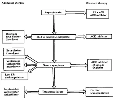
Additional therapy Diuretic* beta-bloeker (lowdow) Beta-blocker (low dote) Ventricular tachycardia amiodarone LowEF: anticoagulation Implan table cardioverter defibrillator Standard therapy I Asymptomatic
Mild to moderate symptoms
Severe ivmptoms
Treatment failure
ACE inhibitor ACE inhibitor
ACE inhibitor +Di ure tics +Digitaha Cardiac transplantation
Figwe 5 Therapeutic strategies based on clinical symptoms in patients with systolic dysfunction.
relaxation and altered passive elastic properties. LV relax-ation can be quantified from the rate of LV pressure decay, and LV stiffness from the simultaneous pressure-volume (chamber stiffness) or stress-strain (myocardial stiffness) relationship. For clinical purposes this differentiation is rarely necessary, but might help to define the severity of diastolic dysfunction in individual patients.
Prognosis
The prognosis of systolic dysfunction is poor, with a 5-year survival rate close to 40% in most studies, compared to 70% in patients with isolated diastolic dysfunction'41.
Diastolic dysfunction often precedes systolic dysfunction and represents an early form of heart failure, not necessar-ily accompanied by clinical symptoms. However, when
sys-Additional therapy Standard therapy I Asymptomatic
Diuretics Mild to moderate symptoms • ^ Atrial fibrillation: an ti coagulation + restoration of sinus rhythm Severe symptoms Treatment failure C alau m-channel blocker or ACE inhibitor or beta-blocker Ca 1 d u m-ch annel blocker or ACE inhibitor or beta-blocker Calcium-channel blocker and/or ACE inhibitor + diuretics Cardiac transplantation
Figure 6 Therapeutic strategies based on clinical symptoms in patients with
tolic dysfunction ensues, the heart failure process is usually in an advanced stage involving both systolic and diastolic function and thus has a poor outcome.
Therapy
Medical treatment in systolic dysfunction (Fig. 5) is pri-marily based on ACE inhibitors, drugs that are effective even in asymptomatic patients with an LV ejection fraction of 35%|161or40% in patients after myocardial infarction'17'.
With further progression of heart failure, diuretics fol-lowed by digitalis are employed'18'. Beta-blockers in low
doses seem to achieve an additional benefit'19'. Calcium
channel blockers are rarely indicated because of their neg-ative inotropic effects. Newer calcium antagonists, such as amlodipine, nisoldipine or felodipine might improve symptoms of systolic dysfunction'20', but this has to be
proven in large scale trials. Atrial fibrillation and ventricu-lar tachycardia are preferably treated by amiodarone. Anticoagulation to prevent systemic embolism is recom-mended in severe systolic dysfunction, especially in dilated cardiomyopathy121'. In treatment failure, cardiac
transplan-tation is possible in appropriate patients; otherwise intractable life-threatening ventricular arrhythmias can be managed by means of an implantable cardioverter defrib-rillator.
Data are sparse for medical treatment of diastolic dys-function. The underlying disease is currently the most important determinant for therapy. In hypertensive heart disease for example (Fig. 6), therapy may start with a cal-cium channel blocker, an ACE inhibitor, or a beta-blocker. All these drugs have been shown to reduce left ventricular hypertrophy122'. A diuretic can be added when symptoms
increase, and in advanced stages a combination of these drugs may be necessary. When atrial fibrillation occurs, restoration of sinus rhythm is often mandatory and is best achieved with electric conversion. For prophylaxis of recurrence, beta-blockers, especially sotalol, which exerts a class III antiarrhythmic effect, are suitable. Cardiac trans-plantation is rarely indicated. The antifibrotic action of spironolactone is currently being investigated: promising animal data show that spironolactone can prevent myocar-dial fibrosis123'. Digitalis can worsen LV relaxation and
should be avoided, except in atrial fibrillation.
References
[1] Hess OM. Pathophysiologie der Herzinsuffizienz. Schweiz med Wschr 1990,120:1833-7.
[2] Hess OM. Hemodynamics in cardiac failure: systolic and diastolic dysfunction. Ther Umsch 1993; 50: 414-8.
[3] Dougherty AH, Naccarelli GV, Gray EL, Hicks CH, Goldstein RA. Congestive heart failure with normal systolic function. Am J Cardiol 1984; 54: 778-82.
[4] Cohn JN, Johnson GJ, Veterans Administration Cooperative Study Group. Heart failure with normal ejection fraction: The V-Heft study. Circulation 1990; 81 (Suppl III): III-48-III-53.
[5] Soufer R, Wohlgelernter D, Vita NA et al Intact systolic function in clinical congestive heart failure. Am J Cardiol 1985; 55:1032-6.
[6] Nixon JV, Burns CA. Cardiac effects of aging and diastolic dysfunction in the elderly. In: Gaasch WH, LeWinter MM, eds. Left ventricular diastolic dysfunction and heart failure. Philadelphia: Lea and Febiger, 1993: 427-35.
[7] Packer M. Pathophysiology of chronic heart failure. Lancet 1992; 340: 88-92.
[8] Grossman W. Diastolic dysfunction and congestive heart failure. Circulation 1990; 81 (Suppl III): III-1-III-7. [9] Hess OM. Diastolische Dysfunktion. Schweiz med Wschr
1991; 1942-5.
[10] Apstein CS, Morgan JP. Cellular mechanisms underlying left ventricular diastolic dysfunction. In: Gaasch WH, LeWinter MM, eds. Left ventricular diastolic dysfunction and heart failure. Philadelphia: Lea and Febiger, 1993: 3-24.
[11] Villari B, Campbell SE, Hess OM et al Influence of collagen network on left ventricular systolic and diastolic function in aortic valve disease. J Am Coll Cardiol 1993; 22:147784. -[12] Little WC, Downes TR. Clinical evaluation of left ventricular diastolic performance. Progr Cardiovasc Dis 1990; 32: 273-90.
[13] Brutsaert DL, Sys SU, Gillebert TH. Diastolic failure: Pathophysiology and therapeutic implications. J Am Coll Cardiol 1993; 22: 318-25.
[14] Jenni R, Vieli A, Hess OM, Anliker M, Krayenbuehl HP. Estimation of left ventricular volume from apical orthogonal 2-D echocardiograms. Eur Heart J 1981; 2: 217-25. [15] Thomas JD. Doppler echocardiography and left ventricular
diastolic dysfunction. In: Gaasch WH, LeWinter MM, eds. Left ventricular diastolic dysfunction and heart failure. Philadelphia: Lea and Febiger, 1993:192-218.
[16] The SOLVD Investigators. Effect of enalapril on mortality and development of heart failure in asymptomatic patients with reduced left ventricular ejection fraction. N Engl J Med 1992; 327: 685-91.
[17] Pfeffer MA, Braunwald E, Moy£ A el al Effect of captopril on mortality and morbidity in patients with left ventricular dysfunction after myocardial infarction. Results of the Survival and Ventricular Enlargement Trial. N Engl J Med 1992; 327: 669-77.
[18] Follath F. Medikamentose Therapie bei Herzinsuffizienz. Ther Umsch 1993; 50: 425-33.
[19] Hjalmarson A, Waagstein F. New therapeutic strategies in chronic heart failure: challenge of long-term beta-blockade. Eur Heart J 1991; 12 (Suppl F): 63-9.
[20] Packer M. Treatment of chronic heart failure. Lancet 1992; 340: 92-5.
[21] Fuster V, Stein B, Halperin JL, Chesebro JH. Antithrombotic therapy in cardiac disease: an approach based on pathogene-sis and risk stratification. Am J Cardiol 1990; 65:38C-44C. [22] DahlOf B, Pennert K, Hansson L. Reversal of left ventricular
hypertrophy in hypertensive patients. A metaanalysis of 109 treatment studies. Am Heart J 1992; 5: 95-110.
[23] Brilla CG, Matsubara LS, Weber KT. Antifibrotic effects of spironolactone in preventing myocardial fibrosis. Am J Cardiol 1993; 71:12A-16A.