ORIGINAL ARTICLE
The phosphorous necrosis of the jaws and what can we learn
from the past: a comparison of
“phossy” and “bisphossy” jaw
C. Jacobsen&W. Zemann&J. A. Obwegeser&
K. W. Grätz&P. Metzler
Received: 5 August 2012 / Accepted: 15 November 2012 / Published online: 28 December 2012 # Springer-Verlag Berlin Heidelberg 2012
Abstract
Introduction The osteopathology of the jaws associated with bone resorption inhibitors is a current topic that engages a variety of clinical specialists. This has increased after the approval of denosumab for treatment of osteopo-rosis and skeletal-related events in patients with solid ma-lignancy. Early after the first publications, there is a possible connection between phosphorous necrosis of the jaws, a dreadful industrial disease mentioned, and bisphosphonate-induced pathology. The nineteenth century was the prime time for phosphorus necrosis of match factory workers. Results This occurrence provides an interesting insight into the medical and surgical profession in the nineteenth centu-ry. There are striking parallels and repetition of current and old ideas in the approach to this“new disease.” There are similar examples in case descriptions when compared with today’s patients of bisphosphonate-related osteonecrosis of the jaws (BRONJ).
Discussion Phosphorus necrosis was first described in Aus-tria. Soon after this, surgeons in German-speaking countries including well-known clinicians Wegner (1872) and von Schulthess-Rechberg (1879) pioneered the analysis, preven-tative measures, and treatment of this disease. The tendency at this time was to approach BRONJ as a“special kind of osteomyelitis” in pretreated and metabolically different bone. Not only the treatment strategy to wait until seques-trum formation with subsequent removal and preventative measures but also the idea of focusing on the periosteum as the triggering anatomical structure may have been adopted from specialists in the nineteenth century. Therefore,
phosphorous necrosis of the jaw is an excellent example of “learning from the past.”
Keywords Phosphorous necrosis . Sequestrum . Infection . Bone resorption inhibitors . Periosteum
Introduction
Since 2003, clinicians have been dealing with a new type of bone pathology that involves the upper and lower jaw: bisphosphonate-related osteonecrosis of the jaws (ONJ) [1]. Today, it is known as a multifactorial disease associated with bone resorption inhibition therapy and with a local triggering factor in most cases. It is clinically usually repre-sented by exposed bone predominantly in the mandibular molar area [2]. Shortly after discovery, the first reports emerged reporting on the similarity of bisphosphonate-related osteonecrosis of the jaws (BRONJ) and a previously well-known disease called phosphorous necrosis of the jaw (PNJ) [3]. The workers in match factories, firework produc-tion companies, and factories producing muniproduc-tions with white phosphorous were affected by PNJ. In 2008, R. Marx demonstrated chemical conversion of phosphorous fumes into bisphosphonates and“uncovered” BRONJ as a replace-ment of phossy jaw [4]. But still, questions about phossy and bisphossy jaw exist after looking back into the nineteenth century with careful analysis of detailed reports of cases, opening some interesting parallels.
The production of matches with white phosphorus in German-speaking countries started in 1833. The first case of PNJ was described soon afterwards in 1838 by Lorinser; he named the disease“phosphorimus chronicus” [5]. Soon afterwards, PNJ became an increasingly public and political problem [6,7]. The first official hearing on necessary con-structive measures in match factories in Switzerland took
C. Jacobsen (*)
:
W. Zemann:
J. A. Obwegeser:
K. W. Grätz:
P. MetzlerDepartment of Oral and Maxillofacial Surgery, University Hospital of Zurich, Frauenklinikstrasse 24, 8091 Zurich, Switzerland
place in 1841 in Zurich [8]. Finally, in 1906 in the Berne Convention, the manufacture and importation of white phosphorus was banned in several European countries, fol-lowed by the USA in 1912 by passing the Match Act [9]. The aim of this article was to retrospectively review the nineteenth century literature to detect and compare interest-ing and strikinterest-ing parallels in dealinterest-ing with a“new” disease and learn from the past.
Material and methods
Patients
To analyze patients with PNJ, we referred to two detailed case series from 1879 to 1893. The first document presented 55 patients with PNJ who were treated at the University Hospital of Bern, beginning in 1867 with a mean follow-up time of 12 years [10]. The second document gave a detailed description of 12 patients with PNJ who were treated at the University Hospital of Zurich, beginning in 1868 [11]. Pa-tient data were compared with retrospectively analyzed data of 110 patients with apparent ONJ who were treated and followed up in the Department of Oral and Maxillofacial Surgery, University of Zurich from 2005 until 2010. Sys-temic factors, etiology, period and kind of drug regimen, local risk factors, symptoms, clinical picture, treatment methods, and response to treatment were carefully analyzed and discussed according to the current knowledge on ONJ.
Literature search
From 2003, an ongoing literature search concerning both ONJ and PNJ was performed. Electronic databases (PubMed, OVID, MEDLINE, and NLM) were searched using a set of predetermined keywords (“phosphorus necro-sis,” “osteonecrosis of the jaws,” “bisphosphonates,” “osteonecrosis,” and “match factory worker”). To find back issues of case reports and reviews on PNJ, an extensive search was performed in Swiss institutes of medical history and libraries in German-speaking countries. Reference lists of articles were additionally searched for any related articles.
Results
Patients
Sixty-seven patients with PNJ were compared with 110 ONJ patients. Descriptive statistics regarding the general infor-mation and details of possible associated risk factors and sites are displayed in Table1.
Clinical picture
Most PNJ patients presented with pain or swelling. The symptoms and clinical picture were described as a“special, dangerous type of infection.” Kocher and von Schulthess-Rechberg characterized this pain of as “persistent, yet pro-gressive with spread to neighboring teeth and jawbone” in contrast to pain of an ordinary infection [10]. Pus formation usually developed and penetrated the skin or oral mucosa with fistula formation, loss of teeth, and recurrent abscesses. Progression persisted, even if the match factory worker was detracted from the “harmful contact to phosphorous” with sequestrum formation about 3 months and necrosis of partly wide areas of the jaw about 6 months after appearance of the first symptoms [10]. Another characteristic finding was extensive periosteal reactions, described as “osteophytes” [11]. The most dangerous area for PNJ was the maxilla with possible spread of infection with subsequent meningitis and death of the patient [12]. The exposed bone areas never showed the entire extent of the lesion.“The disease course was characterized by retraction of the periosteum and oral mucosa extension of necrotic bone into the oral cavity” [11] (Fig. 1). Table 1 summarizes the clinical picture of ONJ patients.
Treatment
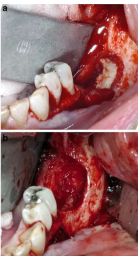
The majority of Kocher’s patients were treated surgically with the intention of removal of infected bone and teeth to eliminate the origin of the disease (Fig. 2a, b). A less invasive method had been performed in Zurich at that time. Demarcation of sequestra was the aim with subsequent sequestrectomy and local revision or resection in all 12 cases without touching the periosteum and osteophytes.
Six of Kocher’s patients died due to direct consequences of the disease or the surgical treatment. A mortality rate of 25 % (n03) and therefore a success rate of 75 % were described by von Schulthess-Rechberg [11]. In 12 patients, regeneration with growth of a neomandible was described.
Of all 110 patients with ONJ, 58 % were treated surgi-cally; if possible, the procedure was minimally invasive (sequestrectomy) after a drug holiday of 3 to 6 months. All patients underwent additional systemic anti-infectious therapy (Fig.3a, b).
Discussion
Analysis of PNJ began in Austria (Vienna) with its first mention in 1838, followed by an official publication by Friedrich Wilhelm Lorinser in 1945 [5]. Phosphorous ne-crosis was one of the most dreaded but accepted industrial diseases, and the origins of many political measures and
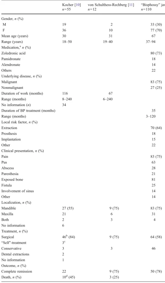
Table 1 Descriptive statistics of patients with phossy jaw and bisphossy jaw including general factors, local risk factors, treat-ment strategy, and outcome
a
Patients may belong to more than one group
b
Type of surgical treatment: maxillectomy 13, hemimaxillec-tomy 7, partial mandibular resec-tion 12, hemimandibulectomy 7, mandibulectomy 10
c“Self-treatment”
mandibulectomy, 3
d
Thereof, six are due to direct consequences of phospho-rous necrosis
Kocher [10] von Schulthess-Rechberg [11] “Bisphossy” jaw
n055 n012 n0110
Gender, n (%)
M 19 2 33 (30)
F 36 10 77 (70)
Mean age (years) 30 31 67
Range (years) 18–50 19–40 37–94 Medication,an (%) Zoledronic acid 80 (73) Pamidronate 18 Alendronate 14 Others 22 Underlying disease, n (%) Malignant 83 (75) Nonmalignant 27 (25)
Duration of work (months) 116 67
Range (months) 8–240 6–240
No information (n) 34
Duration of BP treatment (months) 35
Range (months) 3–120
Local risk factor, n (%)
Extraction 70 (64) Prosthesis 18 Implantation 15 Other 22 Clinical presentation, n (%) Pain 83 (75) Pus 63 Abscess 28 Paresthesia 21 Exposed bone 81 Fistula 25 Involvement of sinus 14 Other 14 Localization, n (%) Mandible 27 (55) 9 (75) 83 (75) Maxilla 21 6 31 Both 2 3 4 No information 6 Treatment, n (%) Surgical 46b(84) 9 (75) 64 (58) “Self”-treatment 3c Conservative 3 3 46 Dental extractions 2 No information 1 Outcome, n (%) Complete remission 22 9 (75) 50 (78) Death, n (%) 10d(45) 3 (25)
laws concerning working conditions and child labor were associated with this disease. Notably, in 1845, about 50 years before the invention of antibiotics, cure of infectious dis-eases was often unsuccessful and time consuming.
Today, severity of infectious diseases differs markedly compared with that 200 years ago. Nevertheless, ONJ has engaged practitioners and scientists for almost 10 years now and remained incompletely understood.
Etiology and pathology
Similarly, during the early recognition of the existence of PNJ, there were vehement discussions about the pathologi-cal pathway of jaw necrosis. A few scientists, like Dr. Georg Wegner from the Institute of Pathology in Berlin, made some important discoveries. Wegner presented different an-imal studies about “the influence of phosphorus on the organism” in 1872 [13] and classified two different effects of phosphorus: an“indirect” reaction after systemic intake of phosphorus and a“direct” reaction, whereas phosphorus fumes had direct contact with the oral mucosa.
After direct contact of the oral mucosa of rabbits with phosphorus fumes, Wegner found swelling of the jaws with collateral infection and indicated the teeth as the origin of this reaction. In some animals, he additionally created a small injury of the periosteum in the jaw area. Interestingly, these animals developed severe periostitis with necrosis of the underlying bone after exposure to phosphorus fumes. This is very similar to recent animal studies by Sonis et al. and others, in which
Fig. 1 A male patient with extensive osteopathology of the maxilla associated with underlying metastasized bladder carcinoma and long-term treatment with zoledronic acid. Detachment (double arrow) and retraction (arrow) of the periosteum are shown clearly to the extent of the infraorbital rim. Massive signs of progressive infection with previ-ous loss of parts of the maxilla are present
Fig. 2 a A 21-year-old female match factory worker, 5 months after mandibulectomy because of PNJ of the entire mandible. b A photo-graph of the resected mandible shows the typical clinical appearance, similar to BRONJ with persistent sockets and no signs of remodeling
Fig. 3 a A female patient with BRONJ in the left mandibular molar area. After a drug holiday of about 6 months, demarcation of necrotic bone could be found. b For surgical treatment, subsequent sequestrec-tomy and local revision of surrounding bone have been performed. Complete remission could be achieved
ONJ-like areas were found after extraction of teeth in rats treated with zoledronic acid [14,15]. Wegner highlighted the importance of direct contact of phosphorus with the periosteum. In a second experiment, Wegner described the indirect reaction as follows:“in all areas where usually bone marrow structure develop, a tissue is present, which looks as cortex of the long bones—absolutely solid and compact all over” [13]. The excess mass was formed at the expense of the volume of vessels in the Haversian canals, possibly leading to necrosis of bone. On the basis of those experiments, the idea of possible intake of phosphorus into the bone was born, and phosphorus was administered for the treatment of diseases with“weak bone development” with questionable success—as a progen-itor of later bisphosphonate therapy.
von Schulthess-Rechberg described the pathological–ana-tomical pathway of PNJ as follows:“specifically acting sub-stances reach the periosteum due to local factors. It inflames, thickens, and is detached from the bone. At the same time, periosteal reaction starts with formation of osteophytes. The infection progresses and affects Haversian canals with osteo-cytes and subsequent necrosis of bone” [11].
Different hypotheses about the pathological pathway of ONJ have been proposed, as for example, avascular necro-sis, genetic influence, direct soft tissue toxicity, or impaired bone remodeling [16, 17]. Interestingly, despite initial op-position, there seems to be a tendency towards acknowledg-ment of a major role of infection [18–21]. The important role of the detached retracted and thickened periosteum in patients with ONJ has been clearly demonstrated clinically and remains to be investigated.
Risk factors
Many different match production workflows had to be per-formed. Some were highly injurious to health, depending on the type and duration of contact to white phosphorus, as there were the dunkers, mixers, and boxers who packed the phosphorus-covered matches into boxes. Interestingly, one characteristic of patients with PNJ, which differed from other
types of jaw necrosis, was the“delayed occurrence” of this disease with an average of about 5 years after the start of phosphorus fume exposure [11, 22]. This fact led to the hypothesis of accumulation of phosphorus in the bone at that time [4]—corresponding to the cumulative dose of
bisphosph-onate as one of the recognized risk factors for ONJ today [2]. Local risk factors (e.g., carious teeth and extraction sockets) were one of the most discussed local factors [22]. Hirt reported an about threefold higher risk of development of PNJ in work-ers with “poor” teeth [22]. Contact of phosphorous with the periosteum was assumed to lead to the development of PNJ.
Local risk factors are acknowledged triggers of the devel-opment of ONJ, including dental infection, dental implants, and ill-fitting dentures. Any factor that involves possible contact with the bone or periosteum of the oral cavity is a possible source of osteopathology, as was the case 200 years ago [23]. In contrast to other authors, spontaneous osteopa-thology was not found in this ONJ patient collective [1].
Clinical picture
Examination of ONJ reveals striking parallels. In 1847, von Bibra and Geist presented a classification of the different courses of PNJ (Table2) [24]. A similar staging system for ONJ was published and updated in 2009 by the American Association of Oral and Maxillofacial Surgery [2].
Geist specifically mentioned the difficulty of diagnosis of first-stage phossy jaw, because there was no difference between patients with common dental pain. Non-exposed bone but apparent osteopathology of the jaws was a frequent finding in our ONJ patient group too (24 %) and has also been described by other authors [25].
Treatment
According to von Schulthess-Rechberg, three main strate-gies were developed during the first years of dealing with PNJ, with interesting parallels to the therapeutic strategies
Table 2 Staging system for PNJ presented from von Bibra and Geist in 1847 and staging system for BRONJ presented by the AAOMS in 2009 Stadium I (stadium morbid primum–stadium invasionis): Beginning of disease with intermittent dental pain and gingival swelling, but the patient is healthy Stadium II (Stadium morbi secundum: Stadium reactionis primum): Time span from the occurrence of symptoms until the exposure of bone.
Patients presented with swelling, pain, and other signs of infection
Stadium III (stadium reactionis secundum): Time span from bone exposure until rejection of necrotic bone Stage 0: No clinical evidence of necrotic bone, but there are nonspecific clinical findings and symptoms Stage 1: Exposed and necrotic bone in asymptomatic patients without evidence of infection
Stage 2: Exposed and necrotic bone associated with infection as evidenced by pain and erythema in the region of the exposed bone with or without purulent drainage
Stage 3: Exposed and necrotic bone in patients with pain, infection, and one or more of the following: exposed and necrotic bone extending beyond the region of alveolar bone (i.e., inferior border and ramus in the mandible, maxillary sinus, and zygoma in the maxilla) resulting in pathologic fracture, extraoral fistula, oral antral/oral nasal communication, or osteolysis extending to the inferior border of the mandible of sinus floor
used in BRONJ, bearing in mind that antibiotic therapy was not available back then.
1. Immediate prevention of progression with exposure prevention and careful intraoral“disinfection” (potassi-um iodine rinse)
2. Abscess drainage, removal of insufficient teeth, and surgical therapy, if necessary
3. Systemic antiphlogistic treatment
Symptoms, especially pain, were usually resolved with these methods—even before the actual surgical treatment [11]—similar to today’s resolution of BRONJ symptoms
after the start of antibiotic treatment.
Two main surgical strategies have been described in about 1850. One group advocated early and radical removal of both affected and parts of healthy bone to prevent inva-sion and systemic proliferation. Another group of surgeons favored surgical restraint until demarcation of sequestrum with subsequent removal and revision of the surrounding bone. This“wait and see” strategy took an average of 1 to 2 years until complete loosening of the sequestrum [11]. The objective of this strategy was to preserve the periosteum, because of“osteoplastic capabilities” with the aim of regen-erating the jawbone. One of the main advocators of this technique was von Langenbeck in Berlin with his students Billroth and Thiersch, who made an effort in operating his patients through creating incisions in the oral cavity instead of having them in the face [11]. The main surgical doctrine after a few years of dealing with PNJ became avoidance of primary resection; this resulted in a decrease of mortality rate to about 18 to 25 % [11,26].
These strategies are again applied today for the treatment of ONJ. Immediate radical resection has shown to be restricting for the patient and did not always result in the expected remission, whereas conservative treatment leads to frequent recurrent symptomatic infections [27]. Therefore, the author’s therapy strategy for BRONJ patients changed during the last years. Drug holidays prior to surgical treat-ment were established; this resulted in rapid sequestrum formation similar to PNJ, followed by sequestrectomy, ex-tensive rinsing with antibiotic solution, and revision of the surrounding bone. With this strategy, satisfactory rates of complete remissions could be achieved with a minimal invasive approach and without restriction of the patients’ quality of life.
Prevention
By 1846, prophylactic measures for prevention of PNJ (e.g., frequent facial and oral washing, ventilation, strict separa-tion of work and break rooms, restricted working hours, and ban of child labor) had been enacted [28]. At that time, corpsmen were responsible for assuring its implementation
in match factories. Restrictions in Zurich also included a prohibition against hiring workers with carious teeth or any type of bone diseases. Each match factory worker under-went monthly clinical and dental examinations by the re-gional specialist [11,28].
Similar measures adapted to present times are included in several guidelines for patients undergoing therapy with bone resorption inhibitors. These measures include eradication of all possible infectious sources before the start of drug ther-apy and frequent dental examinations. Similar to a reported decrease of PNJ after implementation of preventive meas-ures, prevention of ONJ also seems to have shown success [29–31]. This fact again emphasizes the impact of local risk factors such as dental infection or peri-implantitis. This article provides an interesting historical insight into medical and surgical profession in the nineteenth century with strik-ing parallels of ideas and much“repetition” in the approach to a new disease.
Osteopathology of the jaws associated with phosphorus or bone resorption inhibitors is an excellent example of “learning from the past.” The increasing tendency of approaching BRONJ as a “special entity of osteomyelitis” in a pretreated and metabolically different bone could have been adopted from specialists in the nineteenth century, just like the minimally invasive surgical approach. Perhaps, one will soon uncover the periosteum pathophysiologically as the main triggering anatomical structure for bony exposure.
Conflict of interest The authors do not have any conflict of interest.
References
1. Marx RE (2003) Pamidronate (Aredia) and zoledronate (Zometa) induced avascular necrosis of the jaws: a growing epidemic. J Oral Maxillofac Surg 61:1115
2. Ruggiero SL, Dodson TB, Assael LA et al (2009) American Association of Oral and Maxillofacial Surgeons position paper on bisphosphonate-related osteonecrosis of the jaws—2009 up-date. J Oral Maxillofac Surg 67:2
3. Hellstein JW, Marek CL (2005) Bisphosphonate osteochemonec-rosis (bis-phossy jaw): is this phossy jaw of the 21st century? J Oral Maxillofac Surg 63:682
4. Marx RE (2008) Uncovering the cause of“phossy jaw” circa 1858 to 1906: oral and maxillofacial surgery closed case files—case closed. J Oral Maxillofac Surg 66:2356
5. Lorinser FW: Necrose der Kieferknochen in Folge und Einwirkung von Phosphordämpfen. In Oesterreichisch Medizinisches Jahrbuch (ed. Wien 1945:257)
6. Hellstein JW, Marek CL (2004) Bis-phossy jaw, phossy jaw, and the 21st century: bisphosphonate-associated complications of the jaws. J Oral Maxillofac Surg 62:1563
7. Gendrin AN et al.: Recherches sur les maladies des ouvriers employes a la fabrication des allumetes chimiques. In Academie des Sciences (ed., 1846)
8. Meyer-Hofmeister JC: Die Phosphorzündholzfabriken des Cantons Züirch mit Rücksicht auf die in denselben beobachteten Gesundheitsverhältnisse der Arbeiter. Schweizerische Zeitschrift fr Medicin, Chirurgie und Geburtshilfe 1947
9. Myers ML, McGlothlin JD (1996) Matchmakers’ “phossy jaw” eradicated. Am Ind Hyg Assoc J 57:330
10. Kocher T (1893) Zur Kenntnis der Phosphornekrose. Ministry of Trade and Agriculture, Switzerland
11. von Schulthess-Rechberg JA (1879) Über die Phosphornekrose und den Ausgang ihrer Behandlung. In Department of Surgery (ed. Zurich University of Zurich)
12. Hofmokl J (1871) Über die Resection des Ober- und Unterkiefers mit Rücksicht auf 88 darauf bezügliche Krankheitsfälle, welche auf der chirurgischen Klinik von Dumreichers 1852–1870 beo-bachtet und operirt worden sind. Medical Yearbooks, Vienna 13. Wegner G (1872) Einfluss des Phosphors auf den Organismus.
Virchows Archiv. Bd. 55
14. Ali-Erdem M, Burak-Cankaya A, Cemil-Isler S et al (2011) Extraction socket healing in rats treated with bisphosphonate: animal model for bisphosphonate related osteonecrosis of jaws in multiple myeloma patients. Med Oral Patol Oral Cir Bucal 16:e879 15. Sonis ST, Watkins BA, Lyng GD et al (2009) Bony changes in the
jaws of rats treated with zoledronic acid and dexamethasone before dental extractions mimic bisphosphonate-related osteonecrosis in cancer patients. Oral Oncol 45:164
16. Reid IR, Bolland MJ, Grey AB (2007) Is bisphosphonate-associated osteonecrosis of the jaw caused by soft tissue toxicity? Bone 41:318
17. Bertoldo F, Santini D (2007) Lo Cascio V: Bisphosphonates and oste-omyelitis of the jaw: a pathogenic puzzle. Nat Clin Pract Oncol 4:711 18. Kumar SKS, Gorur A, Schaudinn C et al (2010) The role of
microbial biofilms in osteonecrosis of the jaw associated with bisphosphonate therapy. Curr Osteoporos Rep 8:40
19. Lesclous P, Abi Najm S, Carrel J-P et al (2009) Bisphosphonate-associated osteonecrosis of the jaw: a key role of inflammation? Bone 45:843
20. Kassolis JD, Scheper M, Jham B, Reynolds MA (2010) Histopathologic findings in bone from edentulous alveolar ridges: a role in osteonecrosis of the jaws? Bone 47:127
21. Mawardi H, Giro G, Kajiya M et al (2011) A role of oral bacteria in bisphosphonate-induced osteonecrosis of the jaw. J Dent Res 90:1339
22. Hirt L: Die äusseren Krankheiten der Arbeiter Leipzig 1878:117ff 23. Jacobsen C, Metzler P, Rossle M et al.: Osteopathology induced by bisphosphonates and dental implants: clinical observations. Clin Oral Investig 2012. doi:10.1007/s00784-012-0708-2
24. von Bibra E, Geist L: Die Krankheiten der Arbeiter in den Zündhlzfabriken, insbesondere das Leiden der Kieferknochen durch Phosphordämpfe. Vom chemisch-physiologischen, medicinischen-chirurgischen und medicinisch-polizeilichen Standpunkt bearbeitet. 1847
25. Fedele S, Porter SR, D'Aiuto F et al (2010) Nonexposed variant of bisphosphonate-associated osteonecrosis of the jaw: a case series. Am J Med 123:1060
26. Haltenhoff G: De la periostite et de la necrose phosphorique. In Department of Surgery (ed. Zurich University of Zurich 1866) 27. Gevorgyan A, Enepekides DJ (2008) Bisphosphonate-induced
ne-crosis of the jaws: a reconstructive nightmare. Curr Opin Otolaryngol Head Neck Surg 16:325
28. Lesky E (1966) Die Phosphornekrose, klassisches Beispiel einer Berufskrankeheit. Wien Klin Wochenschr 37:601
29. Brock G, Barker K, Butterworth CJ, Rogers S (2011) Practical considerations for treatment of patients taking bisphosphonate medications: an update. Dent Update 38:313
30. Fehm T, Felsenberg D, Krimmel M et al (2009) Bisphosphonate-associated osteonecrosis of the jaw in breast cancer patients: rec-ommendations for prevention and treatment. Breast 18:213 31. Ferlito S, Puzzo S, Liardo C (2011) Preventive protocol for tooth
extractions in patients treated with zoledronate: a case series. J Oral Maxillofac Surg 69:e1
The English in this document has been checked by at least two professional editors, both native speakers of English. For a certificate, please seehttp://www.textcheck.com/certificate/gbqxzp.