HAL Id: inserm-00137179
https://www.hal.inserm.fr/inserm-00137179
Submitted on 19 Mar 2007HAL is a multi-disciplinary open access archive for the deposit and dissemination of sci-entific research documents, whether they are
pub-L’archive ouverte pluridisciplinaire HAL, est destinée au dépôt et à la diffusion de documents scientifiques de niveau recherche, publiés ou non,
Cyclooxygenase involvement in thromboxane-dependent
contraction in rat mesenteric resistance arteries.
Manlio Bolla, Dong You, Laurent Loufrani, Bernard Levy, Sylviane
Levy-Toledano, Aïda Habib, Daniel Henrion
To cite this version:
Manlio Bolla, Dong You, Laurent Loufrani, Bernard Levy, Sylviane Levy-Toledano, et al.. Cyclooxyge-nase involvement in thromboxane-dependent contraction in rat mesenteric resistance arteries.: PGE2 mediated TxA2-vasoconstriction. Hypertension, American Heart Association, 2004, 43 (6), pp.1264-9. �10.1161/01.HYP.0000127438.39744.07�. �inserm-00137179�
1 CYCLOOXYGENASE INVOLVEMENT IN THROMBOXANE-DEPENDENT
CONTRACTION IN RAT MESENTERIC RESISTANCE ARTERIES
Manlio Bolla1*, Dong You2, Laurent Loufrani2$, Bernard I Levy2, Sylviane Levy-Toledano1,
Aïda Habib1#,Daniel Henrion2
Institut National de la Santé et de la Recherche Médicale (INSERM) 1Unit 348 and 2Unit 541, IFR
Circulation-Paris-Nord, Paris, France. Short title: PGE2 mediated TxA2-vasoconstriction
* current address: NicOx SA, 2455 route des Dolines, Sophia-Antipolis, France
# current address : American University of Beirut, Dept of Biochem. and Int. Med., Beirut, Lebanon $ current address: Dept of Physiology – CNRS UMR 6188, Medical School, Angers, France.
Corresponding author and present address: Dr. Daniel HENRION, Pharm.D., Ph.D. INSERM Unit 541, 41 bd de la chapelle 75475 PARIS, FRANCE
tel: 332 41 73 58 45 fax: 332 41 73 58 95
E-mail:daniel.henrion@med.univ-angers.fr ABSTRACT
The influence of cyclooxygenase pathway activation following endoperoxide / thromboxane receptor (TP) stimulation was studied in rat mesenteric resistance arteries (n=6-10 per group). We studied isolated, perfused, and pressurized mesenteric resistance arteries (mean internal diameter 214 µm) using an arteriograph allowing to study arteries in physiological conditions of flow and pressure. Changes in diameter were continuously recorded and contractions were appreciated as internal diameter reduction. Release of cyclooxygenase pathway metabolites was also assessed by EIA analysis of mesenteric bed perfusions. The TxA2 analog, U-46619 (1 µmol/L) induced a significant contraction (108 µm maximal
diameter reduction). Inhibition by 3 chemically different cyclooxygenase inhibitors, flurbiprofen, indomethacin and aspirin, potently reduced the contraction to 27, 25 and 6% of control, respectively. The selective cyclooxygenase-1 inhibitor SC-58560 inhibited U-46619-contraction whereas selective cyclooxygenase-2 inhibition (SC-58236) had no effect. Thromboxane synthase inhibition (furegrelate) did not affect U-46619-induced contraction, but it was reduced by cytosolic phospholipase A2 inhibition. Measurement of cyclooxygenase derivatives produced by the isolated mesenteric bed showed that PGE2 was produced after TxA2-receptor stimulation with U-46619. Exogenous
prostaglandin E2 (in the presence of the TxA2 receptor antagonist SQ 29,548) and U-46619 contracted
mesenteric arteries with a similar potency (EC50: 0.30 and 0.48 µmol/L, respectively). This study
provides the first evidence that TxA2-receptor-dependent contraction in a resistant artery involved
cyclooxygenase- stimulation and, at least in part, PGE2 formation. This mechanism of TxA2-dependent
contraction in resistant arteries might be of importance in the understanding of diseases affecting resistant arteries and involving TxA2, such as hypertension.
This is an un-copyedited author manuscript that was accepted for publication in Hypertension, copyright The American Heart Association.
HAL author manuscript inserm-00137179, version 1
HAL author manuscript
INTRODUCTION
Thromboxane A2 (TxA2), a lipid mediator originating from arachidonic acid (AA) metabolism through
the cyclooxygenase (COX) pathway, is a powerful constrictor of vascular smooth muscle (1). Enhanced TxA2 production has been reported in several cardiovascular diseases such as unstable angina (2), acute
myocardial infarction (2), spontaneous hypertension (3) and pregnancy-induced hypertension (4). In resistance arteries, TxA2 is produced upon flow stimulation (shear stress) of the endothelium and this
production is increased in the case of hypertension (5). Similarly, the involvement of TxA2 also
increases the pressure-induced myogenic tone in hypertension (6).
TxA2 acts through specific G-protein coupled (1). Activation of TxA2 receptor (TP; also known as
thromboxane/endoperoxide receptor) leads to phospholipase C activation, release of inositol triphosphate (IP3), and increase in the intracellular Ca2+ level, thus triggering the smooth muscle contraction (7).
Cyclooxygenases are important enzymes in the formation of prostaglandins and TxA2. They present a
double enzymatic activity, cyclooxygenase before and peroxidase after, producing from the precursor AA, the unstable endoperoxide intermediates PGG2 and PGH2, respectively. PGH2 itself already
possesses activity (1,8), or alternatively it may be metabolized by different isomerases or synthases into prostaglandins, prostacycline (PGI2) or TxA2 (1,9). Two isoforms of COX are known; one
constitutive (named COX-1) and the second one inducible (named COX-2) (10,11).
Resistance arteries are known to play an important role in the generation of peripheral vascular resistance, and are involved in the control of local blood distribution and capillary pressure (12, 13). Their vascular tone is regulated by the sympathetic nervous system, circulating hormones, and vasoactive metabolites produced by endothelial cells, among them TxA2 (14). TxA2 contributes to the
homeostasis of normal resistance arteries, and an alteration of its synthesis and/or release was shown in pathological states, such as hypertension (5,6).
Involvement of COX in the vascular response of resistance arteries to different stimuli has been demonstrated. Vasoactive agents like angiotensin II (15), endothelin (16) and phenylephrine (17) have been shown to activate, and in part to induce contraction via, the COX pathway.
The aim of our work was to evaluate the influence of COX on the response of resistance arteries under conditions of regulated flow and pressure, following TP receptor stimulation by the stable agonist U46619, and to identify the cyclooxygenase metabolite important to the TP-dependent contraction.
METHODS
Mesenteric artery in vitro
Male Wystar Kyoto rats (WKY) (Iffa Credo, France), aged for 8-10 weeks were used. Rats were anesthetized with sodium pentobarbital (50 mg/Kg) and their gut excised. The investigation was in accordance with the European Community standards on the care and use of laboratory animals. A second order mesenteric artery (214 ± 13 µm, internal diameter, 3-5 mm length) was cannulated at both ends and mounted in a video monitored perfusion system as previously described (17) according to Halpern et al. (18). The arterial segment was bathed in a Physiological Salt Solution (PSS) (17) and superfused (2 mL/min). Perfusion of the artery was set at a rate of 90 µL/min. Flow was set at 90 µl/min and pressure at 50 mm Hg. Arterial diameter was measured and recorded continuously using a video monitoring system (Living System Instrumentation Inc). Data was collected (Biopac, La Jolla, CA, USA), recorded and analyzed (AcqKnowledge® software, Biopac) with the results given in µm for arterial inner diameters.
Vessels first were allowed to stabilize for at least 30 min. Vessels were then stimulated with 60 mmol/L KCl in order to verify vessel viability. Testing the vasodilator effect of acetylcholine (1 µmol/L) after preconstriction with phenylephrine (1 µmol/L) then assessed the integrity of the endothelium. The response to U-46619 (0.01 to 10 µmol/L or a single dose of 1 µmol/L), a selective and stable thromboxane receptor agonist, was assessed at least twice to obtain a stable and constant contraction. In some experiments, the preparation was preincubated with one of the following drugs: flurbiprofen, indomethacin, diclofenac and aspirin were used to inhibit COX activity (1), furegrelate to block TxA2
synthesis (19), 58560 to inhibit COX-1, 58236 (20) to prevent COX-2 activation (20), SC-19220, EP1 receptor antagonist (21). Following pretreatment, contraction to U-46619 was repeated and compared to the previous stable contraction to U-46619. At the end of all experiments a contraction to 60 mmol/L KCl was performed, thus showing no change in vessel reactivity compared to the first one. In separate series of experiments, the effect of indomethacin and flurbiprofen was tested on phenylephrine (1 µmol/L), endothelin-1 (10 nmol/L), PGE2 (1 µmol/L), PGF2α (1 µmol/L). These latter
agonists as well as U-46619 were added to the PSS so that the PSS superfusing and perfusing the arteries contained the concentration of drug indicated between the parenthesis.
Mesenteric bed perfusion
Mesenteric beds were prepared as previously described (22, 23). Briefly, the abdomen of anaesthetized rats was opened and the gut exposed. The superior mesenteric artery was separated from surrounding fat tissue in the region of the aorta. The rat was then sacrificed by exanguination cutting the abdominal aorta. A polyethylene catheter (diameter 0.5 mm) was inserted distally into the artery at its origin from the aorta, and the catheter fixed with surgical thread. The intestine was separated from the mesentery and the cannulated mesentery perfused (2.0 mL/min, 37 °C). The perfusion pressure was monitored continuously (cf. upper paragraph).
After a 30 min. stabilization period the preparation was stimulated with 1 µmol/L phenylephrine, followed by 1 µmol/L Ach to check the integrity of the preparations, for vascular smooth muscle and endothelial responses.
The perfusate was collected for 5 min. (10 mL total volume) for the basal and different stimuli.
Perfusate extraction and EIA analysis
To 10 ml of perfusate samples 10 µL of formic acid, 10,000 cpm of [3H]-TxB
2 (NEN) for extraction
recovery and 1 mL of methanol were added. After vortexing, samples were centrifugated at 1,200 x g at 4 °C for 10 min. Supernates were loaded onto C18 Bakerbond silica columns (3 mL), washed with 5 mL water and then eluted with 3 mL of methanol. Samples were dried under a vacuum and resuspended in 1 mL of EIA buffer (EIA buffer: Phosphate buffer 0.1 mol/L, pH 7.4 containing 0.15 mol/L NaCl, 1 mmol/L EDTA and 0.1 % bovine serum albumin). Radioactivity was counted in a 100 µL aliquot for recovery and the sample frozen at –20 °C until measurement of prostanoids.
EIA analysis
Immunoenzyme analysis (EIA) of 6-keto-PGF1α, the breakdown product of PGI2, as well as PGE2 and
PGF2α were performed as previously described (24).
Statistical analysis
Results are expressed as means ± s.e. mean of n measurements. The significance of the different treatments was determined by ANOVA or two tailed Student’s paired T-test. P values less than 0.05 were considered to be significant.
Drugs
SC-58560 and SC-58236 were kindly provided by Dr Peter Isakson (Searle Monsanto, St Louis, USA). HEPES, acetylcholine (ACh), phenylephrine (Phe), acetylsalicylic acid (ASA), indomethacin and flurbiprofen, were purchased from Sigma Chem. Co. (St Louis, MO). Methyl arachidonylfluorophosphate (MAFP) was purchased from Calbiochem. Other reagents were purchased from Prolabo (Paris, France). Other prostanoids were purchased from Cayman Chem. Co. (Ann Arbor, MI).
RESULTS
Isolated mesenteric arteries
Perfused and pressurized rat mesenteric arteries responded to TP receptor stimulation with U-46619 by a concentration-dependent contraction (EC50 = 0.48 ± 0.06 µM, maximal response: 108 ± 8 µm
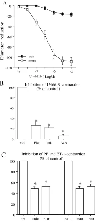
decrease in diameter, n=10). Pretreatment with 3 chemically non-related non-steroidal anti-inflammatory drugs (NSAIDs), flurbiprofen (1 µmol/L), indomethacin (1 µmol/L), and aspirin (10 µmol/L) inhibited U-46619 (1 µmol/L)-induced contraction to 27 ± 6, 25 ± 5 and 6 ± 3% of control, respectively (Fig 1). Indomethacin (1 µmol/L) and flurbiprofen (1 µmol/L) also inhibited, at least in part, phenylephrine (1 µmol/L) and endothelin-1 (10 nmol/L)-induced contraction (fig 1C). PGE2 (1
µmol/L)-induced contraction was also inhibited by aspirin (10 µmol/L, 79 ± 7% of control, n=4), indomethacin (1 µmol/L, 84 ± 8% of control, n=4), and flurbiprofen (1 µmol/L, 84 ± 9% of control, n=4). PGF2α (1 µmol/L)-induced contraction was inhibited by aspirin, (10 µmol/L, 68 ± 7% of control,
n=5), indomethacin (1 µmol/L, 78 ± 9% of control, n=4), but not by flurbiprofen (1 µmol/L, 94 ± 8% of control, n=5).
Removing the endothelium did not affect the inhibitory effect of indomethacin (1 µmol/L) on U-46619-induced contraction (108 ± 9 µm decrease in diameter versus 18 ± 4; n=5).
The selective COX-1 inhibitor SC-58560 concentration dependently decreased CCRC to U-46619, whereas the COX-2 selective inhibitor (SC-58236) was mainly ineffective (Fig 2).
Phospholipase A2 (PLA2) inhibition with MAFP (10 µmol/L) reduced to 13% of the control the maximal response to U-46619 (20 ± 5µm loss in diameter after MAFP versus 155 ± 15 µm in control, n=6). This inhibition completely overlapped inhibition given by the NSAIDs. By contrast, Phe (1 µmol/L)-induced contraction was only reduced to 54±6% (n=4) of the control by MAFP (10 µmol/L). Thromboxane synthase inhibition (furegrelate, 10 µmol/L) left the maximal response unchanged to U-46619 (84 ± 9 µm contraction in the presence of furegrelate versus 89 ± 10 µm in control, n=6). On the other hand, Phe (1 µmol/L) and endothelin-1 (10 nmol/L)-induced contraction were decreased, respectively, to 46 ± 7% (n=4) and 41 ± 7% of control by furegrelate (10 µmol/L).
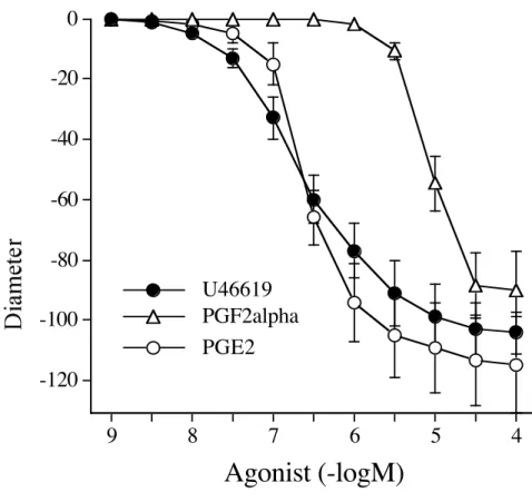
PGF2α and PGE2 are 2 other possible COX-derived compounds that might be involved in
U-46619-induced contraction in MRA. CCRC to exogenous PGF2α showed a contractile response only at high
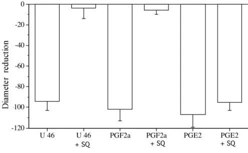
concentrations (fig.3). The response to PGF2α (10 µmol/L) was abolished by the TP receptor antagonist
SQ 29,548, 10 µmol/L (Fig. 4).
The second exogenous metabolite tested was PGE2 (fig.3). The contraction elicited by PGE2 (1 µmol/L)
was not significantly affected by SQ 29,548 (fig.4), suggesting a direct contractile action of PGE2.
The TP receptor antagonist SQ 29,548 induced no significant inhibition of phenylephrine-induced contraction (data not shown), but it induced a 34 ± 5% (n=5) inhibition of endothelin-1-induced tone. The EP1 receptor antagonist SC-19220 (10 µmol/L) significantly inhibited U-46619-induced contraction to a level equivalent to 48% of control (94±7 versus 45±9 µm decrease in diameter, n=6). On the other hand, ET-1-induced contraction was not significantly reduced by SC-19220 (105±17 versus 88±12 µm decrease in diameter, n=4)
In the absence of endothelium SC-19220 (10 µmol/L) inhibited similarly U-46619-induced contraction (112 ± 10 µm decrease in diameter versus 61 ± 7; n=5).
EIA analysis of perfused mesenteric beds
An immunometric measurement for 6-keto-PGF1α (the stable metabolite of PGI2), PGF2α and PGE2 was
performed in the perfusate of isolated mesenteric beds. Unfortunately, U-46619 interferes with the dosage of 6-keto-PGF1α, and PGF2α and the amount of U-46619 present in the PSS collected from
perfused mesenteric beds made it impossible to determine this metabolite. Nevertheless, PGI2 is more
likely to induce dilation in mesenteric resistance arteries (5). However, a rise in 6-keto-PGF1α
immuno-reactivity was shown after of cholinergic receptor stimulation with acetylcholine, confirming that the endothelial layer was intact. A second confirmation was recognized in the fall of perfusion pressure from 81 ± 6 mmHg to 32 ± 5 mmHg in the presence of acetylcholine after phenylephrine-induced precontraction (n=6).
On the contrary, a two-fold rise in PGE2 secretion was observed in the presence of U-46619
stimulation (Fig. 5), with a very low (less than 1% of the final amount calculated) interference of U-46619 in assaying PGE2.
Discussion
The present study brings new insights in the comprehension of the physiological role of thromboxane A2 (TxA2) in resistance arteries. Surprisingly, contractions induced by the stable TP
receptor agonist U-46619 were strongly attenuated by non-selective COX inhibitors and by a selective COX-1 inhibitor. A further analysis showed that PGE2 may be, at least in part, the mediator involved
in U-46619-induced contraction in rat mesenteric resistance arteries.
Resistance arteries determine vascular resistance and hence have a key role in the control of local blood flow (13). There are an increasing number of studies showing that TxA2 may have a significant
role in the vascular homeostasis in several vascular beds (3,5,6). This involvement of TxA2 in the
control of vascular tone is enhanced in pathological situations such as hypertension (3-6). In addition, we have shown in resistance arteries, that TP receptor activation does not desensitize (17). Our finding that U-46619-induced contraction was almost abolished by COX inhibition is surprising as TxA2
production itself depends on COX activation. COX derivatives have been involved in agonist-induced contraction due to angiotensin II (15), endothelin-1 (16) or alpha-adrenoreceptor stimulation (17). In most cases the mediator involved is probably TxA2 and/or PGH2 as contraction was blocked by COX
inhibition and by a TP receptor antagonist. In the present study, TP receptor activation by U-46619 also depended on COX derivatives to produce a contraction. This was confirmed by the use of 3 non-selective and chemically different cyclooxygenase inhibitors (aspirin, flurbiprofen and indometacin) and by the use of a selective COX-1 inhibitor (SC-58560). COX-2 inhibition was ineffective. In addition, PLA2 inhibition also prevented U-46619-induced contraction, and thusly further supporting the involvement of a COX and arachidonic acid-derivative.
Among the COX-derivatives that may be produced in resistance arteries, TxA2, PGF2α and PGE2 are
the main vasoconstrictor products. In the arteries used in the present study, PGF2α was not potent
enough to be involved in U-46619-induced contraction as it only induced a contraction for concentrations higher than 1 µmol/L, possibly interacting with TP receptor (twice the EC50 for
U-46619). On the other hand, PGE2 produced a contraction comparable to that of U-46619. PGE2 has
been shown to enhance vasoconstrictor responses to pressure stimuli in the rat mesenteric artery (24). It is also a potent vasoconstrictor in the rat tail artery (25) and the aorta (26). We performed an immuno-enzymatic assay of PGE2 in mesenteric arterial beds perfused with U-46619 in order to further
investigate the potential involvement of PGE2 in TP receptor activation-dependent contraction. Under
U-46619 stimulation, the PSS perfused in the mesenteric beds contained twice the baseline PGE2
concentration, further supporting that PGE2 may be involved in the contraction induced by TP
receptors stimulation. The technique used to measure COX-derivatives in perfused mesenteric arteries has been previously validated in a study showing that more TxA2 and less PGI2 were produced in the
PSS flowing through the arterial bed from hypertensive rats, compared to normotensive animals (5). In this latter study we have shown that 6-keto-PGF1α, (1.5 ng/mL, the stable metabolite of PGI2), PGF2α
(0.6 ng/mL) and TxB2 (1.5 ng/mL, stable metabolite of TxA2) were also produced by the mesenteric
circulation in the absence of exogenous stimulation. The amount of PGI2, PGF2α and TxA2 previously
found (5) is comparable to the amount of PGE2 found in the present study (1-2 ng/mL).
Interactions between different pathways may also occur. Although COX inhibitors almost completely inhibited U-46619-dependent contraction, they also partially inhibited the contraction induced by
endothelin-1 and phenylephrine, without affecting PGE2-induced tone. In parallel, TP receptor
blockade, which totally inhibited U46169- and PGF2α-induced contractions, also inhibited endothelin-1
and phenylephrine-dependent tone to a lesser extent. Indeed, the contractile effect of these vasoactive agents (U46169, endothelin-1 and phenylephrine) depends on the activation of PLA2 induced by the rise in calcium concentration following receptor activation (1). Then, arachidonic acid release and COX activation lead to the production of different COX-derivatives. Concerning U-46619 in mesenteric resistance arteries, COX activation induced the production of PGE2, which was responsible for a large
part of the contraction. On the other hand, endothelin-1-induced contraction, also inhibited in a great part by PLA2 and COX antagonists, was strongly reduced by TP receptor blockade and unaffected by EP1 inhibition. Thus, TP receptor activation by TxA2/PGH2 is involved in ET-1 induced contraction,
not that to PGE2. This also applies to Phe-induced contraction, but to a lesser extent (PLA2 and COX
inhibition only reduced Phe-induced contraction by 50%). Thus, the different contractile agents studied involved COX derivatives, but to a different degree. In addition, they required either PGE2 or
TxA2/PGH2 to produce a large part of their contractile effect.
Perspective:
This finding might be of importance in the pathophysiology of vascular diseases such as hypertension. In hypertension, TxA2 has been shown to be involved in the control of vascular tone in arterial beds
were it is normally (in the normotensive state) not involved or involved to a lesser degree. For example, a higher TxA2 production in response to shear stress is likely to reduce flow-mediated dilation in
mesenteric resistance arteries (5). Similarly, in rat gracilis muscle, resistance arteries TxA2 contributes to
the elevation in myogenic tone found in SHR (6). Thus, elevated TxA2-production may contribute to
the changes in local blood flow found in hypertension, thereby, participating to the injury produced in end-organs.
Considering the importance of TxA2 in the local control of vascular tone, at least in some arterial beds,
the present finding may enhance our understanding of vascular diseases as well as assist in focusing on potential treatments. In that respect, it may be important to decrease the vasoconstrictor capacity of TxA2 without affecting its other properties. The potential importance of TP receptor blockade in other
diseases has been recently discussed in a review (27), the issue being of importance in atherogenesis (27, 28).
Acknowledgments
M.B. was a recipient of a European Community fellowship (Marie Curie Human Train Mobility of Researcher). L.L. was a recipient of the French Foundation for Medical Research (FRM, Paris France). † This work is dedicated to the memory of Jacques Maclouf who initiated and supported it. He shall always be remembered as a great scientific mentor, collaborator and friend.
References
1. Narumiya S, Sugimoto Y, Ushikubi F. Prostanoid receptors: structures, properties, and functions. Physiol Rev. 1999;79:1193-1226.
2. Noll G, Luscher TF. The endothelium in acute coronary syndromes. Eur Heart J. 1998;19:C30-C38. 3. Cediel E, Vazquez-Cruz B, Navarro-Cid J, De Las Heras N, Sanz-Rosa D, Cachofeiro V, Lahera V. Role of endothelin-1 and thromboxane A2 in renal vasoconstriction induced by angiotensin II in diabetes and hypertension. Kidney Int Suppl. 2002;82:2-7.
4. Paarlberg KM, de Jong CL, van Geijn HP, van Kamp GJ, Heinen AG, Dekker GA. Vasoactive mediators in pregnancy-induced hypertensive disorders: a longitudinal study. Am J Obstet Gynecol. 1998;179:1559-1564.
5. Matrougui K, Maclouf J, Lévy BI, Henrion D. Impaired nitric oxide- and prostaglandin- mediated responses to flow in resistance arteries of Hypertensive rats. Hypertension. 1997;30:942-947.
6. Ungvari Z, Koller A. Endothelin and prostaglandin H(2)/thromboxane A(2) enhance myogenic constriction in hypertension by increasing Ca(2+) sensitivity of arteriolar smooth muscle. Hypertension. 2000;36:856-861.
7. Habib A, Vezza R, Créminon C, Maclouf J, FitzGerald GA. Rapid, agonist-dependent phosphorylation in vivo of human thromboxane receptor isoforms. J Biol Chem. 1997; 272:7191-7200. 8. Ge T, Hughes H, Junquero DC, Wu KK, Vanhoutte PM, Boulanger CM. Endothelium-dependent contractions are associated with both augmented expression of prostaglandin H synthase-1 and hypersensitivity to prostaglandin H2 in the SHR aorta. Circ Res. 1995;76:1003-1010.
9. Reilly M, FitzGerald GA. Cellular activation by thromboxane A2 and other eicosanoids. Eur Heart J. 1993;14(supplK):88-93.
10. FitzGerald GA, Austin S, Egan K, Cheng Y, Pratico D. Cyclo-oxygenase products and atherothrombosis. Ann Med. 2000;32(suppl1):21-26.
11. Bishop-Bailey D, Hla T, Mitchell JA. Cyclo-oxygenase-2 in vascular smooth muscle. Int J Mol Med. 1999;3:41-48.
12. Davis MJ, Hill MA. Signaling mechanisms underlying the vascular myogenic response. Physiol Rev. 1999;79:387-423.
13. Christensen KL, Mulvany MJ. Location of resistance arteries. J Vasc Res. 2001;38:1-12.
14. Shimokawa H.Endothelial dysfunction in hypertension. J Atheroscler Thromb. 1998;4:118-127. 15. Silldorff EP, Hilbun LR, Pallone TL. Angiotensin II constriction of rat vasa recta is partially thromboxane dependent. Hypertension. 2002;40:541-546.
16. White LR, Juul R, Cappelen J, Aasly J. Cyclooxygenase inhibitors attenuate endothelin ET(B) receptor-mediated contraction in human temporal artery. Eur J Pharmacol. 2002;12;448:51-57.
17. Bolla M, Matrougui K, Maclouf J, Loufrani L, Levy BI, Levy-Toledano S, Habib A, Henrion D. p38 MAP kinase activation is required for thromboxane induced contraction in perfused and pressurized rat mesenteric resistance arteries. J. Vasc. Res. 2002;39:353-360.
18. Halpern W, Kelley M. In vitro methodology for resistance arteries. Blood Vessels. 1991;28:245-251.
19. Vila JM, Medina P, Segarra G, Aldasoro M, Noguera I, Lluch S. Endothelin-1-induced potentiation of adrenergic responses in the rabbit pulmonary artery: role of thromboxane A(2). Eur J Pharmacol. 2001;413:247-254.
20. Wang JL, Cheng HF, Zhang MZ, McKanna JA, Harris RC. Selective increase of cyclooxygenase-2 expression in a model of renal ablation. Am J Physiol. 1998;275:F613-F622.
21. Walch L, de Montpreville V, Brink C, Norel X. Prostanoid EP(1)- and TP-receptors involved in the contraction of human pulmonary veins. Br J Pharmacol. 2001;134:1671-1678.
22. Peredo HA. Defibrotide modulates prostaglandin production in the rat mesenteric vascular bed. Prostaglandins Leukot Essent Fatty Acids. 2002;67:211-216.
23. Henrion D, Chillon JM, Capdeville-Atkinson C, Vinceneux-Feugier M, Atkinson J. Chronic treatment with the angiotensin I converting enzyme inhibitor, perindopril, protects in vitro carbachol-induced vasorelaxation in a rat model of vascular calcium overload. Br J Pharmacol. 1991;104:966-972. 24. Malik KU, Ryan P, McGiff JC. Modification by prostaglandins E1 and E2, indomethacin, and arachidonic acid of the vasoconstrictor responses of the isolated perfused rabbit and rat mesenteric arteries to adrenergic stimuli. Circ Res. 1976;39:163-168.
25. Ren J, Karpinski E, Benishin CG. Prostaglandin E2 contracts vascular smooth muscle and inhibits potassium currents in vascular smooth muscle cells of rat tail artery. J Pharmacol Exp Ther. 1995; 275:710-719.
26. Rapoport RM, Williams SP. Role of prostaglandins in acetylcholine-induced contraction of aorta from spontaneously hypertensive and Wistar-Kyoto rats. Hypertension. 1996;28:64-75.
27. Pratico D, Cheng Y, FitzGerald GA. TP or not TP: primary mediators in a close runoff? Arterioscler Thromb Vasc Biol. 2000;20:1695-1698.
28. Cayatte AJ, Du Y, Oliver-Krasinski J, Lavielle G, Verbeuren TJ, Cohen RA. The thromboxane receptor antagonist S18886 but not aspirin inhibits atherogenesis in apo E–deficient mice: evidence that eicosanoids other than thromboxane contribute to atherosclerosis. Arterioscler Thromb Vasc Biol. 2000;20:1724–1728
Figure 1: A: Concentration-response curves to U-46619 obtained in isolated perfused and pressurized rat mesenteric arteries. Concentration-response curves to U-46619 were repeated in the presence of the non-selective COX inhibitor indomethacin (indo 1 µmol/L, n = 6 per group).
B: Inhibitory effect of three chemically non-related non-steroidal anti-inflammatory drugs, indomethacin (1 µmol/L, Indo), flurbiprofen (1 µmol/L, Flur) and aspirin (10 µmol/L, ASA) on U-46619 (1 µmol/L)-induced contraction.
C: Inhibitory effect of indomethacin (1 µmol/L, Indo) and flurbiprofen (1 µmol/L, Flur) on phenylephrine (Phe, 1 µmol/L)- and endothelin-1 (ET-1, 10 nmol/L)-induced contraction.
Contraction was obtained in perfused and pressurized rat mesenteric resistance arteries (n=10 per group). *P <0.05; two tailed Student’s paired T-test.
B
0 20 40 60 80100 Inhibition of U46619-contraction (% of control)
ctrl Flur Indo ASA * * * -120 -100 -80 -60 -40 -20 0 -8 -7 -6 -5 U 46619 (-LogM) A indo control 0 20 40 60 80 100
Inhibition of PE and ET-1-contraction (% of control)
PE indo Flur ET-1 indo Flur
*
*
*
*
C
Figure 2: Changes in diameter in response to increasing concentrations of the TP receptor agonist U-46619, in perfused and pressurized rat mesenteric resistance arteries (n=8 per group, mean ± sem). Concentration-response curves to U-46619 were repeated in the presence of the selective COX-1 inhibitor SC-58560 (upper panel, A) or in the presence of the COX-2 selective inhibitor (SC-58236) (lower panel, B). Control experiments were conducted in the presence of the solvent (<0.1% DMSO). *P <0.05; two-factor ANOVA. -100 -80 -60 -40 -20 0 U46619 [-LogM] SC58560 10 µM SC58560 1 µM SC58560 0.1 µM Control -8 -7 -6 -5
A
*
*
*
-100 -80 -60 -40 -20 0 -8 -7 -6 -5 U46619 (-LogM)B
SC58236 10 µM SC58236 1 µM SC58236 0.1 µM controlFigure 3: Changes in diameter in response to increasing concentrations (1 nmol/L to 0.1 µmol/L) of the TP receptor agonist U-46619, PGF2α or PGE2 in perfused and pressurized rat mesenteric resistance arteries
(n=8 per group, mean ± s.e.m.). *P <0.05; two tailed Student’s T-test.
-120 -100 -80 -60 -40 -20 0 9 8 7 6 5 4
Agonist (-logM)
PGE2 PGF2alpha U46619Figure 4: Inhibitory effect of the TP receptor antagonist SQ 29,548 (SQ, 10 µmol/L) on U-46619 (U46, 1 µmol/L)-, PGF2α (10 µmol/L)- or PGE2 (1 µmol/L)-induced contraction. Contraction was obtained in
perfused and pressurized rat mesenteric resistance arteries (n=8 per group). *P <0.05; two tailed Student’s T-test.
Figure 5: Immunometric analysis of PGE2 were done using specific antibodies and tracers in the
presence or in the absence (control) of U-46619, in the PSS collected from rat perfused mesenteric beds.
*P <0.05; two tailed Student’s T-test.
-120 -100 -80 -60 -40 -20 0
U 46 U 46 PGF2a PGF2a PGE2 PGE2
+ SQ + SQ + SQ 0 1 2 3 PGE2 immunoreactivity (ng/ml) CTRL U46619 1µMU46 Control *