HAL Id: hal-01430037
https://hal-amu.archives-ouvertes.fr/hal-01430037
Submitted on 27 May 2020
HAL is a multi-disciplinary open access
archive for the deposit and dissemination of
sci-entific research documents, whether they are
pub-lished or not. The documents may come from
teaching and research institutions in France or
abroad, or from public or private research centers.
L’archive ouverte pluridisciplinaire HAL, est
destinée au dépôt et à la diffusion de documents
scientifiques de niveau recherche, publiés ou non,
émanant des établissements d’enseignement et de
recherche français ou étrangers, des laboratoires
publics ou privés.
Micellar lipid composition affects micelle interaction
with class B scavenger receptor extracellular loops
Aurélie Goncalves, Brigitte Gontero, Marion Nowicki, Marielle Margier,
Gabriel Masset, Marie Josephe Amiot-Carlin, Emmanuelle Reboul
To cite this version:
Aurélie Goncalves, Brigitte Gontero, Marion Nowicki, Marielle Margier, Gabriel Masset, et al..
Mi-cellar lipid composition affects micelle interaction with class B scavenger receptor extracellular loops.
Journal of Lipid Research, American Society for Biochemistry and Molecular Biology, 2015, 56 (56),
pp.1123-1133. �10.1194/jlr.M057612�. �hal-01430037�
play a major role in lipid transport across cell membranes, although their function at the intestinal level is still a mat-ter of debate.
CD36 is an 88 kDa transmembrane protein with broad specifi city. It interacts with many ligands, including native and oxidized lipoproteins, long-chain fatty acids, cholesterol, anionic phospholipids, carotenoids, and vitamin D [for re-view see ( 1 )], as well as vitamin K ( 2 ). CD36 is highly ex-pressed in the proximal part of the intestine (duodenum and jejunum) ( 3 ), where it is assumed to play a key role in fatty acid ( 4 ) and cholesterol ( 5 ) uptake. Indeed, it has been dem-onstrated both in vitro and in vivo that isolated entero-cytes or proximal intestine from CD36-null mice show reduced fatty acid uptake compared with wild-type mice ( 3 ) and that a CD36 deletion suppresses oleic acid (OA) uptake into the duodenum and reduces it in the jejunum of mice fed triolein ( 6 ). Similarly, CD36-defi cient mice displayed an ac-cumulation of dietary cholesterol in the intestinal lumen and a reduction of dietary cholesterol transport into the lymph ( 7 ). However, it was later shown that CD36 deletion in mice was not rate-limiting for cholesterol absorption ( 8 ). Moreover, it was suggested that the primary role of CD36 was not to mediate fatty acid but to promote the production of large triglyceride-rich lipoproteins ( 9, 10 ).
SR-BI is an 82 kDa transmembrane protein that inter-acts with an equally wide range of ligands such as HDL, anionic phospholipids, cholesterol, carotenoids, and vita-mins D, E ( 1 ), and K ( 2 ). At the intestinal level, SR-BI was shown to facilitate the uptake of free cholesterol as well as esterifi ed cholesterol, phospholipids, and triacylglycerol hydrolysis products ( 11, 12 ). However, the effective role of SR-BI compared with Niemann-Pick C1-like 1 in terms of cholesterol transport is still a matter of debate ( 8 ). It has Abstract Scavenger receptors (SRs) like cluster
determi-nant 36 (CD36) and SR class B type I (SR-BI) play a debated role in lipid transport across the intestinal brush border membrane. We used surface plasmon resonance to analyze real-time interactions between the extracellular protein loops and various ligands ranging from single lipid molecules to mixed micelles. Micelles mimicking physiological structures were necessary for optimal binding to both the extracellular loop of CD36 (lCD36) and the extracellular loop of SR-BI (lSR-BI). Cholesterol, phospholipid, and fatty acid micellar content signifi cantly modulated micelle binding to and dis-sociation from the transporters. In particular, high phospho-lipid micellar concentrations inhibited micelle binding to both receptors ( ⴚ 53.8 and ⴚ 74.4% binding at 0.32 mM com-pared with 0.04 mM for lCD36 and lSR-BI, respectively, P < 0.05). The presence of fatty acids was crucial for micelle in-teractions with both proteins (94.4 and 81.3% binding with oleic acid for lCD36 and lSR-BI, respectively, P < 0.05) and fatty acid type substitution within the micelles was the compo-nent that most impacted micelle binding to the transporters. These effects were partly due to subsequent modifi cations in micellar size and surface electric charge, and could be corre-lated to micellar vitamin D uptake by Caco-2 cells. Our
fi ndings show for the fi rst time that micellar lipid composi-tion and micellar properties are key factors governing micelle interactions with SRs. —Goncalves, A., B. Gontero, M. No-wicki, M. Margier, G. Masset, M-J. Amiot, and E. Reboul.
Mi-cellar lipid composition affects micelle interaction with class B scavenger receptor extracellular loops. J. Lipid Res. 2015. 56: 1123–1133.
Supplementary key words scavenger receptor class B type I • cluster determinant 36 • cholesterol • phospholipids • fatty acid • cholecalcif-erol • intestine • surface plasmon resonance
Scavenger receptors (SRs) such as cluster determinant 36 (CD36) and SR class B type I (SR-BI) are recognized to
A.G. was funded by a CIFRE (Conventions Industrielles de Formation par la REcherche) grant from the ANRT (French national association for research and technology) in partnership with Lesieur Inc. The authors declare no fi nancial confl icts of interest .
Manuscript received 13 January 2015 and in revised form 24 March 2015. Published, JLR Papers in Press, April 1, 2015
DOI 10.1194/jlr.M057612
Micellar lipid composition affects micelle interaction with
class B scavenger receptor extracellular loops
Aurélie Goncalves , * ,†,§ Brigitte Gontero , ** Marion Nowicki , * ,†,§ Marielle Margier , * ,†,§ Gabriel Masset , * ,†,§ Marie-Josèphe Amiot , * ,†,§ and Emmanuelle Reboul 1, * ,†,§
INRA ,* UMR 1260 “Nutrition, Obesity and Risk of Thrombosis,” F-13385 Marseille, France ; INSERM , † UMR 1062, F-13385 Marseille, France ; Aix-Marseille Université , § F-13385 Marseille, France ; and Aix-Marseille Université CNRS,** BIP , UMR 7281, F-13402 Marseille, France
Abbreviations: aa, amino acid; ALA, ␣ -linolenic acid; ARA, arachi-donic acid; CD36, cluster determinant 36; LA, linoleic acid; lCD36, ex-tracellular loop of cluster determinant 36; lSR-BI, exex-tracellular loop of scavenger receptor class B type I; NTA, nitrilotriacetic acid; OA, oleic acid; PA, palmitic acid; RU, response unit; SPR, surface plasmon reso-nance; SR, scavenger receptor; SR-BI, scavenger receptor class B type I.
1
To whom correspondence should be addressed. e-mail: [email protected]
at INRA Institut National de la Recherche Agronomique, on November 18, 2015
www.jlr.org
acid (ARA), cis -5,8,11,14,17-eicosapentaenoic acid (EPA), cis -4,7,10,13,16,19-docosahexaenoic acid (DHA), and vitamin D (cholecalciferol) were purchased from Sigma-Aldrich (Saint-Quentin-Fallavier, France). Nitrilotriacetic acid (NTA) sensor chips were purchased from GE Healthcare Life Sciences (Vélizy-Villacoublay, France). Recombinant human lSR-BI (proline 33 to tyrosine 443 from protein sequence NP_005496.4) and lCD36 (gly-cine 30 to asparagine 439 from protein sequence NP_001001547.1) fused with poly-histidine tag were purchased from Sino Biological Inc. (Beijing, China). The recombinant proteins were produced in human HEK cells, enabling: i ) the formation of the disulfi de bonds as previously described ( 22 ); and ii ) an accurate glycosyl-ation process as demonstrated by the molecular mass of the loops (75–92 kDa ). Both parameters have been shown to be important to maintain the lipid-transporter activity of the proteins ( 22, 23 ).
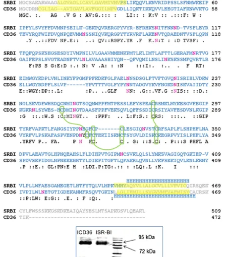
ClustalW sequence alignment of the two recombinant proteins is presented in Fig. 1 : the extracellular loops are represented with blue residues and the deleted amino acids (aas) are in gray. Re-combinant His-tag CP12, used as a control protein for SPR experi-ment, was produced and purifi ed as described previously ( 24 ).
Preparation of analyte solutions
Fatty acid solutions. Stock solutions of OA and DHA from 1 to 100 mM were prepared by dissolving an adequate quantity of each fatty acid in propan-1-ol. Ten microliters of each stock solu-tion was diluted to 1 ml of running buffer [10 mM HEPES, 150 mM NaCl, 50 M EDTA, 0.001% Chaps (pH 7.4)], then soni-cated in a water bath for 10 min and vortexed to obtain homoge-neous solubilization.
recently been hypothesized that the main role of SR-BI was either to facilitate the uptake of other lipid molecules ( 1 ) or to mediate lipid sensing ( 13, 14 ), even if a signifi cant involvement in lipid transport cannot be ruled out.
Numerous experiments on transfected cells have previ-ously shown specifi c binding of lipoproteins, apolipopro-teins, or phospholipids to SR-BI ( 15–17 ) or CD36 ( 18, 19 ), and there have been few attempts to investigate micellar cholesterol binding with one or both SRs at the intestinal level ( 13, 20, 21 ). However, the molecular functions of these two multiligand proteins leading to selective dietary lipid transport remain largely unclear. To gain insight into intes-tinal CD36 and SR-BI function, we used surface plasmon resonance (SPR) to analyze real-time interactions between the isolated extracellular protein loops (lSR-BI and lCD36 ) and various ligands ranging from single lipid molecules to mimetic physiological structures such as mixed micelles.
MATERIALS AND METHODS
Chemicals
Sodium taurocholate, free cholesterol, 2oleoyl1palmitoyl sn 3-phosphocholine (phospholipid), 1-palmitoyl- sn -glycero-3-phosphocholine (lysophospholipid), monoolein, OA, palmitic acid (PA), linoleic acid (LA), ␣ -linolenic acid (ALA), arachidonic
Fig. 1. Sequence alignment of CD36 and SR-BI. ClustalW sequence alignment of the two proteins. Conserved residues between CD36 and SR-BI are pre-sented on the third line. The recombinant proteins used for the binding studies are represented with blue residues. (The gray residues have been deleted and replaced by poly-histidine tags at the C terminus of the proteins). The transmembrane segments are highlighted in yellow, disulfi de bridges are repre-sented by a green junction between cysteines and N-glycosylation are in pink. Inset: Coomassie Blue gel of recombinant human SR-BI and CD36 extracellular loops.
at INRA Institut National de la Recherche Agronomique, on November 18, 2015
www.jlr.org
30 s after the end of the injection (i.e., 150 s after the beginning of injection). The period of time of 120 s was specifi cally chosen dur-ing preliminary tests because most of the sensorgrams reached steady state during injection at this time. The period of time of 30 s after the end of the injection, i.e., during the dissociation phase, was specifi cally chosen because the dissociation was not complete and there were still bound analytes on the ligand, allowing com-parison between different analytes. This was the only way to ana-lyze the data, as the molar concentration of micelles is not known and as micelles are a mixture of different components. Therefore, dissociation (kd) and association (ka) rate constants cannot be de-termined, but R120 and R150 are report points that allow compari-son between different analytes .
Most SPR data came from three independent experiments per-formed on different chips in which the initial amount of protein fi xed could vary substantially, leading to response unit (RU) varia-tions. These data were thus presented as percentage of a reference value set at 100% rather than RU . The percentages of dissociation were calculated as follows: (R120 – R150)/R120 × 100.
For single molecule binding (OA and DHA), values for the dissociation and association rate constants, ka and kd, and the response at equilibrium R eq were determined using the Biacore
software. R eq was then plotted as a function of OA and DHA
con-centration noted [A] and fi tted to a hyperbola using SigmaPlot software (v11): R eq = R max [A]/(K D + [A]), where the equilibrium
constant K D is the dissociation constant (inverse of the
associa-tion or affi nity constant), [A] the analyte concentraassocia-tion, and R max
the maximal response.
Caco-2 cell culture
Caco-2 clone TC-7 cells were cultured in the presence of DMEM supplemented with 16% heat-inactivated FBS, 1% nones-sential aa, and 1% antibiotics (complete medium), as previously described ( 26 ). Cells were seeded and grown on transwells for 21 days, as previously described ( 26 ), to obtain confl uent and highly-differentiated cell monolayers. At 12 h prior to each ex-periment, the medium used in apical and basolateral chambers was a serum-free complete medium.
At the beginning of each experiment, cell monolayers were washed with 0.5 ml PBS. For uptake experiments, the apical side of the cell monolayers received mixed micelles containing 0.5 M of vitamin D (whereas the other side received the serum-free complete medium). Cells were incubated for 60 min at 37°C, and media were harvested at the end of each experiment. Cells were washed twice in 0.5 ml ice-cold PBS to eliminate adsorbed min D, then scraped and collected in 0.5 ml PBS. Absorbed vita-min D was estimated as vitavita-min D in scraped cells plus vitavita-min D in the basolateral side of the cell monolayer, if any ( 26–28 ).
Statistical analysis
Analysis by group. Results are expressed as means ± SD. Differ-ences between more than two groups of unpaired data were tested by the nonparametric Kruskal-Wallis test. The nonpara-metric Mann-Whitney U test was used as a post hoc test when the Kruskal-Wallis test showed signifi cant differences between groups. Differences between two groups of unpaired data were tested using the nonparametric Mann-Whitney U test. Correla-tions between two groups of paired data were tested using the nonparametric Spearman test. Values of P < 0.05 were consid-ered signifi cant. All statistical analyses were performed using Statview software, version 5.0 (SAS Institute, Cary, NC).
ANOVA. Associations between the lipid types (either choles-terol, phospholipid, or free fatty acid), hydrodynamic radius, zeta potential, and the binding and dissociation phases of lCD36 and lSR-BI were further assessed by univariate and multivariate Simple micelles. These micelles were prepared from a mix of
one lipid and the bile salt (taurocholate). An appropriate volume of lipid (either cholesterol, phospholipid, or free fatty acid) was transferred to a glass bottle and carefully evaporated under nitro-gen. The dried residue was solubilized in running buffer contain-ing 5 mM taurocholate and vigorously mixed by sonication at 25 W (Branson 250W Sonifi er, Danbury, CT) for 2 min. The mix-ture obtained was fi ltered using a 0.22 m fi lter (Millipore, Mol-sheim, France).
Mixed micelles. Mixed micelles were prepared as previously described ( 25 ) to obtain the fi nal following concentrations: 0.04 mM phosphatidylcholine, 0.16 mM lysophosphatidylcholine, 0.3 mM monoolein, 0.1 mM free cholesterol, 0.5 mM OA, and 5 mM taurocholate (reference condition). Minor modifi cations were made depending on analyses (either a change in cholesterol or phosphatidylcholine concentrations or a substitution of free fatty acid type).
To assess the effect of mixed micelle quantity on micelle interac-tions with lCD36 and lSR-BI, mixed micelles were prepared with 0.08 mM phosphatidylcholine, 0.32 mM lysophosphatidylcholine, 0.6 mM monoolein, 0.2 mM free cholesterol, 1 mM fatty acid, and 10 mM taurocholate, and progressively diluted. These micelle lots differed by their lipid concentrations from 0.55 to 2.2 mM.
For cell experiments, mixed micelles were enriched with 0.5 M vitamin D.
Phosphatidylcholine and cholesterol content of the micelles was assessed using kits from Biolabo (Maisy, France) according to the manufacturer’s instructions, and results showed that lipid loss induced by fi ltration was less than 5%.
Measurement of mixed micelle size and zeta potential
The intensity-weighted mean hydrodynamic radius (refl ecting particle size) and zeta potential (refl ecting the electric charge on the particle surface) of the micelles were determined by photon correlation spectroscopy at 25°C on a Zetasizer Nano Zs system (Malvern Instruments, Malvern, UK) immediately after prepara-tion of the mixed micelles.
Coomassie Blue gel
Five micrograms of each protein were separated on a 10% acrylamide gel with a Page Ruler Prestained Protein Ladder Plus size marker (Fermentas, Thermo Scientifi c, France). The gel was stained during 3 h in a color bath (50% methanol, 40% water, 10% acetic acid, and 0.25% Coomassie Blue) then destained overnight with washing solution (67.5% water, 25% methanol, and 7.5% acetic acid) to assess protein purity ( Fig. 1 , inset).
SPR
Purifi ed His-Tag lSR-BI, His-Tag lCD36, and His-Tag CP12 were coupled to a carboxymethyl dextran matrix preimmobilized with NTA (chip NTA, BiaCore) following the manufacturer’s instructions.
The interactions of various lipids or lipid micelles with immo-bilized proteins (ligands) were studied in HEPES running buffer (pH 7.4), after a 20 l/min injection of 120 s, using a BiaCore T100 apparatus (GE Healthcare Life Sciences).
Global fi ts of the exponential curves (sensorgrams) were per-formed using the Biacore T100 Evaluation software (v4.1.1). Ana-lyte binding to the ligand during the injection phase was defi ned at 120 s as the association phase, or “binding” (R120). Then, when the injection was stopped and replaced by running buffer, the ana-lyte started to dissociate. This was defi ned as the dissociation phase at 150 s after injection (R150). R120 thus represents the amount of analyte bound 120 s after the beginning of injection, and R150 represents the amount of analyte that remains bound to the protein
at INRA Institut National de la Recherche Agronomique, on November 18, 2015
www.jlr.org
affi nity for the mixed micelles than for the simple micelles. Consequently, mixed micelles were used as a model for the rest of the study.
Effect of mixed-micelle quantity on micelle interactions with lCD36 and lSR-BI
In order to understand whether the amount of ligands can impact their interaction with the proteins, we used a series of different micelle concentrations ( Fig. 2 ). The data highlighted that micelle amount could signifi cantly impact both binding to and dissociation from lCD36. In-deed, high micelle quantities induced a weaker interac-tion with lCD36 than small quantities. Micelle interacinterac-tions with lSR-BI in similar conditions were comparable (data not shown). To understand this result, we assessed the ef-fect of the dilution on mixed-micelle properties, i.e., mi-cellar size and mimi-cellar surface electric charge ( Table 1 ). Low lipid concentrations led to higher particle sizes than high lipid concentrations, and micelle size was signifi -cantly and positively correlated to both lCD36 R120 (Rho = 0.891, P < 0.001) and lSR-BI R120 (Rho = 0.825, P < 0.001), as well as to lCD36 R150 (Rho = 0.827, P < 0.001).
Effect of micellar cholesterol concentration on micelle interactions with lCD36 and lSR-BI
We then investigated the effect of cholesterol concen-trations within the mixed micelles on micelle interactions with the SRs. The absence of cholesterol signifi cantly de-creased mixed-micelle binding to both lCD36 and lSR-BI, as the R120 was reduced by 25.4 and 35.78% compared with their respective control conditions (mixed micelles at 0.1 mM cholesterol; Fig. 4 ). The absence of cholesterol also increased mixed micelle dissociation from SR-BI. Percent dissociation was signifi cantly higher without cho-lesterol (83.5 ± 28.6% vs. 26.9 ± 14.4% in the control condition, P < 0.05). Concentrations higher than 0.1 mM signifi cantly decreased micelle binding to lCD36 without affecting their ability to remain bound to the protein ( Fig. 4A ). In contrast, the increase in cholesterol concentration did not modify the R120 and therefore the same associa-tion for lSR-BI was obtained compared with controls, but ANOVA using SAS software version 9.3 (SAS Institute). The
mul-tivariate model included the three lipid types (cholesterol, phos-pholipid, and free fatty acid) as explanatory variables; partial effect size and signifi cance level were estimated using semi-par-tial Eta-square coeffi cient and type III sum of squares.
RESULTS
Effect of micellar lipid composition on micelle interactions with lCD36 and lSR-BI
Control experiments were performed with CP12, an 8.5 kDa nuclear-encoded chloroplast protein isolated from photosynthetic organisms and having no known lipid bind-ing functionality. Sensorgrams obtained with the His-tagged protein from a green alga (blanks) showed no binding in any condition and did not differ from the sensorgrams ob-tained without protein (data not shown).
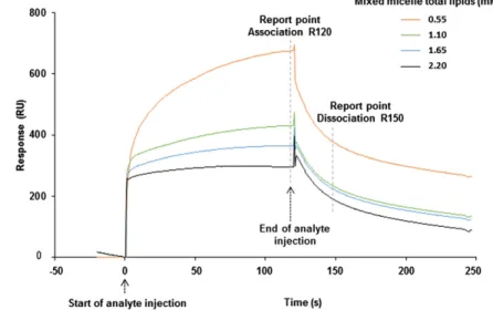
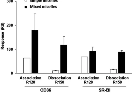
Protein purity of the SRs was fi rst confi rmed by Coo-massie Blue gel ( Fig. 1 , inset). Typical SPR sensorgrams illustrating micelle binding with a SR are shown in Fig. 2 . Micelle capacity to bind and to dissociate from either lSR-BI or lCD36 was dependent on their total lipid composi-tion. Moreover, broad differences were observed between simple micelles containing cholesterol and taurocholate only, and more complex micelles made with a mixture of lipids as found in the gut lumen in postprandial con-ditions (i.e., mixed micelles composed of cholesterol, phospholipids, lysophospholipids, monoolein, OA, and taurocholate) ( Fig. 3 ). The binding of the simple micelles to lCD36 and lSR-BI after a contact time of 120 s (R120) was 3.9-fold and 1.6-fold lower than the binding observed with the mixed micelles. Furthermore, percentage of dis-sociation at 150 s was drastically higher for simple micelles than mixed micelles: 85.0 and 76.3% of ligands dissociated from lCD36 and lSR-BI, respectively, with simple micelles versus 38.0 and 12.9% with mixed micelles ( Fig. 3 ). Simi-lar results were obtained using simple micelles composed with either 0.04 mM of phospholipid or 0.5 mM of OA and taurocholate (data not shown) compared with mixed mi-celles. These results indicated that the SRs have a better
Fig. 2. Sensorgrams comparing abilities of mixed micelles at different dilutions to interact with CD36. Mixed micelles were prepared as described in the Ma-terial and Methods. The sensorgrams illustrate the in-teraction responses obtained when different dilutions of mixed micelles were injected over lCD36. Mixed micelles differed by their lipid concentrations from 0.55 to 2.2 mM. The landmarks used to compare bind-ing (R120) and dissociation (R150) responses between two analytes are represented by a dashed line.
at INRA Institut National de la Recherche Agronomique, on November 18, 2015
www.jlr.org
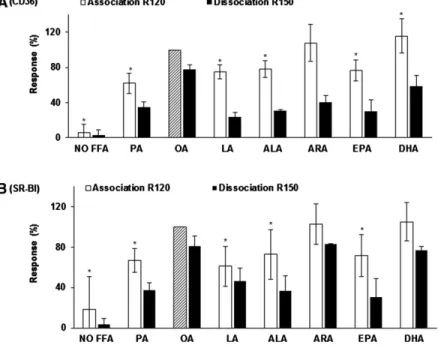
compared with OA micelles ( ⫺ 94.4 ± 9.7% and ⫺ 81.3 ± 32.4%, respectively, P < 0.05). The presence of PA, LA, ALA, or EPA instead of OA also signifi cantly decreased mixed micelle binding to both lCD36 and lSR-BI (up to ⫺ 39%, P < 0.05). Interestingly, the presence of DHA sig-nifi cantly increased micelle binding to lCD36 compared with OA micelles. R150 analyses showed that micelles con-taining either LA, ALA, ARA, EPA, or DHA dissociated more rapidly from lCD36 than the micelles containing OA. Similarly, micelles containing either PA, ALA, or EPA dissociated more from SR-BI than the OA micelles. Our results thus highlighted that micelle binding to CD36 was positively correlated to both chain length (Rho = 0.73; P = 0.0006) and unsaturation (Rho = 0.60; P = 0.0053). Simi-larly, micelle binding to SR-BI was also positively corre-lated to both chain length (Rho = 0.58; P = 0.0062) and unsaturation (Rho = 0.45; P = 0.0338).
As OA and DHA appeared to increase micellar affi nity for the SRs, we then determined their binding affi nity con-stants with both lSR-BI and lCD36 using the single fatty acid. As molar concentrations were known, kinetic analysis was performed. The average apparent equilibrium disso-ciation constant (K D ) for OA was 349 ± 1 M and 520 ±
2 M for SR-BI and CD36, respectively. Surprisingly, the K D
for DHA was about 100-fold lower, at just 2.20 ± 0.01 M and 3.20 ± 0.03 M for lSR-BI and lCD36, respectively ( Fig. 7 ).
ANOVA
Overall, univariate ANOVA ( Table 2 ) fi rst indicated that phospholipid concentration and fatty acid type, but not cholesterol concentration, impacted micellar electric charge ( Table 3 ). Particle size (Table 3) was impacted by fatty acid type only ( Table 2 ).
Univariate ANOVA also indicated that cholesterol and phospholipid concentrations, as well as free fatty acid type, were signifi cantly correlated with both lCD36 and lSR-BI R120 and R150, explaining between 50 and 90% of the variance of levels. Particle size and surface electric charge were also moderately but signifi cantly there was higher dissociation at 0.2 mM cholesterol (54.4
± 5.0% vs. 26.9 ± 14.4% in the control condition, P < 0.05).
Effect of micellar phospholipid concentration on micelle interactions with lCD36 and lSR-BI
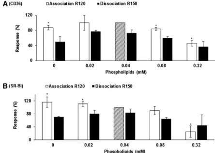
Micelle binding to lCD36 was signifi cantly altered at high phospholipid concentrations (0.08–0.32 mM) but also when micellar composition was phospholipid-free ( Fig.
5A ). Increasing the phospholipid concentration resulted in a linear decrease in mixed-micelle binding to lSR-BI (Rho = ⫺ 0.91, P = 0.0007; Fig. 5B ). Interestingly, the percentages of micelles dissociated were increased for both SRs in the ab-sence of phospholipid compared with the control condi-tion (41.7 ± 20.7% vs. 27.1 ± 7.8% and 38.1 ± 8.0% vs. 16.3 ± 11.8% for lCD36 and lSR-BI, respectively), although the dif-ference was only signifi cant for lSR-BI.
Effect of micellar free fatty acid type on micelle interactions with lCD36 and lSR-BI
As shown in Fig. 6 , the absence of fatty acid drasti-cally decreased micelle binding to both lCD36 and lSR-BI
Fig. 3. Comparison between mixed micelle and cholesterol-taurocholate micelle interactions with lCD36 and lSR-BI. Mixed micelles (white bars) were composed of 0.5 mM OA, 0.16 mM lysocholine, 0.3 mM monoolein, 0.04 mM phosphatidyl-choline, 0.1 mM cholesterol, and 5 mM taurocholate. Cholesterol-taurocholate micelles (simple micelles, black bars) were composed of 0.1 mM cholesterol and 5 mM taurocholate. Data are means ± SD of three assays with three independent micelle batches. Mi-celle binding (R120) and dissociation (R150) were measured as presented in the Material and Methods.
TABLE 1. Size and zeta potential of mixed micelles at various concentrations
Mixed Micelle Taurocholate Concentration (mM)
Mixed Micelle Lipid
Concentration (mM) Size (nm) Zeta Potential (mV) 2.5 0.55 6.0 ± 1.6 ⫺ 37.7 ± 3.0 3 0.66 6.7 ± 1.2 a ⫺ 38.7 ± 3.4 4 0.88 5.0 ± 1.3 ⫺ 37.2 ± 0.5 5 (control) 1.10 3.9 ± 1.1 ⫺ 32.3 ± 4.0 6 1.32 3.5 ± 0.2 ⫺ 36.7 ± 1.2 7 1.54 3.0 ± 0.0 a ⫺ 28.9 ± 9.2 7.5 1.65 2.9 ± 0.3 a ⫺ 38.5 ± 0.8 a 8 1.76 3.1 ± 0.0 a ⫺ 36.6 ± 1.5 9 1.98 2.7 ± 0.0 a ⫺ 38.5 ± 0.1 a 10 2.20 2.6 ± 0.1 a ⫺ 34.1 ± 3.5
The intensity-weighted mean radius and the zeta potential of the mixed micelles at various dilutions were determined by photon correlation spectroscopy at 25°C. Data are means ± SD of three assays.
a
A significant difference ( P < 0.05) with the control (assay
performed with mixed micelles at 5 mM taurocholate and 0.16 mM lysophosphatidylcholine, 0.3 mM monoolein, 0.04 mM phosphatidylcholine and 0.5 mM OA).
at INRA Institut National de la Recherche Agronomique, on November 18, 2015
www.jlr.org
Effect of micellar lipid content on vitamin D uptake by Caco-2 cells
The different mixed micelle lots were enriched with 0.5 M vitamin D, and vitamin D uptake was assayed in Caco-2 differentiated monolayers. Results showed that choles-terol, phospholipids, and fatty acid micellar content sig-nifi cantly impacted vitamin D uptake by cells ( Fig. 8 ). In particular, vitamin D uptake was decreased by high phos-pholipid (up to 69%, P < 0.05) and cholesterol (up to 61%, P < 0.05) micellar concentrations. It was also im-paired in the presence of very long chain polyunsaturated fatty acids compared with OA ( ⫺ 7, ⫺ 19, and ⫺ 20% with ARA, EPA, and DHA, respectively; P < 0.05). When phos-pholipid micellar content was modulated, vitamin D up-take was positively correlated to lSR-BI R120 (Rho = 0.906, P = 0.0007). It was also positively correlated to R150 associated with lCD36 R120 and R150. Finally, micellar
electric charge was modestly but signifi cantly associated with lSR-BI binding and dissociation, whereas particle size had no effect.
Multivariate analysis ( Table 4 ) then showed that free fatty acid type had the biggest effect on micellar electric charge, while both cholesterol concentration and type of free fatty acid were associated with particle size.
Association on (R120) and dissociation from (R150) lCD36 were more impacted by type of free fatty acid than by micellar cholesterol and phospholipid concentrations. In contrast, ligand dissociation from lSR-BI (R120) was mainly affected by phospholipid concentration and fatty acid type, while the dissociation process (R150) was more affected by cholesterol concentration and free fatty acid type than by phospholipid concentration.
Fig. 4. Effect of micellar cholesterol concentration on micelle interaction with lCD36 (A) and lSR-BI (B ). All micelles were composed of 0.16 mM lysophos-phatidylcholine, 0.3 mM monoolein, 0.04 mM phosphatidylcholine, 0.5 mM OA, and 5 mM sodium taurocholate. Cholesterol concentration ranged from 0 to 0.3 mM. Micelle binding (R120) and dissociation (R150) were measured according to the Material and Methods. Data are means ± SD of three assays on three different NTA chips with three independent micelle batches. An asterisk indicates a signifi cant difference ( P < 0.05) in binding compared with the control (mixed micelle at 0.1 mM of cholesterol stan-dardized at 100%, hatched bar).
Fig. 5. Effect of micellar phospholipid concentra-tion on mixed-micelle interacconcentra-tions with lCD36 (A) and lSR-BI (B). All micelles were composed of 0.16 mM ly-sophosphatidylcholine, 0.3 mM monoolein, 0.1 mM cholesterol, 0.5 mM OA, and 5 mM sodium taurocho-late. Phospholipid concentration ranged from 0 to 0.32 mM. Micelle binding (R120) and dissociation (R150) were measured according to the Material and Methods. Data are means ± SD of three assays on three different NTA chips with three independent micelle batches. An asterisk indicates a signifi cant difference ( P < 0.05) compared with the control (mixed micelle at 0.04 mM of phospholipid standardized at 100%, hatched bar).
at INRA Institut National de la Recherche Agronomique, on November 18, 2015
www.jlr.org
proteins. The extracellular loops of SR-BI and CD36 represent 81 and 87% of the proteins (419 and 409 aas), respectively. The deletion of the two transmembrane domains (20 and 19 aas for SR-BI; 23 and 24 aas for CD36) and of the intracellular domains (12 and 37 aas for SR-BI; 6 and 9 aas for CD36) may impact the protein functioning. However, a recent work showed that critical subdomains for receptor-ligand interactions and transport function of SR-BI were located in the extracellular domain ( 29 ).
Mixed micelles were formulated in order to mimic phys-iological conditions and to represent lipid ratios that can be found in the human duodenum (i.e., total bile acid ⵑ 5–10 mM; cholesterol/lysophospholipids ⵑ 0.5 mM; cholesterol/monoglycerides ⵑ 0.4 mM ) ( 25, 30 ), as well as to form micelles mainly according to photon correlation spectroscopy data. Sodium taurocholate appears to con-tinuously self-aggregate over a large range of concentra-tion ( 31 ), but a critical step of aggregaconcentra-tion was shown to occur around 2–2.5 mM in the running buffer as well as in DMEM (data not shown), according to previous report ( 32 ). In the presence of lipids, such as lysophospholipids, critical micellar concentration is usually further reduced ( 33 ). These data support our observation that in our con-ditions, the mixture taurocholate-lipids is mainly present as micelles. These micelles were stable for several hours at room temperature, as determined by photon correlation spectroscopy (data not shown).
(Rho = 0.666, P = 0.0128). This suggested that the more the micelles were binding to lSR-BI, the higher was the uptake of vitamin D. When cholesterol or fatty acid micellar con-tent was modifi ed, vitamin D uptake was negatively corre-lated to lSR-BI R120 (Rho = ⫺ 0.461, P = 0.0478; Rho = ⫺ 0.489, P = 0.0191; for cholesterol and fatty acids, respectively). This suggested that the more the micelles were binding to lSR-BI, the lower was the uptake of vitamin D, and likely highlighted a competition between vitamin D and choles-terol or fatty acids as SR-BI ligands .
DISCUSSION
The molecular mechanisms underpinning the interac-tion between mixed micelles and the SRs CD36 and SR-BI at the brush border membrane level are poorly under-stood. Here, the SPR technique was used for the fi rst time to detect real-time molecular interactions between mobile complex lipid structures, i.e., micelles, and the immobi-lized extracellular loop of CD36 and SR-BI. Because this methodology strictly detects mass, there is no need to label the interacting components, thus ruling out potential al-terations of their molecular properties. We used SR extra-cellular loops and not entire proteins in order to avoid solubility issues as well as nonspecifi c binding of the hy-drophobic analytes to the transmembrane domains of the
Fig. 6. Effect of micellar free fatty acid type on mixed-micelle interactions with lCD36 (A) and lSR-BI (B). All mixed micelles were composed of 0.16 mM ly-sophosphatidylcholine, 0.3 mM monoolein, 0.04 mM phosphatidylcholine, 0.1 mM cholesterol, and 5 mM sodium taurocholate. Only the free fatty acid composi-tion differed between the different condicomposi-tions: no free fatty acid or 0.5 mM of either PA, OA, LA, ALA, ARA, EPA, or DHA. Micelle binding (R120) and dissociation (R150) were measured according to the Material and Methods. Data are means ± SD of three assays on three different NTA chips with three independent micelle batches. An asterisk indicates a signifi cant difference ( P < 0.05) compared with the control (mixed micelle with OA standardized at 100%, hatched bar).
Fig. 7. Determination of binding affi nity constants (K D ) by nonlinear regression fi tting of a one-site
binding model to the SPR data. Fatty acid propan-1-ol solutions from 0.01 to 1 mM were prepared by dissolv-ing 1% of propan-1-ol containdissolv-ing an adequate quan-tity of fatty acid in running buffer. A: Response at equilibrium (R eq ) of OA with lCD36 and lSR-BI. B:
Response at equilibrium (R eq ) of DHA with lCD36 and
lSR-BI affi nity. Filled circles, SRB1; open circles, CD36.
at INRA Institut National de la Recherche Agronomique, on November 18, 2015
www.jlr.org
range of cholesterol concentrations, whereas micelle bind-ing to CD36 was optimal for a cholesterol concentration of around 0.1 mM. This suggests a complementary role of the two proteins during the digestion process, depending on micellar lipid composition. The interaction of micellar cholesterol with brush border membrane vesicles has been previously studied ( 20 ). The authors showed that although Niemann-Pick C1-like 1 has been described as the key me-diator of intestinal cholesterol uptake, the active micelle binding was mediated by SR-BI, according to results ob-tained in Caco-2 cells ( 13 ). Here, the fact that both lSR-BI and lCD36 could effi ciently bind mixed micelles is consis-tent with these data.
We then explored the effect of modulating micellar phosphatidylcholine concentrations on micelle interac-tions with the transporter extracellular loops. Both SR-BI and CD36 were primarily shown to be receptors for an-ionic phospholipids such as phosphatidylserine, but not for phosphatidylcholine ( 35 ). The specifi c binding of phosphatidylserine compared with other phospholipids was confi rmed later by a solid-phase assay using lipid-coated plates ( 36 ). Our data, which show a decrease in micelle-SR loop interaction when phosphatidylcholine mi-cellar content was increased, are in agreement with these results. Moreover, it has been clearly shown by several teams that phospholipids can impair the intestinal absorp-tion of cholesterol or other lipid micronutrients [for re-view see ( 1 )]. The inhibitory effect of phospholipids on other micellar lipid absorption may be due to a decrease in micellar affi nity for their membrane transporters, as high micellar phospholipid concentrations inhibited mi-celle binding to both SR-BI and CD36 extracellular loops.
We also explored the effect of modulating micellar fatty acid type on micelle interactions with the SR loops and showed that free fatty acids were fundamental compo-nents of the structure. Interestingly, micelle binding to both transporters was positively correlated to chain length and unsaturation. This is in line with the concept of long-chain fatty acids being CD36 ligands ( 37 ). Moreover, A key fi nding is that these structures mimicking mixed
micelles, i.e., including various lipids and lipid digestion products, are necessary for optimal binding to both lCD36 and lSR-BI. This is consistent with previous results showing that SR-BI could bind postprandial but not interprandial micelles ( 13 ), and that cholesterol uptake from simple mi-celles was unaffected by the addition of SR-BI inhibitor, in contrast to cholesterol uptake from mixed micelles ( 34 ).
To more specifi cally evaluate the effect of micellar lipid composition on micelle binding to the SRs, we fi rst mod-ulated their cholesterol content, as both proteins may participate partly in cholesterol transport ( 3, 12 ). Mixed-micelle binding to SR-BI was not modifi ed within a wide
TABLE 2. Univariate ANOVA
Explanatory Variable N
Dependent Variables, Model R 2
Zeta Size
lCD36 lSR-BI
R120 R150 R120 R150
Cholesterol ( Fig. 4 ) 18 0.17 0.62 0.91 a 0.67 c 0.66 c 0.75 b
Phospholipids ( Fig. 5 ) 15 0.72 b 0.42 0.85 a 0.78 b 0.91 a 0.52
Fatty acids ( Fig. 6 ) 24 0.79 a 0.74 b 0.90 a 0.92 a 0.72 b 0.90 a
Zeta ( Table 3 ) 57 — 0.03 0.17 b 0.12 c 0.11 c 0.01
Size ( Table 3 ) 57 0.03 — 0.25 a 0.10 c 0.03 0.08 The intensity-weighted mean radius and the zeta potential of the mixed micelles were determined by photon correlation spectroscopy at 25°C. Micelle binding (R120) and dissociation (R150) were measured as presented in the Material and Methods. Results expressed in R-square, e.g., 17% of the variation of zeta was explained by the variation of cholesterol concentration in the micelles.
a P < 0.001. b P < 0.01. c P < 0.05.
TABLE 3. Size and zeta potential of mixed micelles depending on their lipid content
Size (nm) Zeta Potential (mV)
Micellar cholesterol (mM) 0 4.4 ± 0.4 a ⫺ 38.2 ± 0.9 a 0.05 5.3 ± 1.1 a ⫺ 32.3 ± 4.0 0.1 (control) 3.4 ± 0.1 ⫺ 35.1 ± 1.8 0.15 6.1 ± 0.5 a ⫺ 30.3 ± 4.5 0.2 34.4 ± 23.3 a ⫺ 35.5 ± 1.3 0.3 28.2 ± 7.2 a ⫺ 35.5 ± 1.8 Micellar phospholipids (mM) 0 4.2 ± 0.4 ⫺ 35.9 ± 1.3 0.02 4.6 ± 1.2 ⫺ 28.2 ± 3.0 a 0.04 (control) 3.8 ± 0.5 ⫺ 37.4 ± 1.1 0.08 4.8 ± 1.0 ⫺ 34.5 ± 3.6 0.32 6.8 ± 1.9 a ⫺ 31.2 ± 2.7 a Fatty acids 0 28.8 ± 16.5 a ⫺ 14.8 ± 1.1 a PA 5.1 ± 1.7 ⫺ 35.5 ± 4.5 OA (control) 3.9 ± 0.9 ⫺ 37.2 ± 2.3 LA 3.5 ± 1.6 ⫺ 30.2 ± 2.8 a ALA 2.8 ± 0.1 a ⫺ 29.6 ± 2.5 a ARA 3.6 ± 1.5 ⫺ 30.9 ± 5.6 EPA 3.8 ± 1.1 ⫺ 26.9 ± 5.9 a DHA 3.9 ± 1.8 ⫺ 29.7 ± 4.5 a
Mixed micelles were composed of 5 mM taurocholate, 0.16 mM lysophosphatidylcholine, 0.3 mM monoolein, 0.04 mM phospha-tidylcholine or another phosphaphospha-tidylcholine concentration if indicated, 0.1 mM cholesterol or another cholesterol concentration if indicated, and 0.5 mM OA or another fatty acid if indicated. The intensity-weighted mean radius and the zeta potential of the mixed micelles were determined by photon correlation spectroscopy at 25°C. Data are
at INRA Institut National de la Recherche Agronomique, on November 18, 2015
www.jlr.org
tivariate analysis of all experiments showed that surface electric charge of the mixed micelles, and to a lesser extent micellar size, impacted micelle interactions with the SR loops. This is consistent with new data on CD36 and SR-BI structures that recently emerged from the crystallization of another SR family member, lysosomal integral membrane intestinal level displayed an increased uptake of labeled
fatty acids compared with wild-type mice ( 12 ).
Overall, the lipid composition of mixed micelles clearly affects their ability to interact with the extracellular loops of both SRs. However, this is not the only parameter play-ing a key role in such an interaction. Univariate and
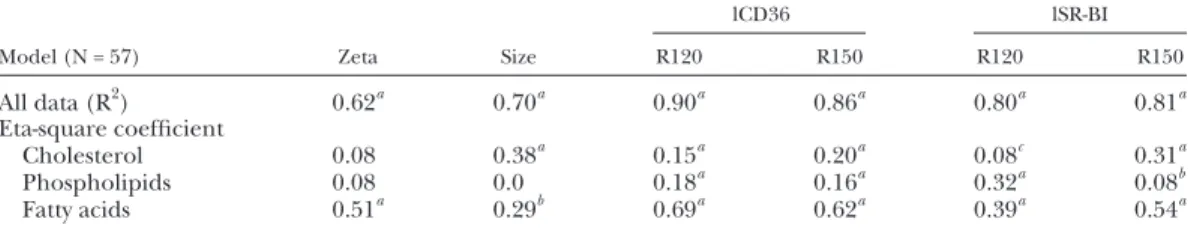
TABLE 4. Multivariate ANOVA
Model (N = 57) Zeta Size
lCD36 lSR-BI
R120 R150 R120 R150
All data (R 2 ) 0.62 a 0.70 a 0.90 a 0.86 a 0.80 a 0.81 a
Eta-square coeffi cient
Cholesterol 0.08 0.38 a 0.15 a 0.20 a 0.08 c 0.31 a
Phospholipids 0.08 0.0 0.18 a 0.16 a 0.32 a 0.08 b
Fatty acids 0.51 a 0.29 b 0.69 a 0.62 a 0.39 a 0.54 a
The intensity-weighted mean radius and the zeta potential of the mixed micelles were determined by photon correlation spectroscopy at 25°C. Micelle binding (R120) and dissociation (R150) were measured as presented in the Material and Methods. Data of the complete model are expressed in R-square, i.e., 62% of the variation of zeta was explained by the combined effect of cholesterol concentration, phospholipid type, and fatty acid composition in the micelles. Eta-square coeffi cient corresponds to the semi partial effect of either cholesterol, phospholipids, or fatty acids in the model, i.e., adjusted for variations of the two other explanatory factors.
a P < 0.001. b P < 0.01. c P < 0.05.
Fig. 8. Effect of lipid micellar content on vitamin D uptake by Caco-2 cells. The apical sides were incu-bated for 60 min with synthetic mixed micelles en-riched with 0.5 M cholecalciferol. The basolateral sides received FBS-free medium. Data are means ± SD of three assays. An asterisk indicates a signifi cant dif-ference ( P < 0.05) with the controls (hatched bars). A: Effect of micellar phospholipid concentration. B: Effect of micellar cholesterol concentration. C: Effect of fatty acid substitution.
at INRA Institut National de la Recherche Agronomique, on November 18, 2015
www.jlr.org
protein II (LIMPII) ( 38 ). The authors showed that the helical bundle face and the apex region forming the head of both proteins were important for ligand binding. We suggest that optimally sized structures would bind more effi ciently to this specifi c area than other structures. More-over, it was also shown that the apex regions of both human SR-BI and CD36 show a signifi cant accumulation of cationic residue ( 38 ). An electrostatic interaction at the apex thus likely contributes to the binding of these receptors to their ligands. Mixed micelles being negatively charged, modifi ca-tions in their lipid composition induce surface electric charge variations that may participate in the modulation of their binding to and dissociation from the SRs.
In order to correlate our SPR data with results in living cells, we performed micellar vitamin D uptake experiment in Caco-2 cells. These cells were specifi cally chosen because: i ) they are resistant mixed micelles (unlike other cell types such as HEK or CHO cells, cytotoxicity tests not shown); ii ) they highly express SR-BI and do not express CD36 ( 25 ); and iii ) micelle binding to their apical membranes is SR-BI-depen-dent ( 20 ). Vitamin D uptake was signifi cantly correlated with micelle binding to SR-BI. In particular, vitamin D uptake di-minished with increasing micellar phospholipid contents, in accordance with the observation that phospholipids inhibit micelle association to SR-BI. Vitamin D uptake also dimin-ished with increasing micellar cholesterol content, and it was negatively correlated to micelle binding to SR-BI. This likely refl ects the fact that cholesterol is a good ligand for SR-BI that directly competes with vitamin D for uptake ( 38 ). Simi-larly, vitamin D uptake also diminished when very long chain fatty acids were substituted to OA and it was negatively cor-related to micelle binding to SR-BI again. This is in agree-ment with our SPR data showing that long chain fatty acids like DHA are better ligands for SR-BI than OA and DHA may compete with vitamin D for uptake/binding.
Our SPR model presents several limits. First, this is an arti-fi cial system and extrapolations to physiological observations should be precautious. Second, the unstirred water mono-layer of the brush border membrane cannot be modeled. Third, the use of a single protein does not represent full com-plexity of the brush border membrane surface, and so any possible cooperation between two or more membrane pro-teins can be evaluated. However, the SRP technique allowed the molecular interactions occurring between mixed mi-celles and intestinal SR extracellular loops to be character-ized for the fi rst time. The use of complex structures is particularly relevant regarding this type of study. Indeed, when OA or DHA was injected alone, the K D of DHA for the
receptors was about 100-fold smaller than the K D of OA,
showing that isolated molecules behave very differently from molecules incorporated in a physiological vehicle. However, it is noteworthy that both K D s were under the range of
con-centrations of fatty acids found in the gut ( 30 ). The smaller K D of DHA compared with the K D of OA was also in
concor-dance with the reduced intake of polyunsaturared n-3 com-pared with monounsaturated fatty acids ( 39 ).
Further investigations on micelle binding to other
intesti-terocyte apical membrane. Further work is also necessary to evaluate other luminal lipid vehicles [vesicles, proteins, etc. ( 1 )] binding to the membrane transport proteins.
Overall, our results show for the fi rst time that the extra-cellular loops of CD36 and SR-BI are effi cient receptors for intestinal mixed micelles. We also demonstrate that micellar lipid composition and micellar properties such as size and electric charge of the particles are key factors gov-erning micelle interactions with intestinal SRs.
The authors are very grateful to Pr. Joël Chopineau and Dr. Maya Allouche for their valuable help and discussions.
REFERENCES
1 . Reboul , E. , and P. Borel . 2011 . Proteins involved in uptake, intra-cellular transport and basolateral secretion of fat-soluble vitamins and carotenoids by mammalian enterocytes. Prog. Lipid Res. 50 : 388 – 402 .
2 . Goncalves , A. , M. Margier , S. Roi , X. Collet , I. Niot , P. Goupy , C. Caris-Veyrat , and E. Reboul . 2014 . Intestinal scavenger recep-tors are involved in vitamin K1 absorption. J. Biol. Chem. 289 : 30743 – 30752 .
3 . Nassir , F. , B. Wilson , X. Han , R. W. Gross , and N. A. Abumrad . 2007 . CD36 is important for fatty acid and cholesterol uptake by the proximal but not distal intestine. J. Biol. Chem. 282 : 19493 – 19501 . 4 . Lobo , M. V. , L. Huerta , N. Ruiz-Velasco , E. Teixeiro , P. de la
Cueva , A. Celdran , A. Martin-Hidalgo , M. A. Vega , and R. Bragado . 2001 . Localization of the lipid receptors CD36 and CLA-1/SR-BI in the human gastrointestinal tract: towards the identifi cation of receptors mediating the intestinal absorption of dietary lipids. J.
Histochem. Cytochem. 49 : 1253 – 1260 .
5 . Werder , M. , C. H. Han , E. Wehrli , D. Bimmler , G. Schulthess , and H. Hauser . 2001 . Role of scavenger receptors SR-BI and CD36 in selective sterol uptake in the small intestine. Biochemistry . 40 : 11643 – 11650 .
6 . Schwartz , G. J. , J. Fu , G. Astarita , X. Li , S. Gaetani , P. Campolongo , V. Cuomo , and D. Piomelli . 2008 . The lipid messenger OEA links dietary fat intake to satiety. Cell Metab. 8 : 281 – 288 .
7 . Nauli , A. M. , F. Nassir , S. Zheng , Q. Yang , C. M. Lo , S. B. Vonlehmden , D. Lee , R. J. Jandacek , N. A. Abumrad , and P. Tso . 2006 . CD36 is important for chylomicron formation and secretion and may mediate cholesterol uptake in the proximal intestine. Gastroenterology . 131 : 1197 – 1207 .
8 . Nguyen , D. V. , V. A. Drover , M. Knopfel , P. Dhanasekaran , H. Hauser , and M. C. Phillips . 2009 . Infl uence of class B scavenger receptors on cholesterol fl ux across the brush border membrane and intestinal absorption. J. Lipid Res. 50 : 2235 – 2244 .
9 . Tran , T. T. , H. Poirier , L. Clement , F. Nassir , M. M. Pelsers , V. Petit , P. Degrace , M. C. Monnot , J. F. Glatz , N. A. Abumrad , et al . 2011 . Luminal lipid regulates CD36 levels and downstream signaling to stimulate chylomicron synthesis. J. Biol. Chem. 286 : 25201 – 25210 . 10 . Masuda , D. , K. Hirano , H. Oku , J. C. Sandoval , R. Kawase , M.
Yuasa-Kawase , Y. Yamashita , M. Takada , K. Tsubakio-Yamamoto , Y. Tochino , et al . 2009 . Chylomicron remnants are increased in the postprandial state in CD36 defi ciency. J. Lipid Res. 50 : 999 – 1011 . 11 . Hauser , H. , J. H. Dyer , A. Nandy , M. A. Vega , M. Werder , E.
Bieliauskaite , F. E. Weber , S. Compassi , A. Gemperli , D. Boffelli , et al . 1998 . Identifi cation of a receptor mediating absorption of dietary cholesterol in the intestine. Biochemistry . 37 : 17843 – 17850 . 12 . Bietrix , F. , D. Yan , M. Nauze , C. Rolland , J. Bertrand-Michel , C.
Comera , S. Schaak , R. Barbaras , A. K. Groen , B. Perret , et al . 2006 . Accelerated lipid absorption in mice overexpressing intestinal SR-BI. J. Biol. Chem. 281 : 7214 – 7219 .
13 . Béaslas , O. , C. Cueille , F. Delers , D. Chateau , J. Chambaz , M. Rousset , and V. Carrière . 2009 . Sensing of dietary lipids by
at INRA Institut National de la Recherche Agronomique, on November 18, 2015
www.jlr.org
2013 . Scavenger receptor class B type I is a plasma membrane cho-lesterol sensor. Circ. Res. 112 : 140 – 151 .
15 . Acton , S. , A. Rigotti , K. T. Landschulz , S. Xu , H. H. Hobbs , and M. Krieger . 1996 . Identifi cation of scavenger receptor SR-BI as a high density lipoprotein receptor. Science . 271 : 518 – 520 .
16 . Williams , D. L. , M. de La Llera-Moya , S. T. Thuahnai , S. Lund-Katz , M. A. Connelly , S. Azhar , G. M. Anantharamaiah , and M. C. Phillips . 2000 . Binding and cross-linking studies show that scaven-ger receptor BI interacts with multiple sites in apolipoprotein A-I and identify the class A amphipathic alpha-helix as a recognition motif. J. Biol. Chem. 275 : 18897 – 18904 .
17 . Thuahnai , S. T. , S. Lund-Katz , D. L. Williams , and M. C. Phillips . 2001 . Scavenger receptor class B, type I-mediated uptake of various lipids into cells. Infl uence of the nature of the donor particle inter-action with the receptor. J. Biol. Chem. 276 : 43801 – 43808 .
18 . Calvo , D. , D. Gomez-Coronado , Y. Suarez , M. A. Lasuncion , and M. A. Vega . 1998 . Human CD36 is a high affi nity receptor for the native lipoproteins HDL, LDL, and VLDL. J. Lipid Res. 39 : 777 – 788 . 19 . Tait , J. F. , and C. Smith . 1999 . Phosphatidylserine receptors: role of CD36 in binding of anionic phospholipid vesicles to monocytic cells. J. Biol. Chem. 274 : 3048 – 3054 .
20 . Labonté , E. D. , P. N. Howles , N. A. Granholm , J. C. Rojas , J. P. Davies , Y. A. Ioannou , and D. Y. Hui . 2007 . Class B type I scaven-ger receptor is responsible for the high affi nity cholesterol bind-ing activity of intestinal brush border membrane vesicles. Biochim.
Biophys. Acta . 1771 : 1132 – 1139 .
21 . van Bennekum , A. , M. Werder , S. T. Thuahnai , C. H. Han , P. Duong , D. L. Williams , P. Wettstein , G. Schulthess , M. C. Phillips , and H. Hauser . 2005 . Class B scavenger receptor-mediated intestinal ab-sorption of dietary beta-carotene and cholesterol. Biochemistry . 44 : 4517 – 4525 .
22 . Yu , M. , T. Y. Lau , S. A. Carr , and M. Krieger . 2012 . Contributions of a disulfi de bond and a reduced cysteine side chain to the intrinsic activity of the high-density lipoprotein receptor SR-BI. Biochemistry . 51 : 10044 – 10055 .
23 . Viñals , M. , S. Xu , E. Vasile , and M. Krieger . 2003 . Identifi cation of the N-linked glycosylation sites on the high density lipoprotein (HDL) receptor SR-BI and assessment of their effects on HDL binding and selective lipid uptake. J. Biol. Chem. 278 : 5325 – 5332 . 24 . Graciet , E. , P. Gans , N. Wedel , S. Lebreton , J. M. Camadro , and B.
Gontero . 2003 . The small protein CP12: a protein linker for supra-molecular complex assembly. Biochemistry . 42 : 8163 – 8170 . 25 . Reboul , E. , L. Abou , C. Mikail , O. Ghiringhelli , M. Andre , H.
Portugal , D. Jourdheuil-Rahmani , M. J. Amiot , D. Lairon , and P. Borel . 2005 . Lutein transport by Caco-2 TC-7 cells occurs partly by a facilitated process involving the scavenger receptor class B type I (SR-BI). Biochem. J. 387 : 455 – 461 .
26 . Reboul , E. , A. Goncalves , C. Comera , R. Bott , M. Nowicki , J. F. Landrier , D. Jourdheuil-Rahmani , C. Dufour , X. Collet , and P. Borel . 2011 . Vitamin D intestinal absorption is not a simple passive diffusion: evidences for involvement of cholesterol transporters. Mol. Nutr. Food Res. 55 : 691 – 702 .
27 . Goncalves , A. , B. Gleize , R. Bott , M. Nowicki , M. J. Amiot , D. Lairon , P. Borel , and E. Reboul . 2011 . Phytosterols can impair vita-min D intestinal absorption in vitro and in mice. Mol. Nutr. Food Res. 55 ( Suppl 2 ): S303 – S311 .
28 . Goncalves , A. , B. Gleize , S. Roi , M. Nowicki , A. Dhaussy , A. Huertas , M. J. Amiot , and E. Reboul . 2013 . Fatty acids affect micellar prop-erties and modulate vitamin D uptake and basolateral effl ux in Caco-2 cells. J. Nutr. Biochem. 24 : 1751 – 1757 .
29 . Kartz , G. A. , R. L. Holme , K. Nicholson , and D. Sahoo . 2014 . SR-BI/ CD36 chimeric receptors defi ne extracellular subdomains of SR-BI critical for cholesterol transport. Biochemistry . 53 : 6173 – 6182 . 30 . Mansbach 2nd , C. M. , R. S. Cohen , and P. B. Leff . 1975 . Isolation
and properties of the mixed lipid micelles present in intestinal con-tent during fat digestion in man. J. Clin. Invest. 56 : 781 – 791 . 31 . Spivak , W. , C. Morrison , D. Devinuto , and W. Yuey . 1988 .
Spectrophotometric determination of the critical micellar con-centration of bile salts using bilirubin monoglucuronide as a mi-cellar probe. Utility of derivative spectroscopy. Biochem. J. 252 : 275 – 281 .
32 . Morita , S. Y. , A. Kobayashi , Y. Takanezawa , N. Kioka , T. Handa , H. Arai , M. Matsuo , and K. Ueda . 2007 . Bile salt-dependent effl ux of cellular phospholipids mediated by ATP binding cassette protein B4. Hepatology . 46 : 188 – 199 .
33 . DeLong , L. J. , and J. W. Nichols . 1996 . Time-resolved fl uorescence anisotropy of fl uorescent-labeled lysophospholipid and taurode-oxycholate aggregates. Biophys. J. 70 : 1466 – 1471 .
34 . Reboul , E. , Z. Soayfane , A. Goncalves , M. Cantiello , R. Bott , M. Nauze , F. Terce , X. Collet , and C. Comera . 2012 . Respective contri-butions of intestinal Niemann-Pick C1-like 1 and scavenger recep-tor class B type I to cholesterol and tocopherol uptake: in vivo v. in vitro studies. Br. J. Nutr. 107 : 1296 – 1304 .
35 . Rigotti , A. , S. L. Acton , and M. Krieger . 1995 . The class B scavenger receptors SR-BI and CD36 are receptors for anionic phospholipids. J. Biol. Chem. 270 : 16221 – 16224 .
36 . Kawasaki , Y. , A. Nakagawa , K. Nagaosa , A. Shiratsuchi , and Y. Nakanishi . 2002 . Phosphatidylserine binding of class B scavenger receptor type I, a phagocytosis receptor of testicular Sertoli cells. J.
Biol. Chem. 277 : 27559 – 27566 .
37 . Drover , V. A. , D. V. Nguyen , C. C. Bastie , Y. F. Darlington , N. A. Abumrad , J. E. Pessin , E. London , D. Sahoo , and M. C. Phillips . 2008 . CD36 mediates both cellular uptake of very long chain fatty acids and their intestinal absorption in mice. J. Biol. Chem. 283 : 13108 – 13115 .
38 . Neculai , D. , M. Schwake , M. Ravichandran , F. Zunke , R. F. Collins , J. Peters , M. Neculai , J. Plumb , P. Loppnau , J. C. Pizarro , et al . 2013 . Structure of LIMP-2 provides functional insights with implications for SR-BI and CD36. Nature . 504 : 172 – 176 .
39 . Rojo-Martinez , G. , F. J. Soriguer , S. Gonzalez-Romero , F. Tinahones , F. Moreno , S. R. de Adana , M. J. Garriga , I. Esteva , J. Garcia-Arnes , J. M. Gomez-Zumaquero , et al. 2000 . Serum leptin and habitual fatty acid dietary intake in patients with type 1 diabetes mellitus. Eur. J. Endocrinol. 142: 263–268.
at INRA Institut National de la Recherche Agronomique, on November 18, 2015
www.jlr.org