Converging Biochemical Pathways in Psychiatric Disorders by
Takahiro Soda
B.S., University of California at Los Angeles (UCLA), (2005) Submitted to the Department of Brain and Cognitive Sciences
In Partial Fulfillment of the Requirements of the Degree of Doctor of Philosophy in the field of
Neuroscience At the
MASSACHUSETTS INSTITUTE OF TECHNOLOGY
June 2012
@2012 Massachusetts Institute of Technology. All rights reserved
ARCHIVES
IMASSACHUSETIS
INSTITUTE OF TECHNO'jL GYUN 2
/
LN2
,RARIF
y
I
Signature of Author... .. .. .. . .. . . ... .. ... ...Department of Brain and Cognitive Sciences May 30, 2012
C e rtifie d b y ... ... : ... Li-Huei Tsai Picower Professor of Neuroscience, Department of Brain and Cognitive Sciences Director, theP/ower Institute for Learning and Memory
Accepted by ...
7
. .. . .. . .. . .Accep ed b ... ... ... Earl K. Miller Picower Professor of Neuroscience Director, BCS Graduate Program
Converging Biochemical Pathways in Psychiatric Disorders by
Takahiro Soda
Submitted to the Department of Brain and Cognitive Sciences on June 1, 2012 in partial fulfillment of the requirements for the Degree of
Doctor of Philosophy in the field of Neuroscience
Abstract:
According to the World Health Organization, neuropsychiatric diseases account for approximately one third of years lost to disability. Yet, despite this huge disease burden, there is a lack of new treatments under development: approved treatments all essentially target the same target(s), if the target itself is known. There is now considerable evidence for a common set of heritable risk for psychiatric disorders including schizophrenia, bipolar disorder, as well as autism. Many of these risk alleles affect genes implicated in neuronal development with known roles at an early stage; these genes would have an effect on the individual before the onset of overt symptoms or diagnosis. Furthermore, many of the genes identified are known to
participate in established pathways that are relevant for neuronal development and function. It is important then to address the causality between these signaling pathways that are important for neurodevelopment, and the risk of developing neuropsychiatric disorder.
The work presented in this thesis represents two projects that aim to work toward this goal. The first project pertains to the mechanisms of transcriptional repression by DISC1 on ATF4-mediated gene transcription. The second project presents some initial steps towards
uncovering the role of BCL9 in neuronal development.
Thesis Supervisor: Li-Huei Tsai
Title: Picower Professor of Neuroscience, Department of Brain and Cognitive Sciences
Acknowledgments:
First and foremost, I would like to express my utmost and sincere gratitude to my advisor, Professor Li-Huei Tsai for her continuous patience and support during my Ph.D study and research. Her drive and dedication is a constant source of inspiration.
I would also like to thank my committee members, Professors Mriganka Sur, Yingxi Lin, and Guoping Feng, who, despite their busy schedules have taken the time to discuss my findings and to help put them into context, as well as Professor. Akira Sawa, who has graciously agreed to travel from Baltimore specifically for my defense.
I would also like to thank my collaborators Yu Park, Dr. Sangki Park, for providing me with strong evidence to further give me confidence regarding my claims, Dr. Ishizuka for proving me with key reagents for testing the hypothesis regarding DISC1 PKA phosphorylation, Dr. Aguet and Dr. Rando for providing us with the BCL9/9L floxed mice. I have been blessed that so many established researchers have graciously extended their assistance.
I also wish to acknowledge Dr. Christopher Frank for initiating the work regarding the
DISC1/ATF4 interaction, and for his patience and advice throughout the development of the project.
I would also like to thank the members of the Tsai lab, past and present, that have helped me both materially, intellectually, and quite possibly, spiritually as I navigated through this process. In no particular order, I would like to thank the following:
Andre Fisher and Farah Sananbezi, for helping guide me through my first immunoprecipitation and fear conditioning experiments as well as providing me with excellent saffron with which to make Chelow.
Lilly Moy, for affirming that it is not abnormal for using a fume hood to prevent having a bubble underneath a screen protector for the computer, as well as showing me without complete disdain that 3% BSA works better than 5% milk, in some contexts.
Jodel Giraud and Zhigang Xie for kindly showing me how to do my first cloning projects, and for being a constant and simultaneous source of levity and knowledge, most often simultaneously Sangki Park for showing how to do neuronal cultures efficiently, and most importantly for helping set a paradigm for psychiatric disorder research
Froylan Calderon for teaching me how things are done in Mexico, and how to take excellent and accurate images on the confocal and epifluorescent microscopes,
Yingwei Mao for showing me the magic behind science, for having many more ideas than he could ever have time to investigate by himself, being a patient mentor and occasional
contributor to the ridiculous bantor that surrounded our bay, and for countless insights throughout the development of the project
Susan Su, who provided me with PSD preps with which I used up to do preliminary experiments without having any clue how long the procedure takes,
Karun Singh, for countless conversations regarding science, and many more conversations that have absolutely nothing to do with anything,
Dinos Meletis, for his excellent sense of time and his patience as he pushed me through the entirety of the BCL9 flox cloning process,
Xuecai Ge, who helped me tremendously during the yeast-2-hybrid process,
Cillian King, my partner in crime for the initial testing phase of the DISC1/ATF4 interaction, Ying Zhou, Mali Eichler, Katie Schlieper, for their patience and mouse mating skills, without which my mouse colonies would have surely gone extinct
Nadine Joseph, Emma Quinn, Ana Rosario, Emily Kilptrick for their excellent demonstration of button response in response to repeated shear force
Ram for Ice cream runs and insights regarding in vitro experiments
Johannes for telling me about Jasper and teaching me European geography
Martin Kahn for being the hardest working graduate student during his tenure in the lab, despite being a summer student
Wen-Yuan Wang for his knowledge of promotors and FACs sorting, helping me design a mutation without inadvertently creating another promotor binding site, and for demonstrating excellent work-life balance
Matt Dobbin for his patience in teaching me the basics of the confocal machine, and helping me get rid of Ampicillin plates
Alyssa Baccarella and Omer Durak, for letting me experience two very different kinds of mentorship relationships, and for helping me achieve my dream of 24 hour continuous lab bench occupancy,
Paola Giusti-Rodriguez, for being a part of the late shift, without which my productivity in lab would have dropped even lower,
Neal White, Scott Adams for making sure I really needed everything I ordered, and making sure
I got them when they were delivered, and for keeping everyone in check
Yea Jin Kaeser-Woo, for agreeing to shepherd the BCL9 project to fruition, Zack Flood and Ester Kwon for breathing new life into the bay,
Sandra Siegert for tips on FACs sorting, Elizabeta Gjoneska for an optimized ChIP protocol, Damien Rei for FACs scheduling, and countless other people that have helped me throughout my tenure in the lab.
I would also like to thank the members, current and former, of Dr. Michael S. Levine's lab, who were kind enough to mentor me through my undergraduate years. Dr. Levine, without this experience I would certainly have not decided to continue to do research. The lab environment was such that it taught me that research can be producting, rewarding, and fun. I would like to especially thank Dr's Carlos Cepeda and Nanping Wu, who introduced me to
electrophysiological techniques and allowed me to use their rigs, and gave me advice throughout my undergraduate years and beyond.
I would also like to thank Dr.'s Kevin Noguchi and Janice Carlson, who were kind enough to allow me to join them as a 1st year college student to assist in their research despite everything
that they had heard and experienced regarding 1st year undergraduates.
I would also like to thank my classmates and colleagues in the HST entering class of 2005, who have kept me grounded and motivated throughout the process, as well as my friends outside the lab, who have always served as a reminder that there is life, outside the lab.
Finally, I would like to thank my family for their continued support and understanding. I realize that my parents probably did not anticipate that I would plan to be a student for this long as well. Their unquestioning trust in my choices and unwavering support throughout the process has been a huge source of both strength and comfort.
Psychiatric diseases: cost, burden and need for basic research
According to the World Health Organization, neuropsychiatric diseases account for approximately one third of years lost to disability (YLD) (Table 1). The major depressive disorders (unipolar and bipolar depression) as well as schizophrenia all separately rank within the top ten causes of YLD and account for roughly half of the YLD due to neuropsychiatric causes (Table 2) (source: http://apps.who.int/qhodata/?vid=1 10001 accessed 04/09/2012).
% total Cause of YLD by Category YLD Neuropsychiatric disorders 31.0 Sense organ disorders 14.5 Unintentional injuries 8.1 Infectious and parasitic diseases 8.0 Musculoskeletal diseases 5.0
Respiratory diseases 4.7
Nutritional deficiencies 4.5
Maternal conditions 3.9
Cardiovascular diseases 3.8 Perinatal conditions (e) 3.4
Digestive diseases 2.5 Congenital abnormalities 1.8 Intentional injuries 1.7 Diabetes mellitus 1.6 Oral diseases 1.3 Respiratory infections 1.0 Nutritional/endocrine disorders 1.0
Diseases of the genitourinary
system 0.8
Malignant neoplasms 0.7
Skin diseases 0.5
Other neoplasms 0.0
Table 1: Cause of years lost to disability (YLD) as recorded by the World Health Organization.
Table 2: Top ten causes of years lost to disability as Organization.
recoraea Dy the woria Hean
These statistics highlight the immense burden these disorders place on the human species as a whole and also the need to better treat the patients that have been identified as having these debilitating disorders. Yet, despite this huge disease burden, there is a lack of new
Cause, Specific Condition %YLD Unipolar depressive 10.9 disorders
Refractive errors 4.6
Hearing loss, adult onset 4.6 Other unintentional injuries 4.0 Alcohol use disorders 3.7
Cataracts 3.0
Schizophrenia 2.7
Osteoarthritis 2.6
Bipolar affective disorder 2.4 Iron-deficiency anemia 2.2
the target itself is known, investment by the private sector into novel therapeutics for these disorders have stalled (GlaxoSmithKline PLC, Sanofi-Aventis, AstraZeneca, and Merck, WSJ http://online.wsi.com/article/SB10001424052748704474804576222463927753954.html
accessed 4/9/2012). The great majority of pharmaceutical companies have halted or refocused their efforts to other disorders, citing high costs, high probability of failure, and the lack of
promising novel therapeutic targets in the treatment of psychiatric disorders. This places an even heavier responsibility on basic scientists in academia to identify the molecular
mechanisms underlying these disorders, as the causes for these conditions remain unknown, and only with a better understanding of the etiology could more drug targets be identified.
The work presented in this thesis pertains to genes with associations to major
depression, bipolar disorder, and schizophrenia. I will briefly introduce the key aspects of each disorder before going into what is currently known about these disorders, based on the genetic studies conducted on affected patients, as well as insights gained from research on the
mechanism of action of currently used therapies.
Major depression, bipolar disorder, and schizophrenia
There are many overlapping features between major depression, bipolar disorder, and schizophrenia, however they can be clinically distinguished by the application of various criteria. Major recurrent depression is diagnosed based on the presence of a major depressive episode including the following symptoms nearly every day for a period of at least two weeks, causing significant distress or dysfunction: depressed mood, loss of interest in enjoying pleasure, loss of energy, thoughts of worthlessness or guilt, poor concentration, sleep disturbances, change in appetite/weight, psychomotor retardation/agitation, and thoughts of suicide, with at least one of the five being either depressed mood or loss of interest or pleasure (American Psychiatric Association., 2000) (Table 3 ). It is estimated that 8% of Americans have had major depressive
episodes, with upwards estimates of prevalence at 11.9% of the population in the United States. Suicide plays a major role in the risk for suicide, which is the leading cause for death in
adolescents and young adults under the age of 30 (Richards, 2011; Kupfer et al., 2012).
A. The presence of at least one major depressive episode, defined as the presence of five or more of the
following symptoms, present most of the day nearly every day for a minimum of two consecutive weeks.
-Depressed mood*
-Loss of interest or pleasure in most or all activities*
-lnsomnia or hypersomnia
-Change in appetite or weight
-Psychomotor retardation or agitation
-Low energy
-Poor concentration
-Thoughts of worthlessness or guilt
* At least one of these must be present
B. The symptoms do not meet criteria for a mixed episode.
C. The mood disturbance is sufficiently severe to cause marked impairment in occupational functioning,
usual social activities, or relationships with others.
D. The symptoms are not due to the direct physiological effects of a substance (eg, a drug of abuse, a
medication, or other treatment) or a general medical condition (eg, hyperthyroidism).
E. The symptoms are not due to bereavement
Table 3. Diagnostic Criteria for Major Depressive Disorder
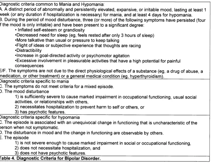
Bipolar disorder, though less common, with an estimated 1-3% of the population affected (Kieseppa et al., 2004; Miller, 2006; Edvardsen et al., 2008; Sachs et al., 2011), nonetheless has a significant impact on society. The diagnosis of bipolar disorder I is made based on the presence of a manic or hypomanic episode, which is defined as a period of abnormally and
persistently elevated mood such as distractibility, inflated self-esteem, racing thoughts, a decreased need for sleep, and risk-taking behavior (Table 4). The presence of a major depressive episode, while not necessary for diagnosis, is very common: in fact, patients with
bipolar I experience depressive episodes approximately 3 times as often as manic episodes. Bipolar II is the diagnosis given to patients that experience hypomania and also have at least one episode of major depression (Benazzi, 2007a). Patients with bipolar 11 experience
depressive symptoms as many as 37 times more frequently than symptoms of hypomania (Judd et al., 2003). A major cause of death in patients with bipolar disorder is suicide, with 15% of
patients diagnosed successfully committing suicide (Miller, 2006; Benazzi, 2007b, a). It should be noted that during episodes of mania, affected individuals could experience delusions, as well as hallucinations, both of which are characteristics of psychosis. Mood stabilizers such as lithium and valproic acid are commonly used to treat bipolar disorder, though several recent reviews of the literature have found that the antipsychotics, which will be mentioned below, are more effective in the treatment of mania than mood stabilizers (Cruz et al., 2010; Chwieduk and Scott, 2011; Sachs et al., 2011; Zupancic, 2011).
Diagnostic criteria common to Mania and Hypomania:
A. A distinct period of abnormally and persistently elevated, expansive, or irritable mood, lasting at least 1
week (or any duration if hospitalization is necessary) for mania, and at least 4 days for hypomania. B. During the period of mood disturbance, three (or more) of the following symptoms have persisted (four if the mood is only irritable) and have been present to a significant degree:
-Inflated self-esteem or grandiosity
-Decreased need for sleep (eg, feels rested after only 3 hours of sleep) -More talkative than usual or pressure to keep talking
-Flight of ideas or subjective experience that thoughts are racing -Distractibility
-Increase in goal-directed activity or psychomotor agitation
-Excessive involvement in pleasurable activities that have a high potential for painful
consequences
E/F. The symptoms are not due to the direct physiological effects of a substance (eg, a drug of abuse, a
medication, or other treatment) or a general medical condition (eg, hyperthyroidism). Diagnostic criteria specific to mania
C. The symptoms do not meet criteria for a mixed episode. D. The mood disturbance
1) is sufficiently severe to cause marked impairment in occupational functioning, usual social
activities, or relationships with others,
2) necessitates hospitalization to prevent harm to self or others, or
3) has psychotic features.
Diagnostic criteria specific for hypomania
C. The episode is associated with an unequivocal change in functioning that is uncharacteristic of the
person when not symptomatic.
D. The disturbance in mood and the change in functioning are observable by others. E. The episode
1) is not severe enough to cause marked impairment in social or occupational functioning,
2) does not necessitate hospitalization, and
3) does not have psychotic features.
Table 4. Diagnostic Criteria for Bipolar Disorder.
Schizophrenia is a chronic debilitating disorder that affects 0.7-3% of the population
synonymous with psychosis and include hallucinations, delusions, and disorganized thought. The negative symptoms are the decrease in, or absence of characteristics such as motivation, affective response, verbal speech, attention, and enjoyment. People with schizophrenia also suffer from cognitive impairment including attention, language, memory, and executive function. Relative cognitive impairment, along with other deficits such as low social function, is often noted before the onset of positive symptoms, and is seen more often seen in people with affected siblings. Schizophrenics also exhibit a moderate and appreciable decline in cognitive function throughout their lifetime. Suicide risk is increased in schizophrenia, particularly amongst those early in the diagnosis and with high cognitive function, with estimates between 5-10% successfully committing suicide (Laursen et al., 2012). Even after taking into account the increased rate of suicide, the diagnosis of schizophrenia is associated with higher mortality; this can only be partially explained to be due to decreased rate of adherence to medication (Saha et al., 2007).
A. Two (or more) of the following, each present for a significant portion of time during a 1-month period
(or less if successfully treated)*:
-Delusions -Hallucinations
-Disorganized speech (eg, frequent derailment or incoherence)
-Grossly disorganized or catatonic behavior
-Negative symptoms, ie, affective flattening, alogia, or avolition
B. For a significant portion of the time since the onset of the disturbance, one or more major areas of functioning such as work, interpersonal relations, or self-care are markedly below the level achieved prior to onset (or when the onset is in childhood or adolescence, failure to achieve expected level of
interpersonal, academic, or occupational achievement).
C. Continuous signs of the disturbance persist for at least 6 months.
1). This 6-month period must include at least 1 month of symptoms (or less if successfully
treated) that meet Criterion A, above (ie, active-phase symptoms) and
2). May include periods of prodromal or residual symptoms during which signs of the disturbance may be manifested by
a). Only negative symptoms or
b). Two or more symptoms listed in Criterion A present in an attenuated form (eg, odd
beliefs, unusual perceptual experiences).
D. Schizoaffective disorder and mood disorder with psychotic features have been ruled out
E. The disturbance is not due to the direct physiological effects of a substance (eg, a drug of abuse, a medication) or a general medical condition.
F. Relationship to a pervasive developmental disorder
If there is a history of autistic disorder or another pervasive developmental disorder, the additional
diagnosis of schizophrenia is made only if prominent delusions or hallucinations are also present for at least a month (or less if successfully treated).
Table 5. Diagnostic Criteria for Schizophrenia
Therapeutic interventions in place for the treatment of major depression, bipolar disorder, and schizophrenia
Recent literature reviews have shown a modest, at best, effect of current therapies for the treatment of severe depression, and no observable effect - beyond placebo- of current pharmacologic therapies on mild to moderate depression, as defined by a Hamilton Depression Rating Scale score of less than 25 (Hamilton, 1980). Most major depressive episodes go undiagnosed (Cepoiu et al., 2008), and most resolve within a matter of months, without treatment. Current pharmacological therapies used in the treatment of major depressive disorder (MDD) include the use of monoamine oxidase inhibitors (MAOi's) which act in the monoamine releasing neurons to inhibit the breakdown of monoamines; tricyclic
antidepressants, which block monoaminergic neurotransmitter reuptake; and more selective reuptake inhibitors, such as selective serotonin reuptake inhibitors (SSRI's), and serotonin-norepinephrine reuptake inhibitors (SNRI's) (Crupi et al., 2011). The first line therapies for treatment have become the selective reuptake inhibitors, because of their relatively modest side
effect profile compared to the MAOi's and tricyclics: in fact, MAOi's are now rarely used, because its use often necessitates dietary restriction and the risk for serotonin syndrome (Gillman, 2006; Sun-Edelstein et al., 2008). However, even the first line therapies come with a number of side effects, including drowsiness, sexual dysfunction, weight gain, insomnia, and anxiety, which are seen in over 10% of patients taking these medications. That all these
medications are effective suggests that all of these neurotransmitter systems play a role in affect and mechanisms downstream of each of these signaling cascades may play a role in the
The reuptake inhibitors all take about a week before improvements in mood can be discerned from placebo (Racagni and Popoli, 2010), and the current theory of this delay in efficacy is due to the requirement of a secondary effect that takes place after the drugs have reached its site of action. Two potential secondary effects have been presented, which are neither mutually exclusive nor comprehensive in their explanations. Several animal model studies (Overstreet et al., 2003; Richardson-Jones et al., 2010) and numerous human brain imaging studies (Hirvonen et al., 2008; Saijo et al., 2010) have implicated the role of presynaptic autoreceptors, 5-HT1A in particular for serotonergic neurons of the dorsal raphe (Serretti et al., 2004) and both 5HT1A and alpha-2-adrenergic receptors in adrenergic neurons of the locus coeruleus (Berrocoso and Mico, 2007; Ortega et al., 2010), in the delay of onset of
antidepressants. The initial blockade of reuptake triggers an increase in extrasynaptic levels of the monoamine neurotransmitters, resulting in an activation of autoreceptors at the presynaptic cell soma of the monoamine-releasing neurons. The activation of autoreceptors at the soma results in a reduced rate of action potentials in these neurons, which leads to a compensatory reduction in the level of released neurotransmitter at their postsynaptic sites of action: it is only with downregulation of these autoreceptors that a functional increase in monoamine
neurotransmitter can be observed (Richardson-Jones et al., 2010). This observation has led to an interest in using a 5HT1A antagonist in conjunction with SSRI's to improve clinical efficacy or abridging the time of antidepressant effect. Some trials have shown promising results (Whale et al., 2010; Portella et al., 2011), while others have shown mixed results (Scorza et al., 2011).
A second reason that has been proposed for the delay in action of reuptake inhibitors, is
that the increase in monoamine levels leads to a cascade of effects postsynaptically that take weeks to manifest. Some of the effects seen after chronic antidepressant treatment that are
hard to appreciate immediately include things such as increase in dendritic arborization in numerous brain regions (Alves et al., 2002; Jones et al., 2009), changes in gene transcription of
numerous factors that have been implicated in cell survival (Mannari et al., 2008), chronic stress response(Aubry et al., 1999; Anacker et al., 2011), metabolism (Duman et al., 2009), and
proliferation (Malberg et al., 2000; Santarelli et al., 2003)). I will group these effects and talk about them as effects on neurons that are already present and established in the neural circuitry underlying depressive disorders, and effects on newly formed cells that become incorporated into the neural circuitry underlying depressive disorders.
Effects on the generation of neurons that become incorporated into circuitry The subgranular zone of the dentate gyrus and subventricular zone have been
established as two areas in which neurogenesis continues into adulthood (Ma et al., 2009). In the rodent brain, 6% of the neurons in the dentate gyrus are estimated to be new neurons
generated within a month (Cameron and McKay, 1998). While a significantly smaller percentage is estimated to generated in humans (Amrein et al., 2011), the same mechanisms are presumed to be important across species. Neurogenesis in the dentate gyrus, in particular, plays a role in
both the development of depression (Snyder et al., 2011) and antidepressant effect (Sahay and Hen, 2007). Therapeutic interventions that induce a depressive phenotype, such as
corticosterone/glucocorticoid treatment (Brummelte and Galea, 2010), chronic stress (Pham et al., 2003), interferon treatment (Kaneko et al., 2006) lead to a significant reduction in
neurogenesis. On the other hand, treatments that have an antidepressant effect increase neurogenesis, whether it be pharmacologic, in the case of MAOi's (Malberg et al., 2000;
Nakagawa et al., 2002a), Tricyclics, SSRI's (Malberg et al., 2000; Santarelli et al., 2003), SNRI's (Shankaran et al., 2006), Lithium (Chen et al., 2000), antipsychotics (Kippin et al., 2005), or electroconvulsive treatment (Madsen et al., 2000).
Antidepressants have also been shown to have effects on neurons independent of neurogenesis. Newly born neurons have a higher rate of survival in the presence of
antidepressants, independent of the effect of SSRI's on progenitor proliferation (Bath et al., 2012). SSRI administration results in structural changes in non-neurogenic brain regions (McCabe and Mishor, 2011). IV Ketamine, currently under investigation in clinical trials for antidepressant treatment, is also known to facilitate the massive reorganization of the dendritic architecture of neurons from numerous cortical brain regions (Surget et al., 2011).
Schizophrenia: Antipsychotics
The treatments for bipolar disorder and schizophrenia fare better than placebo (Leucht et al., 2012) but their efficacy is limited to only a subset of symptoms, and also come with a high-side effect profile that often results in treatment discontinuation, either by the choice of the patient or due to the development of dangerous side effects (Daumit et al., 2008; Caroff et al., 2011).
The first antipsychotic, chlorpromazine, was discovered in 1952, rather accidentally: it was initially developed for use as a surgical anesthetic, but was used in psychiatric patients for its sedative effects. The discovery of chlorpromazine led to a revolution in psychiatric care, as patients that responded to treatment were discharged from confinement in psychiatric inpatient hospitals: previous to this, the diagnosis meant a lifetime of respite and confinement in these wards. The current pharmacological treatments for schizophrenia, referred to as the
antipsychotics have some antagonist activity at the dopamine D2 receptor. The pharmacologic agents available to date for the treatment of the disorder only provide symptomatic relief, and are effective for only a portion of the symptoms (mainly positive). The treatments themselves
have significant adverse effects, including tardive dyskinesia, sedation, metabolic changes including diabetes, and because of this, discontinuation rate is high (Salimi et al., 2009). The
current pharmacologic treatments for bipolar disorder include a combination of mood stabilizers plus an antipsychotic (Correll et al., 2010).
Current hypothesis pertaining to risk for major depressive disorder (MDD)
There are many hypotheses as to what results in the manifestation of these psychiatric conditions, based on neurochemical, neuroanatomical, and epidemiological findings, the pharmacology or effect of treatments, and, most recently, genome-wide association studies (GWAS). The leading hypothesis is that there is dysfunction of numerous brain areas related to the generation and processing of emotions, such as the prefrontal cortex, amygdala,
hypothalamus, medial septal areas and the hippocampus. Significant efforts have been made to examine the role of neurogenesis on the etiology of depression. This is based on the findings that antidepressants, as well as other interventions that have antidepressant like effects such as deep brain stimulation and environmental enrichment in animal models, all increase the number of newly-born neurons in the dentate gyrus, and because the time course for the maturation of adult-born neurons is consistent with the onset of antidepressant effectiveness. However, a change in adult neurogenesis is not sufficient to explain all the activity of antidepressants, as there are neurogenesis-independent actions of antidepressants (Sahay and Hen, 2007). Furthermore, research to date suggests that adult neurogenesis in the hippocampus is a substrate for the behavioral effects of antidepressants, but is not an essential contributor to the etiology of depression. Two large-scale genome-wide association studies have been conducted to date with no significant correlation identified between disease and a genomic region (Muglia et al., 2010). Thus, the etiology of unipolar depression remains a significant challenge to address.
Current hypothesis pertaining to risk for bipolar disorder
The concordance rate of bipolar I disorder in monozygotic twins is around 40% compared to -10% in dizygotic twins and in non-twin siblings (Kieseppa et al., 2004). The
overall heritability of bipolar disorder is 0.71 (Edvardsen et al., 2008). There have been several genome-wide association studies to date that have identified novel genetic loci associated with the disease (2007; Ferreira et al., 2008; Sklar et al., 2008; Purcell et al., 2009). These studies point to the heterogeneity of underlying genetic contributions to the risk of developing bipolar disorder. Recent studies have identified linkage disequilibrium for polymorphisms within the
CACNA1C L-type voltage gated calcium channel subunit and ANK3 (Ferreira et al., 2008), as
well as other genetic regions close to miR137 (Ripke et al., 2011), CDH7 (Soronen et al., 2010), and clOorf26 open reading frames (Ferreira et al., 2008; Sklar et al., 2008; Moskvina et al.,
2009). As ANK3 plays an important role in clustering Na+ and K+ channels at the axon initial
segment (Pan et al., 2006), it is tempting to speculate that changes in channel property or synaptic transmission contribute to the etiology of bipolar disorder. There is, however, a significant proportion of the heritability of the disorder that has yet to be taken into account,
illustrating the need for novel and, as yet unexplored mechanisms to probe for disease heritability that go beyond the sequence of the genes themselves.
Current Hypotheses on Disease Etiology: schizophrenia
Numerous hypotheses to the etiology of schizophrenia have been postulated to date such as the dopamine, glutamate, and GABA hypotheses, based on medications,
pharmacological mimicry and postmortem neuropathological findings, but none are sufficient to explain the onset of the syndrome. The neurodevelopmental hypothesis for schizophrenia has also been gaining traction in the field. This hypothesis predicts that alterations during the development of the brain result in cognitive deficits and renders the individual more susceptible to developing psychiatric disorders at a later age. Indeed, the cognitive deficits seen in patients with schizophrenia are often detected long before the diagnosis is made, and these same deficits are often seen in family members (Gold and Weinberger, 1995). Though there are no pathomnemonic features or tests to date for the disorder, and there is no identified organic
cause, several lines of evidence suggest that such a cause may exist. Endophenotypes, including those from neurological testing and functional brain imaging have been identified in some people with schizophrenia and their family members. These endophenotypes may also
point to a brain with more susceptibility for developing schizophrenia. Genetic abnormalities that result in aberration of neural development have been associated with schizophrenia, as will be discussed below.
There is a 50% concordance rate for schizophrenia between monozygotic twins, and the offspring of affected individuals have a ten-fold increase in risk for developing the disorder relative to the general population, highlighting a genetic basis for the disorder. Recent genetic evidence indicates that microdeletions or duplications of certain chromosomal loci, known as copy number variations (CNVs), increase the risk for schizophrenia. Chromosome 22q1 1.2 microdeletions occur in 1 of every 4,000 live births. These deletions produce a wide range of clinical presentations including velo-cardio-facial syndrome, DiGeorge syndrome, and mental retardation (Debbane et al., 2005). About 30% of carriers develop psychosis (Debbane et al., 2006a). Reduced brain volume, or microcephaly, is a common pathological feature of these carriers (Debbane et al., 2006b). Recently, additional CNVs have been identified that increase the risk for schizophrenia including microdeletions of chromosome 1q21.1, 15q13.3, 15q11.2, 3q29, 16p13.1, and 17p12 and microduplications at 16p11.2 (International Schizophrenia Consortium2008; Stefansson et al., 2008a; Vacic et al., 2011). Interestingly, 1q21.1
microdeletions and duplications are also involved in other neurodevelopmental disorders such as microcephaly/macrocephaly with behavioral abnormalities (Brunetti-Pierri et al., 2008) and autism (Mefford et al., 2008), whereas 15q 13.3 microdeletions and duplications are associated with idiopathic epilepsy, mental retardation, autism, ADHD, and anxiety disorder (Ben-Shachar et al., 2009; Helbig et al., 2009). Another study identified rare CNVs disrupting multiple genes in neurodevelopmental pathways in schizophrenia (Walsh et al., 2008). Collectively, genetic
evidence strongly supports the notion that impaired brain development increases the risk for the manifestation of mental illnesses. The genetic findings are also consistent with the general description of the neuropathology of schizophrenia, namely enlarged lateral ventricles and reduced cortical/hippocampal size (Tamminga and Holcomb, 2005). It remains to be determined why a given genetic lesion gives rise to clinically heterogeneous phenotypes.
A Common set of heritable risk for psychiatric disorders
There is now considerable evidence for a common set of heritable risk for psychiatric disorders including schizophrenia, bipolar disorder, as well as autism. For starters, the
concordance between monozygotic twins, and the increased incidence amongst family members, for bipolar disorder and schizophrenia support the existence of a such a common genetic factor (Lichtenstein et al., 2009). There are a number of recurrent CNV's that are common to both schizophrenic and autistic patients, albeit some go in opposite directions, suggesting a possible gene-dosage effect (Mefford et al., 2008; Stefansson et al., 2008a; Moreno-De-Luca et al., 2010; Malhotra and Sebat, 2012; Rodriguez-Murillo et al., 2012) Further evidence for the shared genetic risk for these psychiatric disorders comes from genome-wide association studies (GWAS) that have identified numerous genetic risk SNPs that are common between disorders(Sklar et al., 2008; Ripke et al., 2011). Current evidence suggests that schizophrenia, bipolar disorder, and autism, either treated separately or treated as a unitary set, would have an inheritance pattern similar to that of several non-neuropsychiatric disorders, including adult-onset diabetes and inflammatory bowel disease, all of which follow a complex inheritance pattern. It is interesting that schizophrenia has common risk variations for both autism and bipolar disorder, both in SNP alleles and recurrent CNV's, almost as if it is an intermediary phenotype.
Many of these risk alleles affect genes implicated in neuronal development with known roles at an early stage; these genes would have an effect on the individual before the onset of overt symptoms or diagnosis. Furthermore, many of the genes identified through GWAS are known to participate in established pathways that are relevant for neuronal development and function.
One could hypothesize that the risk for developing psychiatric disorders can be
accounted for by disruptions to specific biochemical pathways. This type of approach has been successfully implemented for cancer treatment, where the identification of disease targets based on alterations in pathways has led to the development of targeted therapeutics (de Bono and Ashworth, 2010). This would enable the classification of patients based on the presence of risk, grouped by biochemical pathways, as well as provide a more tractable predictor of patient response to medications. Some of the recently identified risk factors affect neural development at an early stage; before the onset of overt symptoms leading to diagnosis with the disorder. Importantly, many of the genes that have been identified through these genetic studies encode proteins that are known to be pivotal for signaling pathways associated with neuronal
development and function. It is important then to address the causality between these signaling pathways that are important for neurodevelopment, and the risk of developing neuropsychiatric disorder.
Several pathways have been clearly implicated in neuronal development: these include the Wnt, Akt, Lis1/NudeL, PKA, and EGR/Map Kinase pathways. These signaling pathways are not straightforward; there is substantial parallel signaling, as well as feed-forward and feedback signaling within one biochemical pathway, as well as substantial crosstalk between these pathways.
In the following paragraphs I will briefly identify some pathways important for neuronal development. I will elaborate on the Wnt pathway moreso than the others because of the intriguing, and notably undocumented, number of genes that are in or near affected gene regions that converge onto the pathway: it is not meant to diminish the importance of the other pathways in neuronal development.
cAMP-PKA-CREB pathway
The cyclic adenosine monophosphate (cAMP)-Protein Kinase A (PKA)-cyclic-AMP response element binding protein (CREB) pathway is a pathway that is activated upon the binding of ligands to G-protein coupled receptors (GPCRs) that are coupled to the GasGpy heterotrimeric G-protein complex. In the baseline state, the G-protein complex is bound to GDP and is inactive. Upon ligand binding to these GPCR, there is an exchange of GDP to GTP,
leading to the dissociation and activation of Gas. Gas -GTP binds to stimulate the activity of adenylyl cyclase, which converts adenosine triphosphate (ATP) into cyclic adenosine
monophosphate (cAMP). cAMP acts as a second messenger, and the increase in the levels of cAMP lead to the activation of several key downstream events. cAMP can bind to and open cyclic-nucleotide gated ion channels, influencing neuronal conductance. It can also activate exchange proteins activated by cAMP (EPACs). It's most well characterized function is the activation of protein kinase A (PKA). PKA normally exists as a holoenzyme consisting of two regulatory and two catalytic subunits. cAMP binds to the regulatory subunits, releasing the catalytic subunits for downstream activity. PKA phosphorylates a plethora of substrates: one of them is CREB, a transcription factor that binds to the cAMP response element (CRE).
Phosphorylated CREB (pCREB) induces the transcription of many genes involved in neuronal development. pCREB is found in almost all newborn neurons and correlates with the
expression of doublecortin (Nakagawa et al., 2002a; Nakagawa et al., 2002b), and is important for newborn neuron survival(Herold et al., 2011). However, the role of CREB in the proliferation
of neural precursors is yet unknown (Merz et al., 2011). However, it is well established that increasing cAMP levels by inhibiting the phosphodiesterase 4 family of enzymes with rolipram, does increase the rate of neuronal progenitor proliferation, raising the possibility that there are other factors that contribute downstream of PKA (Li et al., 2009).
The P13K-AKT-mTOR pathway
The phosphatidylinositol (3,4,5)-triphosphate kinase (Pl3K)-AKT/PKB-mammalian Target of Rapamycin (mTOR) pathway is activated by another class of membrane-bound receptors, the receptor tyrosine kinases. In the baseline state, AKT is bound to phosphatidylinositol(3,4)-bisphosphate (PIP2). Upon activation of a receptor tyrosine kinase, or, in some cases, a G-protein coupled receptor, P13K phosphorylates PIP2 to form PIP3. AKT bound to PIP3 is then phosphorylated by mammalian target of rapamycin complex 2 (mTORC2), then phosphorylated by phosphinositide dependent kinase 1 (PDPK1). The phosphorylation by both kinases
activates AKT to then phosphorylate its targets, including mTOR, implicated in autism, GSK3p, involved in the Wnt signaling cascade, BAD, a pro-apoptotic mitochondrial protein, and IKB
kinase (IKK), which regulates NFKB mediated transcription.
The intersection of the AKT pathway with the pharmacology of psychiatric disorder treatment is notable. Dopamine d2 receptors, which are targets of all antipsychotics to date, have been shown to modulate AKT signaling by recruiting a signaling complex that results in the inactivation of AKT. Upon D2 receptor activation and phosphorylation by G-protein coupled receptor Kinase (GRK), P-arrestin and PP2A are recruited to the cell membrane, where they interact with and dephosphorylate AKT. Antipsychotics inhibit this process, allowing AKT to remain active(Beaulieu et al., 2005; Freyberg et al., 2010).
Lithium, a treatment for bipolar disorder, is thought to exert its action by acting to activate AKT activity on GSK3P. Lithium has been proposed to solubilize AKT from the
P-arrestin/PP2A complex(Beaulieu et al., 2004). Thus, both these treatments secondarily affect the wnt pathway via affecting AKT activity.
PLC-PKC pathway
Phospholipase C is an enzyme that is activated downstream of GaoGpywhich cleaves phosphatidylinositol 4, 5 bisphosphate (PIP2) into Inositol-3-phosphate (IP3) and diacylglycerol (DAG). IP3 binds to receptors that regulate the release of calcium from intracellular stores, such as the smooth endoplasmic reticulum, leading to elevated levels of intracellular calcium, and subsequently activating calcium-dependent signaling cascades such as those involving calcium/calmodulin-dependent protein kinase i (CaMKII), calcineurin and PKC.
Map Kinase/ ERK pathway
The mitogen associated protein kinase (MAPK)/ ERK pathway refers to a general pathway in which a receptor-linked tyrosine kinase activation by ligand activates the GTPase Ras. Ras then activates a MAP kinase kinase kinase (MAP3K), which activates, MAP kinase kinase, which activates MAP kinase (MAPK). MAPK's activate transcriptional activity of pro-survival and proliferative genes via several mechanisms. One mechanism is the
phosphorylation and activation of RSK, a 40s ribosomal protein S6 kinase. This results in the alteration of RNA translation. RSK also phosphorylates CREB as well as c-Myc, c-Fos, NfKB, altering transcription of survival genes. Notably, RSK also phosphorylates GSK3P.
ERK2 is known to be essential for neuronal progenitor proliferation. ERK2 (MAPK1) knockouts(Satoh et al., 2011) die during embryogenesis. Conditional knockout of ERK2 in neuronal progenitors results in mice with decreased cortical thickness, and have a pool of undifferentiated neuronal progenitors (Samuels et al., 2008).
Wnts are secreted lipoproteins that act as morphogens and play important roles in the development and patterning of various limbs and organs. The Wnt ligands bind to their cell surface receptor, the Frizzled (FZ) family of seven transmembrane G-protein coupled receptors. The Wnt ligands were identified by their homology to Wnt, which was named from a
combination of the drosophila gene wingless (Wg) and its mammalian homolog Integration 1 (Int1). To date, nineteen WNTs and ten FZD family members have been identified in humans. The various Wnts as well as the FZDs have distinct, yet overlapping expression patterns throughout the body, which is thought to account for the variability of phenotypes that result upon loss of function (http://www.tcd.ie/Zooloqy/research/WntPathway/wnt.php, accessed April 17, 2012). Furthermore, it has also been shown that different Wnts have different affinities for the different FZs, and that different Wnts activate different downstream signaling cascades (Kikuchi et al., 2009).
Though none of the Wnts have been crystallized for structural analysis, much is known about their general structure. The Wnts are all about 320-400 amino acids long. They all have an N-terminal signaling sequence, which is required for their secretion, followed by a conserved domain consisting of 22-24 cysteines, the first of which is palmitoylated. The secreted form of the protein is highly hydrophobic (Mikels and Nusse, 2006). The loss of palmitoylation reduces Wnt 's potency, which can be overcome with high concentrations of Wnt, suggesting that
palmitoylation is required for Wnt localization(Nusse, 2003; Mikels and Nusse, 2006). Numerous glycosylated forms of Wnt proteins have been detected, supporting the posttranslational
modification of Wnts as a means to regulate Wnt signaling.
The ten members of the frizzled family of seven transmembrane G-protein coupled receptors share an N-terminal extracellular domain, three extracellular and three intracellular loops, and an intracellular C-terminal tail, like the classical G-protein coupled receptors;
cysteines, and Wnts bind to this region (Schulte and Bryja, 2007). The extracellular loops have putative glycosylation sites, a posttranslational modification that is known to play a role in both receptor localization and ligand binding. The intracellular C-terminal region of the receptor contains a PSD-95/DISC Large/POZ-1-homologous (PDZ) binding domain that is crucial for protein-protein interactions needed to mediate its downstream effects. Furthermore, the intracellular loops have numerous consensus sites for serine/threonine as well as tyrosine phosphorylation, reminiscent of receptor tyrosine kinases. There is evidence to suggest that FZD receptors exist as dimers which is facilitated by the cysteine-rich domain, and Wnt ligand binding. Moreover, FZD dimerization appears to be sufficient for activation of downstream pathways (Carron et al., 2003).
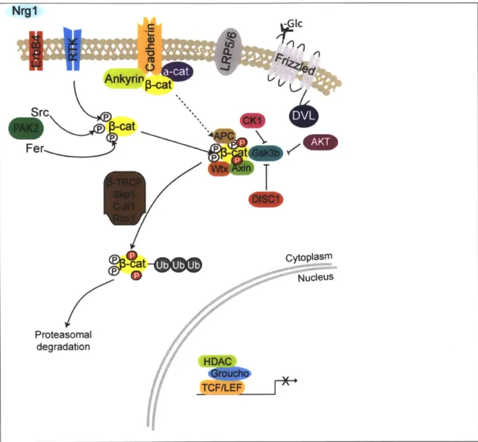
Extensive work in the past 20 years has led to the identification of several Wnt signaling pathways downstream of Wnt ligand binding to FZD or one of its co-receptors: lipoprotein related protein 5/6 (LRP5/6) (Pinson et al., 2000; Tamai et al., 2000; Wehrli et al., 2000), receptor-tyroine kinase-like orphan receptor 2 (Ror2) (Saldanha et al., 1998), and related to receptor tyrosine kinase (Ryk) (Inoue et al., 2004).
Canonical Wnt signaling
Binding of Wnt to LRP5/6- associated frizzled activates a pathway known as the canonical Wnt signaling pathway. The canonical Wnt signaling pathway is defined by their dependence on the transcription co-activator
p-catenin
and the downstream signaling cascades that are associated with it. Briefly, Fzd proteins have a conserved KTxxxW domain that isrequired for interaction with dishevelled (DVL) proteins. In the absence of Wnt stimulation, DVL is part of a destruction complex, in conjunction with AXIN, adenomatous polyposis coli (APC), casein kinase 1 alpha (CK1a), and glycogen synthase kinase 3 (GSK3p). This destruction complex binds and sequentially phosphorylates
p-catenin
at multiple sites, thus increasing theaffinity of P-catenin to
P-TrCP. p-TrCP
serves as one of the F-box proteins in the Skpl-Cullinl-F-box-protein (SCF) E3 ubiquitin ligase complex. Binding ofp-TrCP
top-catenin
leads to the polyubiquitination and subsequent degradation of P-catenin, preventing the accumulation ofp-catenin and limiting its downstream function. Upon Wnt stimulation of the FZD-LRP5/6 complex, DVL is recruited to the complex, where it polymerizes, and through its DIX domain binds to the DIX domain of AXIN. This, in turn, leads to the recruitment of AXIN towards the FZD-LRP5/6 complex, where it mediates interaction with of CK1 a and GSK3P to LRP5/6. Next, the kinases phosphorylate LRP5/6 and increases its affinity for AXIN. The LRP5/6 interaction directly inhibits GSK3p phosphorylation of
P
-catenin (Piao et al., 2008), one of the priming phosphorylations ofp-catenin,
disrupting the ability of the destruction complex to targetp-catenin
for ubiquitination and subsequent degradation. Thus, the activation of the Wnt pathway leads to an increase in cytosolicP-catenin.
p-catenin
is a transcriptional co-activator of TCF/LEF transcription factors, which controlthe dynamics of the cell cycle via regulation of cyclinD1. The accumulation of
p-catenin
in the cytosol leads to its increase in the nucleus and the binding ofP-catenin
and it's associated transcriptional activators to the T-cell factor and lymphoid enhancer-binding protein (TCF/LEF) DNA binding protein family. TCF/LEF has three identified domains, a high mobility group (HMG) box domain that is crucial for DNA interaction, a caspase activated deoxyribonuclease (CAD) domain necessary for binding to Groucho/TLE, and an N-terminalP-catenin
binding domain. In the absence of nuclear P-catenin, the TCF/LEF occupying TCF/LEF DNA binding sites is boundto tetrameric Groucho/TLE, which recruits transcriptional repressors, such as histone
deacetylases (HDACs), keeping the DNA in a condensed, repressed state.
P-catenin
directly competes with, and displaces Groucho/TLE (Daniels and Weis, 2005), and recruits general transcriptional activators such as the histone acetyltransferase CBP, the SWI/SNF complex protein Brg-1, and TATA- binding protein, as well as a more specific core complex consisting ofPygopus and Legless/BCL9 (Belenkaya et al., 2002; Kramps et al., 2002; Thompson et al., 2002). CBP acetylates histone residues in the promoter region, and the recruitment of Pygopus and Legless/BCL9 leads to the methylation of H3K9 residues by SET-1 methyltransferases, altering the chromatin structure from a condensed, transcriptionally repressed one, to a more relaxed and open state, thus allowing transcription (Fiedler et al., 2008).
The canonical Wnt pathway and psychiatric disease gene candidates
Several players in the Wnt signaling pathways have been associated with psychiatric disease. CHD8, which is also recruited during this process, acts to counter the activating activity of p-catenin, by binding to p-catenin and also Histonel, and thus limiting the relaxation of chromatin. CHD8 SNPs are associated with autism, and BCL9 is one of the genes in the
1q21.1 recurrent CNV, with microdeletions associated with schizophrenia and microcephaly and
microduplications associated with autism and macrocephaly. Furthermore, ADA2A, in the 17q12 microdeletion associated with autism, is known to be an acetyltransferase required for
transcriptional activity and proliferative effects mediated by p-catenin (Yang et al., 2008).
Both the wnt ligands and the receptor are glycosylated, and perturbations of
glycosylation are known to affect the function, localization and signaling through them. SNPs in genes that are involved in glycan synthesis (hsa0l 030) associate with schizophrenia. Ankyrin 3, which has noncoding SNPs within its gene that associate with both schizophrenia and bipolar
disorder, is known to be a component of the Cadherin complex, and disruption of the cadherin complex is known to lead to mislocalization of p-catenin (Orsulic et al., 1999).
DLG1 (Sap97) is one of the interactors of Fz and is known to signal downstream of wnt binding by binding to Adenomatous Polyposis Coli (APC), affecting cell proliferation: this gene is found in the 3q29 CNV deletion. ErBB4/Neuregulin, another well-studied candidate that
family members have been shown to activate cascades that phosphorylate and lead to the intracellular localization of
p-catenin:
overexpression of ErbB4 Cyt2 incresed nuclearp-catenin
and TCF-LEF transcriptional activity (Muraoka-Cook et al., 2009). PAK2, also in the 3q29 CNV, also serves to phosphorylate and promote the nuclear localization of
p-catenin
(Zhou et al.,2011).
In addition to the recruitment of DVL, Wnt activates the G-proteins associated with Fz proteins. The FZDs are coupled to the Gao GPy trimeric G-protein complex (Katanaev et al., 2005), and Wnt binding to Fz liberates the Gpy subunits to activate a calcium-dependent pathway that involves the activation of phospholipase C cascade. The perturbation of these signaling pathways can have consequences beyond this pathway, and can impact the canonical Wnt pathway as well. Neurogranin, implicated in bipolar disorder and schizophrenia, is a key regulator of calmodulin, limiting its activity by binding to it; its binding to calmodulin is dependent on its PKC phosphorylation. The association of voltage-gated calcium channel, CACNA1C,with bipolar and schizophrenia is another intriguing risk gene related to calcium signaling.
Binding of Wnt to Fz leads to activation of pathways involved in planar cell polarity, acting through dishevelled, activating RhoA and RACI (Gao et al., 2011; Shafer et al., 2011) (Bo et al 2011 Developmental Cell). RAC1 activation then activates c-Jun N-terminal kinase (JNK) downstream of Wnt5a (Oishi et al., 2003). JNK activation has been shown to be crucial for the
proper development of axons and dendrites (Rosso et al., 2005)Calderon et al 2012), and to enhance or inhibit canonical Wnt activation, depending on the Wnt ligand (Billiard et al., 2005). Tao Kinase 2, in the 16p1 1.2 region, which has CNV's that associate with both autism and schizophrenia (deletion and duplication, respectively), also plays a role in the activation of JNK.
One of the alternative,
p-catenin
independent pathways that is downstream of LRP5/6-Frizzled signaling, depends on the inhibition of GSK3P. GSK3P is known to destabilizemicrotubules by phosphorylating microtubule proteins such as TAU, MAP1 B, and MAP2. Inhibition of GSK3p stabilizes microtubules, promoting axon growth and growth cone
remodeling. In this manner, the wnt pathway is able to exert effects on neuronal morphology.
Binding of Wnt to Ryk has been shown to directly activate the Src pathway, similar to the activation of many other receptor tyrosine kinases such as EGR. The activation of Src, in turn, has been shown to be crucial for axon guidance. Ryk has also been shown to interact directly to FZD8 XXXXXX), as well as DVL, and to potentiate the canonical Wnt signaling pathway,
suggesting that the presence of Ryk in conjunction with Frizzled may either stabilize the DVL-Fz interaction, or enhance AXIN interaction with Fz. The convergence of neuropharmacological and genetic evidence makes the wnt pathway(s), if not a promising target, at least a convenient framework within which the function of psychiatric risk gene polymorphisms and rare variants can be tested.
Src -cat Fer P-cat
t
cat
9O
Proteasomal degradation cytoplasm NucleusFigure 1. Canonical WNT pathway in inactive state. Tyrosine phosphorylation of
p-catenin
results in dissociation of
p-catenin
from its membrane interactors and results in cytosolic localization ofp-catenin.
And binding to destruction complex. Sequential serine phosphorylation by CK1 and GSK3p (represented as red P's).p-catenin
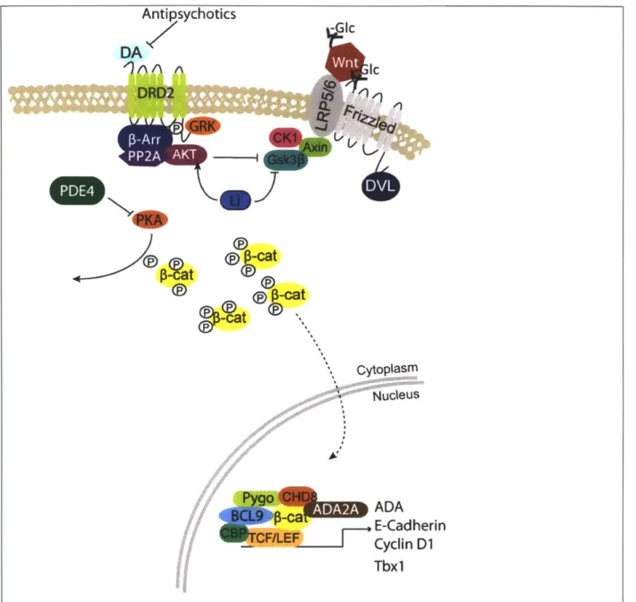
is subsequently polyubiquitinated and degraded by the proteasome.Antipsychotics DA DRQ2 4 Gc
K.]'
0-at ~cat -cat -Mat Cytoplasm SNucleus 0-ca ADA E-Cadherin Cyclin D1 Tbx1Figure 2. Activated canonical Wnt pathway, and select interactions of pathway with psychiatric disorder therapeutics and risk genes. Wnt-Fz/LRP5/6 interaction recruits
destruction complex to the cell surface, and CK1/GSK3p is inactivated. Cytosolic p-catenin bind does not get degraded, gets phosphorylated by PKA, translocates to nucleus where it binds to
TCF/LEF transcription factors, recruiting BCL9, Pygo, CHD8, ADA2A, CBP to initiate polil mediated transcription. Lithium, antipsychotics increase AKT activity, which increases its inhibition of GSK3p, enhancing the activity through this pathway. Phosphodiesterase inhibitors would increase PKA activity, which enhances this pathway by increasing nuclear
p-catenin
translocation and by inactivatinq GSK313.
Disrupted in Schizophrenia 1 (DISC1): a single gene to which many pathways converge
Disrupted in Schizophrenia-1 (DISC1) was identified as the gene that was disrupted on
chromosome 1 in a Scottish family with a high concordance of major psychiatric disorders and a balanced translocation between chromosomes 1 and 11 (Millar et al., 2000). Karyotyping on five generations of this family revealed 18 of the 29 members with this translocation have
schizophrenia, recurrent major depression, or bipolar disorder. This type of near Mendelian segregation for psychiatric disorders had not been previously observed. Mouse models for DISC1 have been generated, and the mice display a variety of phenotypes that can be considered schizophrenia-like behavioral deficits. Mouse models expressing a transgene of human DISC1 mimicking the Scottish translocation mutant exhibit increased ventricle size, decreased gray matter volume, and changes in dendritic arborization in cortical and
hippocampal neurons (Hikida et al., 2007; Li et al., 2007; Pletnikov et al., 2008; Shen et al., 2008). These mice also exhibit behavioral abnormalities such as hyperactivity (Hikida et al., 2007; Pletnikov et al., 2008), increased immobility in the forced swim test (Hikida et al., 2007), decreased sociability (Pletnikov et al., 2008), and working memory (Kvajo et al., 2008; Pletnikov et al., 2008). Clapcote et al characterized ENU induced mouse DISC1 exon 2 mutants and
reported that the Q31L mutant exhibits depression-like and the L100P mutant exhibits schizophrenia like behavior (Clapcote et al., 2007). In addition, reduced embryonic and adult hippocampal neurogenesis have been reported in a DISC1 BAC transgenic mouse model and
DISC1 exon 7-8 deletion mutant mice (Kvajo et al., 2008; Shen et al., 2008). Finally, DISC1 is implicated in the proper integration of adult born neurons in the dentate gyrus circuit (Duan et al., 2007). Collectively, the neuropathology and behavioral phenotypes exhibited by the various
DISC1 mouse models strongly supports a role for DISC1 in mental health.
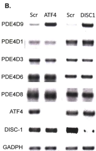
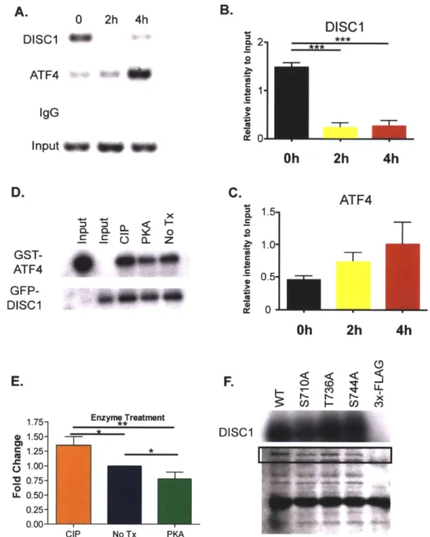
Since its identification as a psychiatric disease risk gene, a large number of DISC1 binding partners have been identified. Some validated interacting partners for DISCI include NDE1/NDEL1, important for neuronal migration, Kalirin-7, important for glutamatergic signaling, ATF4/5, important for neuronal progenitor proliferation, the PDE4family of phosphodiesterases, implicated in depression, and GSK3p, a target of Lithium used in bipolar disorder treatment. These interactions will be described in more detail below.
DISC-1 Structure, features, and Interactions
T1 Transcript 1:11 Translocation Breakpoint
854 sRNA1 shRNA2 4441bp 5'TR Protein structure A ft.DISC-1 L 331-34 166-394 &SS-" 607-628 W02-830 Interaction Partners (Binding regions) *fl 4 PfDE 46 31-65 101-135 190-230 266-290 JE02. 459-479
K<
Leucine-Zipper Coiled-Coil Phosphorylation siteNuclear Localization Sequence Figure 3. Schematic of validated DISCI partners. The interactions relevant for further chapters are colored.
DISC1 and neuronal migration
DISC1 binds to nudE nuclear distribution gene E homolog-like 1 or NDE1/NDEL1, which is known to play a role in neuronal migration, proper neuronal localization and neurite and axonal development (Kamiya et al., 2005). Disrupting DISC1/Ndel1 interaction disrupts neurite outgrowth in PC12 cells (Pletnikov et al., 2007), and also prevents the colocalization of DISC1
and Lis1, which localizes with the dynein heavy chain, indicating that DISC1 plays an important role in the function of Ndell/Lisl complex by regulating its localization. Our lab has shown that DISCI also interacts with this complex via its interaction with DIX domain containing-1 (Dixdcl), the third mammalian gene discovered to contain a Disheveled-Axin (DIX) domain (Dixdcl).
DISC1, Dixdcl and Ndel1 form a tripartite interaction, and the presence of either DISCI or DIXDC1 seems to be able to properly localize Ndel1, indicating that these two proteins may have overlapping/ redundant effects on neuronal migration (Singh et al., 2010). This also indicates that a partial loss of function of either of these two genes can be offset by the function of the other, potentially explaining why differences in the ability of DISC1 snps to bind to NDEL does not result in an appreciable difference in human disease burden.
DISC1 and the WNT signaling pathway
DISC1 has also been shown to bind to, and inhibit the activity of GSK3-p, one of two forms of an enzyme that is known to lead to the phosphorylation of many downstream proteins that are crucial for the pathophysiology of many nervous system disorders. One GSK3-p target is TAU, which forms aggregates in many nervous system disorders including Alzheimer's (TAU forms the neurofibrillary tangles that are used in the Braak and Braak postmortem staging of Alzheimer's), Parkinson's, and tuberous sclerosis: nervous system disorders in which there is TAU aggregation are collectively referred to as tauopathies. Another GSK3-p target is P-catenin.
to a-catenin and the cadherin proteins at the cell membrane, as part of the adherens junction (AJ) complex.
p-catenin
that is not bound to cadherins at the membrane binds to axin, which, in conjunction with APC, complexesp-catenin
with GSK3p. The formation of this complex leads to the phosphorylation ofp-catenin,
which promotes its poly-ubiquitination and subsequentproteasome-mediated degradation. When WNT binds to its receptor, frizzled, disheveled is recruited to the membrane and activated. Activated disheveled inhibits GSK3-P, which subsequently inhibits the phosphorylation and degradation of p-catenin, leading to its accumulation in the cytosol and the activation of downstream pathways, such as the
transcriptions of genes which bind TCF/LEF transcription factors. The activation of this pathway has been shown to be crucial for neuronal progenitor proliferation and the inhibition of the premature differentiation of these neuronal progenitors, and deficits in this pathway have been shown to result in microcephaly(Salcedo-Tello et al., 2011). By inhibiting GSK3p, DISC1 promotes neuronal progenitor proliferation, and the loss of DISC1 leads to decreased neuronal progenitor proliferation (Mao et al., 2009; Ming and Song, 2009).
A molecular switch between the wnt signaling and neuronal migration function of DISC1 has been recently uncovered. DISC1 was found to be a target of PKA-mediated
phosphorylation, downstream of cAMP activation. The study identified two putative PKA
mediated phosphorylation sites on DISC1, and discovered that one of the sites, at serine 710, is responsible for the differential localization and binding properties observed in neuronal
progenitors versus migrating neurons. DISC1 in neuronal progenitors is unphosphorylated at this site, and binds to and inhibits GSK3p activity: upon phosphorylation at this site, DISC1
increases its affinity for BBS1 and localizes at the centrosome. Crucially, the authors
demonstrated that the phosphor-dead form of DISC 1 (S710A) rescued the proliferative but not the migrational phenotype observed in DISC1 knockdown conditions, while the