HAL Id: inserm-01520102
https://www.hal.inserm.fr/inserm-01520102
Submitted on 9 May 2017
HAL is a multi-disciplinary open access
archive for the deposit and dissemination of
sci-entific research documents, whether they are
pub-lished or not. The documents may come from
teaching and research institutions in France or
abroad, or from public or private research centers.
L’archive ouverte pluridisciplinaire HAL, est
destinée au dépôt et à la diffusion de documents
scientifiques de niveau recherche, publiés ou non,
émanant des établissements d’enseignement et de
recherche français ou étrangers, des laboratoires
publics ou privés.
Lipid-oligonucleotide conjugates improve cellular uptake
and efficiency of TCTP-antisense in castration-resistant
prostate cancer
Sara Karaki, Sebastien Benizri, Raquel Mejías, Virginie Baylot, Nicolas
Branger, Tan Nguyen, Brune Vialet, Khalid Oumzil, Philippe Barthélémy,
Palma Rocchi
To cite this version:
Sara Karaki, Sebastien Benizri, Raquel Mejías, Virginie Baylot, Nicolas Branger, et al..
Lipid-oligonucleotide conjugates improve cellular uptake and efficiency of TCTP-antisense in
castration-resistant prostate cancer. Journal of Controlled Release, Elsevier, 2017, 258, Epub ahead of print,
1-9. �10.1016/j.jconrel.2017.04.042�. �inserm-01520102�
Contents lists available atScienceDirect
Journal of Controlled Release
journal homepage:www.elsevier.com/locate/jconrel
Lipid-oligonucleotide conjugates improve cellular uptake and e
fficiency of
TCTP-antisense in castration-resistant prostate cancer
Sara Karaki
a,b,c,d, Sebastien Benizri
e,f,g, Raquel Mejías
a,b,c,d, Virginie Baylot
a,b,c,d,
Nicolas Branger
a,b,c,d, Tan Nguyen
a,b,c,d, Brune Vialet
e,g,g, Khalid Oumzil
e,f,
Philippe Barthélémy
e,f,g, Palma Rocchi
a,b,c,d,⁎aCentre de Recherche en Cancérologie de Marseille (CRCM), INSERM UMR1068, 27 Bd. Lei Roure BP30059, 13273 Marseille, France bInstitut Paoli-Calmettes, 13273 Marseille, France
cAix-Marseille Université, 13284 Marseille, France dCNRS UMR7258, 13009 Marseille, France
eARNA Laboratory, University of Bordeaux, F-33076 Bordeaux, France fINSERM U1212, F-33076, Bordeaux, France
gUMR CNRS 5320, F-33076, Bordeaux, France
A R T I C L E I N F O
Keywords:Antisense oligonucleotides TCTP
Castration resistant prostate cancer Lipid modification
Drug delivery
A B S T R A C T
Translationally controlled tumor protein (TCTP) has been implicated in a plethora of important cellular processes related to cell growth, cell cycle progression, malignant transformation and inhibition of apoptosis. Therefore, TCTP is now recognized as a potential therapeutic target in several cancers including prostate, breast and lung cancers. We previously showed that TCTP is overexpressed in castration-resistant prostate cancer (CRPC), and it has been implicated resistance to treatment. Recently, we developed TCTP antisense oligonucleotides (ASOs) to inhibit TCTP expression. However, the intracellular delivery and silencing activity of these oligonucleotides remains a challenge, and depend on the use of transfection agents and delivery systems. Here we show that lipid-modified ASO (LASOs) has improved penetration and efficiency in inhibiting TCTP expression in the absence of additional transfection agents, both in vitro and in vivo. Transfection with TCTP-LASO led to rapid and prolonged internalization via macropinocytosis, TCTP downregulation and significant decreased cell viability. We also show that lipid-modification led to delayed tumor progression in CRPC xenografts models, with no significant toxic effects observed.
1. Introduction
Prostate cancer (PC) is one of the main health concerns in Western countries, representing the most common type of cancer among men in the US, and the third in Europe[1,2]. Although at early stages PC is a hormone-dependent disease, most patients undergoing androgen depri-vation therapy, by either pharmacological or surgical castration, progress to castration-resistant prostate cancer (CRPC) within 14–30 months [3]. CRPC presents poor prognosis, and therapeutic options are limited. Chemotherapy with docetaxel has demonstrated improved survival in men with CRPC in two different, large, phase III studies[4,5]. However, the increase in overall survival with docetaxel is very modest (2–3 months), and this treatment may affect not only tumor cells, but also healthy tissue, causing serious side effects[6]. For these reasons, in recent years great efforts have been made to develop new, more selective and efficient therapeutic modalities that enable
specific treatment of cancer cells, and restore castration- and chemo-therapy sensitivity without affecting other tissues. One strategy to improve therapies in advanced PC involves targeting genes that are related to androgen deprivation, which may lead to delayed or absent emergence of the resistant phenotype[7].
Recent studies have proposed tpt1, encoding for translationally controlled tumor protein (TCTP) as an androgen-regulated gene implicated in PC[7–9], whose expression correlates with PC grading [10]. TCTP, also known as, histamine releasing factor (HFR), fortilin or p23, is a highly conserved multifunctional protein present in a wide variety of eukaryotic organisms. It is involved in many physiological processes, such as cell proliferation, development, cell cycle progres-sion, stabilization of cytoarchitecture, and protein synthesis; and it shows protective effects against apoptosis and cell stress[11,12]. TCTP can also be secreted to the extracellular space, where it works as a mediator of the immune response by promoting histamine release from
http://dx.doi.org/10.1016/j.jconrel.2017.04.042
Received 9 February 2017; Received in revised form 25 April 2017; Accepted 29 April 2017
⁎Corresponding author.
E-mail address:palma.rocchi@inserm.fr(P. Rocchi).
Available online 01 May 2017
0168-3659/ © 2017 inserm. Published by Elsevier B.V. This is an open access article under the CC BY-NC-ND license (http://creativecommons.org/licenses/BY-NC-ND/4.0/).
basophils, and stimulating B-cell proliferation, and inducing the production of different cytokines and antibodies[13,14]. Although it has a widespread expression profile, TCTP expression is higher in tumors compared to their normal counterparts[15], which suggests a critical role in tumorigenesis. This observation, along with the fact that TCTP silencing may lead to tumor reversion [16,17], indicates that TCTP knockdown might be a very promising strategy for CRPC treatment.
Antisense oligonucleotides (ASOs) have been proven to be a useful tool in molecular biology and also as therapeutic agents, especially in cancer research. ASOs can be used to perform selective knockouts of mRNA functions either in vitro or in vivo. Oligonucleotides mediate their functions by base pairing, where the oligo single stranded DNA binds to its complementary strand of mRNA, making them highly specific. However, some practical obstacles remain unsolved in antisense pharmacology. ASOs, as all nucleic acid-based drugs, show insufficient stability as a result of degradation by nucleases, and poor intracellular delivery due to reduced cell uptake or difficulties in crossing biological membranes to reach the cytoplasm. Moreover, in many cases potential off-target effects and immunostimulation are also observed[18]. In order to reduce the limitations of oligonucleotide-based therapies, different chemical modifications have been developed [18–20]. One of the first modifications introduced in antisense therapy was the phosphorothioate (PS) modification, in which a sulfur atom replaces the non-bridging phosphate oxygen. This modification led to improved stability and extended circulation times in blood, although several toxic effects have been related to the use of this type of molecules. Several sugar modifications, such as 2′O-Methyl (2′O-Me), 2′O-Methoxyethyl (2′O-MOE), 2′F-Arabino Nucleic Acid (2′F-ANA) have been proved to increase the specificity of ASOs, reducing off-target effects. Locked Nucleic Acid (LNA) modifications, in which there is a methylene bridge between 2′ and 4′ positions ‘locking’ the sugar ring in an A-form conformation have shown increased affinity for complementary se-quences. Backbone modifications, such as Morpholino (PMO) or Pep-tide Nucleic Acid (PNA) provide a neutral backbone and work as ribosome steric blockers, efficiently preventing protein translation. Grafting oligonucleotide to small hydrophobic molecules, such as cholesterol[21], lipids[22]orfluorinated chains[23,24], may increase stability and membrane permeation. However, despite all the advan-tages provided by these modifications, improved pharmacokinetics and cell uptake remain a challenge.
In this study we present a new lipid-modified ASO (LASO) that helps overcome the problems related to ASO administration and effective-ness, by increasing the cellular uptake and transfection efficiency, and improving downregulation of TCTP expression in absence of transfec-tion agents in human PC-3 prostate cancer cells both in vitro and in vivo. We also analyzed the effects of TCTP downregulation on cell viability, and assessed in vivo toxicity of ASOs and LASOs, and their effects on tumor size and animal survival in a murine xenograft model of CRPC. Our results indicate that using lipid-modified TCTP ASOs (TCTP-LASO) can be an effective strategy for CRPC treatment.
2. Materials and methods
2.1. Synthesis of antisense oligonucleotides
Oligonucleotides were synthesized using an ABI Expedite 8909 synthesizer (1μmol scale) and an AKTA OligoPilot 10 synthesizer (50μmol scale). The oligonucleotide synthesis was achieved using conventional β-cyanoethyl phosphoramidite chemistry with the stan-dard DNA protocol. Reagents were purchased from Link and Glen Research. HPLC purification was performed on a Hitachi LaChrom Elite HPLC System with gradient standard protocol. The HPLC columns (Nucleosil C4 4 × 250 mm 5μm macherey, nagel) were equilibrated at a flow rate of 1.0 ml/min. A solution containing 100 mM Triethylammonium acetate (TEAA) and 5% acetonitrile at pH 7.0 was
used as solvent A and solvent B contained 20 mM TEAA and 80% acetonitrile. The elution gradient is reported inTable 1. The system was equilibrated for 5 min before the following run. Injection volume for samples was 25μl. Oligonucleotides were detected at the wavelength of 260 nm. TCTP-ASO (5′ AACTTGTTTCCTGCAGGTGA 3′) and control (5′ CGTGTAGGTACGGCAGATC 3′) were synthesized and purified conven-tionally. Same oligonucleotides were synthesized with a lipid conjugate (2′,3′-O-16-hentriacontanyliden-uridine) attached to the 5′ extremity of the oligonucleotide (TCTP-LASO). In the case of labeled oligonucleo-tides, the fluorescein (3′-(6-FAM)-CPG-1-(4,4′-Dimethoxytrityloxy)-3- [O-(N-carboxy-(di-O-pivaloyl-fluorescein)-3-aminopropyl)]-propyl-2-O-succinoyl-lcaa-CPG; Linktech) was inserted at the 3′ extremity (control-ASO FITC, TCTP-ASO FITC, control-LASO FITC, TCTP-LASO FITC). All oligonucleotides were synthesized in phosphorothioate back-bone (PS). All compounds were characterized by mass Spectrometry (Supplementary information) and required the use of a DCTB matrix (trans-2-[3-(4-tert-butylphenyl)-2-methyl-2-propenylidene]-malononi-trile) doped with Ag +(or Na +), at «Centre de Génomique Fonction-nelle» UMR 5248 CBMN.
2.2. Physicochemical characterization of antisense oligonucleotides ASOs and LASOs were analyzed by dynamic light scattering (DLS) using a Nanosizer ZS (Malvern Instruments, UK). Samples were denatured at 90 °C for 5 min, and allowed to cool down to 25 °C for nanoassembly. All the scattered photons were collected at a 173°-scattering angle. The 173°-scattering intensity data was processed using the instrumental software to obtain the hydrodynamic diameter (Dh) and the size distribution in each sample (400μl, 25 °C).
2.3. Transmission electron microscopy (TEM)
Size and morphology of the nano-objects were analyzed by trans-mission electron microscopy (TEM) using a Hitachi H7650 microscope. A drop of the nanomicelle suspension was placed on a formvar grid, and the solvent allowed to evaporate at RT. Counterstaining with uranyl acetate was performed twice (1 min/staining).
2.4. Cell culture
The androgen-independent prostatic cancer cell line PC-3 was obtained from the American Type Culture Collection (Rockville, MD) and cultured in Dulbecco's Eagle's Medium (DMEM, Invitrogen), supplemented with 10% fetal calf serum (FCS). Cells were maintained at 37 °C in a 5% CO2humidified atmosphere.
2.5. Transfection with ASOs and LASOs
PC-3 cells were plated on p100 culture dishes (106cells/dish), and transfected twice (24 and 48 h after seeding) with 100 nM ASOs or LASOs. Unless otherwise indicated, transfection with ASOs was per-formed in free-serum OptiMEM, containing 3 mg/ml oligofectamine (both from Invitrogen), while LASOs were used in free-serum OptiMEM without oligofectamine. After 4-h of incubation, transfection media was replaced with complete culture medium.
Table 1
Elution gradient of solutions A and B, for purification of antisense oligonucleotides by HPLC. Time (min) A (%) B (%) 0 100 0 10 0 100 12 0 100 14 100 0 15 100 0
S. Karaki et al. Journal of Controlled Release 258 (2017) 1–9
2.6. Transfection efficiency analysis
To determine transfection efficiency we used fluorescein-labeled ASOs and LASOs. PC-3 cells were transfected with ASOs or LASOs with and without oligofectamine, and at different time points after transfec-tion, cells were trypsinized and collected in flow cytometry tubes containing 0.5 ml complete culture medium. In each experiment 105 cells per condition were assayed in triplicate. Fluorescence intensity was determined in aflow cytometer (Canto Becton Dickinson) and data were analyzed using the FlowJo software.
2.7. Confocal microscopy
Intracellular distribution of ASOs and LASOs and transfection efficiency were also analyzed by confocal microscopy. One day before transfection, PC-3 cells (8 × 10⁴cells/well) were plated on 8-well Lab-Tek II chamber slides (Nunc-Thermo Scientific). Cells were then transfected withfluorescein-labeled ASOs or LASOs as described, and at different time points after transfection, cells were washed with PBS, fixed with 4% paraformaldehyde for 15 min at 4 °C, and mounted in Prolong Gold mounting medium containing the nuclear counterstain DAPI (Life technologies). For visualization, we used a Zeiss LSM 510 META fluorescence microscope with 405-nm and 488-nm excitation filters.
2.8. Cell viability assay
Cell viability was determined by a 3-(4,5-dimethylthiazol-2-yl)-2,5-diphenyl tetrazolium (MTT) assay. PC-3 cells were plated in 12-well plates (3 × 10⁴cells/well) and transfected the day after with ASOs or LASOs as described. After 48 h, MTT was added to each well (1 mg/ml final concentration) and the plates were incubated for 2 h at 37 °C. Supernatants were then removed and formazan crystals were dissolved in DMSO. The absorbance (595 nm) was evaluated using a Sunrise microplate absorbance reader (Tecan). Cell viability was expressed as the percentage of absorbance of transfected cells compared to untreated cells.
2.9. Western blot
PC-3 cells, transfected with ASOs or LASOs 72 h before analysis, were lysed in lysis buffer (1% v/v Triton X-100, 50 mM HEPES, 150 mM NaCl, 25 mM NaF, 1 mM EDTA, 1 mM EGTA, 10μM ZnCl2, 1 mM sodium orthovanadate) containing 4% v/v protease inhibitor cocktail (Roche) for 30 min at 4 °C. The lysate was centrifuged (60 min, 21,000 ×g) and protein content was quantified using the BCA protein assay kit (Pierce). 40μg of protein from each sample were mixed with Laemmli sample buffer and loaded on 12% SDS-polyacrylamide gels for electrophoresis. Proteins were transferred to PVDF membranes (Millipore), which were blocked with 5% w/v nonfat milk in Tris-buffered saline (TBS). For immunodetection, we used rabbit anti-TCTP polyclonal antibody and rabbit anti-GAPDH polyclonal antibody (both from Abcam) diluted 1:2500 in 0.5% w/v nonfat milk in TBS, and the membranes were incubated overnight at 4 °C. After incubation with a horseradish-conjugated anti-rabbit secondary antibody (Santa Cruz, 1:5000, 1 h, room temperature), specific protein bands were detected using an enhanced chemiluminescence (ECL) WB substrate (Pierce) and developed on Amersham Hyperfilm ECL films (GE Healthcare). 2.10. Mice
Four-week-old male Swiss nude mice (Nu/Nu; Charles River Laboratories) were maintained in the Centre de Recherche en Cancérologie de Marseille (CRCM) animal facility. All animal proce-dures were performed in accordance with protocols approved by French laws, following the European directives, and with appropriate
institu-tional certification.
2.11. In vivo treatment with ASO and LASO
PC-3 cells (107) in 100μl complete culture medium were injected subcutaneously in the rightflank of mice (n = 30). After three weeks, developing tumors were visible and mice were randomly divided into 4 treatment groups: ASO (n = 8), Control-ASO (n = 7), TCTP-LASO (n = 8) and Control-TCTP-LASO (n = 7). Additional mice (n = 5) were added to each group to evaluate the oligonucleotide's effect on TCTP expression after one week of treatment. Each group received daily intraperitoneal injections of 10 mg/kg of the appropriate oligonucleo-tide for up to 11 weeks. All mice were routinely observed for signs of systemic toxicity, and body weights were recorded. Tumor size was measured weekly with a caliper in three perpendicular dimensions (x = width, y = length, z = depth). Tumor volume (mm3) was calcu-lated as (x × y × z)π/6. At the end of the treatment, urine and blood samples were collected for analysis.
2.12. Blood and urine biochemical analysis
Blood samples were maintained at room temperature for 4 h, centrifuged (100 ×g, 30 min) and sera collected. Serum samples were analyzed for alanine aminotransferase (ALT), aspartate aminotransfer-ase (AST) and creatinine by an independent laboratory (Charles River). Urine samples were analyzed for protein, glucose, pH, leucocytes, nitrites, ketones and blood using Multistix 8 SG strips for urine (Siemens).
2.13. Statistical analysis
Gel bands densities were measured with ImageJ software (NIH). Statistical analysis was performed using the GraphPad Prism program (GraphPad Software, San Diego, USA). All data are mean ± SEM. Significance of differences was assessed by a two-tailed Student's t-test. *P≤ 0.05 was considered significant, with **P ≤ 0.01 and ***P≤ 0.001.
3. Results
3.1. ASO and LASO synthesis
In order to downregulate TCTP expression, we chose the ASO strategy. Initially, 28 ASOs targeting TCTP mRNA were designed by gene walk. Among these, 12 were not tested due to high GC percentage or non-specificity to TCTP. Finally, the efficacy to downregulate TCTP of the other 16 ASOs was analyzed by qRT-PCR, and we chose the most efficient for future experiments (Supplementary Fig. S1). The amphi-pathic character of the lipid oligonucleotide was brought to the molecule using the di-C15ketal nucleolipid motif, which provides the hydrophobic driving effect for the self-assembly of the oligonucleotide conjugates[25,26]. No spacer was inserted between the ON head group and the hydrophobic nucleolipid, which is linked to the ON via a phosphodiester linkage. The synthesis of the LASOs used in this study was performed using classical phosphoramidite chemistry in the 3′–5′ elongation. The hydrophobic nucleolipid phosphoramidite[27]used for the preparation of LON was attached in a head to head (5′–5′) fashion when incorporated at the 5′ extremity. In order to observe the impact of the ASO and LASO on biological activities, scramble oligonucleotides (Control-ASO and Control-LASO) were synthesized. All the ASOs and LASOs were purified by HPLC and later characterized by MALDI mass spectrometry (Supplementary Figs. S2–S3).
3.2. Physicochemical characterization
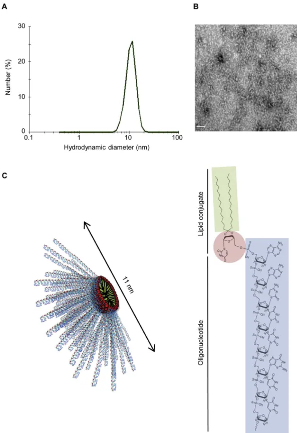
ASOs don't, as revealed by DLS and TEM experiments. The LASOs yielded small spherical objects of ~ 11 nm in diameter (Fig. 1A, B). TEM images of LASO samples dissolved in aqueous media (0.9% NaCl) showed micellar aggregates (Fig. 1B). The architecture of these assemblies can be explained based upon the molecular dimensions of 20 nucleobase-long LASOs (Fig. 1C). LASO molecules areflexible, with cone shape structure and dimensions compatible with the self-assembly
into spherical micelles. Nevertheless, the oligonucleotide sequence was found to influence the aggregation behavior of LASO amphiphiles since Control-LASO led to the stabilization of bigger nano-objects featuring diameters higher than 100 nm. The impact of the ON sequences on the aggregation was previously reported for rich purine or pyrimidine sequences[27]. As expected,ζ-potential measurement revealed nega-tive surface charge (~ 50 mV) of the nanomicelles (data not shown). Fig. 1. Physicochemical characterization of LASO nanomicelles. A) Hydrodynamic diameter of the nanomicelles formed by LASO self-assembly, determined by DLS. B) Representative TEM image of LASO nanomicelles. Scale bar: 100 nm. C) Schematic organization of the nanomicelles obtained (left) and chemical structure of nucleolipid-conjugated antisense oligonucleotides used in this study (right).
S. Karaki et al. Journal of Controlled Release 258 (2017) 1–9
3.3. TCTP-LASO penetrates faster than ASO, and both are internalized via macropinocytosis
It has long been believed that oligonucleotides, can't be efficiently used as silencing tools for in vitro studies, in the absence of a transfection agent such as oligofectamine. Indeed, for effective protein downregulation, it is essential that the oligonucleotides cross the cell membrane, reach the mRNA and inhibit protein translation. The LASOs described in this work might be able to overcome the need to use transfection agents in PS ASO oligonucleotides.
In order to assess the uptake efficiency of each oligonucleotide, we
transfected PC-3 cells by oligonucleotides conjugated tofluorescein, and flow cytometry analysis was performed to evaluate the mean percentage of fluorescent cells. Data showed that, in presence of oligofectamine, both TCTP-ASO and TCTP-LASO penetrated approxi-mately 100% of PC-3 cells. However, in the absence of transfection agents, < 10% of the cells were transfected with TCTP-ASO, whereas ~ 98% of PC-3 cells were successfully transfected with TCTP-LASO (Fig. 2A). Furthermore, confocal imaging experiments revealed the presence of TCTP-LASO inside the cell with or without oligofectamine, while TCTP-ASO was not observed inside the cell without oligofecta-mine (Fig. 2B). These results indicate that TCTP-LASO is able to cross Fig. 2. TCTP antisense uptake by PC-3 cells. A) Flow cytometry was used to quantify the percentage of PC-3 cells transfected with thefluorescent oligonucleotide, 4 h after transfection with and without oligofectamine. Data were normalized to Control-ASO-treated cells, and are shown as mean ± SEM for triplicate samples from a representative experiment of three performed. Two-tailed, unpaired Student's t-test, ***p≤ 0.001. B) Representative images of PC-3 transfected with FITC conjugated ASO or LAS0, 1 h after transfection with and without oligofectamine, and counterstained with DAPI. C) Representative images of PC-3 cells transfected with FITC-conjugated ASO (with oligofectamine) or LASO (without oligofectamine) for 10, 30 and 60 min, and counterstained with DAPI. D) Flow cytometry was used to quantify the percentage of PC-3 cells transfected with thefluorescent oligonucleotide in the presence of specific inhibitors of macropinocytosis (cytochalasin D), caveolae-mediated endocytosis (genistein), and clathrin-mediated endocytosis (chlorpromazine). Data were normalized to control cells (transfected in the absence of inhibitors) and are shown as mean ± SEM for triplicate samples from a representative experiment of three performed. Two-tailed, unpaired Student's t-test, *p≤ 0.05.
the cell membrane, without any transfection agent.
To evaluate the kinetics of TCTP-ASO and TCTP-LASO internaliza-tion, PC-3 cells were transfected with fluorescent TCTP-ASO (with oligofectamine) or TCTPO-LASO (without oligofectamine) for up to 10, 30 and 60 min. The uptake was then analyzed by confocalfluorescence microscopy. A significant cell staining appeared 10 min after transfec-tion with TCTP-LASO, while intracellular TCTP-ASO was detected at 30 min (Fig. 2C).
Finally, we assessed the intracellular delivery pathway of TCTP-ASO and TCTP-LASO in PC-3 cells. Almost 100% internalization was attained within 30 min (Fig. 2D). Neither the clathrin-mediated en-docytosis inhibitor (chlorpromazine) nor the caveolae-mediated endo-cytosis inhibitor (genistein) were able to influence the cellular uptake of TCTP-ASO or LASO oligonucleotides, whereas the macropinocytosis inhibitor (cytochalasin D) significantly decreased the internalization by 50% for TCTP-ASO, and 40% for TCTP-LASO (Fig. 2D), indicating that macropinocytosis might be involved in the uptake of these compounds.
3.4. TCTP-LASO downregulates TCTP expression and reduces cell viability, without additional transfection reagents
To assess the effect of TCTP-ASO and TCTP-LASO on TCTP expres-sion in vitro, we transfected PC-3 cells with different concentrations of ASO and LASO, and Western blot analysis was performed. As shown in Fig. 3, decreased TCTP expression was observed starting at a concen-tration of 70 nM TCTP-ASO, combined with oligofectamine (Fig. 3A). However, TCTP-ASO produced no effect without oligofectamine
(Fig. 3B). In contrast, TCTP-LASO had an inhibitory effect on TCTP expression with or without oligofectamine starting at 70 nM (Fig. 3A, B). To further investigate the efficiency of TCTP-LASO in the absence of Fig. 3. Western blot analysis of TCTP expression in PC-3 cells treated with the indicated
concentrations of Control-ASO, TCTP-ASO, Control-LASO or TCTP-LASO, with oligofec-tamine (A) or without oligofecoligofec-tamine (B). C) MTT viability assay of PC-3 cells untreated or transfected with Control-ASO, TCTP-ASO, Control-LASO or TCTP-LASO in the absence of oligofectamine. Data were normalized to untreated cells, and are shown as mean ± SEM, n = 3. Two-tailed, unpaired Student's t-test, **p≤ 0.01.
A
B
C
**
**
Control TCTP ASO L ASO 0 1000 2000 3000 4000 5000 6000 1 2 3 4 5 6 7 8 9 10 11 M e a n t umor volume ( m m 3) Time (weeks) Control-ASO TCTP-ASO Control-LASO TCTP-LASO***
*
Control-LASO TCTP-LASO TCTP GAPDH Control-ASO TCTP-ASO 22 kDa 37 kDa Co ntrol-ASO TCTP-ASO Co ntrol-L ASO TC TP-L ASO 0 20 40 60 80 100 120**
*
T C T P ex pr es s ion c om par ed t o G A P D H(caption on next page)
S. Karaki et al. Journal of Controlled Release 258 (2017) 1–9
transfection agents, compared to TCTP-ASO, a MTT assay was per-formed (Fig. 3C). A significant decrease of ~40% of cell viability was obtained after transfection with TCTP-LASO. However, no effect was observed for Control-ASO, TCTP-ASO and Control-LASO.
3.5. Lipid modification does not reduce the efficiency of the antisense therapy in murine xenograft models of CRPC
To test TCTP-LASO efficiency in vivo, we used a murine xenograft model. To induce tumors, PC-3 cells were injected subcutaneously into athymic nude mice. PC-3-tumor-bearing mice were divided into 4 groups and treated by serial intraperitoneal injections of Control-ASO, TCTP-ASO, Control-LASO or TCTP-LASO. In order to determine if TCTP-LASO conserves its ability to downregulate TCTP expression in vivo, 5 mice from each group were sacrificed after 1 week of treatment, and their tumors were harvested, protein extracted and TCTP expres-sion analyzed by Western blot. As shown inFig. 4A, decreased TCTP expression was observed for both TCTP-ASO and TCTP-LASO, com-pared to their respective controls. Furthermore, TCTP-LASO produced more inhibition of TCTP expression compared to TCTP-ASO. Treatment was maintained for 11 weeks, and tumor volume was measured weekly. Fig. 4B shows that tumors from mice treated with ASO or TCTP-LASO were significantly smaller at the end of the treatment. Mean tumor volume was ~ 2-fold higher in controls compared with TCTP-ASO- and TCTP-LTCTP-ASO-treated mice (Fig. 4B). Furthermore, tumors from animals treated with TCTP-LASO were smaller than those harvested from TCTP-ASO-treated mice (Fig. 4C), although no statistical signi fi-cance was obtained. Taken together, these data suggest that lipid modification of TCTP-ASO does not affect its capacity to downregulate TCTP expression in vivo, leading to delayed tumor progression. 3.6. In vivo toxic effects of TCTP-ASO and LASO
One of the main needs for these in vivo studies is to test for possible toxic effects resulting from oligonucleotide administration. Mice were routinely observed for signs of systemic toxicity throughout the treatment period (gastrointestinal symptoms, irregular respiration, aggressiveness or immobility); no signs were detected (Table 2).
Mouse body weight was also monitored, showing no obvious changes (Fig. 5A). At the end of the experiment, we tested serum and urine for signs of in vivo toxicity. Analysis of serum from TCTP-ASO- or TCTP-LASO-injected mice compared to controls showed no significant differences in creatinine levels (Fig. 5B).
In TCTP-ASO-treated mice we found increased levels of alanine aminotransferase (ALT), and aspartate aminotransferase (AST),
en-zymes associated mainly with hepatic damage (Fig. 5C). However, these increases did not compromise mouse survival, and induced no signs of animal distress. Treatment with TCTP-LASO did not induced increased aminotransferase levels, suggesting reduced toxicity com-pared to TCTP-ASO (Fig. 5C). Biochemical analysis of urine showed normal results for all parameters analyzed (Table 3).
4. Discussion
While it is easy to confer stability to antisense agents by chemical modifications, the improvement of pharmacokinetic parameters, cellu-lar uptake and bioavailability has proven to be much more challenging. As yet, very few oligonucleotides are on the market. In the present study we have tested a novel ASO lipid modification that might help overcome these limitations.
TCTP is abundantly expressed in many cancer types [28,29], including PC[30]. Although the role of TCTP in prostate cancer cells is not well understood, TCTP has been reported to cause resistance to androgen withdrawal and chemotherapy in PC[7,11]. Furthermore, a study showed that somatostatin, a growth inhibitory hormone, down-regulates the expression of TCTP in LNCaP cells[31], suggesting that manipulating the expression of TCTP can be a useful strategy to control cell growth. We have previously shown that TCTP is overexpressed in CRPC, and p53 expression and function are lost in this type of cancer. Furthermore, we developed and patented a first generation ASO to target and inhibit TCTP expression. We choose the ASO strategy since ASOs can be used for human therapy by inhibiting specifically target genes, especially those difficult to target with small chemical inhibitors or neutralizing antibodies. We have shown that TCTP-ASO significantly inhibits CRPC progression in pre-clinical models and restores p53 expression and function [7]. In order to improve the delivery and bioavailability of TCTP-ASO, we developed novel ASOs, modified using a lipid conjugate attached to the 5′ extremity of the oligonucleotide (Fig. 1). Numerous reports have demonstrated that naked oligonucleo-tides are internalized poorly by cells [32,33]. To improve cellular uptake and activity, a range of chemical modifications have been developed [18–20,22–24]. Indeed, free uptake of a 16-mer LNA PS gapmer ASO targeting bcl-2 was reported in several cell lines, unlike longer 20-mer PS ASO which is ineffectively internalized by cells[34]. Here we show that the addition of a lipid sequence at the 5′ extremity of this type of ASOs enhances its penetration inside the cells, and its efficiency in absence of other transfection agents, since TCTP-LASO uptake was faster compared to TCTP-ASO in PC-3 cells (Fig. 2). Cellular uptake of oligonucleotides is usually achieved by adsorptive endocy-tosis [35,36]. There are several endocytic pathways that could be involved in ASO penetration inside the cell, such as macropinocytosis, clathrin-mediated endocytosis, and caveolae-mediated endocytosis. Our results indicate that TCTP-ASO is internalized via macropinocytosis, confirming what has been described in literature. Indeed, Koller et al. show that unmodified oligonucleotides uptake pathway is endosomal, with the functional uptake pathway being clathrin and caveolin-independent in hepatocytes [37]. Furthermore, we show that lipid addition doesn't affect the internalization mode, since TCTP-LASO is also internalized by macropinocytosis (Fig. 2). Some studies conducted on siRNA delivery, show that diverse lipophilic conjugates, specifically cholesterol can mediate siRNA cellular uptake, through a common mechanism, leading to endogenous gene silencing in vivo[38,39]. The authors support the hypothesis that LDL and HDL particles mediate uptake of cholesterol-siRNA, by receptor-mediated internalization, in tissues[38]. Concerning siRNA formulated with lipoplexes and lipid nanoparticles, Wittrup et al. show by live-cell imaging that they are both taken up by endocytosis and accumulate in late endosomes and lysosomes. They show that siRNA release occurs from maturing endosomes [39]. Understanding the regulation of this release can improve cytosolic delivery of candidate nucleic acids.
In the present study we show that TCTP-ASO downregulates TCTP Fig. 4. Effect of Control-ASO, TCTP-ASO, Control-LASO and TCTP-LASO in PC-3 cell
xenografts. A) Western blot analysis of TCTP expression in PC-3 xenografts after 7 days of treatment with Control-ASO, TCTP-ASO, Control-LASO or TCTP-LASO. Bands were quantified using image J. TCTP expression compared to GAPDH was normalized for TCTP-ASO compared to control-ASO and TCTP-LASO compared to control-LASO. B) Mean tumor volume during treatment with Control-ASO, ASO, Control-LASO or TCTP-LASO. Data show mean ± SEM. Two-tailed, unpaired Student's t-test, *p≤ 0.05, **p≤ 0.01, ***p ≤ 0.001. C) Representative images of tumors collected from mice treated with Control-ASO, TCTP-ASO, Control-LASO or TCTP-LASO after an 11-week treatment.
Table 2
Analysis of mice behavior and signs of systemic toxicity during treatment. Symbols: +, normal; ± , slightly reduced without affecting the ability to obtain food or water.
Control ASO TCTP ASO Control LASO TCTP LASO
Body weight Normal Normal Normal Normal
Diarrhea Neg Neg Neg Neg
Respiration Normal Normal Normal Normal
Aggressiveness Neg Neg Neg Neg
expression when transfection is performed in the presence of oligofec-tamine (Fig. 3). The lipid modification of ASO makes it possible to obtain the same effect without any transfection agent added. We also observed that reduced levels of TCTP correlate with decreased PC-3 cell viability in vitro, confirming TCTP major role in cell growth and survival[28,29,40,41]. These results confirm what has been described in previous studies, in which gene silencing of TCTP by siRNA caused growth suppression and apoptosis of PC cells[8].
TCTP-LASO efficiency was confirmed in vivo. Serial intraperitoneal injections of both TCTP-ASO and TCTP-LASO lead to delayed tumor progression in a murine xenograft model of CRPC (Fig. 4). TCTP expression was downregulated in vivo after 7 days of treatment in both TCTP-ASO and TCTP-LASO groups compared to their respective con-trols (Fig. 4). These results are consistent with our previous work, in which systemic administration of TCTP-ASO suppressed PC-3 and LNCaP tumor growth in vivo, and significantly enhanced docetaxel activity[7].
The high rate of disappointing results of clinical trials using ASOs is mainly due to toxicity. Previous clinical and preclinical studies reported adverse effects of ASOs injury on liver and kidneys, two primary organs of oligonucleotide accumulation, as well as local reactions at the injection site [42–44]. It seems that oligonucleotides as a chemical class are particularly associated with these types of toxicities. Here we
show that, under the experimental conditions used, TCTP-ASO induced an increase in transaminases (Fig. 5). However the levels detected were within the range of normal values for male Swiss nude (Nu/Nu) mice [45], except for AST levels in one TCTP-ASO-treated mouse. In contrast, TCTP-LASO showed no adverse effects, suggesting that the treatment did not cause systemic toxicity to animals.
5. Conclusions
In this study we present a novel lipid modification at the 5′ extremity of ASO molecules. This lipid-modified antisense presents improved cell uptake and efficiency in the absence of additional transfection agents. Furthermore, we provided preclinical proof that the treatment with lipid-modified TCTP-ASO delays tumor progression in a murine xenograft model of PC, with reduced toxicity compared to TCTP-ASO.
Supplementary data to this article can be found online athttp://dx. doi.org/10.1016/j.jconrel.2017.04.042.
Competingfinancial interest
J.A., P.B., K.O. and P.R. are inventors on two patentsfiled by the Institut National de la Santé et de la Recherche Médicale (INSERM, France), entitled“Hydrophobically modified ASOs comprising a ketal group” (patent application number PCT/IB2013/001517) and “Hydrophobically modified antisense oligonucleotides comprising a triple alkyl chain” (patent application number PCT/EP2014/061762). Acknowledgments
S.K. receives a doctoral contract from INSERM and AMU, RM holds a post-doctoral contract from the Société d'Accélération du Transfert de Technologies (SATT) Sud Est. This work was supported by grants from the agence nationale de la recherché (ANR, ANR-11-EMMA-022-02), INSERM Transfert (RSE14046ASA), SATT (276-UAM2-SDV-11-INSE), ARTP (ARTP2014-01), AFU (AFU2015-NB), Vietnamese Goverment Fig. 5. In vivo toxicity of ASOs and LASOs. A) Body weight of Control-ASO-, TCTP-ASO-, Control-LASO- and TCTP-LASO-treated mice was monitored over the duration of the experiment. B, C) Biochemistry of blood samples collected from Control-ASO-, TCTP-ASO-, Control-LASO- and TCTP-LASO-treated mice at the end of the experiment. Serum concentrations of (B) creatinine, and (C) alanine aminotransferase (ALT) and aspartate aminotransferase (AST) were analyzed. Data are shown as mean ± SEM. Two-tailed, unpaired Student's t-test, *p≤ 0.05.
Table 3
Urine biochemical analysis results after treatment. Analysis performed using reactive test strips for urine. Symbols:−, negative; Tr, trace; ± , 10 erythrocytes/μl.
Control ASO TCTP ASO Control LASO TCTP LASO
Leucocytes − −/Tr −/Tr − Nitrite − − − − Protein −/Tr −/Tr −/Tr −/Tr pH 6.0 6.0 6.0 6.0 Blood ± ± ± ± Ketone − − − − Glucose − − − −
S. Karaki et al. Journal of Controlled Release 258 (2017) 1–9
Scolarship (844458G), ITMO (BioSysCall#A12171AS) and Amidex (AM14AVHRXX). Transmission electron microscopy experiments were performed at the Bordeaux Imaging Center, a service unit of the CNRS-INSERM and Bordeaux University, member of France-BioImaging, a national infrastructure for Biological Imaging.
References
[1] M. Malvezzi, G. Carioli, P. Bertuccio, T. Rosso, P. Boffetta, F. Levi, C. La Vecchia,
E. Negri, European cancer mortality predictions for the year 2016 with focus on
leukaemias, Ann. Oncol. 27 (2016) 725–731.
[2] R.L. Siegel, K.D. Miller, A. Jemal, Cancer statistics, 2016, CA Cancer J. Clin. 66
(2016) 7–30.
[3] A. Fusi, G. Procopio, S. Della Torre, R. Ricotta, G. Bianchini, R. Salvioni, L. Ferrari,
A. Martinetti, G. Savelli, S. Villa, E. Bajetta, Treatment options in
hormone-refractory metastatic prostate carcinoma, Tumori 90 (2004) 535–546.
[4] D.P. Petrylak, C.M. Tangen, M.H. Hussain, P.N. Lara Jr., J.A. Jones, M.E. Taplin,
P.A. Burch, D. Berry, C. Moinpour, M. Kohli, M.C. Benson, E.J. Small, D. Raghavan, E.D. Crawford, Docetaxel and estramustine compared with mitoxantrone and prednisone for advanced refractory prostate cancer, N. Engl. J. Med. 351 (2004)
1513–1520.
[5] I.F. Tannock, R. de Wit, W.R. Berry, J. Horti, A. Pluzanska, K.N. Chi, S. Oudard,
C. Theodore, N.D. James, I. Turesson, M.A. Rosenthal, M.A. Eisenberger, Docetaxel plus prednisone or mitoxantrone plus prednisone for advanced prostate cancer, N.
Engl. J. Med. 351 (2004) 1502–1512.
[6] R.J. van Soest, E.S. de Morree, L. Shen, I.F. Tannock, M.A. Eisenberger, R. de Wit,
Initial biopsy Gleason score as a predictive marker for survival benefit in patients with castration-resistant prostate cancer treated with docetaxel: data from the
TAX327 study, Eur. Urol. 66 (2014) 330–336.
[7] V. Baylot, M. Katsogiannou, C. Andrieu, D. Taieb, J. Acunzo, S. Giusiano, L. Fazli,
M. Gleave, C. Garrido, P. Rocchi, Targeting TCTP as a new therapeutic strategy in
castration-resistant prostate cancer, Mol. Ther. 20 (2012) 2244–2256.
[8] M. Gnanasekar, S. Thirugnanam, G. Zheng, A. Chen, K. Ramaswamy, Gene silencing
of translationally controlled tumor protein (TCTP) by siRNA inhibits cell growth and induces apoptosis of human prostate cancer cells, Int. J. Oncol. 34 (2009)
1241–1246.
[9] M. Kaarbo, M.L. Storm, S. Qu, H. Waehre, B. Risberg, H.E. Danielsen, F. Saatcioglu,
TCTP is an androgen-regulated gene implicated in prostate cancer, PLoS One 8
(2013) e69398.
[10] B.J. Rocca, A. Ginori, A. Barone, C. Calandra, F. Crivelli, G. De Falco, S. Gazaneo,
S. Tripodi, G. Cevenini, M.T. del Vecchio, M.R. Ambrosio, P. Tosi, Translationally controlled tumor protein in prostatic adenocarcinoma: correlation with tumor
grading and treatment-related changes, Biomed. Res. Int. 2015 (2015) 985950.
[11] J. Acunzo, V. Baylot, A. So, P. Rocchi, TCTP as therapeutic target in cancers, Cancer
Treat. Rev. 40 (2014) 760–769.
[12] U.A. Bommer, B.J. Thiele, The translationally controlled tumour protein (TCTP),
Int. J. Biochem. Cell Biol. 36 (2004) 379–385.
[13] S.M. Macdonald, Potential role of histamine releasing factor (HRF) as a therapeutic
target for treating asthma and allergy, J. Asthma Allergy 5 (2012) 51–59.
[14] S.M. MacDonald, T. Rafnar, J. Langdon, L.M. Lichtenstein, Molecular identification
of an IgE-dependent histamine-releasing factor, Science 269 (1995) 688–690.
[15] M. Nagano-Ito, S. Ichikawa, Biological effects of mammalian translationally
controlled tumor protein (TCTP) on cell death, proliferation, and tumorigenesis,
Biochem. Res. Int. 2012 (2012) 204960.
[16] R. Amson, S. Pece, J.C. Marine, P.P. Di Fiore, A. Telerman, TPT1/TCTP-regulated
pathways in phenotypic reprogramming, Trends Cell Biol. 23 (2013) 37–46.
[17] M. Tuynder, G. Fiucci, S. Prieur, A. Lespagnol, A. Geant, S. Beaucourt, D. Duflaut,
S. Besse, L. Susini, J. Cavarelli, D. Moras, R. Amson, A. Telerman, Translationally controlled tumor protein is a target of tumor reversion, Proc. Natl. Acad. Sci. U. S.
A. 101 (2004) 15364–15369.
[18] P.M. Moreno, A.P. Pego, Therapeutic antisense oligonucleotides against cancer:
hurdling to the clinic, Front. Chem. 2 (2014) 87.
[19] J.K. Watts, D.R. Corey, Silencing disease genes in the laboratory and the clinic, J.
Pathol. 226 (2012) 365–379.
[20] J. Winkler, Oligonucleotide conjugates for therapeutic applications, Ther. Deliv. 4
(2013) 791–809.
[21] W.R. Epa, P. Rong, P.F. Bartlett, E.J. Coulson, G.L. Barrett, Enhanced
down-regulation of the p75 nerve growth factor receptor by cholesteryl and bis-cholesteryl antisense oligonucleotides, Antisense Nucleic Acid Drug Dev. 8 (1998)
489–498.
[22] G. Godeau, C. Staedel, P. Barthélémy, Lipid-conjugated oligonucleotides via "click
chemistry" efficiently inhibit hepatitis C virus translation, J. Med. Chem. 51 (2008)
4374–4376.
[23] S. Ellipilli, R. Murthy, K.N. Ganesh, Perfluoroalkylchain conjugation as a new tactic
for enhancing cell permeability of peptide nucleic acids (PNAs) via reducing the
nanoparticle size, Chem. Commun. 52 (2016) 521–524.
[24] G. Godeau, H. Arnion, C. Brun, C. Staedel, P. Barthélémy, Fluorocarbon
oligonu-cleotide conjugates for nucleic acids delivery, Med. Chem. Commun. 1 (2010)
76–78.
[25] A. Aime, N. Beztsinna, A. Patwa, A. Pokolenko, I. Bestel, P. Barthelemy, Quantum
dot lipid oligonucleotide bioconjugates: toward a new anti-microRNA
nanoplat-form, Bioconjug. Chem. 24 (2013) 1345–1355.
[26] A. Gissot, C. Di Primo, I. Bestel, G. Giannone, H. Chapuis, P. Barthelemy, Sensitive
liposomes encoded with oligonucleotide amphiphiles: a biocompatible switch,
Chem. Commun. (Camb.) (2008) 5550–5552.
[27] A. Pokolenko, A. Gissot, B. Vialet, K. Bathany, T. Alain, P. Barthélémy, Lipid
oligonucleotide conjugates as responsive nanomaterials for drug delivery, J. Mater.
Chem. B 1 (2013) 5329–5334.
[28] F. Li, D. Zhang, K. Fujise, Characterization of fortilin, a novel antiapoptotic protein,
J. Biol. Chem. 276 (2001) 47542–47549.
[29] M. Tuynder, L. Susini, S. Prieur, S. Besse, G. Fiucci, R. Amson, A. Telerman,
Biological models and genes of tumor reversion: cellular reprogramming through
tpt1/TCTP and SIAH-1, Proc. Natl. Acad. Sci. U. S. A. 99 (2002) 14976–14981.
[30] F. Arcuri, S. Papa, A. Carducci, R. Romagnoli, S. Liberatori, M.G. Riparbelli,
J.C. Sanchez, P. Tosi, M.T. del Vecchio, Translationally controlled tumor protein (TCTP) in the human prostate and prostate cancer cells: expression, distribution,
and calcium binding activity, Prostate 60 (2004) 130–140.
[31] Z. Liu, S. Bengtsson, M. Krogh, M. Marquez, S. Nilsson, P. James, A. Aliaya,
A.R. Holmberg, Somatostatin effects on the proteome of the LNCaP cell-line, Int. J.
Oncol. 30 (2007) 1173–1179.
[32] C.F. Bennett, M.Y. Chiang, H. Chan, J.E. Shoemaker, C.K. Mirabelli, Cationic lipids
enhance cellular uptake and activity of phosphorothioate antisense
oligonucleo-tides, Mol. Pharmacol. 41 (1992) 1023–1033.
[33] G.D. Gray, S. Basu, E. Wickstrom, Transformed and immortalized cellular uptake of
oligodeoxynucleoside phosphorothioates, 3′-alkylamino oligodeoxynucleotides, 2′-O-methyl oligoribonucleotides, oligodeoxynucleoside methylphosphonates, and
peptide nucleic acids, Biochem. Pharmacol. 53 (1997) 1465–1476.
[34] C.A. Stein, J.B. Hansen, J. Lai, S. Wu, A. Voskresenskiy, A. Hog, J. Worm,
M. Hedtjarn, N. Souleimanian, P. Miller, H.S. Soifer, D. Castanotto,
L. Benimetskaya, H. Orum, T. Koch, Efficient gene silencing by delivery of locked nucleic acid antisense oligonucleotides, unassisted by transfection reagents, Nucleic
Acids Res. 38 (2010) e3.
[35] S.L. Loke, C.A. Stein, X.H. Zhang, K. Mori, M. Nakanishi, C. Subasinghe, J.S. Cohen,
L.M. Neckers, Characterization of oligonucleotide transport into living cells, Proc.
Natl. Acad. Sci. U. S. A. 86 (1989) 3474–3478.
[36] L.A. Yakubov, E.A. Deeva, V.F. Zarytova, E.M. Ivanova, A.S. Ryte, L.V. Yurchenko,
V.V. Vlassov, Mechanism of oligonucleotide uptake by cells: involvement of specific
receptors? Proc. Natl. Acad. Sci. U. S. A. 86 (1989) 6454–6458.
[37] E. Koller, T.M. Vincent, A. Chappell, S. De, M. Manoharan, C.F. Bennett,
Mechanisms of single-stranded phosphorothioate modified antisense
oligonucleo-tide accumulation in hepatocytes, Nucleic Acids Res. 39 (2011) 4795–4807.
[38] C. Wolfrum, S. Shi, K.N. Jayaprakash, M. Jayaraman, G. Wang, R.K. Pandey,
K.G. Rajeev, T. Nakayama, K. Charrise, E.M. Ndungo, T. Zimmermann, V. Koteliansky, M. Manoharan, M. Stoffel, Mechanisms and optimization of in vivo
delivery of lipophilic siRNAs, Nat. Biotechnol. 25 (2007) 1149–1157.
[39] A. Wittrup, A. Ai, X. Liu, P. Hamar, R. Trifonova, K. Charisse, M. Manoharan,
T. Kirchhausen, J. Lieberman, Visualizing lipid-formulated siRNA release from
endosomes and target gene knockdown, Nat. Biotechnol. 33 (2015) 870–876.
[40] T.H. Chan, L. Chen, X.Y. Guan, Role of translationally controlled tumor protein in
cancer progression, Biochem. Res. Int. 2012 (2012) 369384.
[41] S.H. Chen, P.S. Wu, C.H. Chou, Y.T. Yan, H. Liu, S.Y. Weng, H.F. Yang-Yen, A
knockout mouse approach reveals that TCTP functions as an essential factor for cell proliferation and survival in a tissue- or cell type-specific manner, Mol. Biol. Cell 18
(2007) 2525–2532.
[42] P.P. Seth, A. Siwkowski, C.R. Allerson, G. Vasquez, S. Lee, T.P. Prakash,
E.V. Wancewicz, D. Witchell, E.E. Swayze, Short antisense oligonucleotides with novel 2′-4′ conformationaly restricted nucleoside analogues show improved
po-tency without increased toxicity in animals, J. Med. Chem. 52 (2009) 10–13.
[43] R. Stanton, S. Sciabola, C. Salatto, Y. Weng, D. Moshinsky, J. Little, E. Walters,
J. Kreeger, D. DiMattia, T. Chen, T. Clark, M. Liu, J. Qian, M. Roy, R. Dullea, Chemical modification study of antisense gapmers, Nucleic Acid Ther. 22 (2012)
344–359.
[44] E.E. Swayze, A.M. Siwkowski, E.V. Wancewicz, M.T. Migawa, T.K. Wyrzykiewicz,
G. Hung, B.P. Monia, C.F. Bennett, Antisense oligonucleotides containing locked nucleic acid improve potency but cause significant hepatotoxicity in animals,
Nucleic Acids Res. 35 (2007) 687–700.
[45] Normal Blood Biochemistry Values for Nude Mice. Charles River Laboratories. http://www.criver.com/files/pdfs/rms/nunu/rm_rm_r_nunu_mouse_clinical_