HAL Id: hal-01902060
https://hal-amu.archives-ouvertes.fr/hal-01902060
Submitted on 23 Oct 2018
HAL is a multi-disciplinary open access
archive for the deposit and dissemination of
sci-entific research documents, whether they are
pub-lished or not. The documents may come from
teaching and research institutions in France or
abroad, or from public or private research centers.
L’archive ouverte pluridisciplinaire HAL, est
destinée au dépôt et à la diffusion de documents
scientifiques de niveau recherche, publiés ou non,
émanant des établissements d’enseignement et de
recherche français ou étrangers, des laboratoires
publics ou privés.
Gene Expression Analysis Reveals Genes Common to
Cerebral Malaria and Neurodegenerative Disorders
Sandrine Cabantous, Ogobara Doumbo, Belco Poudiougou, Laurence Louis,
Abdoulaye Barry, Aboubacar Oumar, Abdoualye Traore, S. Marquet, Alain
Dessein
To cite this version:
Sandrine Cabantous, Ogobara Doumbo, Belco Poudiougou, Laurence Louis, Abdoulaye Barry, et
al.. Gene Expression Analysis Reveals Genes Common to Cerebral Malaria and Neurodegenerative
Disorders. Journal of Infectious Diseases, Oxford University Press (OUP), 2017, 216 (6), pp.771 - 775.
�10.1093/infdis/jix359�. �hal-01902060�
HAL Id: hal-01902060
https://hal-amu.archives-ouvertes.fr/hal-01902060
Submitted on 23 Oct 2018
HAL is a multi-disciplinary open access
archive for the deposit and dissemination of
sci-entific research documents, whether they are
pub-lished or not. The documents may come from
teaching and research institutions in France or
abroad, or from public or private research centers.
L’archive ouverte pluridisciplinaire HAL, est
destinée au dépôt et à la diffusion de documents
scientifiques de niveau recherche, publiés ou non,
émanant des établissements d’enseignement et de
recherche français ou étrangers, des laboratoires
publics ou privés.
Gene Expression Analysis Reveals Genes Common to
Cerebral Malaria and Neurodegenerative Disorders
Sandrine Cabantous, Ogobara Doumbo, Belco Poudiougou, Laurence Louis,
Abdoulaye Barry, Aboubacar Oumar, Abdoualye Traore, S. Marquet, Alain
Dessein
To cite this version:
Sandrine Cabantous, Ogobara Doumbo, Belco Poudiougou, Laurence Louis, Abdoulaye Barry, et
al.. Gene Expression Analysis Reveals Genes Common to Cerebral Malaria and Neurodegenerative
Disorders. Journal of Infectious Diseases, Oxford University Press (OUP), 2017, 216 (6), pp.771 - 775.
<10.1093/infdis/jix359>. <hal-01902060>
Gene Expression Analysis Reveals
Genes Common to Cerebral Malaria
and Neurodegenerative Disorders
Sandrine Cabantous,1 Ogobara Doumbo,3 Belco Poudiougou,3 Laurence Louis,2Abdoulaye Barry,4 Aboubacar A. Oumar,5 Abdoualye Traore,4 Sandrine Marquet,1,a
and Alain Dessein1,a
1INSERM UMR906, GIMP, Labex ParaFrap, and 2INSERM UMR910, GMGF,
Aix-Marseille University, Aix-Marseille, France; 3Malaria Research and Training Center,
Department of Epidemiology of Parasitic Disease, Faculty of Medicine, USTTB,
4Pediatric Wards, Gabriel Toure Hospital, and 5Centre des Oeuvres Universitaires,
University of Bamako, Mali
Cerebral malaria, a reversible encephalopathy affecting young children, is a medical emergency requiring urgent clinical assessment and treatment. We performed a whole-transcrip-tomic analysis of blood samples from Malian children with cerebral or uncomplicated malaria. We focused on transcripts from pathways for which dysfunction has been associated with neurodegenerative disorders. We found that SNCA, SIAH2,
UBB, HSPA1A, TUBB2A, and PINK1 were upregulated
(fold-in-creases, ≥2.6), whereas UBD and PSMC5 were downregulated (fold-decreases, ≤4.39) in children with cerebral malaria, com-pared with those with uncomplicated malaria. These findings provide the first evidence for pathogenic mechanisms common to human cerebral malaria and neurodegenerative disorders.
Keywords. cerebral malaria; human; PBMCs; mRNA;
expression; neurodegenerative disorders; common pathways. Cerebral malaria (CM), one of the most frequent severe compli-cations of Plasmodium falciparum infection, is a diffuse reversible encephalopathy characterized by seizures and a loss of conscious-ness. It mostly occurs in young children and is a major cause of death. One third of survivors develop epilepsy or other neurologi-cal sequels. CM is thought to result from the sequestration of par-asites in the small blood vessels of the brain and the dysregulation of key immune system elements, with high levels of inflammation, breakdown of the blood-brain barrier, and brain swelling. Host genetic determinants make a major contribution to disease sever-ity and the outcome of infection. However, the cellular and molec-ular regulatory mechanisms underlying the pathogenesis of CM are not fully understood. Knowledge of these mechanisms would lead to improvements in diagnosis and treatment and better
clinical outcomes. Analyses of differential gene expression consti-tute a powerful approach for identifying suitable biological indica-tors for predicting the eventual outcome of P. falciparum infection, making it possible to provide appropriate treatment more rapidly. Recent complementary DNA microarray studies have shown significant differences between the gene expression profiles of CM-resistant and CM-susceptible mice [1]. In humans, tran-scriptomic analyses of RNA extracted from the peripheral blood mononuclear cells (PBMCs) of malarial patients infected with
P. falciparum revealed that changes in the expression of human
innate immune pathway genes were correlated with the severity of malaria [2]. Whole-blood transcriptomic analyses have also provided insight into pathogenesis and have been used to identify biomarkers of neurodegenerative and psychiatric disorders [3] or of other infectious diseases, such as leprosy.
The aim of this study was to use blood-based gene expres-sion profiles to identify biological pathways and molecules potentially reflecting the pathological mechanisms of CM. We used a gene selection strategy combining expression profiling and pathway analysis to focus on genes displaying changes in expression also known to be associated with neurological dis-orders. We show here, for the first time, that CM and neurode-generative disorders have pathogenic mechanisms in common. METHODS
This study was approved by the local ethics committees of the Faculty of Medicine, Pharmacy, and Odonto-Stomatology of the University of Bamako and by French ethics committees, including those of INSERM, the Comité de Protection des Personnes (protocol 212 CO2), the Ministère de l’Enseignement Supérieur et de la Recherche (protocol DC-2011-1426), and the Commission Nationale de l’Informatique et des Libertés (pro-tocol 1564177). All experimental methods were performed in accordance with the Declaration of Helsinki. Written informed consent was obtained from all parents.
Patients
CM was defined according to World Health Organization cri-teria as a state of unarousable coma with a Blantyre coma scale score of <2, a hematocrit of >16%, and parasitemia with asex-ual stages of P. falciparum. Meningitis was ruled out by lum-bar puncture. Subjects with uncomplicated malaria (UM) had a thick blood film positive for P. falciparum, a Blantyre coma scale score of 5, and a hematocrit of >26%. The children with UM had never developed CM. Malian children were recruited through the Pediatrics Department of Gabriel Toure Hospital in Bamako, as part of a larger prospective field study.
In this study, we analyzed gene expression in 13 patients with CM (male to female ratio, 7:6; mean age [±SD], 6.2 ± 3.8 years)
aS. M. and A. D. are co–last authors.
Correspondence: S. Marquet, PhD, INSERM UMR906, Faculté de Médecine Timone, 27 bd Jean Moulin, 13385 Marseille, Cedex 5, France (sandrine.marquet@univ-amu.fr). DOI: 10.1093/infdis/jix359
15 September
and 12 patients with UM matched for age and sex (male to female ratio, 6:6; mean age [±SD], 6.5 ± 3.6 years). Blood sam-ples were collected before the treatment of the children.
RNA Extraction
A peripheral blood sample was collected from each participant, and peripheral blood mononuclear cells (PBMCs) were isolated within 1 hour after sample collection. Total RNA was extracted with TRIzol reagent (Life Technologies) according to the man-ufacturer’s instructions but with an additional purification step performed with the RNA Clean and Concentrator kit (Zymo Research). The quality of total RNA was assessed with an RNA LabChip (Agilent 2100 Bioanalyzer, Agilent). Only samples with an RNA integrity number of >8 were used for further analysis.
Whole-Transcriptome Analysis
The samples of high-quality RNA were marked with Cy3 fluo-rescent dye, and then they were hybridized using the SurePrint G3 Human Gene Expression 8 × 60K microarray kit, version 1 (Agilent), according to the manufacturer’s instructions. The slides were composed of 8 high-definition 60-mer oligo-microarrays. Each microarray contained about 22 700 genes based on RefSeq and 7419 large intergenic noncoding RNAs. Fold-changes and P values were calculated for each gene, using Genespring Software (Agilent). The moderated t test and the Benjamini-Hochberg correction were used to obtain the P value. Pathway analysis was also performed with Genespring Software.
Validation of Gene Expression Data by Quantitative Real-Time Polymerase Chain Reaction (PCR) Analysis
We reverse-transcribed 400 ng of total RNA to generate comple-mentary DNA. The expression of selected genes was analyzed by TaqMan PCR, on an ABI 79000 real-time PCR thermocycler (Life Technologies), according to the manufacturer’s instruc-tions. The data were normalized with 2 housekeeping genes (RPLP0 and GAPDH). Data were analyzed by the ΔΔCt method (2-ΔΔCt), with Thermo-Fisher software (cloud).
RESULTS
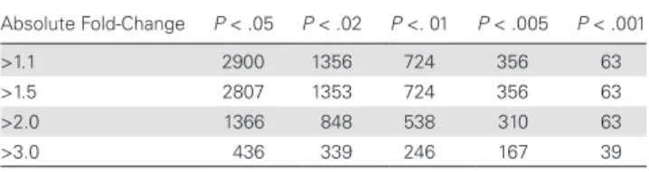
We characterized the biological pathways dysregulated during CM by analyzing the transcriptomes of 7 children with CM and 8 children with UM who were selected at random. In total, 2900 probe sequences differed significantly between the CM and UM groups, with an absolute fold-change of >1.1 and a P value of <.05, even after Benjamini-Hochberg correction (Table 1). In total, 436 probe sequences were identified as differentially expressed, yielding an absolute fold-change of >3 (Table 1): 403 of these sequences were upregulated and 33 downregulated in children with CM. GeneSpring pathway analysis revealed that a number of different Wiki biological pathways were significantly associated with the disease (Table 2). Remarkably, several genes from pathways associated with neurodegenerative disorders
were substantially overexpressed or underexpressed in patients with CM relative to those with UM (Table 2). In particular, 18 genes from the parkin-ubiquitin proteasomal system, 14 from the proteasome degradation pathway, and 6 from the Parkinson disease pathway were found to be differentially expressed between the 2 groups. Six genes from these 3 pathways were sig-nificantly more strongly expressed in children with CM than in those with UM, with fold-increases of >2: SNCA, SIAH2, UBB,
HSPA1A, TUBB2A, and PINK1 (Table 3). Four genes, UBD,
UBE1L, PSMC5, and PSMD5, were downregulated in children
with CM, with fold-decreases of ≤4.39 (Table 3).
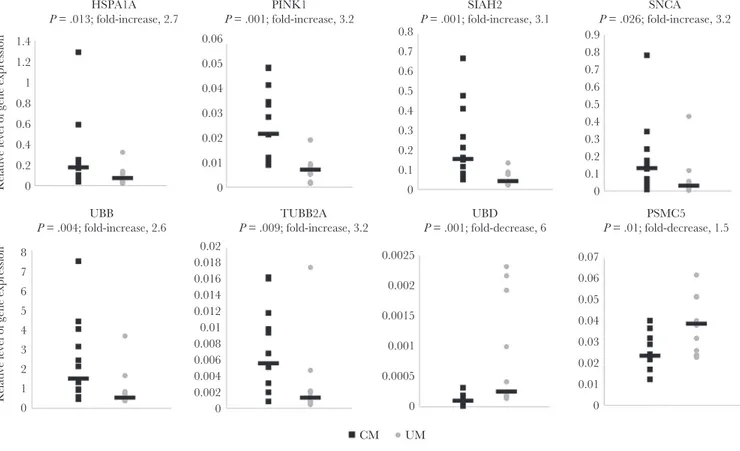
We investigated whether the changes in expression observed on microarray analysis for these 10 selected genes could be con-firmed in a larger sample set of 13 children with CM and 12 with UM. Real-time PCR validation was performed in duplicate and confirmed the significant difference in expression levels for 8 genes. The relative expression levels (2-ΔCT) for these genes are
Table 1. Total Number of Genes Differentially Expressed Between Children With Cerebral Malaria and Those With Uncomplicated Malaria, According to Absolute Fold-Changes in Expression and P Values for Differences in Expression Absolute Fold-Change P < .05 P < .02 P <. 01 P < .005 P < .001 >1.1 2900 1356 724 356 63 >1.5 2807 1353 724 356 63 >2.0 1366 848 538 310 63 >3.0 436 339 246 167 39
P values were determined after Benjamini-Hochberg correction for multiple testing, with
values of <.05 considered statistically significant.
Table 2. Partial Results of Pathways Analysis
Pathway No. of Differentially Expressed Genes Total No. of Genes P Parkin-Ubiquitin Proteasomal system_WP2359_53587 18 73 2.9 × 10⁻⁶ Proteasome Degradation_WP183_45274 14 65 2.7 × 10 -4 Mitochondrial Gene Expression_WP391_48245 7 19 3.3 × 10 -4 Senescence and Autophagy_WP615_47912 19 106 3.9 × 10 -4 Glycolysis and Gluconeogenesis_WP534_51732 10 49 .0034 Synthesis and Degradation of Ketone
Bodies_WP311_43510 3 5 .0039 Inflammatory
Response_WP453_41201
7 33 .0064 Spinal Cord Injury_WP2431_55909 15 102 .008 Tryptophan
Metabolism_WP465_43616 8 47 .022 Parkinson_Disease_WP2371_29836 6 38 .043
The WikiPathways in which the genes were known to be involved are listed. We show only the pathways containing at least 3 dysregulated genes and known to be involved in neurodegenerative disorders or identified in gene expression analysis in a mouse model of malaria.
presented in Figure 1. The difference in expression between sub-jects with CM and those with UM, expressed as a fold-increase, was 3.2 for SNCA (P = .026), 3.1 for SIAH2 (P = .001), 2.6 for UBB (P = .004), 2.7 for HSPA1A (P = .013), 3.2 for TUBB2A (P = .009), and 3.2 for PINK1 (P = .001), whereas for UBD and PSMC5, we obtained a fold-decrease of 6 (P = .001) and 1.5 (P = .01), respec-tively. We then analyzed the relationship between the expression levels of these genes in patients with CM and those with UM. We found significant positive correlations between expression lev-els for each pair-wise comparison between UBB, SIAH2, SNCA,
TUBB2A, and PINK1 (P < .01; Pearson correlation, >0.8). We
found significant negative correlations between expression lev-els in children for UBD and PINK1 (P = .01; Pearson correla-tion, – 0.646) and for UBD and SIAH2, SNCA, TUBB2A, and
UBB (P < .05; Pearson correlation, less than –0.4).
Discussion
In this study, we identified human genes and pathways involved in CM, suggesting possible targets for drug development. The key findings of this study are (1) the efficacy of this approach for identifying changes in the levels in human peripheral blood of transcripts involved in both CM and neurological disor-ders, Parkinson disease, and Alzheimer disease; (2) the over-lap between the pathways dysregulated in both the human and mouse model of CM, such as glycolysis/gluconeogenesis, tryp-tophan metabolism, immune responses, and brain function [1]; and (3) the confirmation that the molecular profiling of PBMCs can reflect physiological and pathological events in the brain, as previously suggested by Mohr et al [4].
We focused here on genes and pathways that have been clearly implicated in neurological disorders: the intercon-nected parkin, Parkinson, and proteasome pathways, in par-ticular. These pathways were considered to be of particular interest because they had never before been studied in human malaria, unlike immune response pathways [2, 5]. We showed that SNCA, SIAH2, HSPA1A, TUBB2A, PINK1, and UBB were expressed significantly more strongly in patients with CM than in patients with UM, whereas UBD and PSMC5 from the pro-teasome degradation pathway were expressed less strongly. Our findings suggest that transcriptomic analyses of peripheral blood can provide important insights into pathogenic mecha-nisms affecting the brain. These findings are entirely consistent with those of Liew et al [6], indicating significant similarities in gene expression between the blood and brain (>80%) and showing that the profiling of gene expression in the blood could be used for diagnostic and prognostic purposes.
SNCA, which encodes α-synuclein, a protein expressed
pre-dominantly in the brain, has been identified as the major causal gene in several neurodegenerative disorders. There is growing evidence to suggest that the overproduction and aggregation of this protein lead to brain dysfunction that is sufficient to cause disease [7]. We found that SNCA was overexpressed in children with CM, suggesting a possible deleterious role of this gene in this disease. Another gene from the same pathway, SIAH2, was also overexpressed in children with CM. This gene encodes an E3 ubiquitin-conjugating enzyme that plays a critical role in the hypoxia response [8], which is frequently observed in patients with CM. Under hypoxia, SIAH2 messenger RNA
Table 3. Genes Related to the Parkin, Parkinson, and Proteasome Degradation Pathways Displaying Differential Expression Between Cerebral Malaria and Uncomplicated Malaria in Children
Pathway(s) Gene Probe Name
Absolute
Fold-Change Regulation Status Pa FDRb
Parkin_WP2359_53587 HSPA1A A_23_P111132 3.27 Upregulated .028 .044 Parkin_WP2359_53587,
Parkinson_WP2371_29836 SNCA A_23_P29939 2.92 Upregulated .028 .044 Parkin_WP2359_53587 SIAH2 A_33_P3358342 2.85 Upregulated .0044 .039 Parkinson_WP2371_29836 PINK1 A_23_P23194 2.59 Upregulated .013 .041 Parkin_WP2359_53587 TUBB2A A_33_P3365002 2.31 Upregulated .028 .044 Parkinson_WP2371_29836,
Proteasome_WP183_45274
UBB A_23_P27215 2.11 Upregulated .04 .047 Proteasome_WP183_45274 UBD A_23_P81898 4.39 Downregulated .010 .040 Proteasome_WP183_45274,
Parkinson_WP2371_29836
UBE1L (UBA7) A_23_P21207 1.87 Downregulated .0068 .039
Proteasome_WP183_45274, Parkin_WP2359_53587 PSMC5 A_23_P164035 1.59 Downregulated .012 .041 Proteasome_WP183_45274, Parkin_WP2359_53587 PSMD5 A_33_P3305254 1.59 Downregulated .015 .042
aBy the moderated t test, with values of <.05 considered statistically significant.
bThe false-discovery rates (FDRs) represent significant results (defined as those with P values of <.05) after Benjamini-Hochberg correction for multiple testing.
Abbreviations: HSPA1A, encodes heat shock 70-kD protein 1; PINK1, encodes PTEN-induced putative kinase 1; PSMC5, encodes proteasome 26S subunit ATPase 5; PSMD5, encodes proteasome 26S subunit non-ATPase 5; SIAH2, encodes SIAH E3 ubiquitin protein ligase family member 1; SNCA, encodes α-synuclein; TUBB2A, encodes tubulin β2A; UBB, encodes ubiquitin B; UBD, encodes ubiquitin D; UBE1L (UBA7), encodes ubiquitin activating enzyme 7.
levels increase and the encoded protein interacts with α-sy-nuclein, promoting its aggregation, which is toxic. The for-mation of these aggregates is prevented by the suppression of endogenous SIAH2 expression. PTEN-induced kinase 1 (PINK1) is a mitochondrial kinase that is strongly expressed in the brain and promotes cell survival, particularly under conditions of oxidative stress. It accumulates on the surface of defective mitochondria and recruits parkin, thereby promot-ing the selective degradation of dysfunctional mitochondria. Abnormal PINK1 expression may be a causal factor in the development of mitochondrial dysfunction leading to neuro-degenerative and neuroinflammatory disorders [9]. Despite its demonstrated protective effects, PINK1 has been shown to be present at abnormally high levels in the brains of individuals with Alzheimer disease or multiple sclerosis [10], as observed in the blood of children with CM. TUBB2A, which encodes β-tubulin, was also overexpressed during CM. Mutations of this gene cause infantile-onset epilepsy and are associated with impaired brain development in humans, but the underlying pathogenic mechanism is unknown [11].
Molecular chaperones and the ubiquitin proteasome system are the first and second lines of defense against protein mis-folding and aggregation. HSP-70, encoded by HSPA1A, is a
chaperone protein that plays an important role in the refold-ing of misfolded proteins and the targetrefold-ing of proteins for proteasomal degradation. A loss of Hsp70 activity has been associated with neurodegeneration and the formation of amy-loid deposits of α-synuclein in Parkinson disease. However, the ability of HSP-70 to inhibit the aggregation of α-synu-clein depends on factors such as nucleotide binding and the presence of the Hip cochaperone. In the presence of ATP (or ADP), Hsp70 overexpression may reduce α-synuclein toxicity but without preventing the highly cytotoxic accumulation of amyloid aggregates in tissue [12]. We found that HSPA1A was overexpressed in children with CM, as in patients with epi-lepsy, in whom HSP-70 levels are correlated with the duration and intensity of seizures, suggesting that this protein may be a marker of seizure-related brain injury [13]. Inflammation and oxidative stress, characteristic features of CM, may account for the increase in HSPA1A transcript levels in this disease and may reflect the severity of the injury. Nevertheless, further studies are required to determine whether HSP-70 is a cause or a consequence in the pathogenesis of CM. The ubiquitin proteasome system is an intracellular protein degradation sys-tem that plays an important role in maintaining intracellular homeostasis [14, 15]. The impairment of this system leads to
1.4 0.06 0.8 0.7 0.6 0.5 0.4 0.3 0.2 0.1 0 0.9 0.8 0.7 0.6 0.5 0.4 0.3 0.2 0.1 0 0.05 0.04 0.03 0.02 0.01 0 0.02 0.0025 0.002 0.0015 0.001 0.0005 CM UM 0 0 0.01 0.02 0.03 0.04 0.05 0.06 0.07 0.018 0.016 0.014 0.012 0.01 0.008 0.006 0.004 0.002 0 1.2 1 0.8 0.6 HSPA1A P = .013; fold-increase, 2.7 0.4
Relative level of gene expression
Relative level of gene expression
0.2 0 8 7 6 5 4 3 2 1 0 PINK1 P = .001; fold-increase, 3.2 SIAH2 P = .001; fold-increase, 3.1 SNCA P = .026; fold-increase, 3.2 UBB
P = .004; fold-increase, 2.6 P = .009; fold-increase, 3.2TUBB2A P = .001; fold-decrease, 6UBD P = .01; fold-decrease, 1.5PSMC5
Figure 1. Real-time polymerase chain reaction–based validation of messenger RNA levels for the 8 most significantly dysregulated genes from the three selected pathways
(parkin, Parkinson and proteasome degradation). Samples from 13 children with cerebral malaria (CM) and 12 with uncomplicated malaria (UM) were analyzed. Relative expression levels were calculated from 2-ΔCt values. Values for children with CM are represented by black squares, and values for children with UM are represented by gray
protein accumulation, resulting in neurodegenerative dis-eases, such as Alzheimer disease and Parkinson disease. The upregulation of UBB and the downregulation of UBD and
PSMC5 in children with CM suggest that the ubiquitin
pro-teasome system is disturbed. Exogenous stress, mitochondrial alterations, and α -synuclein overexpression may also promote disruption of the ubiquitin proteasome system, with poten-tially protein aggregates.
In conclusion, our results suggest that CM has pathogenic mechanisms in common with other brain disorders involving protein aggregation. They also show that transcriptomic analy-sis of PBMCs is an efficient method for investigating changes in the brain. The identification of new molecules involved in CM pathogenesis could improve diagnostic capabilities or facilitate the development of new treatments.
Notes
Acknowledgments. We thank the children and their parents
for participating in this study.
Financial support. This work was supported by the
French Research Ministry, the Institut National de la Santé et de la Recherche Médicale, the European Union (IC18-CT98 0373), and the French Parasitology Alliance for Health Care (ANR-11-LABX-0024-01).
Potential conflicts of interest. All authors: No reported
con-flicts of interest. All authors have submitted the ICMJE Form for Disclosure of Potential Conflicts of Interest. Conflicts that the editors consider relevant to the content of the manuscript have been disclosed.
References
1. Delahaye NF, Coltel N, Puthier D, et al. Gene expression analysis reveals early changes in several molecular pathways in cerebral malaria-susceptible mice versus cerebral malar-ia-resistant mice. BMC Genomics 2007; 8:452.
2. Yamagishi J, Natori A, Tolba ME, et al. Interactive transcrip-tome analysis of malaria patients and infecting Plasmodium falciparum. Genome Res 2014; 24:1433–44.
3. Xu Y, Yao Shugart Y, Wang G, et al. Altered expression of mRNA profiles in blood of early-onset schizophrenia. Sci Rep 2016; 6:16767.
4. Mohr S, Liew CC. The peripheral-blood transcriptome: new insights into disease and risk assessment. Trends Mol Med
2007; 13:422–32.
5. Sobota RS, Dara A, Manning JE, et al. Expression of com-plement and toll-like receptor pathway genes is associated with malaria severity in Mali: a pilot case control study. Malar J 2016; 15:150.
6. Liew CC, Ma J, Tang HC, Zheng R, Dempsey AA. The peripheral blood transcriptome dynamically reflects system wide biology: a potential diagnostic tool. J Lab Clin Med
2006; 147:126–32.
7. Xu L, Pu J. Alpha-synuclein in Parkinson’s disease: From pathogenetic dysfunction to potential clinical application. Parkinsons Dis 2016; 2016:1720621.
8. Nakayama K, Qi J, Ronai Z. The ubiquitin ligase Siah2 and the hypoxia response. Mol Cancer Res 2009; 7:443–51. 9. Matsuda S, Kitagishi Y, Kobayashi M. Function and
charac-teristics of PINK1 in mitochondria. Oxid Med Cell Longev
2013; 2013:601587.
10. Wilhelmus MM, van der Pol SM, Jansen Q, et al. Association of Parkinson disease-related protein PINK1 with Alzheimer disease and multiple sclerosis brain lesions. Free Radic Biol Med 2011; 50:469–76.
11. Cushion TD, Paciorkowski AR, Pilz DT, et al. De novo mutations in the beta-tubulin gene TUBB2A cause sim-plified gyral patterning and infantile-onset epilepsy. Am J Hum Genet 2014; 94:634–41.
12. Roodveldt C, Bertoncini CW, Andersson A, et al. Chaperone proteostasis in Parkinson’s disease: stabilization of the Hsp70/alpha-synuclein complex by Hip. EMBO J 2009; 28:3758–70.
13. Rejdak K, Kuhle J, Rüegg S, et al. Neurofilament heavy chain and heat shock protein 70 as markers of seizure-re-lated brain injury. Epilepsia 2012; 53:922–7.
14. Castellanos-Rubio A, Santin I, Irastorza I, et al. A regula-tory single nucleotide polymorphism in the ubiquitin D gene associated with celiac disease. Hum Immunol 2010; 71:96–9.
15. Gadhave K, Bolshette N, Ahire A, et al. The ubiquitin pro-teasomal system: a potential target for the management of Alzheimer’s disease. J Cell Mol Med 2016; 20:1392–407.
Downloaded from https://academic.oup.com/jid/article-abstract/216/6/771/4036245 by SCDU Mediterranee user