HAL Id: inserm-00258975
https://www.hal.inserm.fr/inserm-00258975
Submitted on 26 Feb 2008
HAL is a multi-disciplinary open access
archive for the deposit and dissemination of sci-entific research documents, whether they are pub-lished or not. The documents may come from teaching and research institutions in France or abroad, or from public or private research centers.
L’archive ouverte pluridisciplinaire HAL, est destinée au dépôt et à la diffusion de documents scientifiques de niveau recherche, publiés ou non, émanant des établissements d’enseignement et de recherche français ou étrangers, des laboratoires publics ou privés.
Role of sarco/endoplasmic reticulum calcium content
and calcium ATPase activity in the control of cell
growth and proliferation.
Larissa Lipskaia, Jean-Sébastien Hulot, Anne-Marie Lompré
To cite this version:
Larissa Lipskaia, Jean-Sébastien Hulot, Anne-Marie Lompré. Role of sarco/endoplasmic reticulum calcium content and calcium ATPase activity in the control of cell growth and proliferation.. Pflügers Archiv European Journal of Physiology, Springer Verlag, 2009, 457 (3), pp.673-85. �10.1007/s00424-007-0428-7�. �inserm-00258975�
Role of sarco/endoplasmic reticulum calcium content and calcium ATPase
activity in the control of cell growth and proliferation.
Larissa Lipskaia, Jean-Sébastien Hulot and Anne-Marie Lompré.
Correspondence to A-M Lompré: INSERM UMR S 621 ; Université Pierre et Marie Curie – Faculté de Médecine Pitié–Salpêtrière, 91, boulevard de l’Hôpital - Paris, F75013 France. Tel : 331 4077 9681, Fax :331 4077 9645, e-mail lompre@chups.jussieu.fr
HAL author manuscript inserm-00258975, version 1
HAL author manuscript
Abstract. Ca2+, the main second messenger, is central to the regulation of cellular growth. There is increasing evidence that cellular growth and proliferation are supported by a continuous store-operated Ca2+ influx. By controlling store refilling, the sarco/endoplasmic reticulum Ca2+ ATPase (SERCA) also controls store-operated calcium entry and thus cell growth. In this review, we discuss data showing the involvement of SERCA in the regulation of proliferation and hypertrophy. First, we describe the Ca2+-related signalling pathways involved in cell growth. Then, we present evidence that SERCA controls proliferation of differentiated cells and hypertrophic growth of cardiomyocytes, and discuss the role of SERCA isoforms. Last, we consider the potential therapeutic applications of increasing SERCA activity for the treatment of cardiovascular diseases and of modulating SERCA and SR content for the treatment of cancer.
Keywords: Sarcoplasmic Reticulum Calcium Transporting ATPases, cell growth processes; calcium signalling.
Abbreviations. ATP – adenosine triphosphate; Ca2+ - ion calcium; CCE – capacitative Ca2+ entry; CDK4 – cyclin-dependent kinase 4; CRAC – calcium-release activated channel; CRACM1 - calcium-release activated channel molecule 1; IP3 –inositol-1,4,5-trisphosphate; IP3R – inositol-1,4,5-trisphosphate receptor; NFAT – nuclear factor of activated T-lymphocytes; PDGF – platelet derived growth factor; PKC – protein kinase C; PMCA – plasma membrane Ca2+ ATPase; Rb – retinoblastoma protein; ROC – receptor operated calcium channels; RyR –ryanodine receptor; SR/ER: sarco/endoplasmic reticulum; SERCA: sarco/endoplasmic reticulum Ca2+ ATPase; SOC – store-operated calcium channels; SRF – serum response factor; STIM1 – stromal interaction molecule 1; TRPC – transient receptor
potential channels; VEGF – vascular endothelial growth factor; VOCC – voltage operated calcium channels; WT – wild type.
Introduction.
The calcium ion, Ca2+, is a ubiquitous second messenger controlling a broad range of cellular functions including growth and differentiation. The plasticity and diverse effects of this signal are based on extensive spatio-temporal compartmentalization. Spatial patterning defined by the amplitude, frequency and duration of the Ca2+ signal, is essential for appropriate intracellular function. There is increasing evidence that cellular growth and proliferation are supported by continuous store-operated Ca2+ influx. Different store-sensitive Ca2+ channels can be mobilized in different cell types, leading to activation of kinases or phosphatases which regulate the activity of transcription factors. One of these factors, the Ca2+-regulated transcription factor NFAT (nuclear factor of activated T lymphocytes), is required for proliferation and hypertrophy. Several data highlight the colocalization of Ca2+ channels, pumps and transducers (protein kinases and phosphatases) with their targets, transcriptions factors, which are essential for proliferation (rev in [66]). Recent data pointed to the essential role of store sensitive Ca2+ entry in proliferation. These Ca2+ channels are
activated by a decrease in the sarcoplasmic reticulum Ca2+ load. The sarcoplasmic reticulum Ca2+ ATPase, SERCA, controls SR refilling, and thereby also controls cellular growth. We begin with a brief overview of different types of Ca2+ events observed in quiescent cells and during induction of proliferation and/or hypertrophic growth. Then, we compile available information concerning the activation of signalling pathways controlled by SERCA, and discuss the physiological role of Ca2+ pumps in the control of cell proliferation and hypertrophy in different cell types. We conclude with a brief consideration of potential therapeutic developments for treatment of hypertropic and proliferative diseases, including cancer.
1. Ca2+-related signalling pathways controlling proliferation and /or hypertrophy: the
role of SR Ca2+ content.
In quiescent cells, the Ca2+ signal consists of a sudden increase in the concentration of cytosolic free Ca2+ ions following the opening of Ca2+ channels either on the cell surface: the voltage-operated Ca2+ channels (VOCC), receptor-operated channels (ROC) and store-operated channels (SOC), or on the sarco/endoplasmic reticulum membranes : the inositol-1,4,5-trisphosphate receptors (IP3Rs) and the ryanodine receptors (RyRs). The free Ca2+ concentration can be rapidly reduced by the Ca2+ pumps on the plasma membrane (plasma membrane Ca2+ ATPase, PMCA) or those on the internal store (sarco/endoplasmic reticulum
Ca2+ ATPase, SERCA). Na+/Ca2+ exchangers also contribute to Ca2+ efflux. These pumps and exchangers ensure that cytosolic Ca2+ remains low and that the stores are loaded with signal Ca2+. There are various isoforms of all the Ca2+-transporting channels and pumps and they are differentially expressed depending on cell type and proliferation state (reviewed by [6, 66, 82]).
In most quiescent cells, the major increase in cytosolic free Ca2+ is provided by the internal store; the role of extracellular Ca2+ influx being limited to a trigger for intracellular calcium release. However, it appears that proliferation is the consequence not of a sudden increase in the intracellular Ca2+ concentration but of a continuous store-operated Ca2+ influx, that corresponds to increased permeability of the plasma membrane to Ca2+.
Recent studies have led to the identification of the key transcriptional Ca2+-regulated pathway controlling proliferation in different cell types. Stimulation of phosphoinositide-coupled receptors by mitogens (hormones, growth factors, signalling molecules) is linked to generation of inositol-1,4,5-trisphosphate (IP3), activation of IP3R leading to Ca2+ release
cytosolic Ca2+ increases (rev. in [66]) (Fig. 1). A long-lasting increase in cytosolic Ca2+ (at least 1-2h) is required for activation of the transcription factor NFAT – the mediator of proliferation in almost all cell types (rev. [13, 66]). Several factors are involved in generating this type of Ca2+ signal: IP3R; the sarcoplasmic reticulum Ca2+ release channel; the sensor molecule STIM1 (Stromal interaction molecule 1), that links store depletion to store-operated channels (CRAC, Orai, TRPC or others channels depending on cell types); and SERCA, that controls store refilling and thus the amplitude and propagation of the Ca2+ signal (Fig.1). Two functions have been attributed to STIM1: 1) the sensor function that initially detects the reduction of Ca2+ content in the lumen of the reticulum; 2) the messenger function provided by STIM1 translocation to the plasma membrane to activate store-operated channels [13]. Ablation of STIM1 inhibited thapsigargin-evoked Ca2+ entry without altering resting Ca2+ levels, Ca2+ release transients or the membrane potential [56, 106]. Furthermore, ablation of STIM1 neither inhibited SERCA activity nor prevented Ca2+ store refilling when cells were stimulated with physiological agonists [56]. These findings suggest that Ca2+ ions can be directly transferred from SOC to SERCA. Ablation of SERCA inhibits thapsigargin-evoked Ca2+ entry, suggesting that abnormally low Ca2+ store content or elevated level of cytosolic Ca2+ inhibit store-operated Ca2+ entry [139]. These various observations suggest the existence of microdomains containing SOC channels on the plasma membrane, STIM proteins on the SR/ER, SERCA pumps and elements of the calcineurin/NFAT signalling pathway (Fig. 1). Interestingly, stimulation of vascular smooth muscle cells with phosphoinositide-coupled agonist for a fewhours resulted in prolongation of cytosolic Ca2+ clearance after Ca2+ release from the ATP-sensitivepool, suggesting inhibition of Ca2+ pump activity during induction of proliferation [69].
The sustained increase in cytosolic Ca2+ due to activation of SOC is necessary to activate calcineurin, a Ca2+/calmodulin-dependent phosphatase, which dephosphorylates
many proteins; one such protein is the transcription factor NFAT and its dephosphorylation results in its rapid import into the nucleus and increased intrinsic DNA binding activity [28, 102]. In the nucleus, NFAT binds to the promoter of various genes as a homodimer or as a heterodimer with other transcription factors including SRF, Fos-Jun, and GATA [5, 41, 74]. Of the five different isoforms, four (NFATc1 to c4) are regulated by calcineurin, but they may well have opposite effects on cell proliferation (rev [47, 66]): NFATc2 exerts tumour suppressor properties, whereas NFATc1, NFATc3 and NFATc4 appear to function as inducers of proliferation/hypertrophy. There is some evidence for an inhibitory role of NFATc2 in the regulation of cellular growth: it represses the expression of the key cell cycle regulatory kinase, cyclin-dependent kinase 4 (CDK4) [4] , and expression of cyclins A2, B1, E and F [12]. By contrast, NFATc1 and NFATc3 favour cell cycle progression by induction of the cell cycle-related genes cyclin D1, cyclin D2, retinoblastoma protein (Rb) and c-myc, which are required for passage through the G1/S checkpoint [65, 89].
In summary, proliferation is associated with a sustained increase in cytosolic Ca2+ due to 1.) enhanced excitability of IP3Rs after IP3 binding; 2.) decreased store refilling probably due to inhibition of SERCA and 3.) enhanced store-operated Ca2+ entry. This sustained increase in cytosolic Ca2+ favours activation of the calcineurin/NFAT complex leading to induction of a genetic programme of proliferation/hypertrophy remodelling.
3. Effect of different SERCA isoforms on cellular growth and proliferation.
SERCA is encoded by three different genes (ATP2A1, ATP2A2 and ATP2A3), each gene giving rise to various isoforms by alternative splicing at the 3' ends of the mRNA [6]. The SERCA isoforms differ mainly by their affinity for Ca2+ (2b>2a=1>>3) [73] and their Ca2+ transport turn-over rates, SERCA2b having the lowest transport capacity of all SERCAs
growth and proliferation is not well understood. For example, in skeletal and smooth muscle, SERCA2a and SERCA2b are present in quiescent differentiated cells, whereas in proliferating cells only the SERCA2b protein is present [21, 65, 69, 125].
SERCA2b differs from SERCA2a by an extension of 46 amino acids that forms an additional transmembrane domain placing the C-terminus of SERCA2b in the ER lumen. In overexpressing systems, the C-terminal domain of SERCA2b interacts with calnexin and calreticulin. This could control the activity of SERCA2b and account for the functional differences in terms of Ca2+ wave properties between SERCA2a and SERCA2b when overexpressed in Xenopus oocytes [55, 104].
There is no available data concerning specific association of SERCA2a or SERCA2b with components of SOC/NFAT signalling. Nevertheless, studies involving gene transfer clearly demonstrated that SERCA2a and SERCA2b are not equivalent in terms of signal transduction. SERCA2a is lost from proliferating VSMC and we have shown that restoring SERCA2a expression to VSMC inhibited VSMC proliferation and neointima formation in rats [65]. Other groups have demonstrated that overexpression of wild-type SERCA2b has no effect on VSMC migration [135]. Both differences in SERCA isoform and in the mechanisms controlling proliferation and migration may explain these results. Unfortunately, there is currently no information available about the effect of SERCA2b overexpression on VSMC proliferation or of SERCA2a on migration.
Data from transgenic mice also clearly demonstrate differences between SERCA2a and SERCA2b in terms of hypertrophic growth of cardiomyocytes [127-129]. Wuytack et coll. have produced transgenic mice where SERCA2a was replaced by the high Ca2+ affinity SERCA2b isoform, resulting in cardiac dysfunction and hypertrophy [129]. In the SERCA2a-deficient animals, expression of phospholamban (PLN) was increased. In SERCA2a/PLN double knock-out mice the phenotype was even more severe with a high risk of cardiac death
after beta-adrenergic stimulation, so an increase in the PLN level may be an adaptation mechanism to lower Ca2+ affinity. The total level of SERCA was lower in SERCA2a-deficient mice and, in the initial study, cardiac hypertrophy could be interpreted as a consequence of down-regulation of total SERCA [129]. However, increasing the cardiac SERCA2b level in these mice did not prevent hypertrophy [127, 128]. Interestingly, SERCA2b/WT heterozygotes in which the natural SERCA2a isoform is the major isoform, do not present hypertrophy.
These results demonstrate that 1) the SERCA2a and SERCA2b isoforms are not equivalent in terms of growth signal transduction in cardiac and vascular myocytes; 2) having, at baseline, a low SERCA2a level or having a SERCA pump with a much higher Ca2+ affinity
may be detrimental for the heart, and 3) replacing SERCA2a, which has low affinity for Ca2+, with the isoform with high affinity, SERCA2b, results in cardiac dysfunction and alteration of Ca2+ signalling pathways.
3. Alterations of Ca2+ signalling during proliferation/hypertrophy in various cell types: the role of SERCA isoforms.
An increase in cytosolic Ca2+ concentration — either oscillatory or sustained depending on cell type — is required for activation of NFAT transcriptional activity. In pathological situations or under the influence of various growth stimuli, the intracellular Ca2+ signal is altered in such a way that a new Ca2+-regulated transcription pathway is activated.
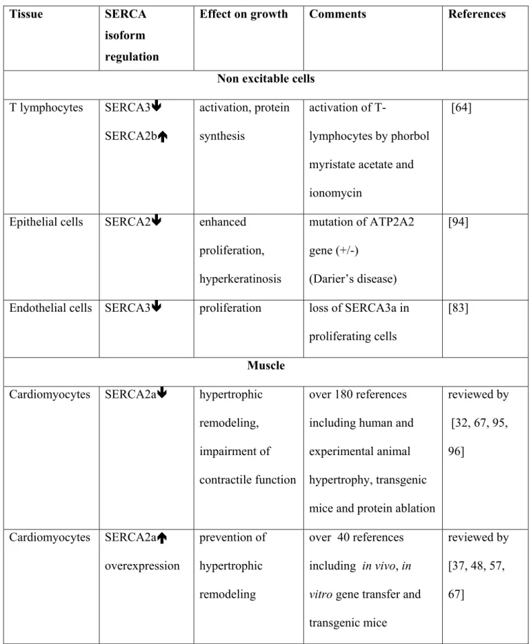
Decrease in SERCA expression and/or activity, reported in various growing cells, may play a role of support for sustained activation of store-operated Ca2+ entry in proliferating cells (Table. 1). This occurs in excitable and non excitable cells but the pathways differ between cell types.
3.1. Non-excitable cells. In non-excitable cells, stimulation with phosphoinositide-coupled
agonists can result in complex global Ca2+ signals organized as regenerative waves. The Ca2+ wave propagation sites are rich in the ER proteins SERCA, calreticulin and IP3Rs (rev in [66]). This oscillatory signal is prevented by inhibition of PKC, SERCA or CCE, or by external Ca2+ removal showing the involvement of IP3R Ca2+ release, SR Ca2+ uptake and Ca2+ entry through SOC in the generation of these waves [80]. Overexpression of SERCA in Xenopus oocytes increases the frequency of IP3-induced waves and narrows the width of individual calcium waves by relieving the inhibitory effect of high Ca2+ on IP3R [14]. These various observations indicate that in non excitable cells SERCA controls the kinetics of Ca2+ wave propagation, and thereby finely regulates Ca2+-dependent transcription pathways.
Interestingly, during activation of T-lymphocytes, SERCA3 expression decreases by 90%, whereas SERCA2b expression approximately doubles [64]. These isoforms have very different Ca2+ sensitivities and may trigger new cellular processes.
In non excitable cells such as lymphocytes, 3T3-L1 preadipocytes, endothelial cells, epithelial cells, and pancreatic beta cells, proliferation is driven by the calcium-dependent calcineurin/NFAT pathway [30, 46, 89, 101, 108, 131, 136]. Also, in non dermal epithelia cells, induction of proliferation by VEGF requires NFAT activation via store-operated STIM-mediated Ca2+ entry [131]. The most complete description of this signalling pathway has been in lymphocytes, in which the importance of all signalling molecules — SERCA, STIM1, a pore subunit named Orai1 (CRACM1) and NFAT — has been validated by ablation, patch clamp experiments and reporter-promoter assays [13, 31, 98, 106, 115, 116, 134, 139, 140].
Together, these findings demonstrate that proliferation in non-excitable cells is driven by NFAT activated by a long-lasting oscillatory store-operated Ca2+ signal. SERCA, controlling the amplitude and the kinetics of Ca2+ wave propagation, also can finely regulate Ca2+-dependent transcription pathways.
3.2. Cardiomyocytes. In cardiomyocytes, depolarization-induced Ca2+ cycling controlling myocyte contraction has no effect on activation of NFAT signalling pathway. By contrast, stimulation with phosphoinositide-coupled agonists induces a slow increase in resting Ca2+ due to the activation of IP3Rs and hypertrophic growth.
A decrease in SERCA activity, associated with a decrease in SERCA2a expression, was described in cardiac hypertrophy in the early 1990s [22, 60, 77, 86]. Since then, numerous papers have been published on the topic. Not all of the authors agreed about decreased expression and discordance between expression of SERCA2a at the mRNA and protein levels has been reported [112]. The decrease in the SERCA2a level is related to the intensity and duration of cardiac overload, but most authors now agree that SERCA2a is down regulated in severe heart failure [2]. Reduced SERCA2a activity and SR Ca2+ uptake lead to abnormal Ca2+ handling in failing cardiomyocytes and this involves an increase in diastolic Ca2+, an abnormally long time course of Ca2+ transients, and a decrease in SR Ca2+ release [20, 42, 43]. Furthermore, reduced SR Ca2+ stores and increased expression of transient receptor potential channels (TRPC) in failing heart favour capacitative Ca2+ entry and sustain
activation of calcineurin [63, 88].
Results from transgenic mice reveal a primordial role for SERCA2a dysfunction in induction of signalling pathways leading to cardiac hypertrophy and failure. Indeed, deletion of the SERCA2 gene (ATP2A2) is lethal but heterozygous mice are viable and develop cardiac hypertrophy [97]. Increasing the cardiac load by aortic banding resulted in faster heart failure in SERCA 2+/- mice than in WT controls [111]. However, in human there is no evidence for cardiac hypertrophy in patients carrying a mutation of the ATP2A2 gene (Darier’s disease)[76, 122]: up regulation of the normal allele is a possible explanation for the absence of cardiac hypertrophy [121].
During phenylephrine-induced hypertrophy in neonatal rat cardiac myocytes, the early and prominent feature of hypertrophic remodelling is the reduction of the abundance of the SERCA2 transcript [99]. The consequence of SERCA2a down-regulation on Ca2+ signalling is compensated by alternate Ca2+ transport mechanisms, and this contributes to the induction of a genetic programme of hypertrophic remodelling. Indeed, reduction of SERCA2a expression by RNA silencing in cardiac myocytes resulted in activation of the calcineurin-dependent complex leading first to increased expression of prohypertrophic transcription factors Sp1, MEF2 and NFATc4, and, subsequently, to up-regulation of Ca2+ handling proteins including the Na+/ Ca2+ exchanger and TRPC [113]. Calcineurin was identified as a central prohypertrophic signalling molecule for the myocardium [45] and its target, NFAT, is necessary and sufficient for mediating pathological skeletal myocyte and cardiac hypertrophy [45, 81, 85, 132].
These studies suggest that hypertrophic stimuli induce a sustained increase in resting Ca2+ in cardiomyocytes due to inhibition of SERCA2a activity. This resting Ca2+ leads to activation of calcineurin/NFAT signalling resulting in hypertrophic remodelling of cardiomyocytes. Furthermore, modulation of expression of Ca2+ transporters in hypertrophic cardiomyocytes leads to functional abnormalities in Ca2+ cycling and in the long term to impairment of cardiac contractile function.
3.3. Smooth muscle. In vascular smooth muscle cells (VSMC)
phosphoinositide-coupled agonists induce a sustained increase of cytosolic Ca2+ due to the generation of repetitive Ca2+ waves [40, 53], inhibition of activity of Ca2+ pumps [69] and increased capacitative Ca2+ entry (CCE) [38, 39].
Proliferation of VSMC is associated, in the rat and rabbit, with loss of the cardiac isoform of the sarcoplasmic reticulum (SR) Ca2+ pump, SERCA2a, and of the SR Ca2+
channel, the ryanodine receptor, RyR [69, 75, 125]. Loss of SERCA2a suggests a decrease in store refilling and this would favour activation of store-operated Ca2+ influx. Indeed, the expression of the store-operated Ca2+ channels TRPC1, TRPC4 or TRPC6 is increased in proliferating VSMC [30, 39, 118, 138]. The ER calcium sensor STIM1 regulates store-operated Ca2+ entry via interaction with TRPC1 in VSMC, and ablation of STIM1 by siRNA inhibits VSMC proliferation [119]. The number of T-type VOC channels is increased in proliferating VSMC [62, 103] and T-type channels are able to replenish the depleted Ca2+ store [34]. Mibefradil, a selective T-type channel blocker, inhibits proliferation of VSMC [68, 105, 110] and NFAT transcriptional activity [68]. Alterations of the Ca2+-handling proteins may be part of the VSMC dedifferentiation process, and has been described for many proteins [92]. It is also plausible that under a growth stimulus, alterations in Ca2+ handling may be a trigger for the activation of new Ca2+-regulated transcription pathways.
The main Ca2+-regulated transcription pathway described in VSMC involves the calcineurin/NFAT pathway. Many receptor tyrosine kinase and G-protein coupled receptor agonists, such as angiotensin II, endothelin 1, and platelet derived growth factor (PDGF-BB), and also very low density lipoproteins, induce VSMC proliferation or migration through activation of the NFAT transcription pathway [40, 41, 69, 71, 117, 133, 137]. NFATc3 is induced by endothelin-1 and ablation of this isoform also inhibits VSMC proliferation [40, 90]. In animal models, restenosis was shown to be prevented by restoring normal SR Ca2+ handling using SERCA2a gene transfer [65] and also by inhibiting the NFAT transcription pathway [72, 137].
These findings indicate that VSMC proliferation is driven by NFAT activation following a sustained increase in cytosolic Ca2+ which is due to inhibition of SERCA activity and increased voltage-independent Ca2+ entry.
3.4. Cancer cells. The calcium-dependent calcineurin/NFAT signalling pathway is
increasingly recognized as a central player in the development of a number of very different malignancies (rev. [9, 82]). Variation in expression of Ca2+ pumps and channels, most frequently an increase in TRPCl expression and decrease in SERCA expression, have been described in numerous cancers (rev. [82]). Furthermore, NFATc1 is commonly overexpressed in pancreatic, breast and colon carcinomas and enhances the malignant potential of tumour cells [10, 54]. Loss of SERCA activity and expression has been detected in many different malignancies. Indeed, changes in SERCA3 expression have been observed in colon cancer: the protein was either absent or present in much lower abundance in colon carcinoma than normal tissue, consistent with a loss of differentiation in tumour cells [8, 36]. Next, SERCA2b expression is very substantially decreased in oral cancers (squamous cell carcinoma), and in thyroid cancer, where it is the major isoform [29, 93]. Alternatively, somatic and germline mutations in lung cancer and germline mutations in colon cancer in the ATP2A2 gene result in the loss or reduction of SERCA2 expression [61]. The direct evidence linking a deficiency of SERCA to tumour genesis has been provided by development of heterozygous SERCA2 mice (ATP2a2+/-). These mice are sensitized to the development of squamous-cell carcinoma, which arises directly as a result of SERCA2 haploinsufficiency [70, 97, 100]. The keratinocytes from Darier’s disease patients, deficient in SERCA2b, as well as normal keratinocytes in which SERCA2b was silenced by siRNA, showed enhanced proliferation supported by up-regulation of the store-operated TRPC1 channel [94]. These observations suggest that haploinsufficiency of the SERCA 2 gene is involved in growth and proliferation of specific tumor cells.
Thus, there is increasing evidence that loss of SERCA activity and store depletion induces proliferation in various normal and cancer cell types via store-operated Ca2+ entry and
NFAT activation. Some groups have reported that in cancer cell lines an increase in SERCA2b expression and in sarco/endoplasmic reticulum Ca2+ content are required for induction of proliferation, but the molecular pathway involved has not been identified [17, 18, 50]. Crepin et al [18] reported that the increased rate of proliferation of immortalized epithelial prostate cells (PNT1A) induced by prolactin is associated with an increase in the SR calcium content and SERCA2b expression. Silencing of SERCA2b expression by siRNA leads to reduction of PNT1A cell proliferation. These observations led the authors to conclude that SERCA2b overexpression is a protagonist of prolactin-induced proliferation. However, in primary human prostate cancer epithelial cells (hPCE), store-operated Ca2+ entry and NFAT activation was identified as the main proliferation pathway [123].
In conclusion, there is a large body of data demonstrating that in various cell types store-operated Ca2+ entry is necessary to activate NFAT signalling, although the exact mechanism of this process is not yet completely understood. SERCA, by controlling store refilling, plays a primordial role in the control of cell growth and proliferation.
4. SR/ER-based therapeutic strategies.
The endoplasmic reticulum is not only a Ca2+ reservoir but is also the site for protein synthesis and folding. This has consequences for the design of any Ca2+ cycling-based therapeutic strategies: the SR/ER Ca2+ load has to be finely tuned to maintain cell integrity.
With this in mind, two types of strategy have been designed with SERCA as a target: one aims to restore normal SR load and SERCA activity and is used in cardiovascular diseases. The other consists of inducing ER stress to cause apoptosis; this strategy is designed to kill cancer cells.
4.1-Increasing SERCA2a activity
Two approaches have been proposed to increase SERCA2a activity: SERCA2a overexpression; and phospholamban (PLN) ablation. They have been particularly well studied in the heart where abnormal Ca2+ cycling is the main determinant of contractile dysfunction and heart failure [67].
SERCA overexpression. Transgenic animals with cardiac-specific expression of either SERCA2a or SERCA1a showed improved contractility under baseline conditions and after pressure overload [3, 84, 120]. The mortality rate after aortic banding in animals overexpressing SERCA2a was identical to that in the WT controls [84]. However, in the same model, Chen et al reported increased mortality after myocardial infarction in association with an increased frequency of arrhythmias [16]. Less persistent arrhythmias were observed after post-ischemic injury in SERCA1a-overexpressing hearts from transgenic rats as well as in hearts overexpressing SERCA2a from an adenoviral vector [24, 120]. The data from transgenic animals indicate that having a high basal level of SERCA (2a or 1a) can improve cardiac function and prevent heart failure.
The enhanced contractility associated with SERCA2a overexpression has been reported to be protective against both heart failure and cardiac hypertrophy [19, 51, 79, 84, 87, 124]. Adenovirus-mediated gene transfer of SERCA2a restored the Ca2+ transient in cardiomyocytes isolated from failing human hearts[23], improved cardiac haemodynamics and increased survival in animal models of heart failure [19, 25]. SERCA2a rescues depressed contractility and survival without adverse effects on energy metabolism [19, 79] or cardiac arrhythmia in animal models [24] but definite proof of SERCA2a gene transfer efficiency in human await the on-going clinical trials.
We have shown that normalization of Ca2+ handling by SERCA2a gene transfer prevents injury-induced vascular remodelling in rats [65]. Thus, preventing SERCA2a loss by
gene transfer is a novel potential strategy for treating restenosis. Coronary restenosis is a major complication of percutaneous coronary balloon angioplasty. It is characterized by neointimal hyperplasia due to proliferation of VSMC. Although the use of drug-eluting stents (DES) limits neointimal hyperplasia, recent data suggest that their use may be associated with adverse clinical effects [7, 35, 126]. Thus, there is a need to discover novel mechanisms governing VSMC proliferation and this information could be used to develop new modalities for treating restenosis. Rapamycin and taxol, used in drug-eluting stents, were designed to induce cell death in proliferating VSMC. The strategy based on SERCA2a gene transfer should preserve cell integrity and prevent loss of the “differentiated/contractile phenotype” of VSMCs .
The same mechanism governs the beneficial effect of SERCA2a overexpression in heart failure and in proliferative vascular diseases: by lowering cytosolic Ca2+, SERCA2a expression inhibits calcineurin activity and the activation of the NFAT pathway [65]. This explains why SERCA2a gene transfer inhibits hypertrophic, hyperplastic and apoptotic signalling pathways mediated by calcineurin. Another reason for the beneficial role of SERCA overexpression might be that it reduces oxidative stress. Indeed, high levels of oxygen-derived free radicals are generated during myocardial ischemia/reperfusion and this damages SERCA2a, potentially contributing to cellular Ca 2+overload and myocardial injury. Similarly, in atherosclerosis, cysteine 674 from SERCA2 is irreversibly oxidized due to prolonged oxidative stress, and consequently the NO-induced S-glutathiolation, activation of SERCA and arterial relaxation are impaired [1].
Phospholamban ablation. Phospholamban (PLN) is a 52-amino-acid protein which controls the affinity of SERCA for Ca2+. It is expressed mainly in cardiac, slow skeletal,
its unphosphorylated form, PLN inhibits SERCA activity and phosphorylation by various protein kinases: PKA, PKG, and Ca2+/Calmodulin kinase relieve this inhibition. Decreasing the inhibitory effect of PLN is another way of enhancing SERCA activity. This has been studied in great detail in heart failure and most findings now indicate that, in hypertrophy and failure, the level of PLN is unchanged or slightly decreased but that PLN is hypophosphorylated [112]. Both an increase in the PLN-to-SERCA ratio and the presence of unphosphorylated PLN should increase the inhibitory function of PLN. Suppressing the inhibitory effect of phospholamban is a promising approach to improving cardiac function. Indeed, the ablation of PLN completely prevents the spectrum of heart failure phenotypes in a mouse model of dilated cardiomyophy [78]. Furthermore, chronic inhibition of PLN using a pseudo-phosphorylated mutant results in favourable changes in cardiac haemodynamics in rat and sheep models of heart failure and prevents cardiomyopathy in a myopathic hamster model [49, 52, 58]. PLN ablation has also been shown to rescue depressed contractile function of calsequestrin-overexpressing hearts [107] and in a mouse line overexpressing a mutant myosin heavy chain [33]. In addition, normalization of the Ca2+ transient and restoration of
cell contractility have been reported in cardiomyocytes isolated from failing human hearts [23]. The excitement generated by these studies has been tempered by the discovery of mutations in PLN, leading to a super-inhibitory PLN, which has been suggested to be causative of human dilated cardiomyopathy [44, 109]. Furthermore, in other genetic models, PLN ablation rescued cardiomyocyte dysfunction but did not prevent ventricular remodelling leading to heart failure [114].
Therapeutic strategies based on normalization of the SR Ca2+ load and of cytosolic Ca2+ by increasing SERCA activity seem promising for preventing hypertrophic growth and vascular proliferative disease. SERCA2a gene therapy for treatment of heart failure is now undergoing clinical trials in USA [67], and prevention of post-injury restenosis by SERCA2a
gene transfer is at the preclinical study stage [65]. The use of pseudophosphorylated PLN in the treatment of heart failure is also being considered in preclinical studies [58]. Furthermore, the development of small molecules for enhancing Ca2+ cycling has now appeared on the horizon and may offer new hope for treatment of Ca2+cycling defects in cardiovascular disease.
4.2 Use of SERCA as a target to induce cell death
The issue of Ca2+ and cancer has been covered recently in a very comprehensive review by G. Monteith [82]. Here we focus on manipulating SERCA and SR Ca2+ load as methods for anticancer therapy. Two strategies could be used: either inducing a general ER stress by depletion of the ER Ca2+ store, or targeting particular SERCA isoforms that are induced or repressed in cancer cells.
An example of the first strategy is provided by the thapsigargin ‘prodrug’ approach to the treatment of prostate cancer [26]. Thapsigargin is a general inhibitor of SERCA, it induces complete SR/ER Ca2+ depletion and apoptosis. To target cancer cells selectively, the drug has
been coupled to a peptide to produce an inactive “prodrug” that is only activated by prostate cancer-specific proteases such as the serine protease prostate-specific antigen [26]. However, thapsigargin resistance has been clearly demonstrated [91] and may hamper the efficacy of this approach. ER stress can also be obtained by blockade of the voltage-independent Ca2+- channels which normally refill the ER after store depletion. Carboxyamido-triazole (CAI), a low molecular weight compound, inhibits these types of channels and is undergoing clinical validation for use as an anticancer agent [82]. CAI acts as an antiangiogenic and antimetastatic agent because it inhibits endothelial cell proliferation [59].
There is a multiplicity of isoforms of SERCA and especially SERCA3 [6] and the
roles in cell adhesion and ER stress [15]. Interestingly, SERCA3 is repressed in highly neoplastic colon cancer cells [8]; consequently, overexpressing SERCA3 in colon cancer to preserve normal ER Ca2+ levels may be of therapeutic value. SERCA2b expression is decreased in skin disorders related to Darier’s disease [11, 27, 121] and in thyroid cancer [93] but because SERCA2b is the ubiquitous isoform it might be difficult to target precisely.
6. Conclusions and future directions
In the past decade, there has been increasing evidence of the role of SERCA in diseases and especially in cardiovascular diseases. As a consequence, SERCA2a gene therapy has now progressed to clinical trials. Furthermore, the development of small molecules to increase SR Ca2+ cycling provides the hope of treatment for Ca2+ cycling defects. The reasons for and consequences of the existence of multiple SERCA isoforms, and especially of SERCA3 isoforms, remain to be elucidated, as does their importance in pathologies such as cancers. Exploitation of siRNA technology to knock-down protein production should help identify the role of these isoforms.
Store-operated calcium channels are increasingly being recognized as central players in the control of hyperthrophic growth and proliferation in almost all cell types including diverse malignancies. The enigma of the cross-talk between depletion of the ER store and refilling by calcium influx through store-operated calcium channels has been highlighted with the discovery of the sensor protein, STIM1, but studies of the role of both STIM1 and SOC in diseases are at an early stage.
The importance of the Ca2+-regulated transcription factor NFAT, which links alteration in Ca2+ cycling to pathological growth and proliferation, has been documented but other Ca2+-regulated pathways may also be involved and remains to be described. Furthermore, new mechanisms of control of gene expression have been uncovered with the
discovery of micro RNAs. Micro RNAs play an important role in pathological growth but the trigger for induction of specific miRNAs as well as the relationship with previously described alterations in Ca2+ signalling remains to be elucidated.
References
1. Adachi, T., Weisbrod, R. M., Pimentel, D. R., Ying, J., Sharov, V. S., Schoneich, C. & Cohen, R. A. (2004) S-Glutathiolation by peroxynitrite activates SERCA during arterial relaxation by nitric oxide, Nat Med. 10, 1200-7.
2. Armoundas, A. A., Rose, J., Aggarwal, R., Stuyvers, B. D., O'Rourke, B., Kass, D. A., Marban, E., Shorofsky, S. R., Tomaselli, G. F. & William Balke, C. (2007) Cellular and molecular determinants of altered Ca2+ handling in the failing rabbit heart: primary defects in SR Ca2+ uptake and release mechanisms, Am J Physiol Heart Circ Physiol. 292, H1607-18. 3. Baker, D. L., Hashimoto, K., Grupp, I. L., Ji, Y., Reed, T., Loukianov, E., Grupp, G., Bhagwhat, A., Hoit, B., Walsh, R., Marban, E. & Periasamy, M. (1998) Targeted
overexpression of the sarcoplasmic reticulum Ca2+-ATPase increases cardiac contractility in transgenic mouse hearts, Circ Res. 83, 1205-14.
4. Baksh, S., Widlund, H. R., Frazer-Abel, A. A., Du, J., Fosmire, S., Fisher, D. E., DeCaprio, J. A., Modiano, J. F. & Burakoff, S. J. (2002) NFATc2-mediated repression of cyclin-dependent kinase 4 expression, Mol Cell. 10, 1071-81.
5. Bert, A. G., Johnson, B. V., Baxter, E. W. & Cockerill, P. N. (2007) A modular enhancer is differentially regulated by GATA and NFAT elements that direct different tissue-specific patterns of nucleosome positioning and inducible chromatin remodeling, Mol Cell Biol. 27, 2870-85.
6. Bobe, R., Bredoux, R., Corvazier, E., Lacabaratz-Porret, C., Martin, V., Kovacs, T. & Enouf, J. (2005) How many Ca(2)+ATPase isoforms are expressed in a cell type? A growing family of membrane proteins illustrated by studies in platelets, Platelets. 16, 133-50.
7. Brilakis, E. S., Banerjee, S. & Berger, P. B. (2007) The risk of drug-eluting stent thrombosis with noncardiac surgery, Curr Cardiol Rep. 9, 406-11.
8. Brouland, J. P., Gelebart, P., Kovacs, T., Enouf, J., Grossmann, J. & Papp, B. (2005) The loss of sarco/endoplasmic reticulum calcium transport ATPase 3 expression is an early event during the multistep process of colon carcinogenesis, Am J Pathol. 167, 233-42.
9. Buchholz, M. & Ellenrieder, V. (2007) An emerging role for Ca2+/calcineurin/NFAT signaling in cancerogenesis, Cell Cycle. 6, 16-9.
10. Buchholz, M., Schatz, A., Wagner, M., Michl, P., Linhart, T., Adler, G., Gress, T. M. & Ellenrieder, V. (2006) Overexpression of c-myc in pancreatic cancer caused by ectopic activation of NFATc1 and the Ca2+/calcineurin signaling pathway, Embo J. 25, 3714-24. 11. Byrne, C. R. (2006) The focal nature of Darier's disease lesions: calcium pumps, stress, and mutation?, J Invest Dermatol. 126, 702-3.
12. Caetano, M. S., Vieira-de-Abreu, A., Teixeira, L. K., Werneck, M. B., Barcinski, M. A. & Viola, J. P. (2002) NFATC2 transcription factor regulates cell cycle progression during lymphocyte activation: evidence of its involvement in the control of cyclin gene expression, Faseb J. 16, 1940-2.
13. Cahalan, M. D., Zhang, S. L., Yeromin, A. V., Ohlsen, K., Roos, J. & Stauderman, K. A. (2007) Molecular basis of the CRAC channel, Cell Calcium. 42, 133-44.
14. Camacho, P. & Lechleiter, J. D. (1993) Increased frequency of calcium waves in Xenopus laevis oocytes that express a calcium-ATPase, Science. 260, 226-9.
15. Chaabane, C., Corvazier, E., Bredoux, R., Dally, S., Raies, A., Villemain, A., Dupuy, E., Enouf, J. & Bobe, R. (2006) Sarco/endoplasmic reticulum Ca2+ATPase type 3 isoforms (SERCA3b and SERCA3f): distinct roles in cell adhesion and ER stress, Biochem Biophys Res Commun. 345, 1377-85.
16. Chen, Y., Escoubet, B., Prunier, F., Amour, J., Simonides, W. S., Vivien, B., Lenoir, C., Heimburger, M., Choqueux, C., Gellen, B., Riou, B., Michel, J. B., Franz, W. M. &
reticulum Ca2+-ATPase delays myocardial failure after myocardial infarction in rats at a cost of increased acute arrhythmias, Circulation. 109, 1898-903.
17. Chung, F. Y., Lin, S. R., Lu, C. Y., Yeh, C. S., Chen, F. M., Hsieh, J. S., Huang, T. J. & Wang, J. Y. (2006) Sarco/endoplasmic reticulum calcium-ATPase 2 expression as a tumor marker in colorectal cancer, Am J Surg Pathol. 30, 969-74.
18. Crepin, A., Bidaux, G., Vanden-Abeele, F., Dewailly, E., Goffin, V., Prevarskaya, N. & Slomianny, C. (2007) Prolactin stimulates prostate cell proliferation by increasing
endoplasmic reticulum content due to SERCA 2b over-expression, Biochem J. 401, 49-55. 19. Davia, K., Bernobich, E., Ranu, H. K., del Monte, F., Terracciano, C. M., MacLeod, K. T., Adamson, D. L., Chaudhri, B., Hajjar, R. J. & Harding, S. E. (2001) SERCA2A
overexpression decreases the incidence of aftercontractions in adult rabbit ventricular myocytes, J Mol Cell Cardiol. 33, 1005-15.
20. Davies, C. H., Davia, K., Bennett, J. G., Pepper, J. R., Poole-Wilson, P. A. & Harding, S. E. (1995) Reduced contraction and altered frequency response of isolated ventricular
myocytes from patients with heart failure, Circulation. 92, 2540-9.
21. De Jaegere, S., Wuytack, F., De Smedt, H., Van den Bosch, L. & Casteels, R. (1993) Alternative processing of the gene transcripts encoding a plasma-membrane and a
sarco/endoplasmic reticulum Ca2+ pump during differentiation of BC3H1 muscle cells, Biochim Biophys Acta. 1173, 188-94.
22. de la Bastie, D., Levitsky, D., Rappaport, L., Mercadier, J. J., Marotte, F., Wisnewsky, C., Brovkovich, V., Schwartz, K. & Lompre, A. M. (1990) Function of the sarcoplasmic reticulum and expression of its Ca2(+)-ATPase gene in pressure overload-induced cardiac hypertrophy in the rat, Circ Res. 66, 554-64.
23. del Monte, F., Harding, S. E., Schmidt, U., Matsui, T., Kang, Z. B., Dec, G. W.,
Gwathmey, J. K., Rosenzweig, A. & Hajjar, R. J. (1999) Restoration of contractile function in
isolated cardiomyocytes from failing human hearts by gene transfer of SERCA2a, Circulation. 100, 2308-11.
24. del Monte, F., Lebeche, D., Guerrero, J. L., Tsuji, T., Doye, A. A., Gwathmey, J. K. & Hajjar, R. J. (2004) Abrogation of ventricular arrhythmias in a model of ischemia and
reperfusion by targeting myocardial calcium cycling, Proc Natl Acad Sci U S A. 101, 5622-7. 25. del Monte, F., Williams, E., Lebeche, D., Schmidt, U., Rosenzweig, A., Gwathmey, J. K., Lewandowski, E. D. & Hajjar, R. J. (2001) Improvement in survival and cardiac metabolism after gene transfer of sarcoplasmic reticulum Ca(2+)-ATPase in a rat model of heart failure, Circulation. 104, 1424-9.
26. Denmeade, S. R. & Isaacs, J. T. (2005) The SERCA pump as a therapeutic target: making a "smart bomb" for prostate cancer, Cancer Biol Ther. 4, 14-22.
27. Dhitavat, J., Dode, L., Leslie, N., Sakuntabhai, A., Lorette, G. & Hovnanian, A. (2003) Mutations in the sarcoplasmic/endoplasmic reticulum Ca2+ ATPase isoform cause Darier's disease, J Invest Dermatol. 121, 486-9.
28. Dolmetsch, R. E., Lewis, R. S., Goodnow, C. C. & Healy, J. I. (1997) Differential activation of transcription factors induced by Ca2+ response amplitude and duration, Nature. 386, 855-8.
29. Endo, Y., Uzawa, K., Mochida, Y., Shiiba, M., Bukawa, H., Yokoe, H. & Tanzawa, H. (2004) Sarcoendoplasmic reticulum Ca(2+) ATPase type 2 downregulated in human oral squamous cell carcinoma, Int J Cancer. 110, 225-31.
30. Fantozzi, I., Zhang, S., Platoshyn, O., Remillard, C. V., Cowling, R. T. & Yuan, J. X. (2003) Hypoxia increases AP-1 binding activity by enhancing capacitative Ca2+ entry in human pulmonary artery endothelial cells, Am J Physiol Lung Cell Mol Physiol. 285, L1233-45.
31. Feske, S., Gwack, Y., Prakriya, M., Srikanth, S., Puppel, S. H., Tanasa, B., Hogan, P. G., Lewis, R. S., Daly, M. & Rao, A. (2006) A mutation in Orai1 causes immune deficiency by abrogating CRAC channel function, Nature. 441, 179-85.
32. Frank, K. F., Bolck, B., Brixius, K., Kranias, E. G. & Schwinger, R. H. (2002)
Modulation of SERCA: implications for the failing human heart, Basic Res Cardiol. 97 Suppl 1, I72-8.
33. Freeman, K., Lerman, I., Kranias, E. G., Bohlmeyer, T., Bristow, M. R., Lefkowitz, R. J., Iaccarino, G., Koch, W. J. & Leinwand, L. A. (2001) Alterations in cardiac adrenergic
signaling and calcium cycling differentially affect the progression of cardiomyopathy, J Clin Invest. 107, 967-74.
34. Gackiere, F., Bidaux, G., Lory, P., Prevarskaya, N. & Mariot, P. (2006) A role for voltage gated T-type calcium channels in mediating "capacitative" calcium entry?, Cell Calcium. 39, 357-66.
35. Garg, P. & Mauri, L. (2007) The conundrum of late and very late stent thrombosis following drug-eluting stent implantation, Curr Opin Cardiol. 22, 565-571.
36. Gelebart, P., Kovacs, T., Brouland, J. P., van Gorp, R., Grossmann, J., Rivard, N., Panis, Y., Martin, V., Bredoux, R., Enouf, J. & Papp, B. (2002) Expression of endomembrane calcium pumps in colon and gastric cancer cells. Induction of SERCA3 expression during differentiation, J Biol Chem. 277, 26310-20.
37. Gianni, D., Chan, J., Gwathmey, J. K., del Monte, F. & Hajjar, R. J. (2005) SERCA2a in heart failure: role and therapeutic prospects, J Bioenerg Biomembr. 37, 375-80.
38. Golovina, V. A. (1999) Cell proliferation is associated with enhanced capacitative Ca(2+) entry in human arterial myocytes, Am J Physiol. 277, C343-9.
39. Golovina, V. A., Platoshyn, O., Bailey, C. L., Wang, J., Limsuwan, A., Sweeney, M., Rubin, L. J. & Yuan, J. X. (2001) Upregulated TRP and enhanced capacitative Ca(2+) entry
in human pulmonary artery myocytes during proliferation, Am J Physiol Heart Circ Physiol. 280, H746-55.
40. Gomez, M. F., Stevenson, A. S., Bonev, A. D., Hill-Eubanks, D. C. & Nelson, M. T. (2002) Opposing actions of inositol 1,4,5-trisphosphate and ryanodine receptors on nuclear factor of activated T-cells regulation in smooth muscle, J Biol Chem. 277, 37756-64.
41. Gonzalez Bosc, L. V., Layne, J. J., Nelson, M. T. & Hill-Eubanks, D. C. (2005) Nuclear factor of activated T cells and serum response factor cooperatively regulate the activity of an alpha-actin intronic enhancer, J Biol Chem. 280, 26113-20.
42. Gwathmey, J. K., Copelas, L., MacKinnon, R., Schoen, F. J., Feldman, M. D., Grossman, W. & Morgan, J. P. (1987) Abnormal intracellular calcium handling in myocardium from patients with end-stage heart failure, Circ Res. 61, 70-6.
43. Gwathmey, J. K. & Morgan, J. P. (1985) Altered calcium handling in experimental pressure-overload hypertrophy in the ferret, Circ Res. 57, 836-43.
44. Haghighi, K., Kolokathis, F., Gramolini, A. O., Waggoner, J. R., Pater, L., Lynch, R. A., Fan, G. C., Tsiapras, D., Parekh, R. R., Dorn, G. W., 2nd, MacLennan, D. H., Kremastinos, D. T. & Kranias, E. G. (2006) A mutation in the human phospholamban gene, deleting arginine 14, results in lethal, hereditary cardiomyopathy, Proc Natl Acad Sci U S A. 103, 1388-93.
45. Heineke, J. & Molkentin, J. D. (2006) Regulation of cardiac hypertrophy by intracellular signalling pathways, Nat Rev Mol Cell Biol. 7, 589-600.
46. Heit, J. J., Apelqvist, A. A., Gu, X., Winslow, M. M., Neilson, J. R., Crabtree, G. R. & Kim, S. K. (2006) Calcineurin/NFAT signalling regulates pancreatic beta-cell growth and function, Nature. 443, 345-9.
47. Horsley, V. & Pavlath, G. K. (2002) NFAT: ubiquitous regulator of cell differentiation
48. Hoshijima, M. (2005) Gene therapy targeted at calcium handling as an approach to the treatment of heart failure, Pharmacol Ther. 105, 211-28.
49. Hoshijima, M., Ikeda, Y., Iwanaga, Y., Minamisawa, S., Date, M. O., Gu, Y., Iwatate, M., Li, M., Wang, L., Wilson, J. M., Wang, Y., Ross, J., Jr. & Chien, K. R. (2002) Chronic suppression of heart-failure progression by a pseudophosphorylated mutant of phospholamban via in vivo cardiac rAAV gene delivery, Nat Med. 8, 864-71.
50. Humez, S., Legrand, G., Vanden-Abeele, F., Monet, M., Marchetti, P., Lepage, G., Crepin, A., Dewailly, E., Wuytack, F. & Prevarskaya, N. (2004) Role of endoplasmic reticulum calcium content in prostate cancer cell growth regulation by IGF and TNFalpha, J Cell Physiol. 201, 201-13.
51. Ito, K., Yan, X., Feng, X., Manning, W. J., Dillmann, W. H. & Lorell, B. H. (2001) Transgenic expression of sarcoplasmic reticulum Ca(2+) atpase modifies the transition from hypertrophy to early heart failure, Circ Res. 89, 422-9.
52. Iwanaga, Y., Hoshijima, M., Gu, Y., Iwatate, M., Dieterle, T., Ikeda, Y., Date, M. O., Chrast, J., Matsuzaki, M., Peterson, K. L., Chien, K. R. & Ross, J., Jr. (2004) Chronic phospholamban inhibition prevents progressive cardiac dysfunction and pathological remodeling after infarction in rats, J Clin Invest. 113, 727-36.
53. Jaggar, J. H. & Nelson, M. T. (2000) Differential regulation of Ca(2+) sparks and Ca(2+) waves by UTP in rat cerebral artery smooth muscle cells, Am J Physiol Cell Physiol. 279, C1528-39.
54. Jauliac, S., Lopez-Rodriguez, C., Shaw, L. M., Brown, L. F., Rao, A. & Toker, A. (2002) The role of NFAT transcription factors in integrin-mediated carcinoma invasion, Nat Cell Biol. 4, 540-4.
55. John, L. M., Lechleiter, J. D. & Camacho, P. (1998) Differential modulation of SERCA2 isoforms by calreticulin, J Cell Biol. 142, 963-73.
56. Jousset, H., Frieden, M. & Demaurex, N. (2007) STIM1 knockdown reveals that store-operated Ca2+ channels located close to sarco/endoplasmic Ca2+ ATPases (SERCA) pumps silently refill the endoplasmic reticulum, J Biol Chem. 282, 11456-64.
57. Kaprielian, R., del Monte, F. & Hajjar, R. J. (2002) Targeting Ca2+ cycling proteins and the action potential in heart failure by gene transfer, Basic Res Cardiol. 97 Suppl 1, I136-45. 58. Kaye, D. M., Preovolos, A., Marshall, T., Byrne, M., Hoshijima, M., Hajjar, R., Mariani, J. A., Pepe, S., Chien, K. R. & Power, J. M. (2007) Percutaneous cardiac recirculation-mediated gene transfer of an inhibitory phospholamban peptide reverses advanced heart failure in large animals, J Am Coll Cardiol. 50, 253-60.
59. Kohn, E. C., Felder, C. C., Jacobs, W., Holmes, K. A., Day, A., Freer, R. & Liotta, L. A. (1994) Structure-function analysis of signal and growth inhibition by carboxyamido-triazole, CAI, Cancer Res. 54, 935-42.
60. Komuro, I., Kurabayashi, M., Shibazaki, Y., Takaku, F. & Yazaki, Y. (1989) Molecular cloning and characterization of a Ca2+ + Mg2+-dependent adenosine triphosphatase from rat cardiac sarcoplasmic reticulum. Regulation of its expression by pressure overload and
developmental stage, J Clin Invest. 83, 1102-8.
61. Korosec, B., Glavac, D., Rott, T. & Ravnik-Glavac, M. (2006) Alterations in the
ATP2A2 gene in correlation with colon and lung cancer, Cancer Genet Cytogenet. 171, 105-11.
62. Kuga, T., Kobayashi, S., Hirakawa, Y., Kanaide, H. & Takeshita, A. (1996) Cell cycle--dependent expression of L- and T-type Ca2+ currents in rat aortic smooth muscle cells in primary culture, Circ Res. 79, 14-9.
63. Kuwahara, K., Wang, Y., McAnally, J., Richardson, J. A., Bassel-Duby, R., Hill, J. A. & Olson, E. N. (2006) TRPC6 fulfills a calcineurin signaling circuit during pathologic cardiac
64. Launay, S., Bobe, R., Lacabaratz-Porret, C., Bredoux, R., Kovacs, T., Enouf, J. & Papp, B. (1997) Modulation of endoplasmic reticulum calcium pump expression during T
lymphocyte activation, J Biol Chem. 272, 10746-50.
65. Lipskaia, L., del Monte, F., Capiod, T., Yacoubi, S., Hadri, L., Hours, M., Hajjar, R. J. & Lompre, A. M. (2005) Sarco/endoplasmic reticulum Ca2+-ATPase gene transfer reduces vascular smooth muscle cell proliferation and neointima formation in the rat, Circ Res. 97, 488-95.
66. Lipskaia, L. & Lompre, A. M. (2004) Alteration in temporal kinetics of Ca2+ signaling and control of growth and proliferation, Biol Cell. 96, 55-68.
67. Lipskaia, L., Ly, H., Kawase, Y., Hajjar, R. & Lompre, A. M. (2007) Treatment of heart failure by calcium cycling gene therapy, Future Cardiology. 3, 413-423.
68. Lipskaia, L., Pinet, C., Fromes, Y., Hatem, S., Cantaloube, I., Coulombe, A. & Lompre, A. M. (2007) Mutation of delta-sarcoglycan is associated with Ca(2+) -dependent vascular remodeling in the Syrian hamster, Am J Pathol. 171, 162-71.
69. Lipskaia, L., Pourci, M. L., Delomenie, C., Combettes, L., Goudouneche, D., Paul, J. L., Capiod, T. & Lompre, A. M. (2003) Phosphatidylinositol 3-kinase and calcium-activated transcription pathways are required for VLDL-induced smooth muscle cell proliferation, Circ Res. 92, 1115-22.
70. Liu, L. H., Boivin, G. P., Prasad, V., Periasamy, M. & Shull, G. E. (2001) Squamous cell tumors in mice heterozygous for a null allele of Atp2a2, encoding the sarco(endo)plasmic reticulum Ca2+-ATPase isoform 2 Ca2+ pump, J Biol Chem. 276, 26737-40.
71. Liu, Z., Dronadula, N. & Rao, G. N. (2004) A novel role for nuclear factor of activated T cells in receptor tyrosine kinase and G protein-coupled receptor agonist-induced vascular smooth muscle cell motility, J Biol Chem. 279, 41218-26.
72. Liu, Z., Zhang, C., Dronadula, N., Li, Q. & Rao, G. N. (2005) Blockade of nuclear factor of activated T cells activation signaling suppresses balloon injury-induced neointima
formation in a rat carotid artery model, J Biol Chem. 280, 14700-8.
73. Lytton, J., Westlin, M., Burk, S. E., Shull, G. E. & MacLennan, D. H. (1992) Functional comparisons between isoforms of the sarcoplasmic or endoplasmic reticulum family of calcium pumps, J Biol Chem. 267, 14483-9.
74. Macian, F., Garcia-Rodriguez, C. & Rao, A. (2000) Gene expression elicited by NFAT in the presence or absence of cooperative recruitment of Fos and Jun, Embo J. 19, 4783-95. 75. Massaeli, H., Austria, J. A. & Pierce, G. N. (2000) Lesions in ryanodine channels in smooth muscle cells exposed to oxidized low density lipoprotein, Arterioscler Thromb Vasc Biol. 20, 328-34.
76. Mayosi, B. M., Kardos, A., Davies, C. H., Gumedze, F., Hovnanian, A., Burge, S. & Watkins, H. (2006) Heterozygous disruption of SERCA2a is not associated with impairment of cardiac performance in humans: implications for SERCA2a as a therapeutic target in heart failure, Heart. 92, 105-9.
77. Mercadier, J. J., Lompre, A. M., Duc, P., Boheler, K. R., Fraysse, J. B., Wisnewsky, C., Allen, P. D., Komajda, M. & Schwartz, K. (1990) Altered sarcoplasmic reticulum Ca2(+)-ATPase gene expression in the human ventricle during end-stage heart failure, J Clin Invest. 85, 305-9.
78. Minamisawa, S., Hoshijima, M., Chu, G., Ward, C. A., Frank, K., Gu, Y., Martone, M. E., Wang, Y., Ross, J., Jr., Kranias, E. G., Giles, W. R. & Chien, K. R. (1999) Chronic phospholamban-sarcoplasmic reticulum calcium ATPase interaction is the critical calcium cycling defect in dilated cardiomyopathy, Cell. 99, 313-22.
79. Miyamoto, M. I., del Monte, F., Schmidt, U., DiSalvo, T. S., Kang, Z. B., Matsui, T.,
transfer of SERCA2a improves left-ventricular function in aortic-banded rats in transition to heart failure, Proc Natl Acad Sci U S A. 97, 793-8.
80. Moccia, F., Berra-Romani, R., Tritto, S., Signorelli, S., Taglietti, V. & Tanzi, F. (2003) Epidermal growth factor induces intracellular Ca2+ oscillations in microvascular endothelial cells, J Cell Physiol. 194, 139-50.
81. Molkentin, J. D., Lu, J. R., Antos, C. L., Markham, B., Richardson, J., Robbins, J., Grant, S. R. & Olson, E. N. (1998) A calcineurin-dependent transcriptional pathway for cardiac hypertrophy, Cell. 93, 215-28.
82. Monteith, G. R., McAndrew, D., Faddy, H. M. & Roberts-Thomson, S. J. (2007) Calcium and cancer: targeting Ca2+ transport, Nat Rev Cancer. 7, 519-30.
83. Mountian, I., Manolopoulos, V. G., De Smedt, H., Parys, J. B., Missiaen, L. & Wuytack, F. (1999) Expression patterns of sarco/endoplasmic reticulum Ca(2+)-ATPase and inositol 1,4,5-trisphosphate receptor isoforms in vascular endothelial cells, Cell Calcium. 25, 371-80. 84. Muller, O. J., Lange, M., Rattunde, H., Lorenzen, H. P., Muller, M., Frey, N., Bittner, C., Simonides, W., Katus, H. A. & Franz, W. M. (2003) Transgenic rat hearts overexpressing SERCA2a show improved contractility under baseline conditions and pressure overload, Cardiovasc Res. 59, 380-9.
85. Musaro, A., McCullagh, K. J., Naya, F. J., Olson, E. N. & Rosenthal, N. (1999) IGF-1 induces skeletal myocyte hypertrophy through calcineurin in association with GATA-2 and NF-ATc1, Nature. 400, 581-5.
86. Nagai, R., Zarain-Herzberg, A., Brandl, C. J., Fujii, J., Tada, M., MacLennan, D. H., Alpert, N. R. & Periasamy, M. (1989) Regulation of myocardial Ca2+-ATPase and phospholamban mRNA expression in response to pressure overload and thyroid hormone, Proc Natl Acad Sci U S A. 86, 2966-70.
87. Nakayama, H., Otsu, K., Yamaguchi, O., Nishida, K., Date, M. O., Hongo, K., Kusakari, Y., Toyofuku, T., Hikoso, S., Kashiwase, K., Takeda, T., Matsumura, Y., Kurihara, S., Hori, M. & Tada, M. (2003) Cardiac-specific overexpression of a high Ca2+ affinity mutant of SERCA2a attenuates in vivo pressure overload cardiac hypertrophy, Faseb J. 17, 61-3. 88. Nakayama, H., Wilkin, B. J., Bodi, I. & Molkentin, J. D. (2006) Calcineurin-dependent cardiomyopathy is activated by TRPC in the adult mouse heart, Faseb J. 20, 1660-70. 89. Neal, J. W. & Clipstone, N. A. (2003) A constitutively active NFATc1 mutant induces a transformed phenotype in 3T3-L1 fibroblasts, J Biol Chem. 278, 17246-54.
90. Nilsson, L. M., Sun, Z. W., Nilsson, J., Nordstrom, I., Chen, Y. W., Molkentin, J. D., Wide-Swensson, D., Hellstrand, P., Lydrup, M. L. & Gomez, M. F. (2007) Novel blocker of NFAT activation inhibits IL-6 production in human myometrial arteries and reduces vascular smooth muscle cell proliferation, Am J Physiol Cell Physiol. 292, C1167-78.
91. O'Neill, J. P., Velalar, C. N., Lee, D. I., Zhang, B., Nakanishi, T., Tang, Y., Selaru, F., Ross, D., Meltzer, S. J. & Hussain, A. (2006) Thapsigargin resistance in human prostate cancer cells, Cancer. 107, 649-59.
92. Owens, G. K., Kumar, M. S. & Wamhoff, B. R. (2004) Molecular regulation of vascular smooth muscle cell differentiation in development and disease, Physiol Rev. 84, 767-801. 93. Pacifico, F., Ulianich, L., De Micheli, S., Treglia, S., Leonardi, A., Vito, P., Formisano, S., Consiglio, E. & Di Jeso, B. (2003) The expression of the sarco/endoplasmic reticulum Ca2+-ATPases in thyroid and its down-regulation following neoplastic transformation, J Mol Endocrinol. 30, 399-409.
94. Pani, B., Cornatzer, E., Cornatzer, W., Shin, D. M., Pittelkow, M. R., Hovnanian, A., Ambudkar, I. S. & Singh, B. B. (2006) Up-regulation of transient receptor potential canonical 1 (TRPC1) following sarco(endo)plasmic reticulum Ca2+ ATPase 2 gene silencing promotes
95. Periasamy, M. & Huke, S. (2001) SERCA pump level is a critical determinant of Ca(2+)homeostasis and cardiac contractility, J Mol Cell Cardiol. 33, 1053-63.
96. Periasamy, M. & Kalyanasundaram, A. (2007) SERCA pump isoforms: their role in calcium transport and disease, Muscle Nerve. 35, 430-42.
97. Periasamy, M., Reed, T. D., Liu, L. H., Ji, Y., Loukianov, E., Paul, R. J., Nieman, M. L., Riddle, T., Duffy, J. J., Doetschman, T., Lorenz, J. N. & Shull, G. E. (1999) Impaired cardiac performance in heterozygous mice with a null mutation in the sarco(endo)plasmic reticulum Ca2+-ATPase isoform 2 (SERCA2) gene, J Biol Chem. 274, 2556-62.
98. Prakriya, M., Feske, S., Gwack, Y., Srikanth, S., Rao, A. & Hogan, P. G. (2006) Orai1 is an essential pore subunit of the CRAC channel, Nature. 443, 230-3.
99. Prasad, A. M., Ma, H., Sumbilla, C., Lee, D. I., Klein, M. G. & Inesi, G. (2007) Phenylephrine hypertrophy, Ca2+-ATPase (SERCA2), and Ca2+ signaling in neonatal rat cardiac myocytes, Am J Physiol Cell Physiol. 292, C2269-75.
100. Prasad, V., Boivin, G. P., Miller, M. L., Liu, L. H., Erwin, C. R., Warner, B. W. & Shull, G. E. (2005) Haploinsufficiency of Atp2a2, encoding the sarco(endo)plasmic reticulum Ca2+-ATPase isoform 2 Ca2+ pump, predisposes mice to squamous cell tumors via a novel mode of cancer susceptibility, Cancer Res. 65, 8655-61.
101. Ranger, A. M., Hodge, M. R., Gravallese, E. M., Oukka, M., Davidson, L., Alt, F. W., de la Brousse, F. C., Hoey, T., Grusby, M. & Glimcher, L. H. (1998) Delayed lymphoid repopulation with defects in IL-4-driven responses produced by inactivation of NF-ATc, Immunity. 8, 125-34.
102. Rao, A., Luo, C. & Hogan, P. G. (1997) Transcription factors of the NFAT family: regulation and function, Annu Rev Immunol. 15, 707-47.
103. Richard, S., Neveu, D., Carnac, G., Bodin, P., Travo, P. & Nargeot, J. (1992) Differential expression of voltage-gated Ca(2+)-currents in cultivated aortic myocytes, Biochim Biophys Acta. 1160, 95-104.
104. Roderick, H. L., Lechleiter, J. D. & Camacho, P. (2000) Cytosolic phosphorylation of calnexin controls intracellular Ca(2+) oscillations via an interaction with SERCA2b, J Cell Biol. 149, 1235-48.
105. Rodman, D. M., Reese, K., Harral, J., Fouty, B., Wu, S., West, J., Hoedt-Miller, M., Tada, Y., Li, K. X., Cool, C., Fagan, K. & Cribbs, L. (2005) Low-voltage-activated (T-type) calcium channels control proliferation of human pulmonary artery myocytes, Circ Res. 96, 864-72.
106. Roos, J., DiGregorio, P. J., Yeromin, A. V., Ohlsen, K., Lioudyno, M., Zhang, S., Safrina, O., Kozak, J. A., Wagner, S. L., Cahalan, M. D., Velicelebi, G. & Stauderman, K. A. (2005) STIM1, an essential and conserved component of store-operated Ca2+ channel
function, J Cell Biol. 169, 435-45.
107. Sato, Y., Kiriazis, H., Yatani, A., Schmidt, A. G., Hahn, H., Ferguson, D. G., Sako, H., Mitarai, S., Honda, R., Mesnard-Rouiller, L., Frank, K. F., Beyermann, B., Wu, G., Fujimori, K., Dorn, G. W., 2nd & Kranias, E. G. (2001) Rescue of contractile parameters and myocyte hypertrophy in calsequestrin overexpressing myocardium by phospholamban ablation, J Biol Chem. 276, 9392-9.
108. Schabbauer, G., Schweighofer, B., Mechtcheriakova, D., Lucerna, M., Binder, B. R. & Hofer, E. (2007) Nuclear factor of activated T cells and early growth response-1 cooperate to mediate tissue factor gene induction by vascular endothelial growth factor in endothelial cells, Thromb Haemost. 97, 988-97.
109. Schmitt, J. P., Kamisago, M., Asahi, M., Li, G. H., Ahmad, F., Mende, U., Kranias, E. G., MacLennan, D. H., Seidman, J. G. & Seidman, C. E. (2003) Dilated cardiomyopathy and heart failure caused by a mutation in phospholamban, Science. 299, 1410-3.
110. Schmitt, R., Clozel, J. P., Iberg, N. & Buhler, F. R. (1995) Mibefradil prevents
neointima formation after vascular injury in rats. Possible role of the blockade of the T-type voltage-operated calcium channel, Arterioscler Thromb Vasc Biol. 15, 1161-5.
111. Schultz Jel, J., Glascock, B. J., Witt, S. A., Nieman, M. L., Nattamai, K. J., Liu, L. H., Lorenz, J. N., Shull, G. E., Kimball, T. R. & Periasamy, M. (2004) Accelerated onset of heart failure in mice during pressure overload with chronically decreased SERCA2 calcium pump activity, Am J Physiol Heart Circ Physiol. 286, H1146-53.
112. Schwinger, R. H., Bohm, M., Schmidt, U., Karczewski, P., Bavendiek, U., Flesch, M., Krause, E. G. & Erdmann, E. (1995) Unchanged protein levels of SERCA II and
phospholamban but reduced Ca2+ uptake and Ca(2+)-ATPase activity of cardiac
sarcoplasmic reticulum from dilated cardiomyopathy patients compared with patients with nonfailing hearts, Circulation. 92, 3220-8.
113. Seth, M., Sumbilla, C., Mullen, S. P., Lewis, D., Klein, M. G., Hussain, A., Soboloff, J., Gill, D. L. & Inesi, G. (2004) Sarco(endo)plasmic reticulum Ca2+ ATPase (SERCA) gene silencing and remodeling of the Ca2+ signaling mechanism in cardiac myocytes, Proc Natl Acad Sci U S A. 101, 16683-8.
114. Song, Q., Schmidt, A. G., Hahn, H. S., Carr, A. N., Frank, B., Pater, L., Gerst, M., Young, K., Hoit, B. D., McConnell, B. K., Haghighi, K., Seidman, C. E., Seidman, J. G., Dorn, G. W., 2nd & Kranias, E. G. (2003) Rescue of cardiomyocyte dysfunction by
phospholamban ablation does not prevent ventricular failure in genetic hypertrophy, J Clin Invest. 111, 859-67.
115. Spassova, M. A., Soboloff, J., He, L. P., Xu, W., Dziadek, M. A. & Gill, D. L. (2006) STIM1 has a plasma membrane role in the activation of store-operated Ca(2+) channels, Proc Natl Acad Sci U S A. 103, 4040-5.
116. Stathopulos, P. B., Li, G. Y., Plevin, M. J., Ames, J. B. & Ikura, M. (2006) Stored Ca2+ depletion-induced oligomerization of stromal interaction molecule 1 (STIM1) via the EF-SAM region: An initiation mechanism for capacitive Ca2+ entry, J Biol Chem. 281, 35855-62.
117. Suzuki, E., Nishimatsu, H., Satonaka, H., Walsh, K., Goto, A., Omata, M., Fujita, T., Nagai, R. & Hirata, Y. (2002) Angiotensin II induces myocyte enhancer factor 2- and calcineurin/nuclear factor of activated T cell-dependent transcriptional activation in vascular myocytes, Circ Res. 90, 1004-11.
118. Takahashi, Y., Watanabe, H., Murakami, M., Ohba, T., Radovanovic, M., Ono, K., Iijima, T. & Ito, H. (2007) Involvement of transient receptor potential canonical 1 (TRPC1) in angiotensin II-induced vascular smooth muscle cell hypertrophy, Atherosclerosis.
119. Takahashi, Y., Watanabe, H., Murakami, M., Ono, K., Munehisa, Y., Koyama, T., Nobori, K., Iijima, T. & Ito, H. (2007) Functional role of stromal interaction molecule 1 (STIM1) in vascular smooth muscle cells, Biochem Biophys Res Commun. 361, 934-40. 120. Talukder, M. A., Kalyanasundaram, A., Zhao, X., Zuo, L., Bhupathy, P., Babu, G. J., Cardounel, A. J., Periasamy, M. & Zweier, J. L. (2007) Expression of SERCA isoform with faster Ca2+ transport properties improves postischemic cardiac function and Ca2+ handling and decreases myocardial infarction, Am J Physiol Heart Circ Physiol. 293, H2418-28. 121. Tavadia, S., Authi, K. S., Hodgins, M. B. & Munro, C. S. (2004) Expression of the sarco/endoplasmic reticulum calcium ATPase type 2 and 3 isoforms in normal skin and Darier's disease, Br J Dermatol. 151, 440-5.
122. Tavadia, S., Tait, R. C., McDonagh, T. A. & Munro, C. S. (2001) Platelet and cardiac function in Darier's disease, Clin Exp Dermatol. 26, 696-9.
123. Thebault, S., Flourakis, M., Vanoverberghe, K., Vandermoere, F., Roudbaraki, M., Lehen'kyi, V., Slomianny, C., Beck, B., Mariot, P., Bonnal, J. L., Mauroy, B., Shuba, Y., Capiod, T., Skryma, R. & Prevarskaya, N. (2006) Differential role of transient receptor potential channels in Ca2+ entry and proliferation of prostate cancer epithelial cells, Cancer Res. 66, 2038-47.
124. Trost, S. U., Belke, D. D., Bluhm, W. F., Meyer, M., Swanson, E. & Dillmann, W. H. (2002) Overexpression of the sarcoplasmic reticulum Ca(2+)-ATPase improves myocardial contractility in diabetic cardiomyopathy, Diabetes. 51, 1166-71.
125. Vallot, O., Combettes, L., Jourdon, P., Inamo, J., Marty, I., Claret, M. & Lompre, A. M. (2000) Intracellular Ca(2+) handling in vascular smooth muscle cells is affected by
proliferation, Arterioscler Thromb Vasc Biol. 20, 1225-35.
126. Van Belle, E., Susen, S., Jude, B. & Bertrand, M. E. (2007) Drug-eluting stents: trading restenosis for thrombosis?, J Thromb Haemost. 5 Suppl 1, 238-45.
127. Vangheluwe, P., Schuermans, M., Raeymaekers, L. & Wuytack, F. (2007) Tight interplay between the Ca2+ affinity of the cardiac SERCA2 Ca2+ pump and the SERCA2 expression level, Cell Calcium. 42, 281-9.
128. Vangheluwe, P., Tjwa, M., Van Den Bergh, A., Louch, W. E., Beullens, M., Dode, L., Carmeliet, P., Kranias, E., Herijgers, P., Sipido, K. R., Raeymaekers, L. & Wuytack, F. (2006) A SERCA2 pump with an increased Ca2+ affinity can lead to severe cardiac hypertrophy, stress intolerance and reduced life span, J Mol Cell Cardiol. 41, 308-17. 129. Ver Heyen, M., Heymans, S., Antoons, G., Reed, T., Periasamy, M., Awede, B.,
Lebacq, J., Vangheluwe, P., Dewerchin, M., Collen, D., Sipido, K., Carmeliet, P. & Wuytack, F. (2001) Replacement of the muscle-specific sarcoplasmic reticulum Ca(2+)-ATPase isoform
SERCA2a by the nonmuscle SERCA2b homologue causes mild concentric hypertrophy and impairs contraction-relaxation of the heart, Circ Res. 89, 838-46.
130. Verboomen, H., Wuytack, F., De Smedt, H., Himpens, B. & Casteels, R. (1992) Functional difference between SERCA2a and SERCA2b Ca2+ pumps and their modulation by phospholamban, Biochem J. 286 ( Pt 2), 591-5.
131. Wang, J., Wang, N., Xie, J., Walton, S. C., McKown, R. L., Raab, R. W., Ma, P., Beck, S. L., Coffman, G. L., Hussaini, I. M. & Laurie, G. W. (2006) Restricted epithelial
proliferation by lacritin via PKCalpha-dependent NFAT and mTOR pathways, J Cell Biol. 174, 689-700.
132. Wilkins, B. J., De Windt, L. J., Bueno, O. F., Braz, J. C., Glascock, B. J., Kimball, T. F. & Molkentin, J. D. (2002) Targeted disruption of NFATc3, but not NFATc4, reveals an intrinsic defect in calcineurin-mediated cardiac hypertrophic growth, Mol Cell Biol. 22, 7603-13.
133. Yellaturu, C. R., Ghosh, S. K., Rao, R. K., Jennings, L. K., Hassid, A. & Rao, G. N. (2002) A potential role for nuclear factor of activated T-cells in receptor tyrosine kinase and G-protein-coupled receptor agonist-induced cell proliferation, Biochem J. 368, 183-90. 134. Yeromin, A. V., Zhang, S. L., Jiang, W., Yu, Y., Safrina, O. & Cahalan, M. D. (2006) Molecular identification of the CRAC channel by altered ion selectivity in a mutant of Orai, Nature. 443, 226-9.
135. Ying, J., Tong, X., Pimentel, D. R., Weisbrod, R. M., Trucillo, M. P., Adachi, T. & Cohen, R. A. (2007) Cysteine-674 of the sarco/endoplasmic reticulum calcium ATPase is required for the inhibition of cell migration by nitric oxide, Arterioscler Thromb Vasc Biol. 27, 783-90.
136. Yoshida, H., Nishina, H., Takimoto, H., Marengere, L. E., Wakeham, A. C., Bouchard,