International Immunology, Vol. 5, No. 6, pp. 695 - 698 © 1993 Oxford University Press
Mouse CD4 binds MHC class II with
extremely low affinity
Susanna Weber and Klaus Karjalalnen
Basel Institute for Immunology, Grenzacherstrasse 487, Postfach, CH-4005 Basel, Switzerland Key words: adhesion, binding, co-receptor
Abstract
Interaction of CD4 with MHC class II molecules plays a crucial role during thymlc development and activation of single-positive CD4 T lymphocytes. The quantltatlon of this Interaction is, therefore, Important for understanding the role of CD4 during these events. To this end, we have developed a rosette assay, which enabled us to study this molecular Interaction. By coupling soluble mouse CD4 onto beads, we could show specific binding of CD4 to MHC class II molecules on A20 B lymphoma cells. These binding studies revealed an extremely low affinity (/C, ^ 104 M"1) between CD4 and MHC class II molecules in mouse.
The CD4 co-receptor is expressed on T cells which are MHC class II restricted and mostly of T helper (Th) cell type (1,2). CD4 is known to bind to a non-polymorphic determinant on MHC class II (2-6). Interaction of CD4 with MHC class II is crucial during thymic ontogeny (for review see 7) and plays a dual role in T cell activation. First, by strengthening the overall interaction between Th cells and antigen presenting cells (APCs) (8-10),
and, second, by transducing signals to T cells (for review see 11). The quantitation of this interaction would provide further insights into the complex interactions occuring in a TCR CD3 CD4 -MHC plus peptide complex during antigen recognition by T cells and is of specific interest since the affinity of mouse TCR to MHC class II plus peptide was recently resolved to be about 105 M"1
(12,13).
The direct binding of CD4 and MHC class II has been studied in the human system (3,6), but no such studies are available for the corresponding murine molecules. Here, we report the development of an in vitro assay, with which direct and specific binding of mouse soluble CD4 (sCD4) to mouse MHC class II on the cell surface is demonstrated. This assay enabled us to estimate the affinity of mouse CD4 for MHC class II to be lower than 104 M " ' . This further supports the hypothesis that the role
of CD4 as a co-receptor probably is more important for signal transduction than adhesion.
To generate soluble CD4 molecules, i.e. CDA-bC/i and CD4-Cx, Ig expression vectors containing the cDNA for the extracellular part of the mouse CD4 gene and the C2 - C4 exons
of human /i heavy chain gene or the mouse Cx exon of light chain genes were transfected into the myeloma cell line J558/L by protoplast fusion (18,19). Stable transfectants were used for mass culture. CD4-hC/t was purified from concentrated supernatants
by gel filtration, as described in Fig. 1, and CD4-Cx by using affinity chromatography (19).
SDS - PAGE analysis of the materials used in this study showed that chimeric sCD4s of predicted molecular nature were produced. Non-reducing SDS-PAGE analysis revealed that CD4-hC/i assembled into a typical pentameric structure of IgM (Fig. 1), corresponding to a valency of 10 for the CD4 part, while CD4-Cx behaved as a monomer.
In addition, all tested mAbs (GK1.5 (20); H129 (21); and RM4-4 and RM4-5, both Pharmingen, San Diego, CA) against different CD4 epitopes recognized the CD4-Qi as well as the CD4-hCx protein. Reciprocally, antibodies raised against CD4-hC/t and CD4-Cx stained CD4+ T cells (data not shown). These results
suggest that antigenically native sCD4s are produced. However, the attempts to detect direct binding of the CD4-hC/i and CD4-Cx to MHC class II positive cells failed consistently, presumably because of low affinity.
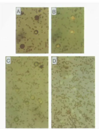
To test this hypothesis, we further increased the valency of sCD4 by coupling CD4-hC/i onto beads and performed a rosette assay by incubating CD4-hC/i- beads with A20 cells, which are known to express high levels of MHC class II molecules (17). Subsequently, rosette formation was analysed by microscopy (Fig. 2). Most of the rosetted cells were completely covered by CD4-hC/i-beads. To demonstrate that the 'bead-berries' are rosetted cells and not aggregated beads, cells were stained with acridine orange, as shown in Fig. 2(A and B).
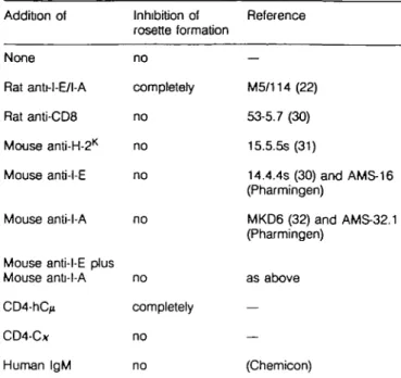
The rosette formation was specific for the C D 4 - M H C class II interaction, since it was completely inhibited by anti-MHC class II mAb M5/114 (22) (Fig. 2 and Table 1). Irrelevant rat anti-mouse mAb (53-6.7) as well as an anti-MHC class I mAb (15.5.5s) did not inhibit rosette formation (summarized in Table 1). M5/114
Correspondence to: K. KarjaJainen
696 Mouse CD4 binding of MHC class II
_ 29
_ 18
Rg. 1. SDS - PAGE analysis of CD4-hQi and CD4-Cx. Purified CD4-hC^, CD4-C*, and, as a control, human IgM (hlgM) protein were analyzed by SDS-PAGE. The gels were subsequently stained with coomassie blue: (A) 7% PA-gel, reducing conditions; (B) 3 - 1 0 % PA-gradient gel; non-reducing conditions. Standard molecular weight markers are indicated on the sides. Method: the part of the mouse CD4 (L3T4) cDNA sequence (14) coding for the extracellular region of the protein was engineered into Ig expression vectors (15,18) upstream of the C2- C4
exons of the human ^ heavy chain gene or the C exons of the mouse x light chain gene The resulting constructs pL3T4-hC^ and pL3T4-Cx (18,19) were introduced into the myeloma cell line J558/L by protoplast fusion as described (16). Stable transfectants, secreting 2 - 6 m g / l CD4-hC/» were adapted to serum free medium (IMDM, 5 mg/1 insulin, 5 mg/l transferrin, 1 x non-essential amino acids, 0.03% primatone). CD4-Cx was purified from supernatant with an anti-moose Cx affinity column and CD4-hC/x was purified from concentrated supernatant by gel-filtration with a FPLC superose 6 column (Pharmacia, Uppsala, Sweden).
recognizes an epitope on MHC class II involving 0, and /32
domains (R. Konig, personal communication) and is conserved in both MHC class II antigens, I-E and I-A. Consistent with this is the recent finding that part of the CD4 binding site to MHC class II molecules is located on the p2 domain and that this
binding site must also be conserved in I-E and I-A antigens. We then tried to carry out further epitope mapping by using a panel of additional anti-MHC class II mAbs. The mAb MKD6 reacting with I-A /S, domain (23) and mAb 14.4.4s, recognizing I-Eor as well as two other mAbs, AMS-16 E) and AMS-32-1 (anti-l-A), with undefined epitopes were used. None of the I-E- or
Fig. 2. Rosette formation. A20 cells were incubated with
CD4-hC>- beads; subsequently, the cells were stained with acridme orange The comparison of (A) light micrograph only versus (B) light micrograph overlaid by fluorescence micrograph reveals that most of the rosetted A20 cells are completely covered by the beads. (C) Rosette formation of A20 cells with CD4-hC/i - beads. (D) Complete inhibition of rosette formation by anti-MHC class II mAb (M5/114, 20 ^g/ml) (x 400). Method. CD4-hC/j-beads were prepared by coupling polyclonal anti-human IgM antibodies (Jackson Immunoresearch, West Grove, PA) onto tosylactivated Dynabeads M-450 (Dynal, Oslo, Norway) overnight, in 0.5 M borate buffer pH 9.5 at 4°C. Washed Dynabeads were then incubated with purified CD4-hC/j overnight in PBS/0 1 % BSA at 4°C. The beads were washed and resuspended in medium (IMDM/5% FCS) to a concentration of 4 x 1 08 beads/ml. The resulting beads
(CD4-hCjt- beads) have thus a layer of CD4, which is oriented to the outside and has a high density of CD4. For negative controls, anti-human IgM-beads were loaded the same way with human IgM (Chemicon, Temecula, CA). The rosette formation assay was performed by mixing 10 ^l of CD4-hC(i - bead suspension (4 x 10%it) with 50 /J A20 cell (17) suspension (2x lO^/ml, IMDM/5% FCS), followed by incubation at room temperature for 1 - 2 h with continuous shaking. To distinguish between bead-covered cells and aggregated beads, the cells were stained with acridine orange. Analysis of rosette formation was performed in a hematocytometer by simultaneous light and fluorescence microscopy. CeHs with more than five attached beads were counted as rosetted cells. The percentage of rosetted cells varied from 40 to 90% of total cells between assays. Anti-human IgM-coupled beads and beads loaded with human IgM protein were included in each assay as negative controls.
l-A-specific mAbs, nor the combination of I-E- and l-A-specific mAbs had inhibitory effects on rosette formation at concentrations up to 250 nM (40 /*g/ml). In contrast, M5/114 inhibited rosette formation completely at concentrations of 15 nM (2 /xg/ml). Thus, it seems that only mAb M5/114 binds MHC class II at an epitope interferring with the interaction of CD4.
Table 1. Specific inhibition of the CD4 - MHC class II interaction Addition of None Rat anU-l-E/l-A Rat anti-CDS Mouse anti-H-2K Mouse anti-l-E Mouse anti-l-A
Mouse anti-l-E plus Mouse anti-l-A CD4-hC^ CD4-C* Human IgM Inhibition of rosette formation no completely no no no no no completely no no Reference — M5/114 (22) 53-5.7 (30) 15.5.5s (31) 14.4.4s (30) and AMS-16 (Pharmingen) MKD6 (32) and AMS-32.1 (Pharmingen) as above — — (Chemtoon)
A20 cells ( H - ^ were preincubated with different antibodies and sCD4 before performing rosette formation assay. Non-blocking antibodies had concentrations up to 250 nM (40 /ig/mf), except human IgM and CD4-C*, where concentrations up to 2 mg/ml respectively did not inhibit rosette formation. ID
E
o2
oc
o
B
120- 100- 80- 60- 40- 20-1 01 0 1 0 " 1 0 " 1 0 71 0 * 1 0 * 1 0- 4 1 0 Inhibitor [M]Rg. 3. Inhibition assay of rosette formation. A20 cells were preincubated
with serial dilutions of anti-MHC class II mAbs (M5/114), CD4-hC^ or CD4-Cx before addition of CD4-hC/j- beads for the rosette formation assay. The arrow indicates the endpoint of CD4-Cx titration. Inhibition was standardized to rosette formation without any inhibitor (0%). A typical example of such an inhibition assay is presented here. Variation between assays was very small.
CD4-hC/i completely inhibited rosette formation, most likely because no washing steps are needed for specific rosette formation. Control human IgM did not inhibit rosette formation, indicating that binding of CD4-hC/x - beads to A20 cells is mediated by the CD4 part of the chimeric molecule. Complete inhibition of rosette formation by CD4-hC/j was obtained at
Mouse CD4 binding of MHC class II 697 concentrations of about 4 /zM. Importantly, a monovalent form of CD4, CD4-C*, was not observed to inhibit the rosette formation at up to 100 iiM (4 mg/ml). We attribute these findings to the increased avidity of the pentameric CD4-hC^, due to its multivalency compared with monovalent CD4-C*. In fact, the pioneering work of Karush has shown that multivalency of IgM can increase the avidity up to 107-fold compared with
monovalent affinity (24).
The concentration values needed for 50% inhibition of the rosette formation can be used to estimate the affinity or avidity of an inhibitor (Fig. 3). This is illustrated by the fact that bivalent M5/114 mAb inhibits 50% of the rosette formation at about 5x 10~9 M (Fig. 3), the value, which corresponds exactly to the
avidity of the bivalent antibody measured by classical Scatchard analyses, Ka - 5 X 1 09 M"1 (21 and our unpublished results).
Thus, the avidity of CD4-hC/» is estimated to be about 7x 10⁷
M~\ since a 100-fold higher concentration of CD4-hC/* is needed for inhibition of rosette formation compared with M5/114. Since extrapolation of affinities from avidity values is not possible (24), we can confidently estimate the upper limit for Ke to be 104
M " ' or below from the fact that the monovalent form of sCD4 did not inhibit rosette formation at concentrations of 100 /iM (10"4 M). This value is in agreement with more indirect
estimates of Hussey etal. (24) in the human system, but much lower than the affinity (KB = 3 x 1 06 M"1) measured by
Cammarota etal. (6), again for the human CD4-MHC class II interaction. The latter authors used both sCD4 and soluble MHC class II molecules to derive their affinity values, and it is conceivable that soluble MHC class II molecules are more accessible to sCD4 than MHC class II molecules embedded in the membrane. This may also explain why human sCD4 has never been shown directly to bind MHC class II positive cells. CD4 has been proposed to have two different roles in antigen-specific interaction between Th cells and APCs: firstly, CD4 as
an accessory molecule is thought to contribute to the general adhesion between Th cells and APCs (8-10), and, secondly,
CD4 as a co-receptor for TCR has been suggested to be essential for optimal Th cell activation, due to its signal transducing
capabilities (for review, see 11). Based on our results, we think that CD4 makes only a very minor contribution to the general adhesion, since the other adhesion systems used by Th cells,
like CD2/LFA-3 and LFA-1/ICAM, have affinities of at least two orders of magnitude higher than that of CD4/MHC class II molecules (26). Therefore, we reason that the co-receptor function is most likely physiologically more significant. Interestingly, it has been shown that CD4 can associate with TCR complexes (27-29) and thus, presumably, it has some affinity to the TCR complex as well. Therefore, due to this dual binding specificity of CD4, it may exhibit much higher affinity to the T C R C D 3 -MHC plus peptide complex, as opposed to its affinities to either TCR-CD3 or MHC class II molecules alone. This would also explain why the CD4, as a signalling molecule during T cell activation, can find its relevant partner in the presence of an excess of potentially irrelevant MHC class II and TCR molecules.
Acknowledgements
We would like to thank D. Avila for technical help with FPLC, and Drs W. Gerhard, P. Lane, and T. Rolink for critical reading of the manuscript. The Basel Institute for Immunology was founded and is supported by F. Hoffmann-La Roche Ltd, Basel, Switzerland.
698 Mouse CD4 binding of MHC class II
Abbreviations
APC sCD4
antigen presenting cell soluble CD4
T helper cell
References
1 Swain, S. 1983. T cell subsets and the recognition of MHC class. Immunol. Rev 74:129.
2 Wilde, D., Marrack, P., Kappler, J., Dialynas, D., and Fitch, F. 1983 Evidence implicating L3T4 in class II MHC antigen reactivity; monoclonal antibody GK1.5 (anti-L3T4a) blocks class II MHC antigen-specific proliferation, release of lymphokines, and binding by cloned murine helper T lymphocyte lines. J. Immunol. 131:2178. 3 Doyle, C. and Strominger, J. 1987. Interaction between CD4 and class
II MHC molecules mediates cell adhesion. Nature 330.256. 4 Gay, D., Buus, S., Pasternack, J., Kappler, J., and Marrack, P. 1988.
The T-cell accessory molecule CD4 recognizes a monomorphic determinant on isolated la. Proc. Natl Acad. Set. USA 85:5629. 5 K6nig, R., Huang, L.-Y., and Germain, R. 1992. MHC class II
interaction with CD4 mediated by region analogous to the MHC class I binding site for CD8. Nature 356:796.
6 Cammarata, G., Scheirle, A., Takacs, B., Doran, D., Knorr, R., Bannwarth, W., Guardiola, J , and Sinigaglia, F. 1992. Identification of a CD4 binding site on the f}2 domain of HLA-DR molecules. Nature
356:799.
7 von Boehmer, H., Kisielow, P., Kishi, H., Scott, B., Borgurya, P., and Teh, H. 1989. The expression of CD4 and CD8 accessory molecules on mature T cells is not random but correlates with the specificity of the a/3 receptor for antigen. Immunol. Rev. 109:143.
8 Marrack, P., Endres, R., Shimonkevitz, R., Zlotnik, A , Dialynas, D., Fitch, F., and Kappler, J. 1983. The major histocompatibility complex-restricted antigen receptor on T cells. II. Role of the L3T4 product J. Exp. Med 158:1077.
9 Biddison, W., Rao, P., Talle, M., Goldstein, G., and Shaw, S. 1984. Possible involvement of the T4 molecule in T cell recognition of class II HLA antigens. Evidence from studies of CTL-target cell binding. J. Exp. Med. 159:783.
10 Gay, D., Maddon, P., Sekaly, R., Talle, M A., Godfrey, M., Long, E., Goldstein, G., Chess, L, Axel, R., Kappler, J., and Marrack, P. 1987. Functional interaction between human T cell protein CD4 and the major histocompatibility complex HLA-DR antigen. Nature 328:626. 11 Janeway, C. 1992. The T cell receptors as a multicomponent signalling machine1 CD4/CD8 coreceptors and CD45 in T cell activation. Annu. Rev. Immunol. 10:645.
12 Weber, S., Traunecker, A., Oftveri, F., Gerhard, W., and Karjalainen, K. 1992. Specific low-affinity recognition of major histocompatibility complex plus peptide by soluble T-cell receptor. Nature 356:793. 13 Matsui, K., Boniface, J., Reay, P., Schild, H., Fazekas de St Groth, B., and Davis, M. 1991. Low affinity interaction of peptide-MHC complexes with T cell receptor. Science 254:1788.
14 Littman.D. and Gettner, S. 1987. Unusual intron in the immunoglobulin domain of the newty isolated murine CD4 (L3T4) gene. Nature 325453.
15 Rutschmann, R. and Karjalainen, K. 1991. Mouse LFA-3 studied with
chimeric soluble CD2 shows preferential expression on lymphoid cells. Eur. J Immunol. 21:1379
16 Oi, V. T., Morrisson, S. L, Herzenberg, L. A., and Berg, P. 1983 Immunoglobufin gene expression in transfected lymphoid ceOs Proc. Natl Acad. Set. USA 80:825.
17 Walker, E., Warner, N., Chestnut, R , Kappler, J., and Marrack, P. 1982. Antigen specific, l-regkxi restricted interactions in vitro between tumor cell lines and T cell hybridomas. J. Immunol. 128:2164. 18 Traunecker, A., Dolder, B., Oliveri, F., and Karjalainen, K. 1989.
Solubflizing the T-cell receptor—problems in solution. Immunol. Today 10:29
19 Weber, S., Traunecker, A., and Karjalainen, K. 1993. Constitutive expression of high levels of soluble mouse CD4 in transgenic mice does not interfere with their immune function. Eur. J. Immunol., 23:511. 20 Dialyas, D., Quan, Z., Wall, K., Pierres, A., Quintas, J., and Loken, M.
1983. Characterization of the murine T cell surface molecule, designated L3T4, identified by monoclonal antibody GK1.5: similarity of L3T4 to the human Leu-3/T4 molecule. J. Immunol. 131:2445. 21 Pierres, A., Naquet, P., Van Agthoven, A , Bekkhoucha, F., Dentzot, F.,
Mishal, Z., Schmitt-Verhulst, A , and Pierres, M. 1984. A rat anti-mouse T4 monoclonal antibody (H129.19) inhibits the proliferation of la-reactive T cell clones and delineates two phenotypically distinct (T4 + , Lyt-2,3", and T 4 " , Lyt2,3+) subsets among anti-lg cytolytic
T cell clones. J. Immunol. 132.2775.
22 Bhattacharya, A., Dorf, M., and Springer, T. 1981. A shared alloantigenic determinant on la antigens encoded by I-A and I-E subregions: Evidence for I region gene duplication. J. Immunol. 127:2488.
23 Braunstein, N. and Germain, R. 1987. Allele-specific control of la surface expression and conformation: Implications for a general model of la structure-function relationship Proc. Natl Acad. Sci USA 84.2921. 24 Karush, F. and Hornick, C 1972. Multivalence and affinity of antibody.
Int. Arch. Allergy 45:130.
25 Hussey, R., Richardson, N., Kowalski, M., Brown, N., Chang, H.-C., Silicano, R., Dorfman, T , Walker, B., Sodroski, J., and Reinherz, E. 1988. A soluble CD4 protein selectively inhibits HIV replication and syncytium formation. Nature 331:78.
26 Springer, T. 1990. Adhesion receptors of the immune system. Nature 346:425.
27 Anderson, P., Blue, M., and Schlossman, S. 1988. Comodulation of CD3 and CD4. Evidence for a specific association between CD4 and approximately 5% of the CD3:T cell receptor complexes on helper T lymphocytes. J. Immunol. 140:1732.
28 Rojo, J., Saizawa, K., and Janeway, C. 1989. Physical interaction of CD4 and the T cell receptor can be induced by anti-T cell receptor antibodies. Proc. Natl Acad. Sci. USA 86:3311.
29 Saizawa, K., Rojo, J., and Janeway, C. 1987. Evidence for a physical association of CD4 and the CD3:a:/3 T cell receptor. Nature 260. 30 Ledbetter, J. A. and Herzenberg, L. A. 1979. Xenogenic monoclonal
antibodies to mouse lymphoid differentiation antigens. Immunol Rev. 47:63.
31 Ozato, K., Mayer, N., and Sachs, D. 1980. Hybridoma cell lines secreting monoclonal antibodies to mouse H-2 and la antigens. J. Immunol. 124.533.
32 Kappler, J., Skidmore, B., White, J., and Marrack, P. 1981. Antigen inducible, H-2-restricted, lnterleukin-2-producmg T cell hybridomas. J. Exp. Med. 153:1198.