HAL Id: hal-02326865
https://hal.archives-ouvertes.fr/hal-02326865
Submitted on 22 Oct 2019
HAL is a multi-disciplinary open access
archive for the deposit and dissemination of
sci-entific research documents, whether they are
pub-lished or not. The documents may come from
teaching and research institutions in France or
abroad, or from public or private research centers.
L’archive ouverte pluridisciplinaire HAL, est
destinée au dépôt et à la diffusion de documents
scientifiques de niveau recherche, publiés ou non,
émanant des établissements d’enseignement et de
recherche français ou étrangers, des laboratoires
publics ou privés.
Phonological grammar shapes the auditory cortex: a
functional magnetic resonance imaging study.
Charlotte Jacquemot, Christophe Pallier, Denis Lebihan, Stanislas Dehaene,
Emmanuel Dupoux
To cite this version:
Charlotte Jacquemot, Christophe Pallier, Denis Lebihan, Stanislas Dehaene, Emmanuel Dupoux.
Phonological grammar shapes the auditory cortex: a functional magnetic resonance imaging study..
Journal of Neuroscience, Society for Neuroscience, 2003, 23 (29), pp.9541-6. �hal-02326865�
Behavioral/Systems/Cognitive
Phonological Grammar Shapes the Auditory Cortex:
A Functional Magnetic Resonance Imaging Study
Charlotte Jacquemot,
1,2Christophe Pallier,
3Denis LeBihan,
3Stanislas Dehaene,
3and Emmanuel Dupoux
11Laboratoire de Sciences Cognitives et Psycholinguistique, Ecole des Hautes Etudes en Sciences Sociales–Ecole Normale Supérieure, Centre National de la Recherche Scientifique, 75006 Paris, France,2Institut National de la Sante´ et de la Recherche Me´dicale, Universite´ Paris XII, Hoˆpital Henri Mondor, 94010 Cedex Cre´teil, France, and3Institut National de la Sante´ et de la Recherche Me´dicale, Service Hospitalier Fre´de´ric Joliot and Commissariat a` l’Energie Atomique, 91401 Cedex Orsay, France
Languages differ depending on the set of basic sounds they use (the inventory of consonants and vowels) and on the way in which these
sounds can be combined to make up words and phrases (phonological grammar). Previous research has shown that our inventory of
consonants and vowels affects the way in which our brains decode foreign sounds (Goto, 1971; Na¨a¨ta¨nen et al., 1997; Kuhl, 2000). Here, we
show that phonological grammar has an equally potent effect. We build on previous research, which shows that stimuli that are
phono-logically ungrammatical are assimilated to the closest grammatical form in the language (Dupoux et al., 1999). In a cross-linguistic design
using French and Japanese participants and a fast event-related functional magnetic resonance imaging (fMRI) paradigm, we show that
phonological grammar involves the left superior temporal and the left anterior supramarginal gyri, two regions previously associated
with the processing of human vocal sounds.
Key words: speech perception; cross linguistic study; language specific illusion; planum temporale; fMRI; phonological grammar
Introduction
Languages differ considerably depending on not only their
inven-tory of consonants (Cs) and vowels (Vs) but also the
phonologi-cal grammar that specifies how these sounds can be combined to
form words and utterances (Kaye, 1989). Regarding the
inven-tory of consonants and vowels, research has shown that infants
become attuned to the particular sound categories used in their
linguistic environment during the first year of life (Werker and
Tees, 1984a; Kuhl et al., 1992). In adults, these categories strongly
influence the way in which foreign sounds are perceived
(Abram-son and Lisker, 1970; Goto, 1971; Miyawaki et al., 1975; Trehub,
1976; Werker and Tees, 1984b; Kuhl, 1991), causing severe
prob-lems in the discrimination between certain non-native sounds.
For instance, Japanese listeners have persistent trouble
discrimi-nating between English /r/ and /l/ (Goto, 1971; Lively et al., 1994).
The current interpretation of these effects is that experience with
native categories shapes the early acoustic–phonetic
speech-decoding stage (Best and Strange, 1992; Best, 1995; Flege, 1995;
Kuhl, 2000). Language experience has been found to modulate
the mismatch negativity (MMN) response, which is supposed to
originate in the auditory cortex (Kraus et al., 1995;
Dehaene-Lambertz, 1997; Na¨a¨ta¨nen et al., 1997; Dehaene-Lambertz and
Baillet, 1998; Sharma and Dorman, 2000).
Regarding phonological grammar, its role has been primarily
studied by linguists, starting with early informal reports
(Po-livanov, 1931; Sapir, 1939) and more recently with the study of
loanword adaptations (Silverman, 1992; Hyman, 1997).
Al-though these studies do not include experimental tests, they
sug-gest a strong effect of phonological grammar on perception. For
instance, Japanese is primarily composed of simple syllables of
the consonant–vowel type and does not allow complex strings of
consonants, whereas English and French do allow these complex
strings. Conversely, Japanese allows a distinction between short
and long vowels, whereas English and French do not (e.g., “tokei”
and “tookei” are two distinct words in Japanese). Accordingly,
when Japanese speakers borrow foreign words, they insert
so-called “epenthetic” vowels (usually /u/) into illegal consonant
clusters so that the outcome fits the constraints of their grammar:
the word “sphinx” becomes “sufinkusu” and the word
“Christ-mas” becomes “Kurisumasu.” Conversely, when English or
French import Japanese words, they neglect the vowel length
distinction: “Tookyoo” becomes “Tokyo” and “Kyooto”
be-comes “Kyoto.” Recent investigations have claimed that such
ad-aptations result from perceptual processes (Takagi and Mann,
1994; Dupoux et al., 1999, 2001; Dehaene-Lambertz et al., 2000).
The current hypothesis is that the decoding of continuous speech
into consonants and vowels is guided by the phonological
gram-mar of the native language; illegal strings of consonants or vowels
are corrected through insertion (Dupoux et al., 1999) or
substi-tution of whole sounds (Massaro and Cohen, 1983; Halle et al.,
1998). For instance, Dupoux et al. (1999) found that Japanese
listeners have trouble distinguishing “ebza” from “ebuza,” and
Dehaene-Lambertz et al. (2000) reported that this contrast does
not generate a significant MMN, contrary to what is found with
Received June 10, 2003; revised Aug. 21, 2003; accepted Aug. 25, 2003.
This work was supported by a Cognitique PhD scholarship to C.J., an Action Incitative grant to C.P., a Cognitique grant, and a BioMed grant. We thank G. Dehaene-Lambertz, P. Ciuciu, E. Giacomeni, N. Golestani, S. Franck, F. Hennel, J.-F. Mangin, S. Peperkamp, M. Peschanski, J.-B. Poline, and D. Rivie`re for help with this work.
Correspondence should be addressed to Charlotte Jacquemot, Laboratoire de Sciences Cognitives et Psycholin-guistique, Ecole des Hautes Etudes en Sciences Sociales, 54 bd Raspail, 75006 Paris, France. E-mail: [email protected].
French listeners. This suggests that the process that turns
non-grammatical sequences of sounds into non-grammatical ones may
take place at an early locus in acoustic–phonetic processing,
probably within the auditory cortex.
In the present study, we aimed at identifying the brain regions
involved in the application of phonological grammar during
speech decoding. We built on previous studies to construct a fully
crossed design with two populations (Japanese and French) and
two contrasts (ebuza– ebuuza, and ebza– ebuza). One contrast,
ebuza– ebuuza, is licensed by the phonological grammar of
Japa-nese but not in French, in which differences in vowel length are
not allowed within words. In French, both ebuza and ebuuza
receive the same phonological representation (ebuza), and
French participants can discriminate these stimuli only by relying
on the acoustic differences between them. The other contrast,
ebza– ebuza, has the same characteristics in reverse. It can be
distinguished phonologically by the French participants but only
acoustically by the Japanese participants. To make acoustic
dis-crimination possible, we presented the contrasts without any
phonetic variability, that is, the tokens were always spoken by the
same speaker and, when identical, were physically identical.
In-deed, previous research has found that phonetic variability
in-creases the error rate for acoustic discriminations considerably
(Werker and Tees, 1984b; Dupoux et al., 1997). Here, our aim
was to obtain good performance on both acoustic and
phonolog-ical discrimination but show that these two kinds of
discrimina-tion nonetheless involve different brain circuits.
French and Japanese volunteers were scanned while
perform-ing an AAX discrimination task. In each trial, three pseudowords
were presented; the first two were always identical, and the third
was either identical or different. When identical, all stimuli were
acoustically the same. When different, the third item could differ
from the other two in vowel duration (e.g., ebuza and ebuuza) or
in the presence or absence of a vowel “u” (e.g., ebza and ebuza).
As explained above, the change that was phonological for one
population was only acoustic for the other (Table 1). Hence, by
subtracting the activations involved in the phonological versus
the acoustic discriminations, the brain areas that are involved in
phonological processing alone can be pinpointed (Binder, 2000).
Such a comparison is free of stimulus artifacts because across the
two populations, the stimuli involved in the phonological and
acoustic contrasts are exactly the same.
Materials and Methods
Participants. Seven native speakers of Japanese 25–36 years of age (mean,
27) and seven native speakers of French 21–30 years of age (mean, 25) were recruited in Paris and participated in the study after giving written informed consent. All Japanese participants had started studying English after the age of 12 and French after the age of 18. None of the French participants studied Japanese. All participants were right-handed
ac-cording to the Edinburgh inventory. None had a history of neurological or psychiatric disease or hearing deficit.
Stimuli and task. The stimuli were the 20 triplets of pseudowords
de-scribed by Dupoux et al. (1999). They followed the pattern VCCV/ VCVCV/VCVVCV (e.g., ebza/ebuza/ebuuza). For the present experi-ment, to present the three stimuli in the 2 sec silent window (see below), the stimuli were compressed to 60% of their original duration using the Psola algorithm in the Praat software (available at http://www. praat.org) so that their final duration was on average 312 msec (⫾43 msec). A fast event-related fMRI paradigm was used. Each trial lasted 3.3 sec and was composed of a silent window of 2 sec during which three stimuli were presented through headphones mounted with piezoelectric speakers (stimulus onset asynchrony; 600 msec), followed by 1.3 sec of fMRI acquisition. Thus, the noise of the gradients of the scanner did not interfere with the presentation of the stimuli. Trials were administered in sessions of 100, with each session lasting 6 min. Trials were of five types: acoustic change, acoustic no-change, phonological change, phonological no-change, and silence. The first four types corresponded to the crossing of two variables: acoustic versus phonological and change versus no-change. The acoustic versus phonological variable was defined as a func-tion of the language of the subject (Table 1). The no-change trials con-tained the same items as the corresponding change trials, except that the three stimuli were physically identical. Within a session, 20 trials of each type were presented in random order. After performing a practice sion, each participant performed between four and six experimental ses-sions during fMRI scanning.
Participants were instructed that they would hear a series of three auditory stimuli, of which the first two would always be identical, and that they had to judge whether the last stimulus was strictly (physically) identical to the first two. They indicated their responses (same or differ-ent) by pressing a response button either with their left or right thumb. The response side was changed at midpoint during the experiment.
Brain imaging. The experiment was performed on a 3-T whole-body
system (Bruker, Ettlingen, Germany) equipped with a quadrature bird-cage radio frequency coil and a head-gradient coil insert designed for echo planar imaging. Functional images were obtained with a T2-weighted gradient echo, echo planar-imaging sequence (repetition time, 3.3 sec; echo time, 40 msec; field of view, 240⫻ 240 mm2; matrix, 64⫻ 64). Each image, acquired in 1.3 sec, was made up of 22 4-mm-thick axial slices covering most of the brain. A high-resolution (1⫻ 1 ⫻ 1.2 mm) anatomical image using a three-dimensional gradient-echo inversion-recovery sequence was also acquired for each participant.
fMRI data analysis was performed using statistical parametric map-ping (SPM99; Welcome Department of Cognitive Neurology, London, UK). Preprocessing involved the following (in this order): slice timing, movement correction, spatial normalization, and smoothing (kernel, 5 mm). The resulting functional images had cubic voxels of 4⫻ 4 ⫻ 4 mm3. Temporal filters (high-pass cutoff at 80 sec; low-pass Gaussian width, 4 sec) were applied. For each participant, a linear model was generated by entering five distinct variables corresponding to the onsets of each of the five types of trials (acoustic change, acoustic no-change, phonological change, phonological no-change, and silence). Planned contrast images were obtained and then smoothed with a 6 mm Gaussian kernel and submitted to one-sample t tests (random effect analysis). Unless specified, the threshold for significance was set at p⬍ 0.001, voxel-based uncorrected, and p⬍ 0.05, corrected for spatial extent.
Results
The analysis of the behavioral results revealed that the
partici-pants (combining Japanese and French groups) were globally
able to detect the change in both conditions (90% correct).
Re-action times and error rates were submitted to ANOVA with the
factors language (Japanese vs French) and condition
(phonolog-ical vs acoustic). The phonolog(phonolog-ical condition was overall easier
than the acoustic condition (error rates, 5.6 vs 13.6%; F
(1,12)⫽
25.1; p
⬍ 0.001; reaction times, 707 vs 732 msec; F
(1,12)⫽ 5.9; p ⬍
0.05 for the phonological vs acoustic condition, respectively).
There were no main effects of language but in the analysis of
Table 1. Two examples of change trials and the condition to which they belong as a function of the native language of the participant
The three auditory stimuli of each example are presented with their spectrogram.
errors only, there was a significant interaction between language
and condition (F
(1,12)⫽ 14.4; p ⬍ 0.01). Post hoc comparisons of
the errors showed that the effect of condition was significant in
the Japanese (3.1 vs 17.2%; p
⬍ 0.01; phonological vs acoustic
condition, respectively) but not in the French group (8 vs 9.9%;
p
⬎ 0.1). Post hoc comparisons of the reaction times showed that
the effect of condition was significant in the French (690 vs 725
msec; p
⬍ 0.05; phonological vs acoustic condition, respectively)
but not in the Japanese group (724 vs 739 msec; p
⬎ 0.1). Such an
asymmetry between speed and accuracy across languages was
already observed by Dupoux et al. (1999) but in both languages,
the conclusion is the same: there is an advantage for the
phono-logical condition relative to the acoustic condition. The overall
size of the phonological effect is smaller than in the Dupoux et al.
study, because we purposefully used a situation with only one
speaker voice to facilitate the discrimination on the basis of
acoustic differences.
In analyzing the fMRI data, we computed three contrasts, one
to identify the circuits involved in the detection of an acoustic
change, one for the circuits involved in the detection of a
phono-logical change, and one for the difference between the two
circuits.
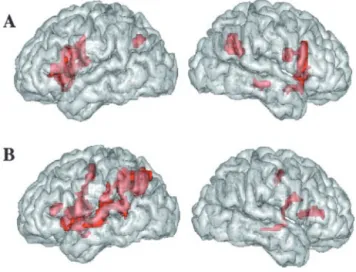
First, we calculated contrast images between the acoustic
change and acoustic no-change conditions. Acoustic change
ac-tivated a large network, comprising the right superior and middle
temporal gyri and, bilaterally, the intraparietal sulci, inferior
frontal gyri, insula, cingulate cortex, and thalamus (Fig. 1 A;
Ta-ble 2), which is congruent with previous studies (Zatorre et al.,
1994; Belin et al., 1998). Second, we calculated the contrast
be-tween the phonological change and phonological no-change
con-ditions. Phonological change caused activation in the perisylvian
areas in the left hemisphere, including the inferior frontal gyrus,
superior temporal gyrus (STG), supramarginal and angular gyri,
and left intraparietal sulcus (Fig. 1 B; Table 3), typically associated
with discrimination tasks involving speech sound analysis
(De´-monet et al., 1992; Zatorre et al., 1992; Burton et al., 2000).
Sig-nificant activation was also observed bilaterally in the cingulate
cortex, insula, and precentral gyrus. To a lesser extent, the right
inferior frontal and the right superior and middle temporal gyri
were also activated. Regions activated in both conditions were the
insula, cingulate cortex, and central sulcus. These regions have
been shown to be involved in the motor and cognitive
compo-nents of an auditory task requiring attention and motor response
(Zatorre et al., 1994). Finally, we calculated the difference
be-tween the phonological and acoustic change circuits. We found
two regions that were significantly more activated by the
phono-logical than the acoustic changes: the left STG and anterior part of
the left supramarginal gyrus (SMG) (Fig. 2). When the threshold
was lowered to p
⬍ 0.01, a region in the right STG also appeared
Figure1. A,B,Activationrenderedonthelefthemisphere(left)andrighthemisphere(right)
of the brain. A, Areas activated by an acoustic change (reaching significance in the comparison of an acoustic change vs no-change conditions). B, Areas activated by a phonological change (reaching significance in the comparison of phonological change vs no-change conditions). Group analysis, voxel-based threshold at p⬍ 0.001, uncorrected; spatial extent threshold,
p⬍ 0.05.
Table 2. Brain areas activated by the detection of an acoustic change
Brain area z-score Peak location in Talairachcoordinates (x, y, z; mm) L inferior frontal gyrus, pars triangularis BA 45 3.85 ⫺44, 28, 4
L inferior frontal gyrus, pars opercularis BA 44 3.54 ⫺44, 12, 8 L inferior frontal gyrus BA 44/45 3.60 ⫺44, 12, 20 L intraparietal sulcus BA 39/40 4.01 ⫺32, ⫺64, 40 L thalamus 3.99 ⫺16, ⫺4, 16 L central sulcus BA 3/4 3.93 ⫺36, 0, 28 L insula 3.80 ⫺28, 20, 4 R inferior frontal gyrus, pars opercularis BA 44 4.02 48, 16, 8 R inferior frontal gyrus BA 44/45 3.73 48, 16, 24 R superior temporal sulcus (anterior) BA
21/22 3.92 48, 4,⫺16
R middle temporal gyrus BA 21 3.87 56,⫺28, ⫺8 R intraparietal sulcus BA 39/40 4.60 28,⫺48, 24 R central sulcus BA 3/4 3.52 40, 0, 20 R insula 4.33 24, 24, 4 R thalamus 4.14 12, 0, 12 Cingulate sulcus BA 32/8/9 3.97 ⫺4, 24, 36 Cingulate sulcus BA 32/9 3.95 4, 24, 32
Coordinates, in standard stereotactic space of Talairach and Tournoux (Talairach and Tournoux, 1988), refer to maxima of the z value within each focus of activation. L, Left; R, right. Approximate Brodmann numbers (BA) associated with anatomical regions are given.
Group analysis: threshold set at p⬍ 0.001, uncorrected, and p ⬍ 0.05, corrected for spatial extent.
Table 3. Brain areas activated by the detection of a phonological change
Brain area z score Peak location in Talairachcoordinates (x, y, z; mm) L intraparietal sulcus BA 31/7 5.23 ⫺32, ⫺60, 36 L supramarginal gyrus (anterior) BA 40 3.91 ⫺56, ⫺28, 28 L supramarginal gyrus (posterior) BA 40 3.81 ⫺44, ⫺40, 40 L angular gyrus BA 39 3.81 ⫺52, ⫺44, 48 L superior temporal sulcus BA 21/22 3.37 ⫺48, ⫺8, ⫺16 L superior temporal sulcus (posterior) BA
21/22 4.22 ⫺48, ⫺48, 8
L superior temporal gyrus (posterior) BA 22/40 4.45 ⫺56, ⫺44, 12 L superior temporal gyrus BA 22/42 4.35 ⫺64, ⫺16, 0 L inferior frontal gyrus, pars triangularis BA 45 3.84 ⫺48, 32, 8 L inferior frontal gyrus, pars opercularis BA 44 4.23 ⫺52, 8, 12 L insula 4.15 ⫺40, ⫺8, 12 L precentral gyrus BA 4/6 4.04 ⫺36, ⫺8, 56 R inferior frontal gyrus, pars triangularis BA 45 3.86 40, 28, 12 R middle temporal gyrus BA 21/22 3.34 60,⫺24, ⫺8 R superior temporal gyrus BA 22 3.36 60,⫺12, ⫺8 R central sulcus BA 3/4 3.53 24,⫺12, 44 R precentral gyrus BA 6/9 3.85 40, 8, 28 R insula 3.62 40, 16, 8 R lingual gyrus BA 17/18 3.41 8,⫺76, 4 Cingulate sulcus BA 32/8 4.32 ⫺4, 8, 52 Cingulate sulcus BA 32/8 3.63 8, 12, 40
Coordinates, in standard stereotactic space of Talairach and Touroux (Talairach and Tournoux, 1988), refer to max-ima of the z value within each focus of activation. L, Left; R, right. Approxmax-imate Brodmann numbers (BA) associated with anatomical regions are given.
(x
⫽ 52; y ⫽ ⫺8; z ⫽ 4; z-score, 3.4; cluster
size, 71; p
⫽ 0.036, corrected). No region
was significantly more activated by the
acoustic changes than by the phonological
changes.
Discussion
We found a phonological grammar effect
in two regions in the left hemisphere: one
in the STG and one located in the anterior
SMG (Fig. 2). There was more activation
in these regions when the stimuli changed
phonologically than when they changed
acoustically. These activations were found
by comparing the same two sets of stimuli
across French and Japanese speakers. In
principle, participants could discriminate
against all stimuli solely on the basis of
acoustic features. However, our results
suggest that a phonological representation
of the stimuli was activated and informed
the discrimination decision. This is
con-firmed by behavioral data that show
per-formance was slightly but significantly
better in the phonological condition than
in the acoustic condition.
The peak activation in the left STG lies
on the boundary between the Heschl gyrus
(HG) and the planum temporale (PT).
At-lases (Westbury et al., 1999; Rademacher
et al., 2001) indicate
⬃40–60% of
proba-bility of localization in either structure
(note that the activation observed in the
right STG when lowering the statistical
threshold is probably located in the Heschl gyrus). Because it is
generally believed that the PT handles more complex
computa-tions than the primary auditory cortex (Griffiths and Warren,
2002), it is reasonable to think that the complex process of
pho-nological decoding takes place in the PT. Yet, the current state of
knowledge does not allow to categorically claim that HG cannot
support this process. Ja¨ncke et al. (2002) observed activations
that also straddled the PT and HG when comparing unvoiced
versus voiced consonants. Numerous studies have revealed
in-creases of PT activations with the spectrotemporal complexity of
sounds (for review, see Griffiths and Warren, 2002; Scott and
Johnsrude, 2003). The present data indicate that PT activations
do not simply depend on the acoustic complexity of speech
sounds but also reflect processes tuned to the phonology of the
native language. This result adds to the converging evidence in
favor of the involvement of the PT in phonological processing.
First, lesions in this region can provoke word deafness, the
inabil-ity to process speech sounds with hearing acuinabil-ity within normal
limits (Metz-Lutz and Dahl, 1984; Otsuki et al., 1998), and
sylla-ble discrimination can be disrupted by electrical interference in
the left STG (Boatman et al., 1995). Second, activity in the PT has
been observed in lip-reading versus watching meaningless facial
movements (Calvert et al., 1997) when profoundly deaf signers
process meaningless parts of signs corresponding to syllabic units
(Petitto et al., 2000) and when reading (Nakada et al., 2001).
Finally, PT activations have also been reported in speech
produc-tion (Paus et al., 1996). These data are consistent with the noproduc-tion
that the PT subserves the computation of an amodal, abstract,
phonological representation.
The second region activated by phonological change was
lo-cated in the left SMG. Focal lesions in this region are not typically
associated with auditory comprehension deficits (Hickok and
Poeppel, 2000) and are not reported when people listen to speech
(Crinion et al., 2003). Yet activations in the SMG have been
ob-served when subjects had to perform experimental tasks
involv-ing phonological short-term memory (Paulesu et al., 1993; Celsis
et al., 1999). A correlation and regression analysis has also
re-vealed that patients impaired in syllable discrimination tend to
have lesions involving the left SMG (Caplan et al., 1995). Thus,
the left SMG activation found in the present study may be linked
to working memory processes and processes translating from
auditory to articulatory representations that can be involved in
speech discrimination tasks (Hickok and Poeppel, 2000).
Remarkably, we did not find that frontal areas were more
involved in the phonological condition than in the acoustic
con-dition, even when the threshold was lowered. This differs from
neuroimaging studies that have claimed that phonological
proc-essing relies on left inferior frontal regions (Demonet et al., 1992;
Zatorre et al., 1992; Hsieh et al., 2001; Gandour et al., 2002).
These studies have used tasks that require the explicit extraction
of an abstract linguistic feature, such as phoneme, tone, or vowel
duration. Such explicit tasks are known to depend on literacy and
engage orthographic representations (Morais et al., 1986;
Poep-pel, 1996). Burton et al. (2000) claimed that frontal activation is
found only in tasks that require explicit segmentation into
con-sonants and vowels and those that place high demands on
work-ing memory. In the present study, the task does not require
seg-mentation of the auditory stimuli.
Previous research on speech processing has focused on the
Figure 2. Areas significantly more activated by a phonological change than by an acoustic change. A, Rendering on a
three-dimensionallefthemispheretemplate.B,C,SectionscenteredonthetwolocalmaximaintheleftSTG( B)(coordinatesinstandard stereotactic space of Talairach and Tournoux: x⫽ ⫺48 mm; y ⫽ ⫺24 mm; z ⫽ 8 mm; z-score, 3.65; cluster size, 14 voxels) and in the left SMG ( C) (coordinates: x⫽ ⫺60 mm; y ⫽ ⫺20 mm; z ⫽ 28 mm; z-score, 3.92; cluster size, 17 voxels). D, Plots of the size of the effect at the two local maxima, as a function of condition and language (Japanese and French). Scale bars show the mean percentage signal change (⫾SE) for each of the following conditions: phonological (phonological change vs phonological no-change) and acoustic (acoustic change vs acoustic no-change).
effects of consonant and vowel categories. These categories are
acquired early by preverbal infants (Werker and Tees, 1984a;
Kuhl et al., 1992; Maye et al., 2002), affect the decoding of speech
sounds (Goto, 1971; Werker and Tees, 1984b), and involve areas
of the auditory cortex (Na¨a¨ta¨nen et al., 1997; Dehaene-Lambertz
and Baillet, 1998). In contrast, the effect of phonological
gram-mar has been less studied but also seems to be acquired early
(Jusczyk et al., 1993, 1994) and shapes the decoding of speech
sounds (Massaro and Cohen, 1983; Dupoux et al., 1999;
Dehaene-Lambertz et al., 2000). At first sight, the regions we
found (left STG and SMG) might be the same as those involved in
consonant and vowel processing. Additional research is needed
to establish whether these regions uniformly represent the
differ-ent aspects of the sound system, or whether separate subparts of
the STG sustain the processing of consonant and vowel categories
on the one hand and phonological grammar on the other. This, in
turn, could help us tease apart theories of perception that posit
two distinct processing stages involving either phoneme
identifi-cation or grammatical parsing (Church, 1987) from theories in
which these two processes are merged into a single step of syllabic
template matching (Mehler et al., 1990).
References
Abramson AS, Lisker L (1970) Discriminability along the voicing continum: cross-language tests. In: Proceedings of the sixth international congress of phonetic sciences, pp 569 –573. Prague: Academia.
Belin P, Zilbovicius M, Crozier S, Thivard L, Fontaine A, Masure MC, Samson Y (1998) Lateralization of speech and auditory temporal processing. J Cogn Neurosci 10:536 –540.
Best C, Strange W (1992) Effects of phonological and phonetic factors on cross-language perception of approximants. J Phonetics 20:305–331. Best CT (1995) Second-language speech learning: theory, findings, and
problems. In: Speech perception and linguistic experience: theoretical and methodological issues (Strange W, Jenkins JJ, eds), pp 171–206. Ti-monium, MD: York.
Binder J (2000) The new neuroanatomy of speech perception. Brain 123:2400 –2406.
Boatman D, Lesser RP, Gordon B (1995) Auditory speech processing in the left temporal lobe: an electrical interference study. Brain Lang 51:269 –290.
Burton MW, Small S, Blumstein SE (2000) The role of segmentation in phonological processing: an fMRI investigation. J Cogn Neurosci 12:679 – 690.
Calvert GA, Bullmore ET, Brammer MJ, Campbell R, Williams SCR, McGuire PK, Woodruff PWR, Iversen SD, David AS (1997) Activation of auditory cortex during silent lipreading. Science 276:593–596. Caplan D, Gow D, Makris N (1995) Analysis of lesions by MRI in stroke
pa-tients with acoustic-phonetic processing deficits. Neurology 45:293–298. Celsis P, Boulanouar K, Doyon B, Ranjeva JP, Berry I, Chollet F (1999)
Differential fMRI responses in the left posterior superior temporal gyrus and left supramarginal gyrus to habituation and change detection in syl-lables and tones. NeuroImage 9:135–144.
Church KW (1987) Phonological parsing and lexical retrieval. Cognition 25:53– 69.
Crinion JT, Lambon-Ralph MA, Warburton EA, Howard D, Wise RJ (2003) Temporal lobe regions engaged during normal speech comprehension. Brain 5:1193–1201.
Dehaene-Lambertz G (1997) Electrophysiological correlates of categorical phoneme perception in adults. NeuroReport 8:919 –924.
Dehaene-Lambertz G, Baillet S (1998) A phonological representation in the infant brain. NeuroReport 9:1885–1888.
Dehaene-Lambertz G, Dupoux E, Gout A (2000) Electrophysiological cor-relates of phonological processing: a cross-linguistic study. J Cogn Neu-rosci 12:635– 647.
De´monet JF, Chollet F, Ramsay S, Cardebat D, Nespoulous JL, Wise R, Rascol A, Frackowiak R (1992) The anatomy of phonological and semantic processing in normal subjects. Brain 115:1753–1768.
Dupoux E, Pallier C, Sebastian-Galle´s N, Mehler J (1997) A destressing “deafness” in French? J Mem Lang 36:406 – 421.
Dupoux E, Kakehi K, Hirose Y, Pallier C, Fitneva S, Mehler J (1999) Epen-thetic vowels in Japanese: a perceptual illusion. J Exp Psychol Hum Per-cept Perform 25:1568 –1578.
Dupoux E, Pallier C, Kakehi K, Mehler J (2001) New evidence for prelexical phonological processing in word recognition. Lang Cogn Proc 16:491–505.
Flege J (1995) Second-language speech learning: theory, findings, and prob-lems. In: Speech perception and linguistic experience: theoretical and methodological issues (Strange W, Jenkins JJ, eds), pp 233–273. Timo-nium, MD: York.
Gandour J, Wong D, Lowe M, Dzemidzic M, Satthamnuwong N, Tong Y, Li X (2002) A cross-linguistic FMRI study of spectral and temporal cues underlying phonological processing. J Cogn Neurosci 7:1076 –1087. Goto H (1971) Auditory perception by normal Japanese adults of the
sounds “l” and “r”. Neuropsychologia 9:317–323.
Griffiths TD, Warren JD (2002) The planum temporale as a computational hub. Trends Neurosci 25:348 –353.
Halle PA, Segui J, Frauenfelder U, Meunier C (1998) Processing of illegal consonant clusters: a case of perceptual assimilation? J Exp Psychol 4:592– 608.
Hickok G, Poeppel D (2000) Towards a functional neuroanatomy of speech perception. Trends Cogn Sci 4:131–138.
Hsieh L, Gandour J, Wong D, Hutchins GD (2001) Functional heterogene-ity of inferior frontal gyrus is shaped by linguistic experience. Brain Lang 3:227–252.
Hyman L (1997) The role of borrowings in the justification of phonological grammars. Stud Afr Ling 1:1– 48.
Ja¨ncke L, Wustenberg T, Scheich H, Heinze HJ (2002) Phonetic perception and the temporal cortex. NeuroImage 4:733–746.
Jusczyk PW, Friederici AD, Wessels JMI, Svenkerud VY, Jusczyk AM (1993) Infants’ sensitivity to the sound pattern of native language words. J Mem Lang 32:402– 420.
Jusczyk PW, Luce PA, Charles-Luce J (1994) Infants’ sensitivity to phono-tactic patterns in the native language. J Mem Lang 33:630 – 645. Kaye JD (1989) Phonology: a cognitive view. Hillsdale, NJ: LEA.
Kraus N, McGee T, Carrell T, King C, Tremblay K (1995) Central auditory system plasticity associated with speech discrimination training. J Cogn Neurosci 7:27–34.
Kuhl PK (1991) Human adults and human infants show a perceptual mag-net effect for the prototypes of speech categories monkeys do not. Percept Psychophys 50:93–107.
Kuhl PK (2000) A new view of language acquisition. Proc Natl Acad Sci USA 97:11850 –11857.
Kuhl PK, Williams KA, Lacerda F, Stevens KN, Lindblom B (1992) Linguis-tic experiences alter phoneLinguis-tic perception in infants by 6 months of age. Science 255:606 – 608.
Lively SE, Pisoni DB, Yamada RA, Tohkura Yi, Yamada T (1994) Training Japanese listeners to identify English /r/ and /l/: III. Long-term retention of new phonetic categories. J Acoust Soc Am 96:2076 –2087.
Massaro DW, Cohen MM (1983) Phonological constraints in speech per-ception. Percept Psychophys 34:338 –348.
Maye J, Werker JF, Gerken L (2002) Infant sensitivity to distributional in-formation can affect phonetic discrimination. Cognition 3:101–111. Mehler J, Dupoux E, Segui J (1990) Constraining models of lexical access:
the onset of word recognition. In: Cognitive models of speech processing: psycholinguistic and computational perspectives (Altmann G, ed), pp 236 –262. Cambridge, MA: MIT.
Metz-Lutz MN, Dahl E (1984) Analysis of word comprehension in a case of pure word deafness. Brain Lang 1:13–25.
Miyawaki K, Strange W, Verbrugge R, Liberman AM, Jenkins JJ, Fujimura O (1975) An effect of linguistic experience: the discrimination of /r/ and /l/ by native speakers of Japanese and English. Percept Psychophys 18:331–340.
Morais J, Bertelson P, Cary L, Alegria J (1986) Literacy training and speech segmentation. Cognition 24:45– 64.
Na¨a¨ta¨nen R, Lehtokovski A, Lennes M, Cheour M, Huotilainen M, Iivonen A, Vainio M, Alku P, Ilmoniemi RJ, Luuk A, Allik J, Sinkkonen J, Alho K (1997) Language-specific phoneme representations revealed by electric and magnetic brain responses. Nature 385:432– 434.
Nakada T, Fujii Y, Yoneoka Y, Kwee IL (2001) Planum temporale: where spoken and written language meet. Eur Neurol 46:121–125.
Otsuki M, Soma Y, Sato M, Homma A, Tsuji S (1998) Slowly progressive pure word deafness. Eur Neurol 39:135–140.
Paulesu E, Frith CD, Frackowiak RSJ (1993) The neural correlates of the verbal component of working memory. Nature 362:342–345.
Paus T, Perry DW, Zatorre RJ, Worsley KJ, Evans AC (1996) Modulation of cerebral blood flow in the human auditory cortex during speech: role of motor-to-sensory discharges. Eur J Neurosci 8:2236 –2246.
Petitto LA, Zatorre RJ, Gauna K, Nikelski EJ, Dostie D, Evans AC (2000) Speech-like cerebral activity in profoundly deaf people processing signed languages: implications for the neural basis of human language. Proc Natl Acad Sci USA 97:13476 –13477.
Poeppel D (1996) A critical review of PET studies of phonological process-ing. Brain Lang 55:317–351.
Polivanov E (1931) La perception des sons d’une langue e´trange`re. Travaux du Cercle Linguistique de Prague 4:79 –96.
Rademacher J, Morosan P, Schormann T, Schleicher A, Werner C, Freund HF, Zilles K (2001) Probabilistic mapping and volume measurement of human primary auditory cortex. NeuroImage 13:669 – 683.
Sapir E (1939) Language: an introduction of study of speech. New York: Harcourt Brace Jovanovich.
Scott SK, Johnsrude IS (2003) The neuroanatomical and functional organi-zation of speech perception. Trends Neurosci 26:100 –107.
Sharma A, Dorman M (2000) Neurophysiologic correlates of cross-languages phonetic perception. J Acoust Soc Am 105:2697–2703. Silverman D (1992) Multiple scansions in loanword phonology: evidence
from Kantonese. Phonology 9:289 –328.
Takagi N, Mann V (1994) A perceptual basis for the systematic phonologi-cal correspondences between Japanese loan words and their English source words. J Phonetics 22:343–356.
Talairach J, Tournoux P (1988) Co-planar stereotaxic atlas of the human brain. New York: Thieme.
Trehub SE (1976) The discrimination of foreign speech contrasts by infants and adults. Child Dev 47:466 – 472.
Werker JF, Tees RC (1984a) Cross-language speech perception: evidence for perceptual reorganization during the first year of life. Infant Behav Dev 7:49 – 63.
Werker JF, Tees RC (1984b) Phonemic and phonetic factors in adult cross-language speech perception. J Acoust Soc Am 75:1866 –1878.
Westbury CF, Zatorre RJ, Evans AC (1999) Quantifying variability in the planum temporale: a probability map. Cereb Cortex 9:392– 405. Zatorre RJ, Evans AC, Meyer E, Gjedde A (1992) Lateralization of phonetic
and pitch discrimination in speech processing. Science 256:846 – 849. Zatorre RJ, Evans AC, Meyer E (1994) Neural mechanisms underlying
me-lodic perception and memory for pitch. J Neurosci 14:1908 –1919.