HAL Id: hal-03053221
https://hal.archives-ouvertes.fr/hal-03053221
Submitted on 10 Dec 2020
HAL is a multi-disciplinary open access
archive for the deposit and dissemination of
sci-entific research documents, whether they are
pub-lished or not. The documents may come from
teaching and research institutions in France or
abroad, or from public or private research centers.
L’archive ouverte pluridisciplinaire HAL, est
destinée au dépôt et à la diffusion de documents
scientifiques de niveau recherche, publiés ou non,
émanant des établissements d’enseignement et de
recherche français ou étrangers, des laboratoires
publics ou privés.
van Tilborg, M Chakravarty, Marc Dhenain, Rick Dijkhuizen, Alessandro
Gozzi, Andreas Hess, et al.
To cite this version:
Francesca Mandino, Domenic Cerri, Clement Garin, Milou Straathof, Geralda van Tilborg, et al..
Animal Functional Magnetic Resonance Imaging: Trends and Path Toward Standardization. Frontiers
in Neuroinformatics, Frontiers, 2020, 13, �10.3389/fninf.2019.00078�. �hal-03053221�
doi: 10.3389/fninf.2019.00078
Edited by: Jan G. Bjaalie, University of Oslo, Norway Reviewed by: Jiaojian Wang, University of Pennsylvania, United States Noam Shemesh, Champalimaud Foundation, Portugal Abraham Z. Snyder, Washington University in St. Louis, United States *Correspondence: Joanes Grandjean [email protected] Received: 29 May 2019 Accepted: 19 December 2019 Published: 22 January 2020 Citation: Mandino F, Cerri DH, Garin CM, Straathof M, van Tilborg GAF, Chakravarty MM, Dhenain M, Dijkhuizen RM, Gozzi A, Hess A, Keilholz SD, Lerch JP, Shih Y-YI and Grandjean J (2020) Animal Functional Magnetic Resonance Imaging: Trends and Path Toward Standardization. Front. Neuroinform. 13:78. doi: 10.3389/fninf.2019.00078
Animal Functional Magnetic
Resonance Imaging: Trends and Path
Toward Standardization
Francesca Mandino
1,2, Domenic H. Cerri
3, Clement M. Garin
4,5, Milou Straathof
6,
Geralda A. F. van Tilborg
6, M. Mallar Chakravarty
7,8, Marc Dhenain
4,5,
Rick M. Dijkhuizen
6, Alessandro Gozzi
9, Andreas Hess
10, Shella D. Keilholz
11,
Jason P. Lerch
12,13, Yen-Yu Ian Shih
3and Joanes Grandjean
1,14*
1Singapore Bioimaging Consortium, Agency for Science, Technology and Research, Singapore, Singapore,2Faculty
of Biology, Medicine and Health, The University of Manchester, Manchester, United Kingdom,3Center for Animal MRI,
Department of Neurology, Biomedical Research Imaging Center, The University of North Carolina at Chapel Hill, Chapel Hill, NC, United States,4Direction de la Recherche Fondamentale, MIRCen, Institut de Biologie François Jacob, Commissariat à
l’Énergie Atomique et aux Énergies Alternatives, Fontenay-aux-Roses, France,5Neurodegenerative Diseases Laboratory,
Centre National de la Recherche Scientifique, UMR 9199, Université Paris-Sud, Université Paris-Saclay,
Fontenay-aux-Roses, France,6Biomedical MR Imaging and Spectroscopy Group, Center for Image Sciences, University
Medical Center Utrecht, Utrecht University, Utrecht, Netherlands,7Department of Psychiatry, Douglas Mental Health
University Institute, McGill University, Montreal, QC, Canada,8Department of Biological and Biomedical Engineering,
Douglas Mental Health University Institute, McGill University, Montreal, QC, Canada,9Functional Neuroimaging Laboratory,
Istituto Italiano di Tecnologia, Centre for Neuroscience and Cognitive Systems @ UNITN, Rovereto, Italy,10Institute
of Experimental and Clinical Pharmacology and Toxicology, Friedrich–Alexander University Erlangen–Nürnberg, Erlangen, Germany,11Department of Biomedical Engineering, Georgia Tech, Emory University, Atlanta, GA, United States,12Hospital
for Sick Children, Department of Medical Biophysics, University of Toronto, Toronto, ON, Canada,13Wellcome Centre
for Integrative NeuroImaging, University of Oxford, Oxford, United Kingdom,14Department of Radiology and Nuclear
Medicine, Donders Institute for Brain, Cognition, and Behaviour, Donders Institute, Radboud University Medical Center, Nijmegen, Netherlands
Animal whole-brain functional magnetic resonance imaging (fMRI) provides a
non-invasive window into brain activity. A collection of associated methods aims to
replicate observations made in humans and to identify the mechanisms underlying the
distributed neuronal activity in the healthy and disordered brain. Animal fMRI studies
have developed rapidly over the past years, fueled by the development of resting-state
fMRI connectivity and genetically encoded neuromodulatory tools. Yet, comparisons
between sites remain hampered by lack of standardization. Recently, we highlighted
that mouse resting-state functional connectivity converges across centers, although
large discrepancies in sensitivity and specificity remained. Here, we explore past and
present trends within the animal fMRI community and highlight critical aspects in study
design, data acquisition, and post-processing operations, that may affect the results
and influence the comparability between studies. We also suggest practices aimed
to promote the adoption of standards within the community and improve
between-lab reproducibility. The implementation of standardized animal neuroimaging protocols
will facilitate animal population imaging efforts as well as meta-analysis and replication
studies, the gold standards in evidence-based science.
INTRODUCTION
A detailed understanding of the mammalian brain structure
and function is one of the greatest challenges of modern
neuroscience. Approaching the complexity of the organ and
the levels of organization of neuronal circuits across several
orders of magnitudes, both spatially and temporally, requires
the collective scientific efforts from multiple teams across several
disciplines. Neuroimaging, especially by means of magnetic
resonance imaging (MRI), is playing a preponderant role
in mapping the human and animal brain, due to its
non-invasiveness, excellent soft-tissue contrast, and multiple readouts.
The human neuroimaging research has accelerated over the past
decade, fueled by numerous discoveries about brain structure
and function and its relation to disorders. In turn, this has led
to population imaging efforts aimed to describe variations in
brain structure and function, and their relation to behavioral
traits, genetic polymorphisms, and pathology. For instance, since
its original description in 1995 (
Biswal et al., 1995
),
resting-state functional connectivity (RS-FC) has been at the center
of numerous population imaging initiatives, such as the 1,000
Functional Connectomes Project (
Biswal et al., 2010
), the
WU-Minn Human Connectome Project (
Van Essen and Ugurbil, 2012
;
Van Essen et al., 2013
), and the UK Biobank (
Miller et al.,
2016
). In addition to providing an important baseline of healthy
cohorts, these initiatives are complemented with population
imaging dedicated to specific psychiatric and neurological
disorders, such as the Alzheimer’s Disease Neuroimaging
Initiative (
Petersen et al., 2010
;
Weiner et al., 2012
), the Autism
Brain Imaging Data Exchange (
Di Martino et al., 2014
), or
Attention-Deficit Hyperactivity Disorder (
HD-200 Consortium,
2012
). Collectively, these resources have significantly advanced
our understanding of neuro- and psychopathologies, as well
as providing an understanding of disorder spectrums at a
population level.
In contrast to the above, functional neuroimaging studies
in animals have remained mostly confined to single centers,
often relying on lab-specific acquisition and processing protocols.
There has been little pressure toward standardization within the
community, and results from different centers have remained
inherently difficult to compare, due to discrepancies related
to animal housing and preparation, recording hardware, and
analysis methodologies. It is now emerging that these preparation
divergences are at the stem of a number of dissensions within the
animal functional neuroimaging community, such as the nature
of unilateral vs. bilateral resting-state networks (RSN) in mice
(
Jonckers et al., 2011
;
Grandjean et al., 2014
;
Mechling et al.,
2014
;
Sforazzini et al., 2014
), the bilateral BOLD response to
non-noxious paw electrical stimulation in mice (
Bosshard et al., 2010
;
Schroeter et al., 2014
;
Shim et al., 2018
), the indirect artifacts
emerging in optogenetics fMRI (ofMRI) through either heating
or vascular photoactivation (
Christie et al., 2013
;
Rungta et al.,
2017
;
Schmid et al., 2017
), or the spatial extent of distributed
networks of translational relevance, such as the rodent “default
mode network” (DMN) reviewed in
Gozzi and Schwarz (2016)
.
Only recently did efforts emerge to combine and compare
structural and/or functional MRI from multiple centers in
monkeys (
Milham et al., 2018
) and in mice (Figure 1;
Grandjean
et al., 2019a
). These initial studies provide solid grounds for the
development of replication studies, meta-analyses, and
multi-center consortia, the gold standards in evidence-based science.
Presently, we aim to describe the current trends in the
field and to examine how these impact the results and
their comparability with the rest of the literature. While
recommendations to enhance reproducibility exists for human
neuroimaging (
Poldrack et al., 2008
), a large number of
acquisition and data processing aspects remain specific to animal
imaging. We systematically assessed the animal fMRI literature
for data acquisition and analysis procedures to provide an
overview of the collective directions taken within the animal
imaging community. We then reviewed the major considerations
taking place in the study design, and how these impact results
and their interpretability. Finally, we use this information to
provide a road map toward the adoption of standards that will
enable animal population studies to inform on the functional
mammalian brain.
METHODS
We searched the Pubmed database
1on February 11, 2019 for
the terms “functional magnetic resonance imaging,” “functional
MRI,” or “fMRI” within the abstract or title, excluding
studies in human and reviews, from 1990 onward, using
the following command.
“Search ((fMRI[Title/Abstract]) OR
functional MRI[Title/Abstract]) OR functional magnetic resonance
imaging[Title/Abstract] Sort by: Best Match Filters: Abstract;
Publication date from 1990/01/01 to 2019/12/31; Other Animals.”
The query returned 2279 entries. The title and abstract from these
were manually screened to exclude studies that did not contain
primary research using MRI to assess brain function in animals.
In total, 868 research article were considered relevant and could
be readily accessed. We recorded the type of study: resting-state
or paradigm free RS-FC recordings, pharmacological-evoked,
opto-/chemogenetic neuromodulation, deep-brain stimulation
(DBS), or stimulus-evoked (including blocks- or events-related
designs with sensory stimulation, gas challenge, etc.). We
recorded animals species, including strain, gender (male, female,
both, N/A), number of animals used, animal preparation (awake,
anesthetized free-breathing, anesthetized ventilated), anesthetic
used for maintenance during fMRI, field strength, fMRI sequence
and contrast, pre-processing softwares, and noted if the datasets
were made available by the authors or in online repositories. The
resulting table is made available in the Supplementary Material.
RESULTS AND DISCUSSION
Experimental Design
Animal fMRI presents the opportunity for new and creative
directions in study design, but care must be taken to ensure that
experimental changes in the fMRI signal are sufficiently robust
1https://www.ncbi.nlm.nih.gov/pubmed/FIGURE 1 | (A) A seed-based analysis of the anterior cingulate area in 98 resting-state fMRI scans reveals the topological distribution of the mouse default-mode network. The regions co-activating with the seed include the dorsal striatum, dorsal thalamus, retrosplenial, and posterior parietal areas. (B) The reproducibility of the default-mode network was assessed in 17 independent datasets consisting of 15 scans each. Overlapping one-sample t-test maps are summarized in a
color-coded overlay. 12/17 datasets present converging topological features, the remaining five failed to present evidence of distal connectivity relative to the seed. Adapted with permission fromGrandjean et al. (2019a).
for detection and that results are not contaminated by procedural
artifacts. Here we highlight evidence supporting standards and
reporting strategies to optimize data quality, interpretation, and
reproducibility for several common animal fMRI paradigms.
Stimulus-Evoked fMRI
In animal studies, stimulus-evoked fMRI usually refers to
externally applied stimuli during fMRI (e.g., electrical forepaw
stimulation), but many principles of study design can be applied
to internally delivered stimuli as well, such as with deep-brain
stimulation (DBS) and optogenetics. Stimuli can be applied in
a block or event-related design. The former alternates between
regular stimulation and no-stimulation conditions, while the
latter uses brief stimuli presented at varying intervals (
Amaro and
Barker, 2006
). Block designs are best suited to test
frequency-related responses and enhance detection power, while
event-related designs are best for determining accurate response-time
courses and/or frequency-independent functional connectivity
(
Amaro and Barker, 2006
;
Van der Linden et al., 2007
;
Maus
and van Breukelen, 2013
;
Allen et al., 2015
;
Schlegel et al., 2015
;
Soares et al., 2016
).
Stimulus frequency has a large influence on stimulus-evoked
fMRI results. In general, higher frequencies will increase the
stimulus input per unit time, thus potentially boosting signal
and ability to detect evoked responses (
Amaro and Barker,
2006
;
Kim et al., 2010
;
Maus and van Breukelen, 2013
), but
excessive electrical or optical stimulation can cause tissue damage
(
Kiyatkin, 2007
;
Lai et al., 2015
;
Acker et al., 2016
;
Cogan et al.,
2016
), heating and related artifacts (
Zeuthen, 1978
;
Kiyatkin,
2007
;
Cardin et al., 2010
;
Christie et al., 2013
;
Lai et al., 2015
;
Stujenske et al., 2015
;
Acker et al., 2016
), and non-specific effects
(
Tuor et al., 2002
;
Christie et al., 2013
;
Schroeter et al., 2014
;
Shih et al., 2014
;
Schlegel et al., 2015
;
Rungta et al., 2017
).
Stimuli may also change basic physiology and therefore alter
the fMRI response (
Tuor et al., 2002
;
Ray et al., 2011
;
Tsubota
et al., 2012
;
Li et al., 2013
;
Schroeter et al., 2014
;
Shih et al.,
2014
;
Reimann C. et al., 2018
), thereby occluding signal from
the stimulus itself. These findings highlight the importance of
carefully monitoring physiology (see below) and establishing
frequency-response curves for the stimuli of choice.
Functional Connectivity MRI
Animal fMRI data acquired in the absence of stimulation or
modulation, RS-FC, is commonly used to probe synchronization
of spontaneously fluctuating signals between combinations of
anatomically, functionally, or procedurally defined brain regions
(
Lowe et al., 2000
;
Lu et al., 2007
;
Zhao et al., 2008
;
van Meer
et al., 2010, 2012
;
Lu and Stein, 2014
;
Pan et al., 2015
;
Guadagno
et al., 2018
;
Grandjean et al., 2019a
). The use of RS-FC in animal
models has rapidly increased over the past decade (Figure 2).
To collect the most robust and interpretable RS-FC data, a
few principles have been proposed. Recent evidence suggests
that brain network components exhibit non-stationary properties
(
Hutchison et al., 2013a
;
Keilholz et al., 2013
;
Liu and Duyn,
2013
;
Liang et al., 2015a
;
Pan et al., 2015
;
Gutierrez-Barragan
et al., 2018
), therefore repetition time should be sufficiently
short (e.g., 1 s) to properly sampled the fluctuations and to
detect these changes, and scan length should produce enough
frames (a minimum of about 300) to account for a large
number of temporal clusters (
Majeed et al., 2011
;
Hutchison
et al., 2013b
;
Jonckers et al., 2015
). Critical aspects for such
analyses are detailed in a later section. Furthermore, if brain
modulation/stimulation is included, additional time should be
added during the transition periods to and from resting-state
to allow for stable connectivity, and subsequent resting periods
following each manipulation should be grouped separately to
account for potential neuroadaptations (
Pawela et al., 2008
;
Zhao
et al., 2008
;
Jonckers et al., 2015
;
Albaugh et al., 2016
;
Chan et al.,
2017
;
Decot et al., 2017
;
Chen et al., 2018
). Importantly, due to the
nature of the signal fluctuations on which RS-FC relies, special
care must be ensured with regard to physiology and anesthesia to
ensure maximal detection. The effects of animal preparations are
further discussed below.
Optogenetics
Many recent stimulus-evoked animal fMRI studies take
advantage of the readily MR-compatible optogenetics toolkit
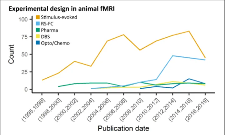
FIGURE 2 | Study design in animal fMRI over time. Stimulus-evoked fMRI (events or blocks related) remain the major component within animal literature. From 2006 and 2010, resting-state fMRI and opto-/chemogenetic fMRI, respectively, have represented an increasing proportion of the animal fMRI studies.
(Figure 2;
Desai et al., 2011
;
Abe et al., 2012
;
Scott and Murphy,
2012
;
Kahn et al., 2013
;
Iordanova et al., 2015
;
Lemieux et al.,
2015
;
Liang et al., 2015b
;
Takata et al., 2015
;
Weitz et al.,
2015
;
Albaugh et al., 2016
;
Chai et al., 2016
;
Ryali et al., 2016
;
Yu et al., 2016
;
Hinz et al., 2017
;
Lohani et al., 2017
;
Albers
et al., 2018
;
Brocka et al., 2018
;
Choe et al., 2018
;
Leong et al.,
2018
;
Grandjean et al., 2019b
). Optogenetics allows for robust
stimulation of specific cellular and/or anatomical populations
(
Zhang et al., 2010
;
Fenno et al., 2011
;
Boyden, 2015
;
Deisseroth,
2015
;
Griessner et al., 2018
), but despite these advantages
this relatively new technique adds layers of complexity over
DBS, thereby requiring more rigorous methodology and
additional controls.
The
light-activated
channels/pumps
expressed
in
optogenetics, also known as “opsins,” provide a great deal
of experimental flexibility (
Fenno et al., 2011
;
Deisseroth, 2015
;
Guru et al., 2015
). There are several opsins to choose from for
optical excitation of cells, including the commonly used ChR2
(
Nagel et al., 2003
;
Boyden et al., 2005
;
Zhang et al., 2006
;
Atasoy
et al., 2008
;
Cardin et al., 2010
) variants activated by penetrating
red-shifted light (
Zhang et al., 2008
;
Lin et al., 2013
;
Klapoetke
et al., 2014
) and ultra-fast variants capable of frequencies up
to 200 Hz (
Lin et al., 2009
;
Gunaydin et al., 2010
;
Hight et al.,
2015
). If stable excitation over even longer periods is required
in fMRI, issues with a continuous light application can be
avoided by using step-function opsins which are temporarily
activated by a single pulse of light (
Berndt et al., 2009
;
Ferenczi
et al., 2016
). Notably, there are also several opsins for cellular
inhibition (
Zhang et al., 2007
;
Berndt et al., 2014
;
Chuong et al.,
2014
), but their application for fMRI is limited as they require
longer periods of illumination prone to heat-related artifacts,
and anesthetized or sedated animals have low baseline levels of
activity (
Lahti et al., 1999
;
Brevard et al., 2003
;
Sicard et al., 2003
).
Injection of viral constructs or expression of foreign genes
can potentially change brain function (
Liu et al., 1999
;
Klein
et al., 2006
;
Zimmermann et al., 2008
;
Lin, 2011
;
Miyashita et al.,
2013
), and light can induce heating and related MRI artifacts,
tissue damage, and non-specific effects (
Elias et al., 1987
;
Christie
et al., 2013
;
Stujenske et al., 2015
;
Schmid et al., 2016
;
Rungta
et al., 2017
) thus it is critical to characterize opsin expression and
activation of the light source with light delivery to empty-vector
(e.g., EYFP) controls. It follows that histological confirmation
of fiber placement and construct co-localization with targeted
promoters is required (
Bernstein and Boyden, 2011
;
Witten et al.,
2011
;
Madisen et al., 2012
;
Zeng and Madisen, 2012
;
Allen et al.,
2015
;
Gompf et al., 2015
;
Lin et al., 2016
;
Decot et al., 2017
).
In addition, given the spatial nature of fMRI, the reporting of
single-point measurements of light power should be avoided
in favor of irradiance (mW/mm
2;
Aravanis et al., 2007
;
Huber
et al., 2008
;
Kahn et al., 2011
;
Yizhar et al., 2011
;
Schmid et al.,
2017
). Finally, light stimulation at frequencies at or below 20 Hz
can produce a visual response by activating the visual-related
network, requiring light masking or careful control comparison
to view experimental effects (
Ferenczi et al., 2016
;
Lin et al., 2016
;
Decot et al., 2017
;
Schmid et al., 2017
).
Chemogenetics
Chemogenetics, initially termed “pharmacogenetics,” utilizes
pharmacologically inert ligands to stimulate genetically encoded
designer receptors, with the aim to produce drug-like sustained
activation or inhibition of specific neuronal populations. Initial
attempts to combine this approach with fMRI have involved
the regional re-expression of pharmacologically targetable
endogenous G-coupled protein receptors (e.g., Htr1a,
Gozzi
et al., 2012
). The recent development of a modular set of
evolved G protein-coupled receptors, termed Designer Receptors
Exclusively Activated by Designer Drugs (DREADDs) has greatly
expanded the capabilities of this approach (
Armbruster et al.,
2007
;
Alexander et al., 2009
;
Lee et al., 2014
;
English and
Roth, 2015
;
Roth, 2016
;
Sciolino et al., 2016
;
Smith et al., 2016
;
Zhu et al., 2016
;
Aldrin-Kirk et al., 2018
). Like optogenetics,
chemogenetics is readily MRI compatible (
Giorgi et al., 2017
;
Roelofs et al., 2017
;
Chen et al., 2018
;
Griessner et al., 2018
;
Markicevic et al., 2018
). Despite its potential, there is, however,
an ongoing debate about the specificity of chemogenetics ligands
both in neurobehavioral studies (
MacLaren et al., 2016
;
Gomez
et al., 2017
;
Mahler and Aston-Jones, 2018
;
Manvich et al.,
2018
) and in chemo-fMRI applications (
Giorgi et al., 2017
),
thereby requiring rigorous methodology to control for potential
off-target effects.
Both hM3Dq and hM4Di DREADDs are classically activated
with infusion of the effector clozapine-N-oxide (CNO)
(
Armbruster et al., 2007
;
Alexander et al., 2009
;
Roth, 2016
;
Smith et al., 2016
;
Giorgi et al., 2017
;
Markicevic et al., 2018
),
but new evidence suggests that CNO does not cross the
blood-brain barrier and instead is back-metabolized
in vivo into its
precursor, clozapine (
Gomez et al., 2017
;
Mahler and
Aston-Jones, 2018
;
Manvich et al., 2018
). Importantly, unlike CNO,
clozapine is a psychoactive drug, that possesses an affinity for
many endogenous receptors. As a result, the use of high CNO
doses may result in a plethora of undesirable off-target effects
(
Ashby and Wang, 1996
;
Selent et al., 2008
;
MacLaren et al.,
2016
;
Roth, 2016
), including unspecific fMRI response (
Giorgi
et al., 2017
). Overall, it is apparent that chemogenetics effects
cannot be interpreted without proper non-DREADD expressing
controls. Specifically, the effect of effector administration
should be compared between DREADD expressing, and
non-DREADD expressing animals and/or hemispheres. Finally,
as with optogenetics, validation of DREADD expression and
co-localization with target promoters is essential for data
interpretation (
Farrell et al., 2013
;
Smith et al., 2016
;
Giorgi et al.,
2017
;
Gomez et al., 2017
;
Roelofs et al., 2017
;
Aldrin-Kirk et al.,
2018
;
Chen et al., 2018
;
Markicevic et al., 2018
).
Pharmacological fMRI
Modulating the brain with pharmacological agents during animal
fMRI has a wide variety of traditional applications such as
studying the global effects of compounds and their target
neurotransmitter systems (
Mueggler et al., 2001
;
Shah et al.,
2004
;
Ferrari et al., 2012
;
Razoux et al., 2013
;
van der Marel
et al., 2013
;
Jonckers et al., 2015
). This approach does not
require surgical methods, and is apt for identifying global or
regional changes in function associated with new or existing drug
therapies for neurotransmitter-related brain disorders (
Leslie
and James, 2000
;
Martin and Sibson, 2008
;
Canese et al.,
2011
;
Bifone and Gozzi, 2012
;
Klomp et al., 2012
;
Minzenberg,
2012
;
Medhi et al., 2014
), or to map the effect of exogenously
administered neuromodulators. In addition, pharmacological
challenges can be used to probe how targets and neurotransmitter
systems modulate BOLD responses evoked by other stimuli or
pharmacological agents (
Marota et al., 2000
;
Hess et al., 2007
;
Schwarz et al., 2007
;
Knabl et al., 2008
;
Rauch et al., 2008
;
Shih
et al., 2012a
;
Squillace et al., 2014
;
Shah et al., 2016
;
Decot et al.,
2017
;
Bruinsma et al., 2018
;
Griessner et al., 2018
). However,
functional imaging with pharmacological agents may not be
ideal for dynamic or repetitive studies as effects are dependent
on diffusion and receptor kinetics (
Steward et al., 2005
;
Ferris
et al., 2006
;
Mandeville et al., 2013
;
Bruinsma et al., 2018
), and
subject to receptor desensitization and downregulation (
Chen
et al., 1999
;
Arey, 2014
;
Berg and Clarke, 2018
); which in some
instances may be species-specific (
Knabl et al., 2008
).
It is important to consider dose-response effects and the
pharmacokinetics of each drug used in the experimental design.
Ideally several doses of drug, and sufficiently long time series
should be included in order to interpret the results according
to dose-response and absorption/elimination functions (
Leslie
and James, 2000
;
Marota et al., 2000
;
Mueggler et al., 2001
;
Steward et al., 2005
;
Ferris et al., 2006
;
Rauch et al., 2008
;
Jenkins, 2012
;
Minzenberg, 2012
;
Jonckers et al., 2015
;
Shah
et al., 2015
;
Bruinsma et al., 2018
). Indeed, many pharmacological
agents have known systemic effects which can influence animal
physiology and the BOLD signal (
Shah et al., 2004
;
Wang et al.,
2006
;
Martin and Sibson, 2008
;
Ferrari et al., 2012
;
Klomp
et al., 2012
), and some drugs have direct effects on the vascular
endothelium in the brain, which could alter properties of the
hemodynamic response (
Luo et al., 2003
;
Gozzi et al., 2007
;
Shih et al., 2012b
). It is imperative to closely control and
monitor animal physiology, and use appropriate doses in order
to control for unwanted side effects. Importantly, vehicle controls
are necessary for any pharmacological fMRI study, as increased
blood flow/volume and increased blood pressure from systemic
infusions can alter the MRI signal (
Kalisch et al., 2001
;
Tuor et al.,
2002
;
Gozzi et al., 2007
;
Reimann H. M. et al., 2018
).
Species, Sample Size, and Gender
Distribution
We assessed studies performed using animals, i.e., all species
except homo sapiens. The rat and specifically the Sprague–
Dawley strain was the most common species and strain used in
fMRI studies, representing 55% of the total studies considered
presently (Figures 3A,B). Non-human primate (NHP) studies
were second and mostly relied on the macaques (23%). Studies
involving medium-sized domestic mammals (cats, dogs, sheeps,
pigs, and rabbits) presented 9% of the total literature considered.
Studies on males (54%) had a higher incidence than studies in
females (14%). A sizable number of studies (22%) omitted to
specify the gender. This gender bias reflects a greater trend found
throughout neuroscience and other biomedical disciplines (
Beery
and Zucker, 2011
) and should require a greater consideration
within the animal neuroimaging community. Finally, the total
number of animals was assessed within the studies considered.
It should be noted that this was done irrespective of the number
of groups. There, we found that nearly half the studies were
carried out on ten or fewer subjects (Figure 3C). This was
particularly marked in studies with NHP (Percentiles 25, 50,
75 = [2, 3, 5]). While sample size depends on the goals of each
study and appropriate power calculation, it remains unclear how
group sizes were determined in most of these studies. The small
group sizes reported here are consistent with general trends
in neuroscience toward underpowered studies.
Button et al.
(2013)
estimated that the median power level in neuroscience
was at 21%. Hence these trends need to be carefully taken into
consideration in the initial stages of study design so that the
required animals are used to their full potential.
The wide range of experimental animals available for
research offers unique opportunities to study evolutionary
trends on distributed neuronal networks. To date, however,
interspecies comparisons have remained a difficult task. fMRI
has provided numerous descriptions of the network organization
in mammals. Specifically, RSNs have been mainly studied in
mammals to develop translational models of human diseases
and to understand the mechanisms underlying their functional
alterations. RSNs’ organization has been described in numerous
mammalian species (usually under anesthesia) including rodents
(
Hutchison et al., 2010
;
Jonckers et al., 2011
;
Sforazzini et al.,
2014
;
Grandjean et al., 2017b
), ferrets (
Zhou et al., 2016
), rabbits
(
Schroeder et al., 2016
), dogs (
Kyathanahally et al., 2015
), prairie
vole (
Ortiz et al., 2018
), and NHP (
Vincent et al., 2007
;
Hutchison
et al., 2010
;
Mantini et al., 2011
;
Belcher et al., 2013
). Particularly
active at rest, one of the most widely investigated networks
is the DMN (
Raichle, 2015
;
Buckner and DiNicola, 2019
).
This network comprises distributed polymodal cortices that are
thought to be involved in memory consolidation and higher
cognitive functions. Homologs of the human DMN (
Raichle
et al., 2001
) have been identified in a variety of species including
NHP (
Vincent et al., 2007
;
Mantini et al., 2011
), rats (
Lu et al.,
2012
), mice (
Sforazzini et al., 2014
;
Stafford et al., 2014
) and
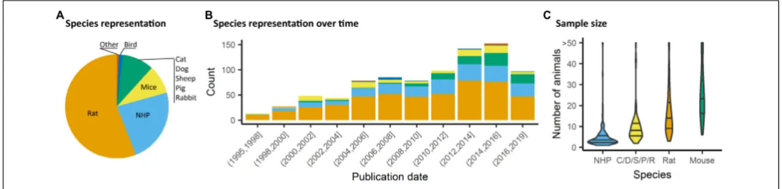
FIGURE 3 | Species distribution and sample size. (A) Animal representation in the documented studies. (B) Animal species occurrence in the literature over time. Rats and non-human primate (NHP) represent the major species used, however, since 2008, mice have been used in a growing proportion of animal fMRI studies. (C) Number of animals used per fMRI study irrespective of number of groups or classes. NHP studies are carried out with fewer animals (Percentiles25,50,75= [2, 3, 5]), whereas studies involving mice involved larger number of animals (Percentiles25,50,75= [17, 24, 34]).
rabbits (
Schroeder et al., 2016
). The hypothesis of two separated
DMNs (anterior and a posterior) has been evoked in dogs
(
Kyathanahally et al., 2015
) and ferrets (
Zhou et al., 2016
).
The description of each species’ functional architectures has
been based on a variety of acquisitions, analyses, and anesthesia
or awake protocols. This lack of interspecies standardization is
often justified by the variety of brain sizes, different response
to anesthesia, and anatomical organizations observed within
mammals. Throughout evolution, brain regions could have
duplicated, fused, reorganized or expanded (
Hutchison and
Everling, 2012
). A few studies have compared the connectivity
between different species and with similar approaches. Using
ICA, Jonckers et al. found that the extracted components,
i.e., functional network regions, were more unilateral in mice
compared to rats (
Jonckers et al., 2011
), however, this effect failed
to be replicated in numerous follow-up studies in mice (e.g.,
Grandjean et al., 2014
;
Sforazzini et al., 2014
). In mouse lemur
primates and humans, the cortical large-scale networks repertoire
presents important similarities but the regional organization
into networks highlighted compositional and structural
divergences (
Garin et al., 2019
). Strong interhemispheric
functional connectivity (FC) between homotopic regions has
been consistently observed in humans and primates suggesting a
phylogenetically preserved mammalian characteristic (
Hutchison
and Everling, 2012
). However, lateralized networks (i.e.,
fronto-parietal resting-state network) remain a phenomenon which
has only been demonstrated in humans. According to the few
comparative studies on mammals functional organization,
humans seem to display the strongest variety of functional
networks. The complexity and diversity of the animal behaviors
are probably related to this large repertoire of networks. This
complexity is also reflected by the white matter fiber tracts
network (
Nadkarni et al., 2018
). Moreover, direct evidence
is in favor of a close relationship between the structural and
functional organization in humans (
Damoiseaux and Greicius,
2009
), in primates (
Miranda-Dominguez et al., 2014
) and in mice
(
Stafford et al., 2014
;
Grandjean et al., 2017b
). However, a recent
systematic review showed that structure-function correlations
in mammalian brains depend on the connectivity measures,
which differ across methods and scales (
Straathof et al., 2019
).
The structure-function correspondence observed in multiple
species is an important step in favor of the neural origin
underlying the BOLD signal and provides a key to understanding
neural network development through the evolution of complex
brain structure.
Other universal properties of the brain topology have also
emerged recently with graph analysis. One of them is the
small-world feature which maximizes the efficiency of information
transferred within a network. This network property has been
found in multiple species including humans (
Bullmore and
Sporns, 2009
), NHP (
Barttfeld et al., 2015
;
Garin et al., 2019
),
rodents (
Mechling et al., 2014
), and ferrets (
Zhou et al.,
2016
). Moreover, graph-based approaches have clearly revealed a
modular nature of human (
Sporns and Betzel, 2016
), and rodent
(
Liska et al., 2015
) rsfMRI networks, along with evidence of
strongly functionally interconnected polymodal areas, exhibiting
hub-like properties (
Buckner et al., 2009
;
Liska et al., 2015
).
Concerning highly connected regions in human, macaque and
mouse lemur, the posterior cingulate cortex was found to
be critical in these three species with its major functional
hubs located in the DMN (
Garin et al., 2019
). Interestingly,
these areas seem to be instead shifted anteriorly in rodents,
in which the anterior cingulate and prefrontal areas exhibit
robust hub-like properties (
Liska et al., 2015
;
Gozzi and
Schwarz, 2016
;
Garin et al., 2019
). This finding is consistent
with rodent species lacking an evolutionary homolog of the
primate posterior cingulate cortex (
Vogt and Paxinos, 2014
).
Determining the fine-grained topology and contribution of
regions critical for network organization and stability across
species and evolution could highlight functional patterns that
are especially relevant for network stability. Despite the lack of
consensus concerning a standardized methodology in mammals
fMRI, cross-species studies could provide essential clues toward a
better understanding of brain physiology and evolution.
Animal Preparation and Anesthesia
Animal Preparation Impact on Motion and Stress
Functional MRI traditionally relies on temporal changes
in hemodynamic parameters, e.g., blood oxygenation
level-dependent contrast (BOLD), cerebral blood volume (CBV),
or cerebral blood flow (CBF). Functional MRI signals inform
on neuronal activity through the evaluation of hemodynamic
response i.e., the adaptability of local capillaries to deliver oxygen
to active neurons at a greater rate than to inactive neurons. BOLD
signal, the most commonly used fMRI parameter, is dependent
on the relative levels of oxyhemoglobin and deoxyhemoglobin
(oxygenated or deoxygenated blood), which is modulated by
local blood volumes and flow. In addition, fMRI acquisitions
are highly sensitive to subject movement, specifically at tissue
boundaries. In humans, several studies showed that small head
motions can produce spurious but spatially structured patterns
which drastically impacts RS-FC (
Power et al., 2014
).
In animals, as well, it is critical to control for head motion.
As animals are non-compliant species, the most widely used
method to control for head stability is to anesthetize the animals
and to stabilize the head with bite bar and ear bars (78%,
Figure 4A
). However, training for awake restraint techniques has
been developed in rodents and primates (22%, Figure 4A). These
procedures may include acclimation in a scanner environment
with an increase of the exposure periods of time. Atraumatic
devices such as cylindrical head-holder or flat ear bars can be
used to fix the head (
Liang et al., 2011
). Moreover, head fixes
attached to the skull with dental cement provide alternatives that
do not require lengthy animal training (
Yoshida et al., 2016
). In
primates, individualized plastic helmets have been constructed
based on 3D anatomical images for better stabilization of the head
(
Belcher et al., 2013
). The quality of the mechanical set-up to
fix the head is critical: according to
Kalthoff et al. (2011)
, even
with carefully fixed heads, motion remains the main source of
noise in rat fMRI at 11.75T and it contributes to 30% of the
non-neuronal signal variance (60% being attributed to residual noise).
This residual motion is related to respiration that represents 5%
of the total variance of RS-FC signal (
Kalthoff et al., 2011
). It
can be minimized by artificially ventilating and paralyzing the
animal, a process that results in excellent control of the motion
artifacts (
Ferrari et al., 2012
). Beyond motion, either spontaneous
or related to ventilation, cardiac motion induces low-frequency
BOLD fluctuations and is another source of noise for fMRI
signal interpretation (
Murphy et al., 2013
). In some instances,
cardiac responses can eclipse the neuronal response, especially
in response to potentially stressful stimuli (
Schroeter et al.,
2014
). Hence decisions to mitigate these strong confounding
sources and variations between laboratories remain a major
obstacle toward the standardization in animal imaging protocols,
decisively more so than in human corresponding experiments.
Impact of Anesthesia on Animal Physiology
The global BOLD signal is modulated by heart rate, arterial CO
2concentration, and temperature. Different anesthetics modulate
various targets in the brain and have different impact on
peripheral receptors acting on respiratory or cardiac regulation.
Thus, they have different impact on BOLD signal and other
hemodynamic readouts. For example, mechanically ventilated
rats, for which arterial blood gases (PaCO
2, PaO
2) and pH were
maintained constant, showed decreased T2
∗contrast between
veins and parenchyma when anesthetized with isoflurane 2%
as compared to medetomidine or ketamine/xylazine. This was
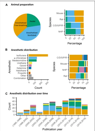
FIGURE 4 | Animal preparation and anesthesia trends. (A) Animal fMRI relies mainly on anesthesia to help restrain animals. NHP remain the major species acclimated to awake fMRI. (B) Isoflurane is the principal anesthetic used for maintenance during fMRI recordings. However, the distribution of other agents change with species. (C) Medetomidine is growing to become the second most used agent behind isoflurane.
explained by increased CBF and vasodilatation in animals
under isoflurane (
Ciobanu et al., 2012
). The use of mechanical
ventilation has the advantage of avoiding hypercapnia (increased
paCO2) which has an impact on fMRI reproducibility (
Biswal
et al., 1997
;
Ramos-Cabrer et al., 2005
). Hypercapnia also leads to
vasodilation and increased CBF (
Xu et al., 2011
). The modulation
of CBF could explain the decrease of the BOLD response
specificity to neuronal activity induced by stimuli (
Uhrig et al.,
2018
). Interestingly, Uhrig et al. showed different impacts of
various anesthetics on blood oxygenation in different brain
regions. For example, ketamine leads to higher oxygenation in
the cortex as compared to the thalamus while the opposite
occurs for propofol (
Uhrig et al., 2014
). This variability may
affect the ability to detect networks connecting these regions.
The impact of anesthesia on other physiological parameters, such
as body temperature and peripheral cardiovascular activity can
modulate the quality of the measured functional connectivity.
Both these parameters represent strong benefits to be registered
and kept stable to assure normal physiological conditions during
the acquisition. The body temperature is usually controlled
with a heating cradle, pad or any additional heating system,
leading to stable reported temperatures. In light of the above,
controlling for the temperature, the paCO
2and the movement
parameters remains essential in assuring the animal stability
and the quality of the data. Finally, anesthesia can tightly
impact CBF autoregulation in response to peripheral blood
pressure changes (
Gozzi et al., 2007
). Peripheral blood pressure
recordings, and the presence of autoregulation, are parameters
of critical importance for studies of neuromodulation using
drugs, optogenetics and/or chemogenetics-fMRI (e.g.,
Giorgi
et al., 2017
), as well as in the case of somatosensory stimulation
(
Schroeter et al., 2014
). This is because transmitter-induced
peripherally evoked blood pressure changes, in the absence of
physiological CBF autoregulation, can give rise to seemingly
regionalized fMRI responses (
Gozzi et al., 2007
;
Reimann H.
M. et al., 2018
). Future research is required to understand to
which extent commonly used anesthetic regimens in rodents do
preserve CBF autoregulation. While technically challenging, and
invasive, blood pressure recordings can be carried out via femoral
arterial cannulation (
Ferrari et al., 2015
), hence making it possible
to understand whether peripheral cardiovascular response and
central fMRI activity are temporally correlated.
Several anesthetics are used for animal studies (Figure 4B).
They have been classified into several classes according to their
targets: GABA
Areceptors, NMDA receptors, two-pore-domain
K+ channels, and other modes of actions. GABA
Areceptors
are the most widely used targets for anesthetics. They are
chloride channels that hyperpolarize neurons, making them less
excitable and thus inhibiting the possibility of an action potential.
Widely used anesthetics as isoflurane, propofol and barbiturates
are GABA
Areceptors agonists (
Franks, 2008
;
Garcia et al.,
2010
). Each drug within this category displays a subtly unique
pharmacological characteristic. For example, isoflurane and
sevoflurane have opposite metabolic activities on cerebral blood
flow and glucose consumption in various brain regions (
Lenz
et al., 1998
).
α-chloralose is widely used in the context of BOLD
fMRI because it provides robust metabolic and hemodynamic
responses to functional stimulation and is also expected to act
on GABA
Areceptors (
Garrett and Gan, 1998
). NMDA receptors
are other widely used targets. The use of antagonists for these
receptors, such as ketamine, is supposed to block excitatory
synaptic activity probably leading to anesthesia. This latter may
be related to the fact that ketamine binds preferentially to
the NMDA receptors on GABAergic interneurons. Ketamine,
however, leads to a “dissociative anesthesia” during which
the perception of pain is dissociated from the perception of
noxious stimuli. Besides, it has psychotomimetic effects at low
concentrations, leading to auditory and visual hallucinations
(
Franks, 2008
). Ketamine and other NMDAr antagonists increase
regional brain activity and cerebral blood volume, mainly
in the anterior cingulate, the thalamus, the putamen, and
the frontal cortex (
Långsjö et al., 2003
;
Gozzi et al., 2008
;
Bonhomme et al., 2012
). Two-pore-domain K+ channels are
targeted by volatile anesthetics (isoflurane, halothane, nitrous
oxide) which have different affinities for subfamilies (TREK-1
or TASK) of these receptors (
Patel et al., 1999
). These channels
modulate the potassium conductance that contributes to the
resting membrane potential in neurons. Their opening, therefore,
facilitates a hyperpolarizing current, which reduces neuronal
excitability and anesthetizes. Among other targets,
α2-adrenergic
receptor agonists are targeted by xylazine, medetomidine,
dexmedetomidine (
Sinclair, 2003
). The activity of these drugs is
related to their action on receptors located in the locus coeruleus
and its projections. At this level, they reduce the release of
norepinephrine, a neurotransmitter that is necessary for arousal.
The anesthesia induced by these compounds resembles the state
of non-REM sleep, i.e., the first four of the five stages of the sleep
cycle (
Franks, 2008
).
Impact of Anesthetics on Neuronal Network
Organization in Rodents
In rodents, isoflurane and medetomidine are currently the
most commonly used anesthetics (Figures 4B,C). Importantly,
isoflurane is almost systematically used for anesthesia induction,
specifically in rodents. Variations in the induction time may
lead to a lasting effect on brain function, even though
anesthesia is switched to another agent (
Magnuson et al.,
2014
). In addition to their different mechanisms of action
(GABA
Areceptors agonist for isoflurane and
α2 adrenergic
receptor agonists for medetomidine), they have opposite
vaso-properties (vasodilatation for isoflurane and vasoconstriction
for medetomidine) which could impact neurovascular coupling
differently. In rodents, isoflurane seems to preserve the
interhemispheric and cortico-cortical FC but only at low doses
(∼1%) (
Wang et al., 2011
;
Grandjean et al., 2014
;
Uhrig et al.,
2014
;
Bukhari et al., 2017
). Medium to high doses induce
burst-suppression effects which are reflected in an increase in the
global signal (
Liu et al., 2011, 2013
;
Grandjean et al., 2014
).
Medetomidine seems to present opposite effects such as a
cortico-cortical disruption and a pronounced striatal FC (
Grandjean
et al., 2014
;
Bukhari et al., 2017
;
Paasonen et al., 2018
). The
effect of isoflurane and medetomidine and other anesthetics on
the thalamo-cortical FC is still debated. Several studies suggested
that a combination of isoflurane and medetomidine (med/iso) at
low doses is the best compromise (Table 1, med/iso) to preserve
FC and to recapitulate network properties of the awake state
(
Grandjean et al., 2014
). However, this combination appears to
inhibit thalamo-frontal cortical connectivity, when compared to
connectomic estimates of the mouse connectome (
Grandjean
et al., 2017b
). A number of studies in control and transgenic
mouse models have been carried out with low doses of halothane
(
Sforazzini et al., 2014
;
Liska et al., 2015, 2018
;
Bertero et al.,
2018
;
Gutierrez-Barragan et al., 2018
;
Pagani et al., 2019
). This
inhalational anesthetic produces stable and long-lasting
RS-FC correlation recapitulating patterns of connectivity observed
with med/iso combination (
Grandjean et al., 2017b
), with the
advantage of robustly preserving thalamo-frontal connectivity,
an effect that makes it especially apt for the investigation
of prefrontal circuitry and the rodent default mode network
(
Bertero et al., 2018
). However, the hepatotoxic properties of this
compound have led its banning in most countries, preventing
widespread use of this anesthetic regimen. Other anesthetics used
in rodents (propofol, urethane,
α-chloralose) are presented in
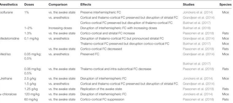
TABLE 1 | Anesthetics effects on the functional connectivity in rodents.
Anesthetics Doses Comparison Effects Studies Species
Isoflurane 1% vs. the awake state Preserve interhemispheric FC Jonckers et al. (2014) Mice vs. anesthetics Cortical and thalamo-cortical FC preserved but disruption of striatal FC Grandjean et al. (2014)
Cortico-cortical FC preserved but disruption of thalamo-cortical FC Bukhari et al. (2017)
1–2% Increasing doses Disruption of interhemispheric FC with increasing doses Bukhari et al. (2018)
1.3% vs. the awake state Cortico-cortical and striatal FC increase Paasonen et al. (2018) Rats Medetomidine 0.1 mg/kg vs. anesthetics Disruption of thalamo-cortical FC but pronounced striatal FC Grandjean et al. (2014) Mice Thalamo-cortical FC preserved but disruption cortico-cortical FC Bukhari et al. (2017) Mice vs. the awake state Cortico-cortical FC decreased Paasonen et al. (2018) Rats Med/iso 0.05 mg/kg;
0.5%
vs. anesthetics Preserved FC Grandjean et al. (2014) Mice
Bukhari et al. (2017)
0.06 mg/kg; 0.5%
vs. the awake state Thalamo-cortical and intra-subcortical FC decrease Paasonen et al. (2018) Rats
Urethane 2.5 g/kg vs. the awake state Disruption of interhemispheric FC Jonckers et al. (2014) Mice 1.5 g/kg vs. anesthetics Cortical and thalamo-cortical FC preserved but disruption of striatal FC Grandjean et al. (2014)
1.25 g/kg vs. the awake state Replication of the awake state Paasonen et al. (2018) Rats α-chloralose 120 mg/kg vs. the awake state Disruption of interhemispheric FC Jonckers et al. (2014) Mice 60 mg/kg vs. the awake state Cortico-cortical FC suppression Paasonen et al. (2018) Rats Review of five studies between 2014 and 2018.
Table 1
. They are not further discussed here as they showed
ambiguous effects on RS-FC and are no longer recommended.
Notably, RSNs in mice were shown to converge in a multi-center
comparison (Figure 1;
Grandjean et al., 2019a
), irrespective
of anesthesia regimen, indicating to some extent that network
properties are retained between different conditions.
Impact of Anesthetics on Neuronal Network
Organization in Primates
In primates, isoflurane is the most used anesthetic (
Vincent
et al., 2007
;
Hutchison et al., 2013b
;
Miranda-Dominguez et al.,
2014
;
Grayson et al., 2016
). As in rodents, lower dose and
shorter anesthesia duration are associated with an increased
ability to detect RS-FC (Table 2;
Barttfeld et al., 2015
;
Uhrig et al.,
2018
). Also, one should keep in mind that a direct comparison
of the impact of anesthetics on cerebral networks is difficult
because anesthesia depth also modulates networks and can lead
to misinterpretation of the results.
Data Acquisition
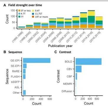
Contrary to human fMRI, which is carried mostly at 1.5T,
3T, and in rarer cases at 7T, animal fMRI is carried at a
variety of field strengths, with 7T and 9.4T being the most
frequently encountered field strength (26 and 25% respectively,
Figure 5A
). The availability of ultra-high field strength
small-bore systems in rodents further increase this range, with
fMRI being recorded as high as 15.2T (
Jung et al., 2019
).
While fewer animal MRI system vendors exist compared to
human systems, this apparent similarity is compounded with
a greater range of coil designs, including home-made coils or
cryogenic coils (
Baltes et al., 2011
), which provide an additional
source of variation among the animal studies. Whilst these
factors are determined by the center where the acquisitions
are performed, even greater variability comes in in the form
of sequence parameters and the resulting contrasts across the
different studies. This is exemplified in a report by Grandjean
et al. which indicated cortical signal-to-noise ratios ranging
from 20 to 400 in mice fMRI acquired at different centers
(
Grandjean et al., 2019a
).
Neuronal activity induces vasodilation in surrounding
capillaries and arterioles, which may propagate further up- and
downstream toward arteries and draining veins. The resulting
increase in CBF and CBV and blood oxygenation forms the basis
of imaging strategies for fMRI. The most commonly used fMRI
method is based on the BOLD contrast (
Ogawa et al., 1990
).
BOLD contrast results from the paramagnetic properties of
deoxyhemoglobin, which causes magnetic susceptibility effects
inside blood vessels as well as in their surrounding tissue that
can be detected with T2- or T2
∗-weighted sequences (
Norris,
2006
;
Kim and Ogawa, 2012
). Deoxyhemoglobin concentration
increases dramatically from the arterial (
<5%) to the venous side
(∼40%) of the vascular tree due to the extraction of oxygen in the
capillaries (
Vovenko and Sokolova, 1998
), which makes BOLD
imaging particularly sensitive to capillaries, venules and veins.
In healthy brain tissue, the neuronal activity typically induces
an increase in CBF with resultant increased oxygen delivery
that exceeds the decrease in oxygen due to capillary oxygen
extraction. As a result, deoxyhemoglobin concentration in the
capillaries and veins decreases, giving rise to a positive BOLD
response in T2- or T2
∗-weighted images.
The most frequently used BOLD-weighted fMRI sequence in
rodents is T2
∗-weighted gradient echo (GE) echo planar imaging
(EPI) (Figure 5B). GE-EPI provides a relatively high
contrast-to-noise ratio (CNR), which increases with field strength. At
field strengths ≥ 7T, the intravascular contribution to the GE
BOLD signal is negligible and signal changes scale almost linearly
with echo time (TE) (
Yacoub et al., 2003
;
Han et al., 2019
). For
optimal BOLD CNR, TE is typically set equal to the average
TABLE 2 | Anesthetics effects on the functional connectivity in primates.
Anesthetics Doses Comparison Effects Studies Species
Isoflurane 1–2.75% Increasing doses Disruption of interhemispheric FC after 1.5% Hutchison et al. (2014) Macaca fascicularis 0.89–1.19% Duration effect Reduction of the DMN FC with a prolonged administration Li and Zhang (2018) Macaca mulatta Ketamine 20 mg/kg vs. the awake state Preservation of positive FC but average positive FC reduced Uhrig et al. (2018) Macaca mulatta Sevoflurane 2.2–4.4 vol% vs. the awake state Average positive FC reduced Uhrig et al. (2018) Macaca mulatta Review of five studies between 2014 and 2018.
gray matter tissue T2
∗value (for an overview of brain tissue
T2 and T2
∗values we refer to
Uluda˘g et al. (2009)
and
Han
et al. (2019)
. The disadvantage of using GE-EPI for rodent fMRI,
however, is its sensitivity to susceptibility artifacts, which are
most prominent near air cavities such as the ear canals and
around the olfactory bulb, particularly at long TE and high field
strength. Furthermore, GE-EPI is highly sensitive to large veins
(
Uluda˘g et al., 2009
), which makes this sequence spatially
non-specific as neurovascular coupling occurs at the level of the
capillaries. This has been clearly demonstrated by fMRI studies
in rats subjected to electrical stimulation of the forepaws, where
the highest GE-EPI BOLD response is observed in the outer
layer of the somatosensory cortex where pial vessels are located
(
Mandeville and Marota, 1999
;
Han et al., 2019
), while neuronal
activation mostly occurs in deeper cortical layers. The relative
contribution of capillaries to the BOLD signal increases with field
FIGURE 5 | Data acquisition. (A) There is a general trend toward higher strength of the main magnetic field in animal fMRI over time. In the past decade, the majority of studies were performed on 7T and 9.4T systems. (B) The acquisitions relied mainly on gradient echo EPI for the acquisition, while older studies either used FLASH or RARE sequences. (C) BOLD is the most commonly used contrast in animal studies. The availability of iron nanoparticles in animal studies explains the relative high incidence of CBV contrasts.