HAL Id: hal-00963727
https://hal.archives-ouvertes.fr/hal-00963727
Submitted on 27 Mar 2014HAL is a multi-disciplinary open access archive for the deposit and dissemination of sci-entific research documents, whether they are pub-lished or not. The documents may come from teaching and research institutions in France or abroad, or from public or private research centers.
L’archive ouverte pluridisciplinaire HAL, est destinée au dépôt et à la diffusion de documents scientifiques de niveau recherche, publiés ou non, émanant des établissements d’enseignement et de recherche français ou étrangers, des laboratoires publics ou privés.
Susceptibility and chemical shift in MRI
Serge Langlois, Michel Desvignes, Jean-Marc Constans, Marinette Revenu
To cite this version:
Serge Langlois, Michel Desvignes, Jean-Marc Constans, Marinette Revenu. Susceptibility and chemical shift in MRI. Fifth International Conference on Functional Brain Mapping, 1999, Dusseldorf, Germany. pp.S161. �hal-00963727�
Susceptibility and chemical Shift in MRI.
Serge Langlois, Michel Desvignes, Jean Marc Constans, Marinette Revenu GREYC-ISMRA CNRS UPRESA 6072, IRM-CHU Caen, GIN Cyceron
Introduction:
Geometrical distortion in Magnetic Resonance Images can be classified in two main categories[1]: non-linear gradient fields and static and patient induced field heterogeneity. The first kind is machine dependent and can be corrected by calibration or post processing [2]. The second kind will be the one discussed in this paper. It can be important near the skull. Since some registration technique use these landmarks, field heterogeneity must be corrected for accurate registration.
Material and Methods
The static (Machine dependent) field heterogeneity can be reduced by regularly shimming the magnet[3]. Patient induced field heterogeneity arises because of the difference in chemical shift between water and fat contained in biological tissues and also wherever a sharp variation in the magnetic susceptibility exists i.e. near the surface of the skin or near the tissue-air interface. The effect is a local displacement of the structures on the image
compared to their real physical position that must be estimated and corrected. Several methods have been described to image fat and water individuality using selective saturation pulse or Dixon method. Three points Dixon technique [4] uses 3 images to estimate fat and water images and static field heterogeneity. We propose a method to estimate and correct the effects of the field heterogeneity with only 2 acquisitions of the same object. The method is based on two acquisitions with time echoes TE1 and TE2 such as:
∆ω TE1 = ∆ω TE2 + (2k+1)Π,
where ∆ω is the chemical shift between water and fat (220Hz at 1,5Tesla). It can be shown that the module of the sum of these images is proportional to the quantity of water. The module of the difference of these images is proportional to the quantity of fat. The phase of the sum is an image of the field variations used to compute the displacements associated to water and fat components. An efficient phase unwrapping algorithm enables to correct the phase jumps that occur in phase images during the reconstruction process.
Results
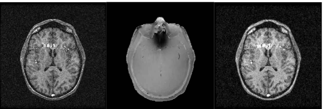
Two gradient echo images have been acquired at time echo TE1 = 9,09ms (fat and water in phase) and at TE2 = 11,37ms (fat and water out of phase). Shifts are presented in figure 1b and corrected image is presented in figure 1c. The corrected image has a better SNR and chemical shift is clearly reduced: the fat (white signal near the skull), which was inside the LCR in figure 10a is replaced in figure 10c. Echo planar images have also been processed by this method.
References
[1] : T.S. Sumanaweera et al., Characterization of Spatial Distortion in Magnetic Resonance Imaging and Its Implication for Stereotactic Surgery, Neurosurgery, vol. 35(4), 1-9, 1994
[2] : S. Langlois, M. Desvignes, J.M. Constans, M. Revenu,"Modelization of the gradient field nonlinearities in Magnetic Resonance Imaging",In Proceeding World Congress on Medical Physics and Biomedical Engineering, Nice (Fr), vol. 35(2), p. 765, 1997.
[3] : E. Schneider, G. Glover, Rapid in Vivo Proton Shimming, Magnetic Resonance in Medicine, vol. 18, 898-904, 1995
[4] : Y. Wang et al., A Three-Point Dixon Method for Water and Fat Separation Using 2D and 3D Gradient Echo Techniques, Journal of Magnetic Resonance Imaging, vol. 8, 703-710, 1998