HAL Id: hal-01170351
https://hal.sorbonne-universite.fr/hal-01170351
Submitted on 1 Jul 2015
HAL is a multi-disciplinary open access
archive for the deposit and dissemination of
sci-entific research documents, whether they are
pub-lished or not. The documents may come from
teaching and research institutions in France or
abroad, or from public or private research centers.
L’archive ouverte pluridisciplinaire HAL, est
destinée au dépôt et à la diffusion de documents
scientifiques de niveau recherche, publiés ou non,
émanant des établissements d’enseignement et de
recherche français ou étrangers, des laboratoires
publics ou privés.
Distributed under a Creative Commons Attribution| 4.0 International License
does not modulate dopaminergic neurodegeneration
Anne-Sophie van Rompuy, Marusela Oliveras-Salvá, Anke van der Perren,
Olga Corti, Chris van den Haute, Veerle Baekelandt
To cite this version:
Anne-Sophie van Rompuy, Marusela Oliveras-Salvá, Anke van der Perren, Olga Corti, Chris van
den Haute, et al.. Nigral overexpression of synuclein in the absence of parkin enhances
alpha-synuclein phosphorylation but does not modulate dopaminergic neurodegeneration. Molecular
Neu-rodegeneration, BioMed Central, 2015, 10 (1), pp.23. �10.1186/s13024-015-0017-8�. �hal-01170351�
R E S E A R C H A R T I C L E
Open Access
Nigral overexpression of alpha-synuclein in
the absence of parkin enhances alpha-synuclein
phosphorylation but does not modulate
dopaminergic neurodegeneration
Anne-Sophie Van Rompuy
1, Marusela Oliveras-Salvá
1, Anke Van der Perren
1, Olga Corti
2,3,4,
Chris Van den Haute
1,5and Veerle Baekelandt
1*Abstract
Background: Alpha-synuclein is a key protein in the pathogenesis of Parkinson’s disease. Mutations in the parkin gene are the most common cause of early-onset autosomal recessive Parkinson’s disease, probably through a loss-of-function mechanism. However, the molecular mechanism by which loss of parkin function leads to the development of the disease and the role of alpha-synuclein in parkin-associated Parkinson’s disease is still not elucidated. Conflicting results were reported about the effect of the absence of parkin on alpha-synuclein-mediated neurotoxicity using a transgenic approach. In this study, we investigated the effect of loss of parkin on
alpha-synuclein neuropathology and toxicity in adult rodent brain using viral vectors. Therefore, we overexpressed human wild type alpha-synuclein in the substantia nigra of parkin knockout and wild type mice using two different doses of recombinant adeno-associated viral vectors.
Results: No difference was observed in nigral dopaminergic cell loss between the parkin knockout mice and wild type mice up to 16 weeks after viral vector injection. However, the level of alpha-synuclein phosphorylated at serine residue 129 in the substantia nigra was significantly increased in the parkin knockout mice compared to the wild type mice while the total expression level of alpha-synuclein was similar in both groups. The increased
alpha-synuclein phosphorylation was confirmed in a parkin knockdown cell line.
Conclusions: These findings support a functional relationship between parkin and alpha-synuclein phosphorylation in rodent brain.
Keywords: Parkinson’s disease, Alpha-synuclein, Alpha-synuclein phosphorylation, Parkin, Adeno-associated viral vectors, Knockout
Background
Parkinson’s disease (PD) is the second most common neurodegenerative disorder. Neuropathologically, it is characterized by the progressive loss of dopaminergic neurons in the substantia nigra (SN) and the presence of proteinaceous intracellular inclusions called Lewy bodies (LBs) and Lewy neurites in the surviving neurons [1]. Al-though the etiology of sporadic PD remains still unclear,
the discovery of genes linked to familial forms of the dis-ease has improved our understanding of the pathogenic mechanisms leading to PD.
Point mutations and multiplications of theα-synuclein (α-SYN) gene, SNCA, cause a rare familial autosomal dominant form of PD [2]. α-SYN is a small protein of 140 amino acids that is widely expressed in the brain and localizes predominantly to presynaptic terminals [3]. The biological function ofα-SYN remains unknown, al-though it’s involvement in dopamine transmission and biosynthesis [4,5], synaptic plasticity [6] and turnover of synaptic vesicles has been suggested [7]. Under pathological
* Correspondence:Veerle.Baekelandt@med.kuleuven.be
1
Laboratory for Neurobiology and Gene Therapy, Department of Neurosciences, KU Leuven, Flanders, Belgium
Full list of author information is available at the end of the article
© 2015 Rompuy et al. This is an Open Access article distributed under the terms of the Creative Commons Attribution License (http://creativecommons.org/licenses/by/2.0), which permits unrestricted use, distribution, and reproduction in any medium, provided the original work is properly credited. The Creative Commons Public Domain Dedication waiver (http:// creativecommons.org/publicdomain/zero/1.0/) applies to the data made available in this article, unless otherwise stated.
conditions, including mutations and increased expression levels, α-SYN has the propensity to adopt a β-sheet rich conformation which leads to the formation of oligomers and fibrillar aggregates [8]. This fibrillar form ofα-SYN is the main protein component of LBs and Lewy neurites, in-dicating thatα-SYN plays a crucial role in the pathogen-esis of PD [9]. Moreover, animal models based on overexpression of wild type (WT) or mutant α-SYN, re-capitulate some of the main hallmarks of PD, including neurodegeneration, motor dysfunction and inclusion for-mation [10].
Mutations in the parkin gene are the major cause of early-onset autosomal recessive PD [11]. Parkin has been identified as an E3 ubiquitin-ligating enzyme, catalyzing the attachment of ubiquitin to substrate proteins, which consequently leads to their proteasomal degradation [12-14]. It was therefore suggested that the loss of parkin function due to disease-causing mutations might damage neurons through the accumulation of toxic proteins. On the other hand, evidence is accumulating that parkin ubi-quitin ligase activity also contributes to non-degradative poly- and mono-ubiquitination, which are involved in al-ternative cellular pathways, including mitochondrial qual-ity control [15] and signal transduction cascade activation [16,17]. However, the molecular mechanism by which loss of parkin function leads to the development of PD is still not completely elucidated. More specifically, the role of α-SYN in parkin-associated PD remains unclear, although there are lines of evidence suggesting a potential link be-tween parkin and α-SYN. On one hand it has been re-ported that the unmodified form of α-SYN does not interact with parkin [18] and no accumulation ofα-SYN was detected in the brain of parkin knockout (parkin−/−) mice [19-21]. On the other hand, in about two thirds of the seventeen neuropathological reports of patients with parkin mutations published to date, no LBs were found, indicating that parkin might play a role in LB forma-tion[22]. Furthermore, overexpression of parkin provides protection against α-SYN toxicity in a variety of cellular and animal models [23-29]. Conflicting results were pub-lished in a number of studies using a transgenic approach to investigate the effect of parkin on α-SYN-induced neurotoxicity. The neurodegenerative phenotype was un-expectedly delayed in the absence of parkin in A30P α-SYN transgenic mice [30], while no effect of loss of parkin on neuropathology was found in A53Tα-SYN transgenic mice [31]. In a third publication, an increase in damaged mitochondria in neurons of the SN and a reduction of complex I activity was reported in old double mutant mice generated by the crossing of parkin−/− mice with double mutated A53T and A30Pα-SYN overexpressing mice [32]. However, these results in transgenic mice showed that the absence of parkin did not clearly affect α-SYN-induced neurodegeneration, except for some minor changes.
In the present study, we chose an alternative approach to investigate the effect of the absence of parkin on α-SYN-induced cell death in adult rodent brain using viral vector technology. Therefore, we overexpressed human WT α-SYN in the SN of parkin−/−and wild type (parkin+/+) mice with recombinant adeno-associated viral (rAAV) vectors. Subsequently, we analyzed the degree of neurodegeneration and synucleinopathy in the SN of those mice.
Results
Absence of parkin does not increase the sensitivity to dopaminergic degeneration induced by a high dose of rAAV2/7-WTα-SYN
In a previous study, we showed that rAAV2/7-mediated overexpression of WTα-SYN in the SN of mice resulted in a dose-dependent, progressive dopaminergic neurode-generation [33]. Therefore, to study the effect of the ab-sence of parkin on α-SYN induced dopaminergic cell death, in a first experiment we stereotactically injected a rAAV2/7 vector encoding WTα-SYN in the right SN of adult parkin−/− and parkin+/+ mice at a vector titer of 4E + 11 genome copies (GC)/ml (high vector titer, see experimental design in Table 1).The animals were ana-lyzed at 1 week and 4 weeks after injection. As add-itional control, a group of parkin−/−and parkin+/+mice was injected with a similar titer of rAAV2/7 vector cod-ing for enhanced green fluorescent protein (eGFP).
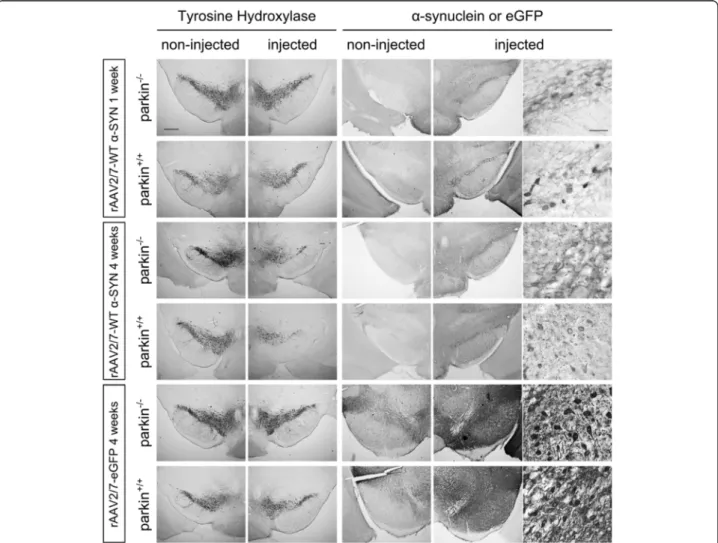
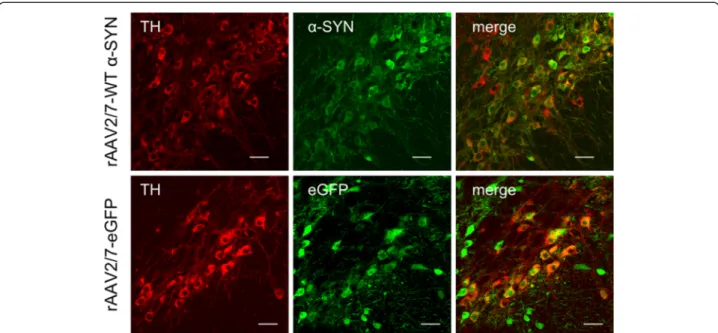
Immunohistochemical stainings for α-SYN and eGFP revealed high transgene expression in the SN for both vectors (Figure 1). With confocal analysis we observed a transduction efficiency of the dopaminergic neurons of approximately 85% (Figure 2). To investigate the degree of dopaminergic neurodegeneration, we stereologically quantified the number of tyrosine hydroxylase (TH)-positive cells in the SN. At 4 weeks after injection, the rAAV2/7-WT α-SYN induced a dopaminergic lesion of 59 ± 6% compared to the non-injected side in the parkin
+/+
mice, which is in agreement with our previous study [33] (Figure 1 and 3A). In the parkin−/−mice a compar-able dopaminergic cell loss of 52 ± 6% was observed. The rAAV2/7-eGFP injected mice did not show any loss of TH-positive cells (Figure 1 and 3B). The loss of dopa-minergic terminals in the striatum was comparable be-tween parkin+/+(24 ± 5.2%) and parkin−/−(33 ± 6.9%) mice (Figure 3C). Thus, we conclude that the sensitivity of par-kin−/−mice and parkin+/+ mice to dopaminergic degener-ation induced by a high dose of rAAV2/7-WT α-SYN is similar.
Overexpression ofα-SYN with rAAV2/7 increases phosphorylation ofα-SYN at serine residue 129 in parkin
−/−mice compared to parkin+/+
mice
In a next step, we determined the number of cells in the SN that were positive forα-SYN phosphorylated at serine
Table 1 Experimental design
Group
Analysis time point (Immunohistochemistry) rAAV2/7-WTαSYN rAAV2/7-eGFP rAAV2/7-WTαSYN (4E + 11 GC/ml) (4E + 11 GC/ml) (1E + 11 GC/ml) highα-SYN dose lowα-SYN dose parkin+/+ parkin−/− parkin+/+ parkin−/− parkin+/+ parkin−/−
1 week n = 4 n = 4 n = 2 n = 3 4 weeks n = 15 n = 16 n = 5 n = 6 n = 10 n = 13
8 weeks n = 6 n = 5
16 weeks n = 6 n = 6
Adult 2 to 4-month-old parkin+/+
and parkin−/−mice were stereotactically injected in the right SN with 2μl of rAAV2/7-WT α-SYN vector at a titer of 4E + 11 GC/ml (highα-SYN dose) or 1E + 11 GC/ml (low α-SYN dose) or rAAV2/7-eGFP vector at a titer of 4E + 11 GC/ml. At the mentioned time points after injection, animals were perfused for immunohistochemical analysis.
Figure 1 Overexpression ofα-SYN or eGFP in mouse SN with high titer rAAV2/7 vectors. Immunohistochemical staining for TH (left panels) shows cell loss in the injected side of both the parkin−/−and the parkin+/+mice at 4 weeks after injection with a high titer of rAAV2/7-WTα-SYN. Immunohistochemical stainings forα-SYN and eGFP (middle and right panels) show wide expression of these proteins in the injected side of the SN. Right panels are magnifications of the adjacent left panel. Scale bars overviews = 400μm and magnifications = 50 μm.
residue 129 (P-S129), a form ofα-SYN which is considered to be the pathological form ofα-SYN and the most abun-dant modification ofα-SYN in LBs [34,35]. Therefore, we performed an immunohistochemical staining with an anti-body specifically recognizing this phosphorylated form of α-SYN [34] and stereologically quantified the number of positive cells in the injected side of the whole SN. No P-S129 positive cells were observed in the non-injected side of the SN, indicating that only phosphorylation of the overexpressed humanα-SYN is within the limits of detec-tion. At 1 week after injection, no difference was observed between parkin−/− mice (3563 ± 259 cells) and parkin+/+ mice (3371 ± 416 cells). At 4 weeks after injection, the number of cells positive for P-S129α-SYN was increased
in both groups when compared to 1 week. Interestingly, this number was significantly higher in the parkin−/−mice (7632 ± 291 cells) than in the parkin+/+mice (6288 ± 495 cells) (p = 0,027) (Figure 4A-B). This higher number of P-S129 α-SYN positive cells in the parkin−/− mice was not caused by higher expression levels of α-SYN, since the total number ofα-SYN positive cells in the SN was similar in the parkin+/+ and parkin−/− mice at both time points (Figure 4C). Differences in the affinities of the P-S129 α-SYN antibody and α-SYN antibody explain the apparent lower number of α-SYN positive cells compared to the number of P-S129 α-SYN positive cells. We also stained the striatum for P-S129 α-SYN to check if the dopamin-ergic terminals also contain phosphorylated α-SYN. We
Figure 2 High transduction efficiency of mouse dopaminergic neurons with rAAV2/7-WTα-SYN and rAAV2/7-eGFP. Fluorescent double stainings for TH andα-SYN at 1 week or TH and eGFP at 4 weeks after injection of rAAV2/7-WT α-SYN or rAAV2/7-eGFP respectively, demonstrate that the majority of the dopaminergic neurons of the injected side of the SN is transduced. Scale bar = 25μm.
Figure 3 A high dose of rAAV2/7-WTα-SYN induces similar dopaminergic degeneration in parkin−/−and parkin+/+mice. Stereological quantification of the number of TH-positive neurons in the SN of parkin+/+and parkin−/−mice at (A) 1 week and 4 weeks after injection with a high titer of rAAV2/7-WTα-SYN and at (B) 4 weeks after injection with rAAV2/7-eGFP. (C) quantification of TH staining in striatum (injected over non-injected side) of parkin+/+and parkin−/−mice 4 weeks after injection. Asterisks depict significant decrease respective to 1 week, unless specified otherwise. (Mean ± SEM, two-way ANOVA followed by Bonferroni post-hoc test, ** p < 0.01 ***p < 0.001, 1 week rAAV2/7-WTα-SYN: n = 4; 4 weeks rAAV2/7-WTα-SYN: n = 15-16; 4 weeks rAAV2/7-eGFP: n = 5-6).
detected P-S129 α-SYN-positive neuritic inclusions (Figure 4E) but no difference in the number of these inclu-sions was observed between parkin+/+and parkin−/−mice at 4 weeks after injection (Figure 4D).
A low dose of rAAV2/7-WTα-SYN induces slower but similar dopaminergic degeneration and increasedα-SYN phosphorylation in parkin−/−mice
The enhanced phosphorylation ofα-SYN in the parkin−/− mice observed in the previous experiment was not paral-leled by an increase in dopaminergic cell death. We rea-soned that this might be due to the very fast and robust degenerative process, precluding the detection of subtle differences. Therefore, we decided to repeat the experi-ment with a 4 times lower dose of rAAV2/7-WTα-SYN (1E + 11 GC/ml, low vector titer). In this experiment, animals were analyzed at 1 week, 4 weeks, 8 weeks and 16 weeks after injection (see experimental design in Table 1).
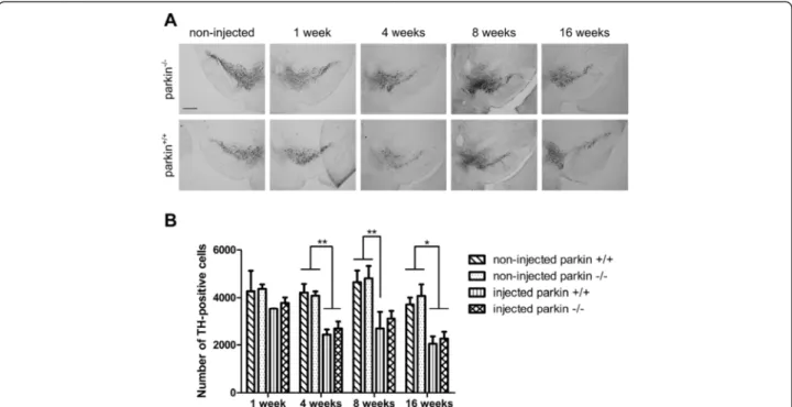
As expected, stereological quantification of the num-ber of surviving dopaminergic neurons revealed a milder dopaminergic cell loss at 4 weeks (approximately 40%) compared to the high titer of rAAV2/7-WT α-SYN. At 8 weeks and 16 weeks, in both groups the dopaminergic degeneration was not further progressive, suggesting that with the low titer of rAAV2/7-WTα-SYN the maximum amount of degeneration was already reached at 4 weeks after injection. However, the TH-positive cell loss was again comparable between the parkin−/− and parkin+/+ mice at all time points (e.g. at 4 weeks 35 ± 7% and 41 ± 5% TH-positive cell loss, respectively compared to the non-injected side) (Figure 5A-B). These results confirm that the absence of parkin does not alter the susceptibility toα-SYN induced dopaminergic cell death.
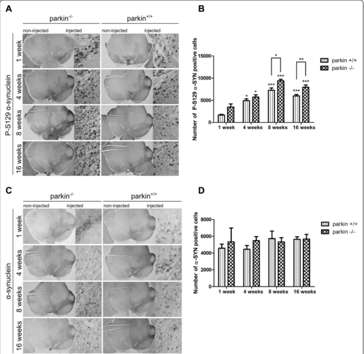
We also investigated the effect of absence of parkin on α-SYN phosphorylation in the low dose α-SYN set-up. The number of P-S129α-SYN positive cells in the injected side progressively increased over time until 8 weeks and remained stable at 16 weeks after viral vector delivery to the SN (Figure 6A-B). Here again, we observed a sig-nificantly higher level of α-SYN phosphorylation in the parkin−/− mice compared to the parkin+/+ mice at 8 weeks and 16 weeks after injection (respectively
9389 ± 337 cells versus 7263 ± 532 cells at 8 weeks, p = 0,0179; 7954 ± 518 cells versus 6000 ± 313 cells at 16 weeks, p = 0,009). Immunohistochemical staining forα-SYN con-firmed expression ofα-SYN up to 16 weeks after injection (Figure 6C). As seen before, the total number of α-SYN positive cells was similar in the parkin−/− mice and the parkin+/+mice at the 4 different time points (Figure 6D).
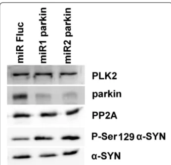
To investigate the mechanism behind this increased α-SYN phosphorylation in the absence of parkin we induced stable parkin knockdown using microRNA (miR)-based lentiviral vectors in human SHSY5Y neuroblastoma cells overexpressing WT humanα-SYN. Two miRparkin lenti-viral vectors induced efficient knockdown of parkin as shown by Q-RT-PCR (data not shown) and Western blot-ting (Figure 7). In agreement with the in vivo data we observed an increase ofα-SYN phosphorylation at serine residue 129 without affecting the totalα-SYN levels in cell culture (Figure 7). Since Polo-Like-Kinase-2 (PLK2) was shown to phosphorylate α-SYN at S129 [36] and protein phosphatase-2A (PP2A) is a known α-SYN phosphatase at S129 [37], we verified the levels of PLK2 and PP2A in both parkin knockdown and the control cell lines but no differences were observed in expression of either protein (Figure 7).
We can conclude that overexpression ofα-SYN in the absence of parkin enhances phosphorylation ofα-SYN at serine residue 129 but does not affect the degree of dopaminergic neurodegeneration.
Discussion
This study was designed to investigate the effect of the loss of parkin onα-SYN induced neurotoxicity in rodent brain. We found that the absence of parkin did not alter the vulnerability of dopaminergic neurons to WTα-SYN induced neurodegeneration. However, the number of P-S129 α-SYN positive cells in the SN of parkin−/− mice was increased compared to parkin+/+mice. This increase in the number of P-S129α-SYN positive cells in the par-kin−/− mice was not due to differences in expression level ofα-SYN, since the total number of α-SYN-positive cells was similar in both groups, These results were repro-duced in a second, independent experiment performed with a 4 times lower titer of rAAV2/7-WTα-SYN.
(See figure on previous page.)
Figure 4 Increased phosphorylation ofα-SYN at S129 in parkin−/−mice compared to parkin+/+mice. (A) Representative images of P-S129α-SYN expression in the SN of parkin−/−and parkin+/+mice at 1 week and 4 weeks after injection with a high titer of rAAV2/7-WTα-SYN. Right panels are magnifications of the overviews of the injected side (middle panels). Scale bar overviews = 400μm and magnifications = 50 μm. (B) Stereological quantification of the number of P-S129α-SYN positive cells in the injected side of the SN of parkin+/+and parkin−/−mice at 1 week (n = 4) and 4 weeks (n = 15-16) after injection with a high titer of rAAV2/7-WTα-SYN. (Mean ± SEM, two-way ANOVA followed by Bonferroni post-hoc test, # p < 0.05 versus parkin+/+, **p < 0.01 versus 1 week, ***p < 0.001 versus 1 week). (C) Stereological quantification of the number ofα-SYN positive cells in the injected side of the SN of parkin+/+and parkin−/−mice at 1 week (n = 4) and 4 weeks (n = 15-16) after injection with a high titer of rAAV2/7-WTα-SYN. (Mean ± SEM). (D) Quantification of the number of P-S129 α-SYN positive neuritic inclusions in the striatum at 4 weeks (n = 14–15, Mean ± SEM). (E) Picture of the P-S129 α-SYN staining in the striatum (left: 10 x) and magnification (right: 40 x) of neuritic inclusions. Scale bar = 50μm.
A number of previous studies already addressed the question if the absence of parkin affects the development of α-synucleinopathy using a transgenic strategy. How-ever, inconsistent results were reported, since in one study no effect was found in A53T α-SYN transgenic mice [31], whereas in a second study the loss of parkin unexpectedly mitigated the α-SYN phenotype in A30P α-SYN transgenic mice [30]. In the present study, we opted for an alternative approach with viral vector-mediated overexpression of WT α-SYN in the SN of parkin−/− and parkin+/+ mice. rAAV vectors are an at-tractive tool for gene delivery in the brain, since they provide several advantages: specific brain regions can be targeted, the transduction efficiency in dopaminergic neurons is high, and a long-lasting and stable expression of the transgene at different doses can be achieved with a single delivery [38]. In addition, in a previous study in our own research group, we showed that rAAV2/7-mediated WTα-SYN overexpression in mouse SN leads to a dose-dependent, progressive dopaminergic cell death [33].
With our strategy, we did not observe a difference in sensitivity to WTα-SYN induced dopaminergic cell death between parkin−/−and parkin+/+mice. The strength of our study is that it allowed us to specifically investigate the sen-sitivity of the dopaminergic cell population to α-SYN tox-icity in the absence of parkin. Since the majority ofα-SYN transgenic mice do not develop a robust dopaminergic
phenotype, they are less suitable to study the effect of par-kin deficiency and α-synucleinopathy in nigral dopamin-ergic neurons [39,40]. On the other hand, our finding that loss of parkin does not exacerbate dopaminergic degener-ation is partly consistent with the results in the transgenic mice, since in those studies, no difference in dopaminergic cell survival was found between theα-SYN overexpressing mice and the parkin−/−-α-SYN double transgenic mice, in-dicating that the complete absence of parkin does not affect dopaminergic cell survival [30,31]. A possible explanation for this somewhat unexpected observation might be the presence of compensatory mechanisms in the parkin−/− mice, which may counterbalance for the total loss of parkin protein occurring already during embryonic development. Reports of an increased sensitivity of striatal metabotropic glutamate receptors and elevated levels of reduced glutathi-one in parkin−/−mice indicate that such compensatory ad-aptations exist [41,42]. Locoregional downregulation of parkin with viral vectors or the generation of conditional parkin knockout animals may be a valuable strategy to overcome this issue. Indeed, it was recently reported that adult depletion of parkin in the SN of conditional parkin−/− mice resulted in a progressive loss of dopaminergic neurons up to 40%, a phenotype that has never been observed in the constitutive parkin−/−mice [21,43].
The lack of increased vulnerability of parkin−/−mice to WTα-SYN induced dopaminergic cell death was observed
Figure 5 A low dose of rAAV2/7-WTα-SYN induces similar dopaminergic degeneration in parkin−/−and parkin+/+mice. (A) Representative images of immunohistochemical staining for TH in the SN at 1 week, 4 weeks, 8 weeks and 16 weeks after injection with a low titer of rAAV2/7-WTα-SYN. Scale bar = 400μm. (B) Stereological quantification of the number of TH-positive neurons in the SN of parkin+/+and parkin−/−mice at 1 week, 4 weeks, 8 weeks and 16 weeks after injection with a low titer of rAAV2/7-WTα-SYN. (Mean ± SEM, two-way ANOVA followed by Bonferroni post-hoc test, *p < 0.05, **p < 0.01, n = 2-3 at 1 week, n = 10-13 at 4 weeks, n = 5-6 at 8 weeks, n = 6 at 16 weeks).
Figure 6 Increased P-S129α-SYN in parkin−/−mice after injection with a low titer of rAAV2/7-WTα-SYN. (A) Representative images of P-S129 α-SYN expression in the SN of parkin−/−and parkin+/+mice at 1 week, 4 weeks, 8 weeks and 16 weeks after injection with a low titer of rAAV2/7-WTα-SYN. Right panels are magnifications of the overview of the injected side (middle panels). Scale bar overviews = 400μm and magnifications = 50 μm. (B) Stereological quantification of the number of P-S129α-SYN positive cells in the injected side of the SN of parkin+/+and parkin−/−mice at 1 week (n = 2-3), 4 weeks (n = 10-13), 8 weeks (n = 5-6) and 16 weeks (n = 6) after injection. Asterisks depict significant increase respective to 1 week, unless specified otherwise. (Mean ± SEM, two-way ANOVA followed by Bonferroni post-hoc test, student’s T-test followed by Benjamini-Hochberg to compare parkin+/+and parkin−/−separately at the different time points, *p < 0.05, **p < 0.01, ***p < 0.001). (C) Representative images ofα-SYN expression in the SN of parkin−/−and parkin+/+mice at 1 week, 4 weeks, 8 weeks and 16 weeks after injection with a low titer of rAAV2/7-WTα-SYN. Right panels are magnifications of the overview of the injected side (middle panels). Scale bar overviews = 400μm and magnifications = 50 μm. (D) Stereological quantification of the number ofα-SYN positive cells in the injected side of the SN of parkin+/+and parkin−/−mice at 1 week (n = 2-3), 4 weeks (n = 10-13), 8 weeks (n = 5-6) and 16 weeks (n = 6) after injection with a low titer of rAAV2/7-WTα-SYN. (Mean ± SEM).
with two different doses of WTα-SYN. In the second ex-periment, we opted for a lower dose, because we reasoned that the dramatic dopaminergic cell loss achieved with the highest dose of WTα-SYN might hide small differences in sensitivity between parkin−/− and parkin+/+ mice. Al-though the lower dose of WTα-SYN resulted in a milder dopaminergic cell death, the degree of degeneration was still considerable (approximately 40% at 4 weeks). There-fore, we cannot exclude that subtle differences in sensitiv-ity might emerge when using even lower overexpression of α-SYN or at later time points than analyzed in this study.
In a next step, we wondered whether the absence of par-kin would influence the phosphorylation of α-SYN at serine residue 129, since the level of P-S129 α-SYN is highly elevated in the brains of PD patients [34,35]. We found that the number of nigral cells positive for P-S129 α-SYN was significantly higher in the parkin−/−mice
com-pared to parkin+/+ mice. This was not the case for the dopaminergic terminals in the striatum. This discrepancy might suggest that mainly the non-dopaminergic neurons show an increased phosphorylation in the parkin−/−mice or that the dopaminergic neurons with increased P-S129 α-SYN have relatively lower terminal densities. This in-creased phosphorylation is in apparent contradiction with the findings of Lo Bianco et al. who reported an increase in P-S129 α-SYN positive aggregates after overexpression of parkin together with A30Pα-SYN in the rat SN using
lentiviral vectors [25]. That observation was associated with a protective effect of parkin overexpression on A30P α-SYN induced dopaminergic degeneration. Furthermore, no increase in P-S129 α-SYN abundance was noticed in A30P α-SYN transgenic mice on a parkin−/−background [30]. On the contrary, and in agreement with our results, lentiviral vector-mediated co-expression of parkin with WTα-SYN in the striatum of rats reduced the levels of P-S129 α-SYN [24]. A similar result was found in the stri-atum of macaque monkeys when parkin and WTα-SYN were overexpressed by means of rAAV vectors, although in this study a decrease in totalα-SYN was also reported [29]. In the study with the A53Tα-SYN transgenic mice crossed with parkin−/− mice phosphorylation of α-SYN has not been investigated [31]. So far, we cannot clearly explain the inconsistencies between these reports, al-though the most obvious explanation is differences in ex-perimental conditions. Altogether, the studies performed with WTα-SYN, including ours, point towards a correl-ation between decreased levels of parkin and increased phosphorylation ofα-SYN at S129, whereas in the studies with mutant A30Pα-SYN either the reverse or no effect is suggested. Thus, it is possible that the influence of parkin on S129-phosphorylation is different for WTα-SYN than for A30P α-SYN, since the two forms also differ in other properties including aggregation [44] and membrane-binding [45].
At this point, we can only speculate about the mech-anism behind the increased phosphorylation of WT α-SYN in the absence of parkin. No direct binding of parkin to P-S129 α-SYN was observed in brain extracts of A30Pα-SYN transgenic mice [30]. We also quantified the percentage of ubiquitin and α-SYN double-positive cells but no difference was seen between parkin+/+ and parkin−/− mice (data not shown). This suggests that the increased phosphorylation of WT α-SYN is not a conse-quence of a difference in ubiquitination of α-SYN. It is possible that parkin modulates kinases and phosphatases regulating the phosphorylation of α-SYN at S129. In agreement with this hypothesis it was reported that overexpression ofα-SYN in the brain of rats resulted in an increase in the level of Polo-Like-Kinase-2 (PLK2) and this increase was annihilated when parkin was co-expressed with α-SYN [24]. Absence of parkin might then result in a more pronounced increase in PLK2 levels and therefore increased S129 phosphorylation of α-SYN, since PLK2 is known to phosphorylate α-SYN at S129 [36]. We performed stainings for PLK2 on brain sections, but we failed to reliably detect endogenous ex-pression levels of PLK2 (data not shown). Therefore we induced stable parkin knockdown in human SHSY5Y neuroblastoma cells overexpressing WTα-SYN. Interest-ingly, the increased S129 phosphorylation ofα-SYN after parkin knockdown was replicated in this cell culture
Figure 7 Increased P-S129α-SYN in human SHSY5Y neuroblastoma cells after parkin knockdown. Western blotting against PLK2, parkin, PP2A, P-Ser129α-SYN and α-SYN. The miRs against parkin induced both parkin knockdown and an increase in P-Ser129α-SYN signal without affectingα-SYN, PLK2 and PP2A levels. A miR against firefly luciferase (Fluc) was used as control.
model, without effect on total α-SYN level. However, PLK2 protein levels were not altered after parkin knock-down in cell culture. Another potential mechanism in-volves protein phosphatase-2A (PP2A) that has been shown to dephosphorylate α-SYN at S129 [37]. Indeed, the level of protein phosphatase-2A (PP2A) was reportedly increased when parkin was co-expressed with α-SYN compared to expression ofα-SYN alone in rat striatal ex-tracts [24]. Absence of parkin might then decrease PP2A levels in these conditions, resulting in increased S129 phosphorylation of α-SYN. However, we failed to detect alterations in PP2A expression in the parkin knockdown cells. Thus, our data suggest that the mechanism behind the increasedα-SYN phosphorylation might be independ-ent from the PLK2 or PP2A pathway, although we cannot exclude changes in activity of either PLK2 or PP2A.
Furthermore, it has been demonstrated that 26S protea-somal activity is decreased in parkin−/−mice and parkin null Drosophila [46]. Also proteasomal dysfunction and S129 phosphorylation of α-SYN have been linked before. First, two independent studies describe that proteasomal inhibition increases casein kinase 2 activity, another kinase mediating phosphorylation ofα-SYN at S129 [47], result-ing in enhanced S129 phosphorylation ofα-SYN [48,49]. Second, it was demonstrated that inhibition of the prote-asome pathway resulted in the accumulation of P-S129 α-SYN without alteration in the total levels of α-SYN, suggesting that P-S129α-SYN specifically undergoes deg-radation by the proteasome pathway [50].
In the present study, the increased level of P-S129 α-SYN in the parkin−/− mice was not associated with in-creased dopaminergic degeneration, which is intuitively in contradiction with the knowledge that ± 90% of α-SYN within LBs from PD patients is phosphorylated at S129 [34,35]. The toxicity of P-S129 α-SYN in vivo has been studied extensively in the last years using mutated forms ofα-SYN in which S129 is replaced either with an aspartate (S129D), to mimic phosphorylation or with an alanine (S129A), to block phosphorylation. However, conflicting results were reported. Expression of S129D α-SYN in Drosophila resulted in an enhanced toxicity, whereas no dopaminergic cell loss was observed if S129A α-SYN was expressed [51]. On the contrary, Caenorhabditis elegans overexpressing S129A α-SYN showed severe motor dysfunction and synaptic abnor-malities, unlike worms overexpressing S129D α-SYN that exhibited a nearly normal phenotype [52]. Two studies using rAAV vectors in rats found expression of S129A α-SYN to be more toxic than S129D α-SYN [53,54]. In a third study, there was no difference between the two forms of α-SYN [55]. In a recent study, S129D α-SYN expression in rat SN resulted in an accelerated striatal dopaminergic fiber loss and an earlier appearance of motor deficits compared to S129A α-SYN, although
the nigral degeneration was similar [56]. However, the S129D mutant might not completely mimick the constitu-tively phophorylatedα-SYN. In another study phosphoryl-ation of A53T α-SYN was induced by rAAV-mediated overexpression of G-protein-coupled receptor kinase 6 in rats, which resulted in accelerated A53T α-SYN induced neurodegeneration [57], in contrast to our data. Species differences might be involved, since we observed that rats are more sensitive to rAAV-α-SYN-induced neurodegen-eration compared to mice [33,58]. Altogether, the role of S129 phosphorylation ofα-SYN in neurodegeneration still remains unclear.
Conclusions
In the present study, we have shown that the vulnerabil-ity of mouse dopaminergic neurons to α-SYN toxicity is not altered in the absence of parkin, but that the loss of parkin enhances phosphorylation of α-SYN at S129. Additional studies will be required to elucidate the mo-lecular mechanism behind our findings and the signifi-cance for the pathogenesis of parkin-associated PD.
Methods
Cloning of rAAV vector plasmids
The plasmids for the rAAV vector production were pre-viously described [38,59]. These plasmids include the construct for the AAV2/7 serotype, the AAV transfer plasmid and the pAdvDeltaF6 adenoviral helper plasmid. The transfer plasmids encoded as transgene eGFP or human WTα-SYN. The transgenes were under the con-trol of the CMVie-enhanced Synapsin1 promotor.
Recombinant rAAV vector production and purification
Vector production and purification were performed as previously described [38]. Briefly, the triple transfection into HEK293T cells was carried out using linear poly-ethylenimine solution. Vector particles were harvested from the supernatant and concentrated using tangential flow filtration. The concentrated supernatant was further purified using a discontinuous iodixanol step gradient. The final sample was aliquoted and stored at −80°C. Characterization of the rAAV stocks included real-time quantitative PCR analysis for genomic copy determin-ation (presented as GC/ml) and silver-stained sodium dodecyl sulfate-polyacrylamide gel electrophoresis ana-lysis for vector purity.
Animals and stereotactic neurosurgery
All animal experiments were carried out in accordance with the European Communities Council Directive of 24 November 1986 (86/609/EEC) and approved by the Bio-ethical Committee of the KU Leuven (Belgium).
The transgenic parkin−/−mice carry a homozygous dele-tion of exon 3 [41]. Parkin+/+mice in the whole first 'high
α-SYN dose’ experiment (n = 19), including rAAV2/7-eGFP (n = 5), and part in the second ‘low α-SYN dose’ experiment (1 week all and 4 weeks: 6 out of 10) were age- and gender-matched C57BL/6 J mice (Janvier, Le-Genest-Saint-Isle, France) (see Table 1). Rest of parkin+/+ mice in the ‘low α-SYN dose’ experiment were age- and gender-matched wild type littermates. Parkin genotyping was performed as described in [30]. No differences were observed in number of TH-positive cells, Ser-129-P α-SYN positive cells and α-SYN positive cells between the C57Bl6 mice and wild type littermates at 4 weeks after in-jection with the low titer of rAAV2/7-WT α-SYN (data not shown). Therefore, we concluded that potential minor differences in background did not influence our outcome. The age of the mice was 2 months in the first experiment and varied between 2 and 4 months in the second experi-ment. The mice were housed under a normal 12-h light/ dark cycle with free access to pelleted food and tap water. All surgical procedures were performed using aseptic techniques and ketamine (75 mg/kg IP, KETALAR®, Pfizer, Elsene, Belgium) and medetomidine (1 mg/kg, DORMI-TOR®, Pfizer) anesthesia. Following anesthesia the mice were placed in a stereotactic head frame (Stoelting Co, Wood Dale, IL, USA). The skull was exposed by a midline incision and a small hole was drilled in the appropriate lo-cation, using bregma as reference point. 2 μl of rAAV vector were injected at a rate of 0.25 μl/min with a 30-gauge needle on a 10μl Hamilton syringe. Stereotactic co-ordinates used for the right mouse SN were anteroposter-ior −3.1 mm and medio-lateral −1.2 mm relative to bregma, and dorsoventral−4.0 mm, from the dura surface. The needle was left in place for an additional 5 min be-fore being slowly retracted. After surgery, anesthesia was reversed with an intraperitoneal (IP) injection of atipamezol (0.5 mg/kg, ANTISEDAN®, Pfizer).
Histology
At different time points after injection, animals were deeply anesthetized with an IP injection of pentobarbital (60 mg/kg, NEMBUTAL®, CEVA Santé Animale, Libourne, France) and transcardially perfused with phos-phate buffered saline (PBS) followed by ice-cold 4% paraformaldehyde. After removal of the brain and over-night postfixation, 50 μm thick coronal sections were cut using a vibrating microtome (HM 650 V, Microm, Walldorf, Germany) and stored at 4 °C. Immunohisto-chemistry was performed under uniform conditions on free-floating sections using antibodies raised against eGFP (rabbit polyclonal 1:10000, in-house), TH (rabbit polyclonal 1:5000, Millipore AB152, Darmstadt, Germany), α-SYN (rabbit polyclonal 1:5000, Millipore AB5038) or P-S129 α-SYN (mouse monoclonal 1:5000, 11A5, Anderson et al. 2006 [34]; Elan Pharmaceuticals, South San Francisco, CA, USA). Sections were pretreated with 3% hydrogen peroxide
for 10 min and incubated overnight with primary antibody in 10% normal goat or swine serum (DakoCytomation, Heverlee, Belgium). As secondary antibody, we used bio-tinylated anti-rabbit or anti-mouse IgG (1:600 (α-SYN), 1:300 (others), DakoCytomation), followed by incubation with streptavidin-horseradish peroxidase complex (1:1000, DakoCytomation). eGFP,α-SYN and P-S129 αSYN immu-noreactivity was visualized using 3,3-diaminobenzidine (0,4 mg/ml, Sigma-Aldrich, St. Louis, MO, USA). TH im-munoreactivity was visualized using Vector SG (SK-4700, Vector laboratories, Burlingame, CA) as a chromogen. After being rinsed and mounted, sections were coverslipped with DPX (Sigma-Aldrich).
For fluorescent double staining, sections were rinsed three times in PBS and then incubated overnight in PBS-0.1% triton X-100, 10% goat serum, and the following anti-bodies: TH (mouse monoclonal 1:500, Merck Millipore MAB318), eGFP (rabbit polyclonal 1:1000, in-house) and α-SYN (rabbit polyclonal 1:1000, Millipore AB5038). After three rinses in PBS-0.1% triton X-100 the sections were in-cubated in the dark for 2 h in fluorochrome-conjugated secondary antibodies: goat anti-rabbit Alexa 488 (1:500, Molecular Probes™, Invitrogen, Gent, Belgium) and goat anti-mouse Alexa 555 (1:500, Molecular Probes™, Invitro-gen). After being rinsed in PBS and mounted, the sections were coverslipped with Mowiol. Fluorescent double stain-ing for TH and eGFP orα-SYN was visualized by confocal laser scanning microscopy (Fluoview FV1000, Olympus, Tokyo, Japan) using a 40x lens.
Stereological quantification
The total number of immunoreactive positive cells in the SN was estimated by stereological measurements using the optical fractionator method in a computerized system (StereoInvestigator; MicroBright-Field, Magde-burg, Germany) and a Leica DMR optical microscope as described before [60]. The SN pars compacta was delin-eated based on visual observation and morphology. Every fifth section throughout the rostro-caudal extent of the SN was analyzed, with a total of six sections for each ani-mal. The coefficients of error, calculated according to the procedure of Schmitz and Hof as estimates of precision [61] varied between 0.07 and 0.16. The conditions of the experiment were blinded to the investigator. To determine the terminal density in the striatum pictures of TH stained sections were taken of 3 sections spaced 250μm apart and analyzed using ImageJ. The number of P-S129 α-SYN-positive neuritic inclusions in the striatum was counted on a representative section 4 weeks after injection from the animals of the‘high α-SYN dose’ experiment.
Knockdown of parkin in SHSY5Y cells
To induce knockdown of parkin in cell culture different lentiviral vectors expressing a microRNA-based
short-hairpin RNA (miR) against human parkin were generated as described [62]. The 2 target sequences resulting in the most potent knockdown on Q-RT-PCR (data not shown), miR1 parkin: CCAGAGGAAAGTCACCTGCGAA and miR2 parkin: ATGTAAAGAAGCGTACCATGAA, were used for further experiments. A miR against firefly lucifer-ase is used as control [62]. Human dopaminergic SHSY5Y neuroblastoma cells stably overexpressing human WT α-SYN were transduced with lentiviral vectors expressing the miRs against parkin or control. Cells were maintained in DMEM (Invitrogen) supplemented with 15% fetal calf serum (Harlan Sera-Lab, International Medical), 1% non-essential amino acids (Invitrogen) and 50μg/ml gentamy-cin (Invitrogen) and selected with blasticidin (10 μg/ml, Invitrogen). After selection cell extracts were generated in 1% SDS supplemented with protease cocktail inhibitor (Roche Diagnostics) and 8.5 μg of protein was loaded on gel. After electrophoresis and protein transfer the PVDF membranes (Bio-Rad) were blocked with 5% milk powder in PBS supplemented with 0.1% Tween 20. The membranes were incubated with the different primary antibodies against parkin (4211, 1/1000, Cell Signaling), P-S129α-SYN (11A5, 1/2000), α-SYN (15G7, 1/1000, Enzo), protein phosphatase 2A (1/5000, kind gift of Dr. Veerle Janssens, Laboratory of Protein Phosphorylation and Pro-teomics, KU Leuven) and PLK2 (1/500, H-90, Santa Cruz). All samples were run on the same gel. Detection was performed after incubation with the appropriate HRP-conjugated secondary antibodies (Dako) using chemilu-minescence (ECL+, Pierce).
Statistical analysis
Statistical analysis was performed using GraphPad Prism 5.0 (GraphPad Software, La Jolla, CA, USA). Results are expressed as means ± standard error of the mean (SEM). Statistical analysis of the number of TH-positive cells, the number of Ser-129-P α-SYN positive cells and the number of α-SYN positive cells was carried out using two-way analysis of variance (ANOVA) followed by the Bonferroni post-hoc test for intergroup comparisons. To compare the number of P-S129 α-SYN positive cells at the different time points between the two genotypes in the experiment with the low titer of rAAV2/7-WT α-SYN, a Student’s T-test was performed and P-values were adjusted with the Benjamini-Hochberg procedure, with the false discovery rate set at 0.05, to avoid accu-mulation ofα-errors in multiple T-tests.
Statistical significance level was set as follows: * if P < 0.05, ** if P < 0.01, *** if P < 0.001.
Abbreviations
ANOVA:Analysis of variance;α-SYN: Alpha-synuclein; eGFP: Enhanced green fluorescent protein; GC: Genome copies; IP: Intraperitoneal; LBs: Lewy bodies; PD: Parkinson’s disease; parkin−/−: parkin knockout; parkin+/+: parkin wild type; PBS: Phosphate buffered saline; P-S129α-SYN: α-SYN phosphorylated at
serine residue 129; rAAV: recombinant adeno-associated viral vector; SEM: Standard error of the mean; SN: Substantia nigra; TH: Tyrosine hydroxylase; WT: Wild type.
Competing interests
The authors declare that they have no competing interests.
Authors’ contributions
A-SVR designed, performed and analyzed allin vivo experiments and wrote the manuscript. MOS contributed to the design of thein vivo experiments and the optimization of the dose of rAAV2/7-WTα-SYN. AVdP cloned the rAAV transfer plasmids and assisted in the optimization of the viral vector production process. OC provided the parkin−/−and parkin+/+mice, has been involved in the design of the study and revised critically the manuscript. CVdH participated in the design of thein vivo experiments, performed the cell culture experiments and has been involved in drafting the manuscript. VB conceived the study, participated in its design and helped to write the manuscript. All authors read and approved the final manuscript.
Acknowledgements
The authors thank Caroline van Heijningen and Sylvie De Swaef for excellent technical assistance and dr. Veerle Janssens for the in-house PP2A monoclonal antibodies. Confocal microscopy was performed at the lab of Molecular Imaging and Photonics at the KU Leuven.
A-SVR is a doctoral fellow supported by the Flemish Research Foundation FWO-Vlaanderen. MOS is a doctoral fellow supported by the European FP7 ITN NEUROMODEL (PITN-GA-2008-215618). This work was supported by the FWO Vlaanderen (G.0768.10), the FP7 RTD project MEFOPA (HEALTH-2009-241791), and the KU Leuven (IOF-KP/07/001, OT/08/052A).
Author details
1Laboratory for Neurobiology and Gene Therapy, Department of
Neurosciences, KU Leuven, Flanders, Belgium.2Inserm, U 975, CRICM, Hôpital
de la Pitié-Salpêtrière, F-75013 Paris, France.3UPMC Université Paris 06,
UMR_S975, F-75013 Paris, France.4CNRS, UMR 7225, F-75013 Paris, France. 5Leuven Viral Vector Core, KU Leuven, 3000 Leuven, Belgium.
Received: 19 September 2013 Accepted: 10 April 2015
References
1. Lees AJ, Hardy J, Revesz T. Parkinson's disease. Lancet. 2009;373:2055–66. 2. Schulte C, Gasser T. Genetic basis of Parkinson's disease: inheritance,
penetrance, and expression. Appl Clin Genet. 2011;4:67–80.
3. Maroteaux L, Campanelli JT, Scheller RH. Synuclein: a neuron-specific protein localized to the nucleus and presynaptic nerve terminal. J Neurosci. 1988;8:2804–15.
4. Abeliovich A, Schmitz Y, Farinas I, Choi-Lundberg D, Ho WH, Castillo PE, et al. Mice lacking alpha-synuclein display functional deficits in the nigrostriatal dopamine system. Neuron. 2000;25:239–52.
5. Perez RG, Waymire JC, Lin E, Liu JJ, Guo F, Zigmond MJ. A role for alpha-synuclein in the regulation of dopamine biosynthesis. J Neurosci. 2002;22:3090–9.
6. George JM, Jin H, Woods WS, Clayton DF. Characterization of a novel protein regulated during the critical period for song learning in the zebra finch. Neuron. 1995;15:361–72.
7. Lotharius J, Brundin P. Pathogenesis of Parkinson's disease: dopamine, vesicles and alpha-synuclein. Nat Rev Neurosci. 2002;3:932–42. 8. Deleersnijder A, Gerard M, Debyser Z, Baekelandt V. The remarkable
conformational plasticity of alpha-synuclein: blessing or curse? Trends Mol Med. 2013;19:368–77.
9. Spillantini MG, Schmidt ML, Lee VM, Trojanowski JQ, Jakes R, Goedert M. Alpha-synuclein in Lewy bodies. Nature. 1997;388:839–40.
10. Chesselet MF.In vivo alpha-synuclein overexpression in rodents: a useful model of Parkinson's disease? Exp Neurol. 2008;209:22–7.
11. Kitada T, Asakawa S, Hattori N, Matsumine H, Yamamura Y, Minoshima S, et al. Mutations in the parkin gene cause autosomal recessive juvenile parkinsonism. Nature. 1998;392:605–8.
12. Imai Y, Soda M, Takahashi R. Parkin suppresses unfolded protein stress-induced cell death through its E3 ubiquitin-protein ligase activity. J Biol Chem. 2000;275:35661–4.
13. Shimura H, Hattori N, Kubo S, Mizuno Y, Asakawa S, Minoshima S, et al. Familial Parkinson disease gene product, parkin, is a ubiquitin-protein ligase. Nat Genet. 2000;25:302–5.
14. Zhang Y, Gao J, Chung KK, Huang H, Dawson VL, Dawson TM. Parkin functions as an E2-dependent ubiquitin- protein ligase and promotes the degradation of the synaptic vesicle-associated protein, CDCrel-1. Proc Natl Acad Sci U S A. 2000;97:13354–9.
15. Exner N, Lutz AK, Haass C, Winklhofer KF. Mitochondrial dysfunction in Parkinson's disease: molecular mechanisms and pathophysiological consequences. EMBO J. 2012;31:3038–62.
16. Fallon L, Belanger CM, Corera AT, Kontogiannea M, Regan-Klapisz E, Moreau F, et al. A regulated interaction with the UIM protein Eps15 implicates parkin in EGF receptor trafficking and PI(3)K-Akt signalling. Nat Cell Biol.
2006;8:834–42.
17. Henn IH, Bouman L, Schlehe JS, Schlierf A, Schramm JE, Wegener E, et al. Parkin mediates neuroprotection through activation of IkappaB kinase/ nuclear factor-kappaB signaling. J Neurosci. 2007;27:1868–78. 18. Chung KK, Zhang Y, Lim KL, Tanaka Y, Huang H, Gao J, et al. Parkin
ubiquitinates the alpha-synuclein-interacting protein, synphilin-1: implications for Lewy-body formation in Parkinson disease. Nat Med. 2001;7:1144–50. 19. Goldberg MS, Fleming SM, Palacino JJ, Cepeda C, Lam HA, Bhatnagar A, et al.
Parkin-deficient mice exhibit nigrostriatal deficits but not loss of dopaminergic neurons. J Biol Chem. 2003;278:43628–35.
20. Ko HS, von Coelln R, Sriram SR, Kim SW, Chung KK, Pletnikova O, et al. Accumulation of the authentic parkin substrate aminoacyl-tRNA synthetase cofactor, p38/JTV-1, leads to catecholaminergic cell death. J Neurosci. 2005;25:7968–78.
21. Oliveras-Salva M, Van Rompuy AS, Heeman B, Van den Haute C, Baekelandt V. Loss-of-function rodent models for parkin and PINK1. J Parkinsons Dis. 2011;1:229–51.
22. Doherty KM, Silveira-Moriyama L, Parkkinen L, Healy DG, Farrell M, Mencacci NE, et al. Parkin disease: a clinicopathologic entity? JAMA Neurol. 2013;70:571–9.
23. Haywood AF, Staveley BE. Mutant alpha-synuclein-induced degeneration is reduced by parkin in a fly model of Parkinson's disease. Genome. 2006;49:505–10.
24. Khandelwal PJ, Dumanis SB, Feng LR, Maguire-Zeiss K, Rebeck G, Lashuel HA, et al. Parkinson-related parkin reduces alpha-Synuclein phosphorylation in a gene transfer model. Mol Neurodegener. 2010;5:47.
25. Lo Bianco C, Schneider BL, Bauer M, Sajadi A, Brice A, Iwatsubo T, et al. Lentiviral vector delivery of parkin prevents dopaminergic degeneration in an alpha-synuclein rat model of Parkinson's disease. Proc Natl Acad Sci U S A. 2004;101:17510–5.
26. Petrucelli L, O'Farrell C, Lockhart PJ, Baptista M, Kehoe K, Vink L, et al. Parkin protects against the toxicity associated with mutant alpha-synuclein: proteasome dysfunction selectively affects catecholaminergic neurons. Neuron. 2002;36:1007–19.
27. Yamada M, Mizuno Y, Mochizuki H. Parkin gene therapy for alpha-synucleinopathy: a rat model of Parkinson's disease. Hum Gene Ther. 2005;16:262–70.
28. Yang Y, Nishimura I, Imai Y, Takahashi R, Lu B. Parkin suppresses dopaminergic neuron-selective neurotoxicity induced by Pael-R in Drosophila. Neuron. 2003;37:911–24.
29. Yasuda T, Miyachi S, Kitagawa R, Wada K, Nihira T, Ren YR, et al. Neuronal specificity of alpha-synuclein toxicity and effect of Parkin co-expression in primates. Neuroscience. 2007;144:743–53.
30. Fournier M, Vitte J, Garrigue J, Langui D, Dullin JP, Saurini F, et al. Parkin deficiency delays motor decline and disease manifestation in a mouse model of synucleinopathy. PLoS One. 2009;4, e6629.
31. von Coelln R, Thomas B, Andrabi SA, Lim KL, Savitt JM, Saffary R, et al. Inclusion body formation and neurodegeneration are parkin independent in a mouse model of alpha-synucleinopathy. J Neurosci. 2006;26:3685–96. 32. Stichel CC, Zhu XR, Bader V, Linnartz B, Schmidt S, Lubbert H. Mono- and
double-mutant mouse models of Parkinson's disease display severe mitochondrial damage. Hum Mol Genet. 2007;16:2377–93.
33. Oliveras-Salva M, Van der Perren A, Casadei N, Stroobants S, Nuber S, D'Hooge R, et al. rAAV2/7 vector-mediated overexpression of alpha-synuclein in mouse substantia nigra induces protein aggregation and progressive dose-dependent neurodegeneration. Mol Neurodegener. 2013;8:44.
34. Anderson JP, Walker DE, Goldstein JM, de Laat R, Banducci K, Caccavello RJ, et al. Phosphorylation of Ser-129 is the dominant pathological modification
of alpha-synuclein in familial and sporadic Lewy body disease. J Biol Chem. 2006;281:29739–52.
35. Fujiwara H, Hasegawa M, Dohmae N, Kawashima A, Masliah E, Goldberg MS, et al. Alpha-Synuclein is phosphorylated in synucleinopathy lesions. Nat Cell Biol. 2002;4:160–4.
36. Inglis KJ, Chereau D, Brigham EF, Chiou SS, Schobel S, Frigon NL, et al. Polo-like kinase 2 (PLK2) phosphorylates alpha-synuclein at serine 129 in central nervous system. J Biol Chem. 2009;284:2598–602.
37. Lee KW, Chen W, Junn E, Im JY, Grosso H, Sonsalla PK, et al. Enhanced phosphatase activity attenuates alpha-synucleinopathy in a mouse model. J Neurosci. 2011;31:6963–71.
38. Van der Perren A, Toelen J, Carlon M, Van den Haute C, Coun F, Heeman B, et al. Efficient and stable transduction of dopaminergic neurons in rat substantia nigra by rAAV 2/1, 2/2, 2/5, 2/6.2, 2/7, 2/8 and 2/9. Gene Ther. 2011;18:517–27.
39. Lee MK, Stirling W, Xu Y, Xu X, Qui D, Mandir AS, et al. Human alpha-synuclein-harboring familial Parkinson's disease-linked Ala-53– > Thr mutation causes neurodegenerative disease with alpha-synuclein aggregation in transgenic mice. Proc Natl Acad Sci U S A. 2002;99:8968–73. 40. Neumann M, Kahle PJ, Giasson BI, Ozmen L, Borroni E, Spooren W, et al.
Misfolded proteinase K-resistant hyperphosphorylated alpha-synuclein in aged transgenic mice with locomotor deterioration and in human alpha-synucleinopathies. J Clin Invest. 2002;110:1429–39.
41. Itier JM, Ibanez P, Mena MA, Abbas N, Cohen-Salmon C, Bohme GA, et al. Parkin gene inactivation alters behaviour and dopamine neurotransmission in the mouse. Hum Mol Genet. 2003;12:2277–91.
42. Martella G, Platania P, Vita D, Sciamanna G, Cuomo D, Tassone A, et al. Enhanced sensitivity to group II mGlu receptor activation at corticostriatal synapses in mice lacking the familial parkinsonism-linked genes PINK1 or Parkin. Exp Neurol. 2009;215:388–96.
43. Shin JH, Ko HS, Kang H, Lee Y, Lee YI, Pletinkova O, et al. PARIS (ZNF746) repression of PGC-1alpha contributes to neurodegeneration in Parkinson's disease. Cell. 2011;144:689–702.
44. Li J, Uversky VN, Fink AL. Conformational behavior of human alpha-synuclein is modulated by familial Parkinson's disease point mutations A30P and A53T. Neurotoxicology. 2002;23:553–67.
45. Jo E, Fuller N, Rand RP, St George-Hyslop P, Fraser PE. Defective membrane interactions of familial Parkinson's disease mutant A30P alpha-synuclein. J Mol Biol. 2002;315:799–807.
46. Um JW, Im E, Lee HJ, Min B, Yoo L, Yoo J, et al. Parkin directly modulates 26S proteasome activity. J Neurosci. 2010;30:11805–14.
47. Ishii A, Nonaka T, Taniguchi S, Saito T, Arai T, Mann D, et al. Casein kinase 2 is the major enzyme in brain that phosphorylates Ser129 of human alpha-synuclein: Implication for alpha-synucleinopathies. FEBS Lett. 2007;581:4711–7. 48. Visanji NP, Wislet-Gendebien S, Oschipok LW, Zhang G, Aubert I, Fraser PE,
et al. Effect of Ser-129 phosphorylation on interaction of alpha-synuclein with synaptic and cellular membranes. J Biol Chem. 2011;286:35863–73. 49. Waxman EA, Giasson BI. Specificity and regulation of casein kinase-mediated
phosphorylation of alpha-synuclein. J Neuropathol Exp Neurol. 2008;67:402–16. 50. Machiya Y, Hara S, Arawaka S, Fukushima S, Sato H, Sakamoto M, et al.
Phosphorylated alpha-synuclein at Ser-129 is targeted to the proteasome pathway in a ubiquitin-independent manner. J Biol Chem. 2010;285:40732–44. 51. Chen L, Feany MB. Alpha-synuclein phosphorylation controls neurotoxicity
and inclusion formation in a Drosophila model of Parkinson disease. Nat Neurosci. 2005;8:657–63.
52. Kuwahara T, Tonegawa R, Ito G, Mitani S, Iwatsubo T. Phosphorylation of alpha-synuclein protein at Ser-129 reduces neuronal dysfunction by lowering its membrane binding property in Caenorhabditis elegans. J Biol Chem. 2012;287:7098–109.
53. AzeredodaSilveira S, Schneider BL, Cifuentes-Diaz C, Sage D, Abbas-Terki T, Iwatsubo T, et al. Phosphorylation does not prompt, nor prevent, the formation of alpha-synuclein toxic species in a rat model of Parkinson's disease. Hum Mol Genet. 2009;18:872–87.
54. Gorbatyuk OS, Li S, Sullivan LF, Chen W, Kondrikova G, Manfredsson FP, et al. The phosphorylation state of Ser-129 in human alpha-synuclein determines neurodegeneration in a rat model of Parkinson disease. Proc Natl Acad Sci U S A. 2008;105:763–8.
55. McFarland NR, Fan Z, Xu K, Schwarzschild MA, Feany MB, Hyman BT, et al. Alpha-synuclein S129 phosphorylation mutants do not alter nigrostriatal toxicity in a rat model of Parkinson disease. J Neuropathol Exp Neurol. 2009;68:515–24.
56. Febbraro F, Sahin G, Farran A, Soares S, Jensen PH, Kirik D, et al. Ser129D mutant alpha-synuclein induces earlier motor dysfunction while S129A results in distinctive pathology in a rat model of Parkinson’s disease. Neurobiol Dis. 2013;56:47–58.
57. Sato H, Arawaka S, Hara S, Fukushima S, Koga K, Koyama S, et al. Authentically phosphorylated alpha-synuclein at Ser129 accelerates neurodegeneration in a rat model of familial Parkinson’s disease. J Neurosci. 2011;31:16884–94.
58. Van der Perren A, Toelen J, Casteels C, Macchi F, Van Rompuy A-S, Sarre S, et al. Longitudinal follow-up and characterization of a robust rat model for Parkinson’s disease based on overexpression of alpha-synuclein with adeno-associated viral vectors. Neurobiol Aging. 2015;36:1543–58.
59. Gao G, Vandenberghe LH, Wilson JM. New recombinant serotypes of AAV vectors. Curr Gene Ther. 2005;5:285–97.
60. Baekelandt V, Claeys A, Eggermont K, Lauwers E, De Strooper B, Nuttin B, et al. Characterization of lentiviral vector-mediated gene transfer in adult mouse brain. Hum Gene Ther. 2002;13:841–53.
61. Schmitz C, Hof PR. Design-based stereology in neuroscience. Neuroscience. 2005;130:813–31.
62. Osorio L, Gijsbers R, Oliveras-Salva M, Michiels A, Debyser Z, Van den Haute C, et al. Viral vectors expressing a single microRNA-based short-hairpin RNA result in potent gene silencingin vitro and in vivo. J Biotechnol.
2014;169:71–81.
Submit your next manuscript to BioMed Central and take full advantage of:
• Convenient online submission
• Thorough peer review
• No space constraints or color figure charges
• Immediate publication on acceptance
• Inclusion in PubMed, CAS, Scopus and Google Scholar
• Research which is freely available for redistribution
Submit your manuscript at www.biomedcentral.com/submit