HAL Id: hal-01482708
https://hal.archives-ouvertes.fr/hal-01482708
Submitted on 29 Mar 2017HAL is a multi-disciplinary open access archive for the deposit and dissemination of sci-entific research documents, whether they are pub-lished or not. The documents may come from teaching and research institutions in France or abroad, or from public or private research centers.
L’archive ouverte pluridisciplinaire HAL, est destinée au dépôt et à la diffusion de documents scientifiques de niveau recherche, publiés ou non, émanant des établissements d’enseignement et de recherche français ou étrangers, des laboratoires publics ou privés.
Distributed under a Creative Commons Attribution - NonCommercial - NoDerivatives| 4.0 International License
One-week in vivo sustained release of a peptide
formulated into in situ forming implants
Marianne Parent, Igor Clarot, Sébastien Gibot, Marc Derive, Philippe
Maincent, Pierre Leroy, Ariane Boudier
To cite this version:
Marianne Parent, Igor Clarot, Sébastien Gibot, Marc Derive, Philippe Maincent, et al.. One-week in vivo sustained release of a peptide formulated into in situ forming implants. International Journal of Pharmaceutics, Elsevier, 2017, 521 (1-2), pp.357-360. �10.1016/j.ijpharm.2017.02.046�. �hal-01482708�
1
One-week in vivo sustained release of a peptide formulated into in situ forming
1
implants
2
3
Marianne PARENTa,*, Igor CLAROTa, Sébastien GIBOTb, Marc DERIVEc, Philippe
4
MAINCENTa, Pierre LEROYa, Ariane BOUDIERa
5
6
a Université de Lorraine, CITHEFOR, EA 3452, Nancy, France
7
b Université de Lorraine, INSERM U1116, Vandœuvre-lès-Nancy, France.
8
c INOTREM, Vandœuvre-lès-Nancy, France.
9
* corresponding author: Dr Marianne Parent, Université de Lorraine, CITHEFOR, EA3452, 5 rue 10
A. Lebrun, BP 80403 F-54001 Nancy Cedex (France), phone (+33) 3 72 74 73 07, 11 [email protected] 12 GRAPHICAL ABSTRACT 13 14
2
ABSTRACT
15
The LR12 peptide has been reported to reduce the size of infarct and improve both cardiac function 16
and survival in myocardial infarction in murine models, after daily repeated intraperitoneal 17
injections. In order to protect peptide from degrading and to prolong its release, in situ implants 18
based on biocompatible biodegradable polymers were prepared and both in vitro and in vivo 19
releases were evaluated after subcutaneous administration to Wistar rats. A progressive and 20
complete release was obtained in vitro in 3 weeks. In vivo, a 7-day sustained release was 21
demonstrated after administrating the formulation once; bioavailability was improved by 22
protecting the peptide against the degradation identified as a dimerization through disulfide bond 23
formation. As a conclusion, in situ forming formulations are a suitable alternative for the 24
therapeutic use of this peptide. 25 26 KEYWORDS 27 LR12 28
TREM-1 inhibitory peptide 29 Sustained delivery 30 In situ-forming depot 31 Bioavailability 32 Dimerization 33 34
3
TEXT
35
With 17.3 million deaths per year (a number expected to grow to more than 23.6 million by 2030), 36
cardiovascular disease is the leading global cause of death. Related direct and indirect costs are 37
estimated to be higher than $320.1 billion, including health expenditures and loss of productivity
38
(Mozaffarian et al. 2015). As the immune system and the inflammation are now recognized as key
39
players in the establishment and exacerbation of cardiovascular diseases, new therapeutic strategies
40
have been emerging to control them. In this context, the triggering receptor expressed on
myeloid-41
cells-1 (TREM-1) has been identified as an interesting target. This immune-receptor is expressed
42
by neutrophils, macrophages and mature monocytes, and acts as an amplifier of the innate immune
43
response during both infectious and sterile inflammation (Bouchon et al. 2001, Gibot et al. 2009,
44
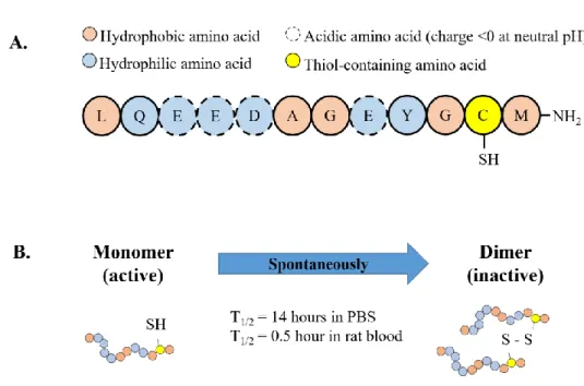
Zhou et al. 2013). Recently, the modulation of TREM-1 signaling by an inhibitory dodecapeptide
45
(LR12, Figure 1A) reduced the size of infarct and improved both cardiac function and survival in
46
a murine model of myocardial infarction (Boufenzer et al. 2015). In that study, LR12 (5 mg/kg)
47
was intraperitoneally (IP) administered once a day for 5 days. This therapeutic scheme should be
48
advantageously replaced by a single injection of a sustained-release formulation. Moreover, LR12
49
oxidizes spontaneously in aqueous media with a short half-life (t1/2) (approximately 14 hours in
50
phosphate buffer saline (PBS) in vitro, and 0.5 hour in blood ex vivo): a disulfide bridge is formed
51
between two peptides, generating a dimer (Figure 1B). While still to be confirmed by further
52
investigation, preliminary experiments on cells suggested that the dimer is devoid of
53
pharmacological activity. Consequently, the formulation should both sustain LR12 release for at
54
least a 5-day period and, as much as possible, protect the drug from degrading by dimerization.
55
In this contribution, the in vitro and in vivo release of LR12 (both the monomer and its main product 56
of degradation, i.e. the dimeric form) from in situ forming implants have been evaluated. In situ 57
4
forming implants are liquid formulations which, when injected into aqueous environments, 58
precipitate as solid polymeric matrices entrapping the drug (Parent et al. 2013a). Sustained releases 59
obtained with in situ forming implants have been described in the literature for a wide range of 60
drugs with various physicochemical properties. Herein, poly-lactide-co-glycolide (PLGA, 61
Resomer RG502H, 50:50 ratio LA:GA) and poly-lactide (PLA, Resomer R203S) were used as 62
biocompatible biodegradable polymers due to their frequent use in in situ forming formulations 63
and their lack of toxicity. Polymers were solubilized (18.7% w/w) in triacetin (TA, 74.8% w/w) 64
before adding the drug (6.5% w/w). The concentration of peptide used in the formulation allowed 65
to subcutaneously inject a dose of 80 mg LR12/kg to the animals. This can be compared to the total 66
dose of 25 mg/kg of free LR12 administered via several IP injections in the study, which 67
demonstrated the benefit of LR12 in myocardial infarction (Boufenzer et al. 2015). This dose is 68
compatible with a slow delivery of LR12 from the reservoir formulation during the time of 69
experiment, and it is likely without safety issues. Additionally, with this concentration, the 70
viscosity of the formulation remained suitable for an easy injection. 71
Formulations were prepared and in vitro release experiments were performed in physiological 72
buffer saline according to previously reported protocols (Parent et al. 2013b). An HPLC-UV 73
method was developed, then validated for selectivity, precision, accuracy and linearity according 74
to the FDA guidelines, and used to quantify LR12 (monomer and dimer) released in the aqueous 75
medium (Figure 2). The chromatographic system was the same as previously described (Parent et 76
al. 2016). The elution was isocratic (86/14 % v/v of water/acetonitrile + 0.1% trifluoroacetic acid) 77
with 20 µL of injected sample. The detection was set at 220 nm. Linearity was verified for 78
monomer and dimer between 1.0 and 50.0 µg/mL (y = 14.30x-17.55 and R² = 0.994 for monomer, 79
y = 14.15x -11.87 and R² = 0.996 for dimer). Peptides remaining in the implants were also 80
5
quantified with the same method after being extracted (dissolution of implants in ethyl acetate then 81
liquid-liquid extraction with PBS added with 0.1% trifluoroacetic acid, recoveries of 102.5 ± 6.2 82
% for the monomer and 100.3 ± 4.2% for the dimer, n = 3). 83
84
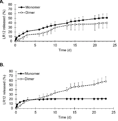
In vitro experiments demonstrated a 3-week sustained release of LR12 monomer from PLGA
85
implants (Figure 2A), without any burst, as expected when using a solvent with low water 86
solubility such as TA (Camargo et al. 2013). LR12 dimer also appeared progressively in the 87
medium. The release profile from PLA implants was different (Figure 2B): the monomer release 88
reached a plateau after 3 days, while the concentration of the dimer increased until the end of the 89
experiment. Dimer appears, resulting from the degradation of monomer, which can occur either 90
inside the implant or in the aqueous medium after its release. In the conditions of the in vitro release 91
test, non-formulated LR12 monomer spontaneously oxidized within time to form the disulfide 92
dimer, with a short half-life of 14 hours. Moreover, at the end of the release experiments (after 3 93
weeks), remaining LR12 (< 5.0% of the initial load) was extracted and was shown to be mainly 94
under the monomer form (95.0 ± 1.3% for PLGA and 93.4 ± 1.1% for PLA). The very low 95
proportion of dimer inside the implants suggests that the degradation of LR12 likely occurs in the 96
release medium rather than inside the formulations. To conclude, in situ implants offered a 97
sustained in vitro release of LR12 monomer up to 3 weeks, while efficiently protecting the drug 98
from dimerization. 99
100
In a previous study, blood concentrations of LR12 (monomer and dimer) after intraperitoneal 101
injection (5 mg/kg) to male Wistar rats were monitored (Parent et al. 2016). The same protocol and 102
6
method were applied to quantify LR12 in blood after one single subcutaneous injection of in situ 103
implants (80 mg LR12/kg) to male Wistar rats (280-350 g). Rats were subcutaneously injected with 104
in situ forming formulation prepared with PLGA or PLA (final dose of 80 mg LR12/kg, 21 G
105
needle used for injection). Under isoflurane anesthesia, blood was collected 1h, 1, 3, 7 or 14 days 106
after the treatment, before sacrificing the animal and retrieving the implants for residual LR12 107
quantitation. Three animals were used for each time point for each polymer (total = 30 rats). Blood 108
samples were treated and analyzed with the HPLC-fluorescence method as previously reported 109
(Parent et al. 2016). Remaining LR12 was also extracted from the retrieved implants and quantified 110
as described above for the in vitro study. At autopsy, no sign of irritation or inflammation of the
111
tissues surrounding the implants was observed, whatever the polymer used (Representative
112
photograph of extraction is supplied in Supplementary data S1).
113
After one single administration of in situ forming implants, LR12 monomer was detected at 114
therapeutic concentrations (between 100 and 400 ng/mL) for a week (Figure 3). The area under 115
the concentration-time curve (AUC07 days) was calculated using the trapezoidal rule and was
116
normalized by the received dose (Table 1). The highest observed plasma concentration (Cmax) and
117
the time required to reach Cmax (Tmax) were obtained from the concentration-time curves. Results
118
indicated that in situ forming formulations significantly improved LR12 monomer bioavailability 119
by a factor 50, without any difference between PLGA and PLA. This can be explained by the 120
sustained-release properties of the in situ forming formulations, which increase the circulation time 121
of LR12, and by the protection of monomer from degradation because it was encapsulated into the 122
polymeric matrices. Although LR12 remaining inside the implants was mainly under the monomer
123
form, the proportion of dimer inside the in vivo implants increased within time (4.3 ± 1.7 % at day
124
1 to 27.3 ± 2.4% at day 7 for PLGA and 3.5 ± 3.0% at day 1 to 26.1 ± 10.9% at day 7 for PLA).
7
After one week, more than 95% of the initial load was released. Compared with these in vivo
126
results, better LR12 protection (> 90% remaining as monomer after 3 weeks in the implants) and
127
more sustained release (up to 3 weeks) with in situ forming implants were observed in vitro.
128
129
In the literature, only a few studies deal with in vivo peptide sustained delivery with in situ forming 130
formulations. A 48 h – in vivo release was for example reported for enfuvirtide, an anti-HIV fusion 131
inhibitor of 36 amino acids, when incorporated into implants made of PLGA and a mixture of 132
DMSO and triacetin (Kapoor et al. 2012). A sustained effect of S-nitrosoglutathione, a nitric oxide 133
donating tripeptide, was also observed over the same time length, using PLGA implants with N-134
methyl-2-pyrrolidone (NMP) as solvent (Parent et al. 2015). Regarding leuprolide acetate (9 amino 135
acids), a 14-days in vivo release in rats was described from PLGA/NMP implants (Mashayekhi et 136
al. 2013), but similar marketed formulations (Eligard®) allow therapeutic efficiency in humans 137
from 1 to up to 6 months after a single administration. In this study, in situ forming formulations 138
administered to healthy rats demonstrated their ability to deliver the dodecapeptide LR12 according 139
to a smooth and sustained profile at therapeutic concentrations for 7 days. As a result, this single 140
injection could be an interesting alternative to the current therapeutic scheme (free drug 5 mg/kg, 141
daily injection for 5 days) proposed in myocardial infarction (Boufenzer et al. 2015). PLGA 142
formulations should probably be preferred over PLA ones, because they will be degraded faster, 143
while offering the same monomer bioavailability in this case, but with lower burst and less dimer 144
blood exposure (dimer AUC/dose increased by 12 for PLGA implants compared to IP 145
administration and by 18 for PLA ones, Table 1). Modifications of the peptide itself or of the 146
formulation could also be envisaged to further increase the in vivo duration of release. For example, 147
the PEGylation of a natural polysaccharide enhanced the in vivo mean retention time from 1.0 h to 148
8
2.8 days, and this result was drastically improved (up to 13 days) when the conjugate was 149
formulated into PLGA in situ implants (Shi et al. 2014, 2015). 150
To conclude, this study demonstrates that in situ formulations are promising candidates for the 151
therapeutic use of LR12, a TREM-1 inhibitory dodecapeptide useful in many conditions involving 152
inflammation and exacerbated immune response. However, pharmacokinetics of LR12 could be 153
modified in pathological situations, for example by the apparition of the soluble form of TREM-1 154
in the blood (Boufenzer et al. 2015). The benefit of in situ formulations for LR12 treatment should 155
therefore be confirmed in animals suffering from myocardial infarction for example. 156
157
ACKNOWLEDGEMENTS
158
This work was supported by the “Fondation pour la Recherche Médicale”, grant number 159
DBS20131128445, to Philippe Maincent. 160
Authors thank F. Dupuis and M.L. Bouressam for their assistance during blood sampling. They 161
also acknowledge M. Alcazar-Duque, M. Girardon and C. Verebi for their contribution in in vitro 162
release experiments and HPLC-UV validation. They are also very grateful to Marjorie Antoni 163
(lifelong learning division of Université de Lorraine) for helping to improve the level of English in 164 the manuscript. 165 166 REFERENCES 167
Bouchon, A., Facchetti, F., Weigand, M.A., Colonna, M., 2001. TREM-1 amplifies inflammation 168
and is a crucial mediator of septic shock. Nature 410, 1103–1107. 169
9 170
Boufenzer, A., Lemarié, J., Simon, T., Derive, M., Bouazza, Y., Tran, N., Maskali, F., Groubatch, 171
F., Bonnin, P., Bastien, C., Bruneval, P., Marie, P.Y., Cohen, R., Danchin, N., Silvestre, J.S., Ait-172
Oufella, H., Gibot, S., 2015. TREM-1 mediates inflammatory injury and cardiac remodeling 173
following myocardial infarction. Circ. Res. 116, 1772-1782. 174
175
Camargo, J., Sapin, A., Nouvel, C., Daloz, D., Leonard, M., Bonneaux, F., Six, J.L., Maincent, P., 176
2013. Injectable PLA-based in situ forming implants for controlled release of ivermectin, a BCS 177
class II drug: solvent selection based on physico-chemical characterization. Drug Dev. Ind. Pharm. 178
39, 146-155. 179
180
Gibot, S., Massin, F., Alauzet, C., Derive, M., Montemont, C., Collin, S., Fremont, S., Levy, B., 181
2009. Effects of the TREM 1 pathway modulation during hemorrhagic shock in rats. Shock 32, 182
633–637. 183
184
Kapoor, D.N., Katare, O.M., Dhawan, S, 2012. In situ forming implant for controlled delivery of 185
an anti-HIV fusion inhibitor. Int. J. Pharmaceut. 426, 132-143. 186
187
Mashayekhi, R., Mobedi, H., Najafi, J., Enayati, M., 2013. In vitro/in vivo comparison of 188
leuprolide acetate release from an in situ forming PLGA system. DARU J. Pharm. Sci. 21:57, 189
doi:10.1186/2008-2231-21-57 190
191
Mozaffarian, D., Benjamin, E.J., Go, A.S., Arnett, D.K., Blaha, M.J., Cushman, M., de Ferranti, 192
S., Despres, J-P., Fullerton, H.J., Howard, V.J., Huffman, M.D., Judd, S.E., Kissela, B.M., 193
10
Lackland, D.T., Lichtman, J.H., Lisabeth, L.D., Liu, S., Mackey, R.H., Matchar, D.B., McGuire, 194
D.K., Mohler, E.R. 3rd, Moy, C.S., Muntner, P., Mussolino, M.E., Nasir, K., Neumar, R.W., 195
Nichol, G., Palaniappan, L., Pandey, D.K., Reeves, M.J., Rodriguez, C.J., Sorlie, P.D., Stein, J., 196
Towfighi, A., Turan, T.N., Virani, S.S., Willey, J.Z., Woo, D., Yeh, R.W., Turner, M.B., on behalf 197
of the American Heart Association, Statistics Committee and Stroke Statistics Subcommittee, 198
2015. Heart disease and stroke statistics—2015 update: a report from the American Heart 199
Association. Circulation 131, e29–e322. 200
201
Parent, M., Nouvel, C., Koerber, M., Sapin, A., Maincent, P., Boudier, A., 2013a. PLGA in situ 202
implants formed by phase inversion: critical physicochemical parameters to modulate drug release. 203
J. Control. Release 172, 292-304. 204
205
Parent, M., Boudier, A., Dupuis, F., Nouvel, C., Sapin, A., Lartaud, I., Six, J.L., Leroy, P., 206
Maincent, P., 2013b. Are in situ formulations the keys for the therapeutic future of S-nitrosothiols? 207
Eur. J. Pharm. Biopharm. 85, 640-649. 208
209
Parent, M., Boudier, A., Maincent, P., Gibot, S., Ait-Oufella, H., Boufenzer, A., Jolly, L., Derive, 210
M., Kouach, M., Goossens, J.F., Leroy, P., Clarot, I., 2016. LR12-peptide quantitation in whole 211
blood by RP-HPLC and intrinsic fluorescence detection: validation and pharmacokinetic study. 212
Biomed. Chromatogr., doi: 10.1002/bmc.3877. 213
214
Parent, M., Boudier, A., Perrin, J., Vigneron, C., Maincent, P., Violle, N., Bisson, J.F., Lartaud, I., 215
Dupuis, F., 2015. In situ microparticles loaded with S-nitrosoglutathione protect from stroke. PLoS 216
One 10, e0144659. 217
11 218
Shi, X., Lin, X., Zheng, X.W., Feng, Y., Shen, L., 2014. Injectable long-acting systems for Radix 219
Ophiopogonis polysaccharide based on mono-PEGylation and in situ formation of a PLGA depot. 220
Int. J. Nanomed. 9, 5555-5563. 221
222
Shi, X., Lin, X., Yao, C.X., Shen, L., Feng, Y., 2015. Injectable long-acting in situ forming systems 223
for Radix Ophiopogonis polysaccharide. Int. J. Biol. Macromol. 72, 553-559. 224
225
Zhou, J., Chai, F., Lu, G., Hang, G., Chen, C., Chen, X., Shi, J., 2013. TREM-1 inhibition attenuates 226
inflammation and tumor within the colon. Int. Immunopharmacol. 17, 155-161. 227
228
FIGURES
229
230
Figure 1: A) Murine sequence of LR12 monomer peptide (Mr 1341 g/mol, calculated pI ~ 3.6)
231
and B) main pathway of degradation by dimerization. 232
12 233
Figure 2: In vitro release profiles of LR12 (monomer and dimer) obtained from in situ implants
234
made of TA and of either PLGA (A) or PLA (B). Results are presented as mean ± sd of three 235
experiments. 236
13 237
238
Figure 3: Blood concentrations of LR12 (monomer form, panel A and dimer form, panel B) after
239
administration of LR12 monomer (unformulated, 5 mg/kg, intraperitoneally vs formulated into in 240
situ implants, 80 mg/kg, subcutaneously). Limit of quantification of the method was 50 ng/mL. All
241
experiments were performed in accordance with the European Community guidelines 242
(2010/63/EU) for the use of experimental animals, and protocols were approved by the regional 243
and national ethical committees (project “Slow-release”, APAFIS#1146-2015071313458604 v3). 244
14
TABLE 1
245
Pharmacokinetic parameters of LR12 after administration to male Wistar rats.
246 247 Intraperitoneal injection of LR12 monomer Subcutaneous injection of LR12 monomer-in situ implants
PLGA PLA Monomer AUC07 days (ng. mL-1.h) 18 17 467 17 388 AUC/Dose (L-1.h.g body weight) 4 218 217 Tmax (h) 0.08 1 1 Cmax (ng/mL) 181 ± 35 261 ± 95 468 ± 252 Dimer AUC07 days (ng. mL-1.h) 197 38 414 57 499 AUC/Monomer dose (L-1.h.g body weight) 39 480 719 Tmax (h) 0.25 24 24 Cmax (ng/mL) 300 ± 162 379 ± 74 367 ± 39 248 Supplementary data 249 250
(S1): Representative photograph of the extraction of one in vivo in situ forming implant (here, PLA
251
implant, one week after injection). 252