HAL Id: hal-02375861
https://hal.archives-ouvertes.fr/hal-02375861
Submitted on 26 Nov 2019HAL is a multi-disciplinary open access archive for the deposit and dissemination of sci-entific research documents, whether they are pub-lished or not. The documents may come from teaching and research institutions in France or abroad, or from public or private research centers.
L’archive ouverte pluridisciplinaire HAL, est destinée au dépôt et à la diffusion de documents scientifiques de niveau recherche, publiés ou non, émanant des établissements d’enseignement et de recherche français ou étrangers, des laboratoires publics ou privés.
Involvement of ion channels and transporters in
carcinoma angiogenesis and metastasis
Sonia Martial
To cite this version:
Sonia Martial. Involvement of ion channels and transporters in carcinoma angiogenesis and metastasis. American Journal of Physiology - Cell Physiology, American Physiological Society, 2016, 310, pp.710 - 727. �10.1152/ajpcell.00218.2015�. �hal-02375861�
Involvement of ion channels and transporters in carcinoma angiogenesis
1
and metastasis
2 3
Author: Sonia MARTIAL 4
5
Affiliation: 6
IRCAN, CNRS UMR 7284, Inserm U1081, Université Nice-Sophia Antipolis, NICE, FRANCE 7
8
Running title: Ion transport modulates tumor angiogenesis and metastasis 9 10 Corresponding author: 11 Dr. Sonia MARTIAL 12
CNRS UMR 7284 – INSERM U 1081 – UNS 13
Institute for Research on Cancer and Aging, Nice 14 33 avenue Valombrose 15 06189 NICE CEDEX 2 16 FRANCE 17 e-mail : sonia.martial@unice.fr 18 Tel : +33 4 92 03 12 27 / +33 4 92 03 12 28 19 Fax : +33 4 92 03 12 35 20 21
ABSTRACT
2223
Angiogenesis is a finely tuned process, which is the result of the equilibrium between pro- and anti-angiogenic 24
factors. In solid tumor angiogenesis, the balance is highly in favor of the production of new, but poorly 25
functional blood vessels, initially intended to provide growing tumors with nutrients and oxygen. Among the 26
numerous proteins involved in tumor development, several types of ion channels are overexpressed in tumor 27
cells, as well as in stromal and endothelial cells. Ion channels thus actively participate in the different hallmarks 28
of cancer, especially in tumor angiogenesis and metastasis. Indeed, from their strategic localization in the 29
plasma membrane, ion channels are key operators of cell signaling, as they sense and respond to environmental 30
changes. This review aims to decipher how ion channels of different families are intricately involved in the 31
fundamental angiogenesis and metastasis hallmarks, which lead from a nascent tumor to systemic 32
dissemination. An overview of the possible use of ion channels as therapeutic targets will also be given, 33
showing that ion channel inhibitors or specific antibodies may provide effective tools, in a near future, in the 34
treatment of carcinomas. 35
36 37
KEYWORDS: ion channel, tumor vascularization, cell signaling, ion channel-targeted therapy 38
39 40
ABBREVIATIONS: HIF, hypoxia-inducible factor; CRC, colorectal carcinoma; ccRCC, clear cell renal cell 41
carcinoma; CAIX, carbonic anhydrase IX; RVD, regulatory volume decrease; PDAC, pancreatic ductal 42
adenocarcinoma; VEGFR, VEGF receptor; TSP, thrombospondine; MMP, matrix metalloproteinase; SOCE, 43
store-operated calcium entry; CRAC, Ca2+-release activated channel; ROCE, receptor-operated calcium entry;
44
IP3, inositol 1,4,5-trisphosphate; EMT, epithelial-mesenchymal transition; TME, tumor microenvironment; 45
STIM, stromal interaction molecule; MAM, mitochondrion-associated ER membrane; NMDAR, N-methyl, D-46
aspartate Receptor; CML, chronic myeloid leukemia; ER, endoplasmic reticulum; BiP, binding immunoglobulin 47
protein; NSAID, non-steroid anti-inflammatory drug; GPCR, G protein-coupled receptor. 48
I.
Introduction
50Carcinoma is a solid cancer developing from epithelial cells and is the most widely spread form of human 51
cancer. Among the phenomena leading to the development and progression of this type of cancer, one of the 52
most important is angiogenesis. Indeed, tumor vascularization is essential for a growing tumor to obtain oxygen 53
and nutrients needed for its ongoing growth, in situ. Tumor neovascularization is also a means for the tumor to 54
grow away from its primary site towards a remote neo-colonized structure (metastasis). 55
In the last 10 to 15 years, many efforts have been made on the elucidation of which genes might be directly 56
involved or affected during the different steps of tumorogenesis, described by Hanahan and Weinberg as the 57
hallmarks of cancer (95, 96). Multiple studies demonstrated that, among these genes, plasma membrane ion 58
channels and transporters are clearly modulated during carcinogenesis, in different types of carcinomas (9, 20, 59
31, 75, 78, 93, 133, 147, 201, 207). Several different roles have been ascribed to ion channels during 60
carcinogenesis, depending on the step of the tumorization process and on the organ in which it takes place. 61
In this review, we do not intend to make an exhaustive list of channels involved in cancer development. Instead, 62
we choose to focus on the involvement and/or alteration of some ion channels in angiogenesis, from the 63
vascularization of recently formed tumors to the metastasis cascade. Indeed, neo-formation of tumors involves 64
pH changes, which are associated with proton exchange through the plasma membrane (127, 209, 217) and 65
water transport (17, 162). The subsequent tumor growth needs neovascularization, associated with Ca2+, K+ and
66
Na+ channels (5, 61, 201). Finally, Na+, K+, Ca+ and Cl- channels are implicated in the transition to metastasis
67
(32, 73, 75). 68
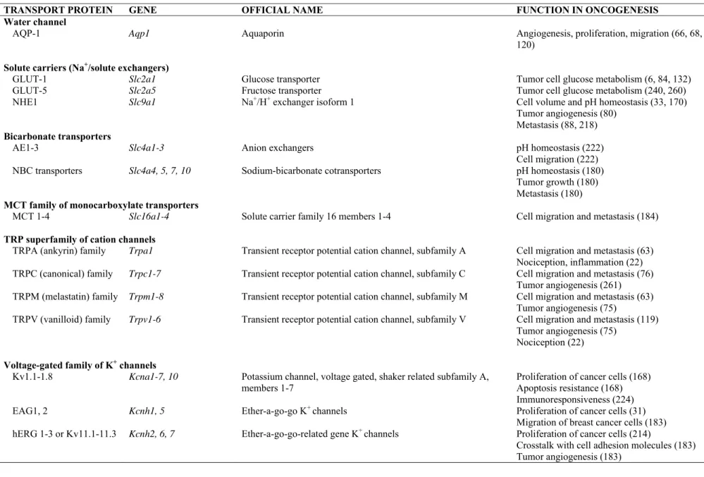
The main ion channels involved in tumor development processes and mentioned in this review are listed in 69
Table 1. 70
II.
Neoangiogenesis
72A.
The fate of new tumors
73In the early steps of its formation, a tumor depends solely on the host blood vessels (104). However, this initial 74
peripheral vasculature quickly disappears, leaving the tumor and its microenvironment in a hypoxic state (84, 75
97). Folkman (77) was the first to show that, under these conditions, the tumor growth is limited to a few cubic 76
millimeters, unless reorganization of a new blood supply is promoted (neoangiogenesis). 77
The lowered local O2 pressure (pO2) observed near cancer cells and the resulting tumor microenvironment
78
acidification activate hypoxia-inducible factors, especially HIF-1 (44, 58, 115). HIF-1α is stabilized by 79
phosphorylation (172) and dimerizes with the constitutively expressed HIF-1β to form the transcription factor 80
HIF-1, which in turn stimulates a set of genes involved in tumor development. Conversely, recent reports 81
present HIF-2 as the tumorigenic form of HIF, whereas HIF-1 would be a tumor suppressor (227, 263). 82
Initially, the cancer cells undergo metabolic changes, among which increased glucose consumption and lactate 83
production (the Warburg’s effect) lead to acidosis of the cells. In conjunction with the higher need for glucose, 84
several authors report a HIF-dependent expression of the GLUT-1 glucose transporter in breast or colorectal 85
carcinoma cell (CRC) models (25, 84, 132) and a repressed mitochondrial function (175). In clear cell renal cell 86
carcinomas (ccRCC), some isoforms of glucose transporters are equally increased, including the GLUT-5 87
fructose transporter (240). Moreover, in order to prevent acidosis induced by the accumulation of lactate, cancer 88
cells increase the amount and/or activity of several families of acid-extruding plasma membrane proteins. In 89
several carcinoma models, these HIF-1 increased proteins include: NHE1, the Na+/H+ exchanger (79, 170, 231),
90
the MCT1 and MCT4 forms of monocarboxylate transporters, dedicated to lactate extrusion (62, 184, 246), the 91
SLC4A4 (NBC) Na+/HCO
3- cotransporter (1, 180), involved in the bicarbonate reabsorption which contributes
92
to intracellular pH recovery. Acid-sensing ion channels (ASIC1, 2, 3) are also up-regulated in adenoid cystic 93
carcinomas, following the acidification of the extracellular space (257). Finally, carbonic anhydrases (especially 94
CAIX, the main carbonic anhydrase found in hypoxic tumors (64)), dedicated to microenvironment pH 95
modulation are also upregulated (132, 171, 180). The different ion channels involved at this stage are 96
summarized in Figure 1. Activation of all these transport proteins eventually contribute to a modest elevation of 97
the tumor cell intracellular pH, while the extracellular medium is acidified. 98
Subsequently, the newly constituted tumor will only grow larger if it manages to regenerate a constant blood 99
supply to provide O2 and nutrients to the tumor cells and to rid the cells of metabolites. Two main families of
100
plasma membrane ion channels have been proposed to be involved in this tumor developmental stage and 101
regulated by HIF. Transient receptor potential (TRP) channels contribute to calcium homeostasis and are 102
upregulated in several cancer cell types, thus playing an important part in Ca2+ influx, regulatory volume
103
decrease (RVD) and cell cycle progression (63, 254). A variety of K+ channels are also involved in the
HIF-104
induced regulation of proliferation and revascularization, namely the voltage-dependent “ether-à-gogo” 105
potassium channel (EAG1, KCNH1, Kv10.1) (31, 177), and the two-pore domain potassium channels TASK 106
(30) and TREK (243). In pancreatic ductal adenocarcinoma (PDAC), the Ca2+-dependent potassium channel of
107
intermediate conductance, IKCa (KCa3.1) is up-regulated and contributes to cell proliferation (114).
108 109
B.
Angiogenic switch
110Tumor angiogenesis is the key step for tumor growth, invasion and metastasis. Tumor angiogenesis mostly 111
relies on the same processes as those involved in physiological angiogenesis (27, 45, 176). Particularly, 112
ischemia and hypoxia are in both cases, major initiators of angiogenesis processes (“on” switches). Moreover, 113
tumor angiogenesis and physiological angiogenesis heavily depend on VEGF secretion, as was described 114
elsewhere and as we will describe here for tumor-depending angiogenesis. The main differences are: 1) 115
Pathological angiogenesis is more dependent upon VEGF than physiological angiogenesis (45); and 2) 116
Physiological angiogenesis events recede when vessel perfusion starts and upon disappearance of the stimuli 117
that gave rise to neoangiogenesis (“off” switch), whereas tumor angiogenesis is a continuous process, fueled by 118
the steady secretion of tumor-induced angiogenesis factors (45). 119
The concept of “angiogenic switch” was formulated in order to characterize angiogenesis as a balance between 120
processes that tend to favor and processes that tend to prevent angiogenesis (94). When tumor cells enter a 121
hypoxic state, activation of HIF leads to the subsequent activation of a multiple array of pro-angiogenic factors; 122
thus, the regular balance of angiogenesis is lost: the angiogenic switch has been turned “ON”. The most studied 123
HIF-regulated pro-angiogenesis factor is probably VEGF, the angiogenesis cytokine that regulates endothelial 124
cell proliferation and blood vessel formation. Both VEGF and its receptors VEGFR1 (FLT-1) and VEGFR2 125
(KDR/FLK1), which are located principally on the surface of endothelial cells, are upregulated by HIF (123, 126
185). Fibroblast Growth Factor (FGF) is also regulated by hypoxia and may act synergistically with VEGF to 127
amplify angiogenesis (125). 128
The “angiogenic switch” is turned “ON”, in several types of cancer cells, including mouse colon carcinoma, 129
mouse melanoma and human neuroblastoma, when several types of ion channels are modulated. P2X7R, the
130
non-selective cationic pore, has been shown to promote tumor growth and VEGF release (4, 5, 7). TRPC 131
channels are functionally coupled to VEGF or FGF, playing important roles in cancer development steps (76, 132
156). Moreover, in renal cell carcinoma (RCC), the TRPC4 Ca2+ channel expression is decreased, which impairs 133
Ca2+ metabolism, hence preventing the production of thrombospondin-1 (TSP1), an angiogenesis inhibitor
134
(238). Numerous studies report overexpression of most families of K+ channels in human cancers. In particular,
135
the closely related voltage-dependent hERG and EAG1 K+ channels are both involved in the processes leading
136
to angiogenesis. Indeed, hERG channels regulate angiogenesis in CRC by increasing the HIF-1 activated VEGF 137
expression and secretion in a β1-integrin-dependent manner and via a PI3K/Akt pathway (46). This regulation 138
has been confirmed in transgenic mice expressing hERG channels and treated with azoxymethane. These mice 139
present increased staining for VEGF in immunohistochemistry (IHC) studies and a higher total number of blood 140
vessels, consistent with neoangiogenesis, as well as colorectal lesions, on the whole showing acceleration of the 141
colorectal cancer phenotype (74). EAG1, but not EAG2 contributes to tumor progression, as demonstrated by 142
xenograft tumor formation resulting from implanted MDA-MB-435S breast cancer cells, EAG1-transfected 143
CHO cells and EAG1- or EAG2-transfected NIH-3T3 cells. Furthermore, EAG1 stimulates VEGF secretion, 144
measured in CHO- or 3T3-transfected cells. Finally, EAG1 increases HIF-1 amount in EAG1-expressing cells, 145
thus demonstrating that EAG1 is involved in the angiogenic switch and the subsequent neoangiogenesis (61). 146
The role of Ca2+ channels in conjunction with angiogenic switch is worth to mention. Indeed, it has been
147
demonstrated that VEGF utilizes Ca2+ signaling to promote endothelial cell proliferation and subsequent
148
neoangiogenesis, which we largely discuss in the next section. However, it is noteworthy that some Ca2+
149
channels, mostly of the TRP family, are upregulated by hypoxia in cancer cells (76). The subsequent increase in 150
intracellular Ca2+ concentration is at the basis of cancer cell hallmarks such as cell proliferation or migration
151
(76). 152
C.
Neovascularization
154Tumor vasculature is a crucial feature of carcinoma development. New blood vessels develop by sprouting 155
(angiogenesis) or intussusception from pre-existing vessels (144, 199). They can also emerge from the assembly 156
of endothelial precursors derived from the bone marrow (vasculogenesis) (189). Sprouting is a very well studied 157
process, which is involved in tumor blood vessel formation. Angiogenesis is a multistep process that begins 158
when endothelial cell VEGF receptors are stimulated by VEGF (19, 34) to give rise to new blood vessels 159
growing towards the avascular tumor that initially secreted the stimulating cytokine. The different steps of the 160
process are detailed below: 161
162
1. VEGF-induced blood vessel dilatation 163
Most research on tumor angiogenesis has focused on VEGF-A. VEGF-A signaling occurs through two VEGF 164
receptors, VEGFR1 and VEGFR2 (19, 144). Although VEGF-A has the highest affinity for VEGFR1, the 165
higher tyrosine kinase activity of VEGFR2 makes it the primary receptor involved in angiogenesis processes. 166
VEGFR1, conversely, is involved in macrophage chemotaxis, as well as in tumor cell survival and invasion 167
(136, 205, 255). Additionally, VEGFR1 may also have a negative role on angiogenesis and tumorogenesis 168
processes, only acting as a decoy receptor (102, 142). VEGF-A binds to its receptor and this first results in 169
blood vessel dilatation, as recently shown by in vivo studies of breast tumors, by sonographic examination (41). 170
Like most mammalian cells, endothelial cells express cell-swelling activated Cl- channels, called the
Volume-171
Regulated Anion Channel (VRAC) (166, 167). Interestingly, receptor tyrosine kinases such as VEGFRs can 172
serve as mechanosensors (40). As such, they are involved in the VRAC signaling pathway. Thus, one might 173
infer that VEGF signaling in endothelial cell is mediated by VRAC Cl- channels, as shown by the suppression of
174
angiogenesis by VRAC inhibitors (146). 175
176
2. Disorganization of the vascular bed 177
Angiogenesis partly proceeds by the VEGF stimulation of endothelial cell matrix metalloproteinases (MMPs) 178
(188). Secretion of these MMPs, more precisely MMP-2 and MMP-9 (18, 250), triggers the dissolution of the 179
basement membrane surrounding these endothelial cells, thus provoking the destabilization of the entire blood 180
vessel wall and leading to vessel leak (34). Among the multiple mechanisms that cooperate for the close 181
regulation of MMP activity, Ca2+ signaling is one of the most important (121). The work, led by Kato,
182
demonstrates that under extracellular acidification, B16 mouse melanoma cell line displays increased voltage-183
gated Ca2+ channel expression and activity, resulting in an increase in intracellular Ca2+ concentration and in
184
MMP-9 induction. Thus, Ca2+ signals and channels are potentially the most important points in the regulation of
185
the VEGF-triggered blood vessel reorganization. However, the work conducted by Kato and colleagues 186
addresses the other types of Ca2+ channels that may be involved in the complex processes going from VEGF
187
upregulation to basement membrane disorganization. Indeed, under a hypoxic stimulus, the formation of a 188
VEGF-VEGFR complex at the endothelial cell plasma membrane can also result in intracellular Ca2+ increase,
189
due to the release of intracellular Ca2+ stores (Fig. 2). The depletion of these stores in turn results in Ca2+ influx,
190
via plasma membrane Ca2+ channels which results in store-operated calcium entry or SOCE (for review, see
191
(75)). Several Ca2+ channels of the transient receptor potential (TRP) superfamily appear to be involved in
192
SOCE and help explain how SOCE contributes to VEGF-induced basement membrane disruption. Among the 193
TRPCs (canonical), TRPC1 activation magnifies the VEGF-induced increase in endothelial cell monolayer 194
transepithelial permeability (111, 178). Conversely, inhibition of the TRPC1 activity reduces this transepithelial 195
permeability increase (111). TRPC4 is a necessary intermediate in the response of human pulmonary artery 196
endothelial cells to VEGF stimulation (71). TRPC6 is also presented as a good candidate in the process of 197
VEGF-induced vascular permeability increase. Indeed, as shown in individually perfused frog microvessels, 198
stimulation of TRPC6 generates an increase in the hydraulic conductivity (Lp) of the vessels (186). The Ca2+
-199
release activated Ca2+ (CRAC) channel (composed of the plasma membrane Orai1 subunit coupled with the
200
endoplasmic reticulum STIM subunit) is also involved in VEGF-dependent SOCE (139). Indeed, in human 201
umbilical vein endothelial cells, inhibition or disruption of Orai1 reduces VEGF-induced calcium entry, cell 202
migration and in vitro angiogenesis. The increase in transepithelial permeability may result from both in-cell 203
and in-between-cell pore creation (43, 98). Finally, VEGF effects can also occur via receptor-operated-calcium 204
entry (ROCE), as shown on human microvascular endothelial cells (42); TRPC3 and TRPC6 are two examples 205
of channels involved in calcium-mediated vascular permeabilization. 206
In conclusion, VEGF-induced calcium-dependent events leading to the basement membrane disorganization, 207
though resulting in the same final outcome, appear to be the consequence of several calcium entry pathways. 208
209
3. Endothelial cell proliferation, migration and organization 210
Breakdown of the blood vessels (i.e. dissolution of the basement membrane) leads endothelial cells to be 211
released into the extracellular matrix (ECM) where they proliferate and organize to generate new blood vessels, 212
growing towards the tumor that initially emitted stimulating signals. Angiogenic sprouting is a guided process in 213
which the vasculature expands outwards from the preexisting vessel, following a gradient of angiogenic factors 214
such as VEGF-A (85) or soluble VEGFR1 (38). In this regard, Gerhardt and colleagues demonstrated that the 215
first cell of the column, the so-called “endothelial tip cell,” consists of a single, non-proliferative, highly 216
polarized endothelial cell, which is the sensor of the emitted elongation signal. The cell migration along this 217
signal (chemotaxis) paves a path for the cells that follow, the “stack cells”, which undergo high rate mitosis to 218
form the newly elongating blood vessel. At the endothelial cell level, it is now recognized that VEGFR2 is the 219
main actor of the angiogenic cascade. VEGF, when binding to VEGFR2, activates a signaling cascade starting 220
from auto-phosphorylation of VEGFR2, subsequent phosphorylation and activation of phospholipase C-γ (PLC-221
γ), increased production of inositol 1,4,5-trisphosphate (IP3) and resulting increase in the Ca2+ release from
222
endoplasmic reticulum. The subsequent rise in calcium influx by the aforementioned SOCE process involves 223
TRPC channels, as already discussed (see chapter II.C.2) and promotes endothelial cell migration via a 224
Ca2+/calmodulin/calcineurin pathway (72). The role of SOCE was also established in bovine artery endothelial
225
cells for the different mechanisms of angiogenesis (i.e., cell sprouting, cell proliferation and cell migration) and 226
involves the Ca2+/calmodulin/protein kinase cascade (15).
227
One of the fundamental roles of ion channels in endothelial Ca2+ signaling is the fine-tuning of the
228
electrochemical gradient for Ca2+ (118). Several types of ion channels cooperate in elaborating the cell
229
membrane potential, which regulates calcium entry into the endothelial cells (118). Among those, K+ channels,
230
especially Ca2+-activated K+ channels (251), inwardly rectifying K+ channels (K
ir) and voltage-dependent K+
231
channels, are the major classes of ion channels involved in setting the membrane potential (165), which in turn 232
influences the driving force for the Ca2+ via SOCE. Indeed, blockers of K+ channels, as well as plasma 233
membrane depolarization, stop the cell cycle in G1 phase, thus reducing cell proliferation (153, 165). However, 234
among the Ca2+-dependent K+ channels, the Ca2+-dependent intermediate conductance K+ channel IK1 (SK4,
KCNN4) increases cell proliferation with a completely different mechanism, in a HEK overexpressing system. 236
Indeed, the potassium current itself does not seem to mediate the effect, since a non-transporting mutant of the 237
channel induces the same proliferation. So it seems that the IK1-induced proliferative effect is not SOCE-238
mediated (152). The cell membrane potential is partly regulated by Cl- channels (247). Hence, volume-regulated
239
anion channels (VRAC), Ca2+-activated Cl- channels (CaCC), as well as cystic fibrosis transmembrane regulator
240
(CFTR) (145) might be important regulators of SOCE, which drives the proliferation step of angiogenesis. 241
242
4. Maturation of the new blood vessel 243
Two steps are essential for completion of angiogenesis: 244
(1) A new vessel is formed when the elongating sprout lumenizes to create a tube, which will eventually be able 245
to transport fluid and the multiple blood components: this step is called tubulogenesis (117, 143). At least two 246
models of tubulogenesis have been described. The “cell hollowing” model (currently the most studied) evolves 247
from a chain of single endothelial cells (117). This model involves intracellular vesicle formation, then fusion of 248
these vesicles into vacuoles, first intracellularly, and then from cell to cell, all along the sprout, when the 249
vacuoles get bigger. The future lumen vacuole membrane finally acquires hallmarks of an apical membrane, 250
thus defining the apical-basolateral polarity of the neo-formed vessel. In the less studied “cord hollowing” 251
model, which happens in a multi-cellular elongated stack, the cells first lose their initial polarity, then 252
basolateral membrane markers are accumulated at the membrane facing the extracellular matrix, while vesicles 253
are accumulated and get fused on the opposite membrane, hence forming an initial lumen that expands 254
afterwards (179, 248). In the “cell hollowing” model, the formation of vacuoles is linked to the activation of 255
intracellular chloride channels of the CLIC family. The chloride intracellular channel (CLIC) family of proteins 256
contains six mammalian members (CLIC1-6), from which only CLIC1 and CLIC4 are expressed in endothelial 257
cells (230). CLIC proteins can change from a soluble state to a membrane-linked state. In the latter case, when 258
inserted in such organelles as mitochondria, Golgi membranes, nuclear membranes, etc., CLICs function as 259
chloride channels, although very different in structure from the other known Cl- channels (65, 233). In the “cell
260
hollowing” model of tubulogenesis, CLIC4 is present in human umbilical vein endothelial cells and promotes 261
several processes involved in angiogenesis, including lumenal formation (229). Particularly, endothelial cells 262
derived from transgenic mice, in which the Clic4 gene has been invalidated, fail to proceed to the vacuole 263
creation and extension process. Tung and colleagues show that the enlarging vacuoles acidify during their 264
formation, involving an electrogenic vacuolar proton-ATPase (vH+-ATPase), and propose that CLIC4 supports
265
this acidification by creating an electrical gradient for the proton transfer. The same article proposes that CLIC1 266
may partially compensate for the absence of CLIC4 in the process of tubulogenesis, although CLIC1 seems 267
mainly committed to proliferation and migration processes rather than to morphogenesis of endothelial cells 268
(230). 269
(2) The new hollow tube thus created is finally stabilized by the formation of a new basement membrane. 270
Indeed, in physiological angiogenesis, accessory cells, such as pericytes and smooth muscle cells are recruited 271
to the newly developed vessel, to form a new basement membrane (54). In tumor angiogenesis, however, the 272
newly formed blood vessels are very heterogeneous in structure and function (54). Their architecture is very 273
chaotic and disorganized, with abnormal branching, inflated cells and tortuous courses. Subsequently, the 274
association between endothelial and mural cells is altered, becoming loose and leading to highly permeable 275
vessels, which, paradoxically, are not adapted to a fluent and regular irrigation of the tumor, hence favoring its 276
dissemination to other, supposedly more welcoming, locations (35). The leaky vessels allow the tumor to escape 277
the noxious environment in which it grows (metastasis). Again, Ca2+ channels of the TRP family, namely
278
TRPV4 Ca2+ channels, are key determinants in the balance between normal and defective vasculature (2).
279
Indeed, tumoral endothelial cells naturally contain low levels of TRPV4. Conversely, overexpression or 280
pharmacological activation of TRPV4 normalizes the vasculature and restores the endothelial cell layer physical 281
and biophysical properties. 282
283
III. Metastasis
284A.
The tumor microenvironment (TME): role and ion channel profile
285The extracellular environment of tumor cells is viewed as a complex network of stromal cells, deregulated 286
vasculature, extracellular membrane (ECM) proteins, growth factors and cytokines, all of which generated or 287
activated by the tumor cells, acting together to influence the growth, behavior and malignancy of the tumor 288
(236), and constantly evolving while the tumor grows and becomes more aggressive. The physical and chemical 289
characteristics of the TME is cancer-type dependent, but there are a number of typical, cancer type-independent 290
features, among which hypoxia, acidic pH, high levels of lactate and low levels of glucose (21, 82). In this 291
context, ion channels and transporters are important specialized proteins involved in the regulation of signaling 292
pathways associated with these properties. All the cells composing the tumor microenvironment (TME) are 293
laden with several types of missexpressed/overexpressed ion channels, which participate in the crosstalk 294
described by Arcangeli, between the tumor and its TME (8). These ion channels also contribute to maintain the 295
harsh conditions that allow tumor growth (33). Concerning the vasculature, we have already discussed the 296
channels involved in neoangiogenesis and mentioned the importance of endothelial cell Ca2+ homeostasis
297
throughout this process. Mesenchymal cells (fibroblasts, myofibroblasts, immune cells) also express ion 298
channels, particularly lymphocytes that infiltrate the tumors express K+ channels of the K
v1.3 and KCa3.1 types,
299
which are involved in T cell activation via Ca2+ signaling (124). Interestingly, tumor-associated macrophages
300
express P2X7R non-selective cation channels and inwardly-rectifying KIR K+ channels; both families are
301
involved in the Ca2+-dependent activation of macrophages (8).
302 303
B.
Role of the epithelial-mesenchymal transition (EMT) in the metastasis process
304Metastasis of epithelial cancer cells is a multistep process, which involves mobilization of the cells from their 305
original location, migration of the cells, invasion of surrounding locations, intravasation of cancer cells into 306
newly formed tumoral blood vessels, cell survival in the blood flow and seeding to more distant new niches, 307
where the cells start proliferating again, thus creating secondary tumors. Multiple oncogenic signals originating 308
from the TME convert the cells into their new metastatic and invasive phenotype. These signals lead the tumor 309
cells to acquire several biological capabilities described as hallmarks of cancer (95, 96) and mediated by an 310
epithelial-mesenchymal transition (EMT) process, otherwise called the metastatic cascade. During this cascade, 311
cells, which originally display epithelial cell features (i.e., adhesion to each other and to the basement 312
membrane, high level of E-cadherin involved in the epithelial integrity (16)) are progressively transformed into 313
mesenchymal cells (116). Briefly, during this process, polarized epithelial cells acquire a new phenotype, which 314
includes increased resistance to death, enhanced migratory capacities and invasiveness. The complete spectrum 315
of signaling agents that contribute to EMT remains an open question. EMT is a complicated process and ion 316
channels from several different families play a particularly important role in this transition (249). 317
318
1. Mobilization of the cells: involvement of ion channels 319
Loss of cell-cell contacts is the first step of EMT. This phenotype is associated with repression of cell junction 320
proteins, such as E-cadherin, and correlated with stimulation of both vimentin (an intermediate filament protein) 321
and N-cadherin (neural cadherin, normally expressed in migrating non-tumoral cells) synthesis (107, 164, 226). 322
This process has been shown in breast cancer cells to be Ca2+-dependent (52). In fact, induction of EMT is
323
clearly related to the expression of TRPM7, the melastatin-like TRP Ca2+ channel. Indeed, upregulation of
324
TRPM7 regulates the expression of the EMT marker vimentin. Conversely, downregulation of the channel 325
prevents EMT induction in breast cancer cells. In colorectal carcinoma cells, the Ca2+-activated KCNN4
326
(KCa3.1; SK4) channels have been shown to be involved in EMT promotion, probably through participation in
327
the cell hyperpolarization, subsequent induction of Ca2+ entry via voltage-dependent Ca2+ channels and an
328
increase in intracellular Ca2+ concentration (129).
329
Tumor cell mobilization is the next step in the metastatic cascade. This step involves migration of the cells from 330
their initial site and invasion of the surrounding stroma and vasculature. This is a complex process requiring the 331
coordination of multiple macromolecules involved in adhesion, cytoskeletal dynamics and digestion of the 332
ECM. Among the extracellular elements favoring EMT, hypoxia and microenvironment acidification have been 333
the most studied and are involved in tumor cell mobilization. As discussed earlier in this review, tumor acidity 334
is a direct result of hypoxia (83, 237). Indeed, low pO2 raises the rate of glycolysis, which produces elevated
335
levels of lactate and protons in tumor cells. In order to maintain pHi within a tight range (pH 7.2 – 7.4), the
336
tumor cells upregulate H+ extrusion, which, in turn, results in microenvironmental acidosis, down to pH 6.5 (50)
337
or even to pH 5.8 as shown in highly metastatic MDA-MB-435S breast tumor cells (208). Extracellular acidosis 338
increases even more due to hypoxia-induced activation of carbonic anhydrases, especially CAIX (223). 339
Consequently, tumor cells are equipped with mechanisms that allow them to sense and react to the low pHe to
340
which they have been exposed. pH sensors of several types are present in the plasma membrane of mammalian 341
tumor cells and these pH sensors are categorized into G-protein coupled receptors (GPCRs) and non-GPCR pH 342
sensors (50). GPCR signaling pathways are manifold: ERK pathway/Ca2+ release (105), G
s-protein/cAMP,
343
G12/13-protein/Rho, and Gq-protein/phospholipase C pathways (140). pH sensing results in reorganization of the
344
cytoskeleton (e.g. actin rearrangement, stress fiber synthesis, etc.), thus contributing to tumor migration and 345
eventually to tumor progression (39). Non-GPCR proteins include ion channels of several families and 346
participate in migration and invasion phenotypes. Indeed, expression of the tumor cell cation-permeable Acid 347
Sensing Ion Channels (ASICs) is pH-dependent. ASIC1a is associated with tumor cell migration and invasion in 348
hepatocellular carcinoma cells (112); ASIC2a and ASIC3 have been identified in the plasma membrane of 349
adenoid cystic carcinoma cells and proposed to be involved in the acquired invasive capabilities of the cells 350
(257). The Ca2+ channel TRPV1 is also activated by low pH (259) and is correlated with tumor cell proliferation
351
in human prostate cancer cell lines (159). Both families of channels are involved in cancer-associated pain (57, 352
259) and plasma membrane phospholipids are proposed to be modulators of ASIC and TRPV1 activities (128). 353
Other acid-sensitive channels comprise several members of the Ca2+ channel TRP family, ionotropic
354
purinoceptors (P2X), inward rectifier K+ channels (K
ir), voltage-activated K+ channels (Kv family), L-type Ca2+
355
channels (Cav, CACN), hyperpolarization-activated and cyclic nucleotide-gated cation channels (HCN), gap
356
junction channels, and Cl− channels. They are involved in the process of tumor cell migration (217).
357
Interestingly, the Ca2+-activated, high conductance, BKCa K+ channels are not involved in breast cancer
358
progression, as shown by the absence of effect of iberiotoxin (an inhibitor of BKCa) in the processes of 359
proliferation, survival, migration or invasion in several breast cancer cell lines (200). 360
Migratory cells are polarized along their longitudinal axis, with hypoxia- and TME acidosis-dependent 361
protrusion of lamellipodia at the cell front edge and coordinated retraction of the rear edge (213). This process is 362
intracellular Ca2+ concentration-sensitive, thus involving Ca2+ release from intracellular stores and SOCE
363
channels (228). It creates the amoeboid movement characteristic of migrating tumor cells which is highly 364
dependent on ion and water flux (210). Indeed, the formation of lamellipodia is accompanied by relocalization 365
at the leading edge of a number of plasma membrane transport proteins, among which CAIX, NHE, MCT (all 366
involved in pH modulation), anion exchangers (such as NBC transporters or the anion exchanger AE2) and 367
water channels (aquaporins; AQP). It is proposed that the coordinated action of all these channels and 368
transporters makes it possible for the cells to establish the osmotic gradient that subsequently induces AQP-369
mediated water flux at the leading edge of the cell. Water extrusion by AQP, at the rear end of the cell, is 370
mediated by the combined activity of Ca2+-activated K+ channels (K
Ca3.1) and Volume-Regulated Anion
371
Channels (VRACs) and leads to regulatory volume decrease (RVD) and shrinkage of the cells (206). 372
373
2. Invasion of surrounding TME and vasculature 374
Invasion of healthy tissues by tumor cells is critically dependent on the local degradation of ECM and basement 375
membrane, which otherwise would constitute a barrier to tumor metastasis. The degradation process thus creates 376
the escape pathway by which single tumor cells leave their primary location and colonize surrounding areas. 377
This degradation results from the low extracellular pH produced by tumor cells and relies on the secretion or 378
membrane expression of acid-dependent proteases (MMPs and cathepsins). Soluble (MMP2, MMP9) or 379
membrane-bound (MMP14) MMPs are key enzymes in tumor cell invasiveness, involved in both ECM and 380
basement membrane degradation (187, 193, 253) and involved as well in the angiogenic sprouting (18, 53). 381
MMP activity is promoted by low pHe localized at the front part of migrating cells. As stated above, NHE1 is
382
one of the main actors of this extracellular acidification. While the acidification process appears as a crucial step 383
to weaken and break the ECM barrier, the physical progression of aggressive tumor cells relies on the formation 384
of ECM-degrading protrusions, called invadopodia, at the leading edge of the cells. NHE1 has been localized 385
within these invadopodia (29) and this localization has been correlated with extracellular acidification and MMP 386
expression and activity (88), thus suggesting the invadopodia as the major sites of ECM digestion and NHE1 as 387
the main channel involved in this process. 388
Voltage-gated sodium channels are highly expressed in epithelial tumors. In the MDA-MB-231 breast cancer 389
cell line, the Nav1.5 Na+ channel, by driving Na+ influx and depolarizing the cells, greatly increases NHE1
390
potency to promote extracellular acidification and tumor invasion (23, 24, 86). 391
Calcium channels are also involved in the migration and invasion processes. Indeed, TRP channels, which 392
mediate SOCE Ca2+ entry and regulate intracellular [Ca2+], especially TRPV2 (158) and TRPM8 (169),
393
positively influence carcinoma invasion by upregulating MMP2 and MMP9 synthesis and production. Thus, 394
MMP production may be an intracellular [Ca2+]-dependent process. Interestingly, high concentrations of ATP
395
are observed in the TME of most hypoxic solid tumors (181). ATP acts as a signaling agent by activating 396
plasma membrane ligand-gated receptors of the P2X family, namely P2X7R cation channels promote migration
and invasion, as shown in T47D breast cancer cells (253). P2X7R cation channels mediate Ca2+ influx in tumor
398
cells thus contributing to the increase in intracellular [Ca2+]. In the breast cancer cell line MDA-MB-435S, ATP
399
activation of P2X7R cation channels activates Ca2+-dependent SK3-mediated potassium currents, thus favoring
400
the elongation of cell protuberances, which, in turn, promote cell migration (110). In this model, the P2X7
R-401
promoted invasive phenotype also relies on the activation of proteolytic enzymes, essentially cathepsin B. 402
Correlatively, pharmacological inhibition or downregulation of P2X7R channels in PC-3M human prostate
403
carcinoma-derived cell lines inhibit ATP-driven migration and invasion (193). This Ca2+-dependent process
404
requires activation of either PI3/AKT or ERK1/2 signaling pathway. It reduces the expression of proteins 405
involved in the maintenance of cell-cell contacts (e.g. E-cadherin and claudin-1) and correlatively increases the 406
expression of Snail (an inhibitor of E-cadherin), interleukin 8 (IL-8, a promoter of migration and angiogenesis) 407
and MMPs, thus contributing to the invasion phenomenon. The metastasis- and malignancy-promoting status of 408
P2X7R, however, is still an important field in cancer research. Indeed, recent studies have been designed to
409
explore the effects of P2X7R on tumor development and metastatic outcome in vivo. Hofman and collaborators
410
demonstrate that colitis-associated cancers (colon adenocarcinoma, colon carcinoma) are favored by P2X7R
411
silencing or pharmacological inhibition (103). A different study reveals that P2X7R restricts tumor progression
412
by developing antitumoral immunity (4). 413
414
3. Metastasis: transit in blood vessels and extravasation 415
Tumor cell metastasis occurs when the cells successfully escape from their primary location, overcome the step 416
of transit in the circulation as unattached cells and finally disseminate to their secondary sites by extravasation 417
from the vessels and reattachment to a new ECM support. 418
Circulating tumor cells do not always survive long enough to metastasize. It is estimated that only 0.01% of the 419
cells that enter the vasculature will eventually survive the harsh conditions to which they are submitted (87). 420
Tumor cells must resist anoikis whereby apoptosis is triggered by inadequate or loss of cell anchorage. Among 421
the multiple signals involved in anoikis resistance, several types of ion channels have been described (137). 422
Indeed, to effectively resist detachment-induced apoptosis, tumor cells utilize mechanisms that eventually limit 423
the rise in intracellular [Ca2+]. Ca2+ channels are thus downregulated, especially those acting via SOCE (130).
Depletion in other Ca2+ channels (TRPM2, TRPV6) also tends to reduce cell apoptosis (211). Activity of
425
caspases and other DNA-degrading enzymes, as well as a decrease in membrane potential have been linked to 426
loss of intracellular [K+] by K+ channels. Cell shrinkage involved in apoptotic volume decrease (AVD) results
427
from a loss of KCl by K+ and Cl- channels, with a concomitant loss of water. Thus, downregulation of K+ (K v,
428
KCa, KATP, KIR, K2P) and Cl- channels also prevents apoptosis (26). Alternatively, anoikis-resistant cells can
429
develop regulatory volume increase (RVI) whereby Cl- channels of the volume-regulated (VRAC) family,
430
especially prostate cancer cell ClC-3 (138), which are involved in the maintenance of cell volume under 431
hyperosmotic stress, are upregulated in cells that resist apoptosis. Interestingly, Ca2+-activated Cl- channels 432
(CLCA) may be either activated or not activated in anoikis-resistant cells, as shown in mammary gland cells 433
(67, 244), thus suggesting a different function of these channels in cancer cells. 434
Tumor cells are subjected to mechanical forces resulting from the blood stream, especially in narrow capillaries. 435
Mechanical stress is the main destruction pathway that tumor cells must avoid in order to be able to form 436
metastases. To prevent destruction, they develop integrin-involving adhesion processes and eventually get 437
attached to the endothelial bed of the vessel in which they are traveling (81). This interaction between 438
circulating tumor cells and endothelial cells is fundamental for the rest of the metastasis process, as it 439
determines the site where the cells will eventually exit from the vessel, hence initiating the extravasation 440
process. Gout and Huot have described the whole process of extravasation (87). Briefly, this process involves a 441
first step of tumor cell adhesion to endothelial cells, utilizing specific receptors. Tumor cells rolling along the 442
endothelial layer characterize this “light adhesion” step. A second step of firmer cell attachment then takes 443
place, mediated by cytokines and cell adhesion molecules. Extravasation (or diapedesis) finally occurs when the 444
cells cross the endothelial barrier through cell-cell junctions. As for several steps of the tumorogenesis process, 445
the importance of intracellular [Ca2+] and of the associated Ca2+ channels is also demonstrated for metastasis of
446
tumor cells. Indeed Orai1, the pore-forming component of the store-operated Ca2+ channel and its associated
447
STIM1 Ca2+ sensor are associated with cell migration, invasion and metastasis in human breast carcinoma cells
448
(256). In vivo studies in a zebrafish model corroborate the importance of the Orai1/STIM1 partners and of 449
SOCE Ca2+ entry in the process of cell extravasation (262). Indeed, inhibition of the store-operated Ca2+ entry or
450
knockdown of Orai1 in nasopharyngeal carcinoma cells prevents extravasation of the cells from vasculature in a 451
zebrafish hematogenous metastasis model. 452
Finally, diapedesis is the result of tumor cells passing in between insufficiently tight endothelial cell junctions 453
(due to damaged vasculature). The alterations in cell shape necessary for diapedesis are most likely explained by 454
K+ and Cl- fluxes and by the following water fluxes. Indeed, it has been already shown that glioma cell
455
migration and invasion are supported by K+ and Cl- channels, which, by governing transmembrane water
456
movements and the resulting cell shape, are important for glioma invasion through narrow spaces (215). 457
Colonization of the secondary site by the tumor is not simply a matter of gaining access to this site. Tumor cells 458
will only be able to colonize compatible target tissues. The host tissue must then adapt to be able to foster the 459
colonizing tumor cells; this is the concept of “pre-metastatic niche” (212). Tumor cells must also adapt to their 460
new stroma and become able to secrete all the molecules necessary for their efficient attachment and ulterior 461
vascularization (91). Hence, there are many barriers to overcome before the tumor actually metastasizes. 462
463
IV. Sigma 1 receptor: the main organizer?
464In the present review, we will skip the long history of sigma-1 receptor (Sig-1R), which has been widely studied 465
on the functional, pharmacological, structural and mechanistic points of view for the last 30 – 40 years (11, 148, 466
245). Similarly, we will not open this discussion to the sigma-2 receptor, which has not been cloned yet, 467
although it was shown to be involved in cancer development (234). Instead, we will focus on the specific role of 468
chaperone of Sig-1R that is of major interest in the context of cancer development. 469
Sig-1R is a 25 kD, endoplasmic reticulum (ER)-resident, ligand-regulated protein, which is involved in many 470
diseases, ranging from toxic substance addiction to stroke and cancer (10, 11, 148). It is mainly present in 471
normal brain, heart and liver, but is highly expressed in tumor cell lines and human cancer tissues of various 472
origins, including lung, colon, prostate carcinomas, sarcomas, breast tumors, brain tumors, and melanomas (10, 473
11, 113, 173, 241), in which it interferes with cell cycle and proliferation (10, 197). Interestingly, in breast 474
cancer cells, Sig-1R positively correlates with the metastatic grade of the cells, i.e. highly metastatic cells 475
contain higher levels of Sig-1R mRNA and protein than their less aggressive counterparts (10). 476
Sig-1R is anchored in the ER membrane, in a specific region called mitochondrion-associated ER membrane 477
(MAM). At rest, it is in a “dormant” state, associated with a chaperone protein, called GRP78 or BiP (100). BiP 478
is a main regulator of ER function: it has a high Ca2+-buffering capacity and serves to store Ca2+, which is
479
essential to many signaling pathways. Under specific ligand stimulation or under prolonged ER stress such as 480
ER Ca2+ depletion, Sig-1R is up-regulated, dissociates from BiP and becomes able to act as a chaperone (100)
481
after translocation to other ER areas or to the plasma membrane (101). In the plasma membrane, Sig-1R 482
interacts with – and regulates the activity of – several ion channels (Ca2+, K+, Na+, Cl-), receptors (NMDA
483
receptor) or kinases (Src) (148). 484
In the context of cancer development, as we discussed earlier in this review, many ion channels of diverse 485
classes (voltage-gated, calcium-gated, volume activated) are involved in a number of tumor cell processes. 486
Voltage-gated ion channels are involved in cell processes including proliferation, apoptosis, angiogenesis, 487
adhesion to ECM and migration. Interestingly, Sig-1R interacts with most of these channels, as demonstrated in 488
several works (12, 13, 126, 197). Patch-clamp studies and western blot assays in the presence of Sig-1R ligands 489
(e.g. (+)-pentazocine, igmesine, and 1,3-di(2-tolyl)guanidine [DTG]) were conducted in small cell lung cancer 490
(SCLC) derived cell lines (NCI-H146 and NCI-H209) and in T-cell leukemia-derived Jurkat cells. These studies 491
demonstrate that ligand-mediated inhibition of Sig-1R results in cell cycle arrest in G1 phase with no stimulation
492
of apoptosis (197). This effect correlates with the reduction of voltage-operated K+ currents in all these cell
493
lines, indicating that Sig-1R-mediated cell growth arrest is the result of the negative regulation of K+ channels
494
(197). Cell cycle arrest also correlates with the negative modulation of Volume-Regulated Chloride Channels 495
(VRCC), both effects leading to the overall inhibition of the Regulated Volume Decrease (RVD) process, (196), 496
which is known to govern cell cycle progression and numerous cellular functions such as proliferation, 497
apoptosis, migration, … (202). The Xenopus oocyte system allowed direct reconstitution (in the absence of Sig-498
1R ligands) of the interaction that exists between Sig-1R and potassium channels (Kv1.4 and Kv1.5) and 499
established that Sig-1R directly acts as an auxiliary subunit of a few channels to govern several cell functions 500
(12). In order to provide further evidence of this direct interaction, I participated in a study (47) showing that in 501
chronic myeloid leukemia (CML)-derived cells (K562) – which overexpress Sig-1R – the abnormally expressed 502
cardiac K+ channel hERG co-immunoprecipitated with Sig-1R. In this model, Sig-1R extinction by a specific
503
shRNA or Sig-1R inhibition by Sig-1R ligands led to the reduction of hERG K+ currents, whereas co-injection
504
of both hERG and Sig-1R in Xenopus oocytes increased hERG current when compared to oocytes injected with 505
hERG alone. This modulation proceeds from the regulation of hERG channel maturation and stability, which is 506
in adequation with the role Sig-1R as a chaperone of hERG K+ channel in cancer cells (47).
507
Recently, using the technique of atomic force microscopy, Sig-1R was shown to physically associate, in a four-508
fold symmetry (1 channel molecule vs. 4 Sig-1R molecules), with the voltage-gated Na+ channel Na
v1.5 (13),
509
previously proven to promote invasiveness in breast cancer cells (24, 86). With the same technique, the same 510
team demonstrated physical interaction between Sig-1R and acid-sensing ion channels (ASIC1a; (36)) and 511
provided evidence of Sig-1R association with the heteromeric GluN1/GluN2A NMDA receptor, via specific 512
binding of Sig1R to the GluN1 subunit (14). 513
These data raise the following question: how can an ER protein interact with plasma membrane ion channels of 514
so many different families and govern so many cell functions in oncogenesis? A plausible explanation resides in 515
the following observations: 1) Sig-1R is localized in ER, a cell area where Ca2+ signaling is crucial; 2) Sig1R is
516
highly mobile in the ER membrane and Sig-1R overexpression, cellular stress or pharmacological treatment lead 517
to dissociation of Sig-1R from its chaperone BiP and subsequent translocation to the plasma membrane (100, 518
149, 221), where it interacts with its client proteins (ion channels, receptors). Indeed, during cancer progression, 519
the tumor cells undergo harsh conditions (hypoxia, low pH, starvation in nutrients) or even treatments that 520
create cell stress and alter ER protein expression levels or generate abnormal secretion of ER-resident 521
chaperones (157). Indeed, inasmuch as ER chaperones such as Sig-1R contribute to the correct folding of newly 522
synthetized or conformationally distorted proteins, or to protect proteins that are prone to de misfolded, their 523
role becomes crucial in case of cell stress. Moreover, it is worth pointing out that the BiP-Sig-1R association is 524
tightly regulated in an ER Ca2+/Mn2+-dependent manner. Any cell signal that results in lowering ER Ca2+
525
concentration below physiological concentration (0.5 mM) leads to disassembly the two chaperones, thus 526
increasing the activity of each (100). Under cell stress conditions, Sig-1R stabilizes the conformation of type3-527
IP3 receptor in the ER, thus stimulating Ca2+ transport from the ER to the mitochondria (100) and to the cytosol
528
(252). Hence, Sig-1R orchestrates Ca2+ signaling between ER and mitochondria, by fueling the production of
529
ATP by the mitochondria, which conversely regulates Ca2+ concentration in the ER (99). Sig1R also maintains
530
proper folding and proper function of the plasma membrane proteins challenged by cell stress: it thus plays a 531
major role in cell survival (Figure 3). Sig-1R has since then been proposed as a modulator of inter-organelle 532
signaling (220). 533
Subsequently, several lines of evidence demonstrate that Sig-1R interacts with different types of proteins 534
including ER stress sensors, cytokines, ion channels and that this interaction constitutes the mechanism whereby 535
Sig-1R governs cancer cell behavior in response to alterations of the tumor microenvironment. Sig-1R 536
modulates cell survival (160), apoptosis (60, 151) and cell cycle (196, 197). Recently, I participated in a study 537
presenting evidence that Sig-1R is a key regulator of the signaling events that occur between the tumor cell and 538
its microenvironment. Indeed, in chronic myeloid leukemia- and colon carcinoma-derived cells (resp. K562 and 539
HCT-116 cells), we showed that extracellular matrix triggered a Sig-1R-dependent increase in cell adhesion, 540
cell migration, cell invasiveness, as well as VEGF secretion (48) and that a hERG K+ channel-β1 integrin
541
interaction mediated these effects in both cell lines. Silencing of Sig-1R in K562 and HCT-116 cells resulted in 542
in vivo reduction of angiogenesis, invasion and extravasation (48). Sig-1R thus appears as a major regulator of
543
tumor development. Further studies are necessary to assess whether Sig-1R might be envisioned as a therapeutic 544
target and whether Sig-1R ligands might be designed as antitumoral treatments. 545
546
V.
Antiangiogenic therapy and therapy resistance
547Cancer is one of the deadliest pathologies, with approximately 14 million of new cases and 8 million of cancer-548
related deaths in 2012; negative WHO predictions expect annual cancer cases to rise from 14 million in 2012 to 549
22 million in the next two decades. Carcinomas (lung, liver, stomach, colorectal, breast, esophageal) are among 550
the most common causes of cancer deaths http://www.who.int/mediacentre/factsheets/fs297/en/. 551
Conventional global anti-tumoral therapies (chemotherapies) are only partly efficient because of the lack of 552
responsiveness of some cancers and because these therapies sometimes induce systemic toxicity or resistance, 553
ending up in treatment discontinuation. For vascularized cancers, a more precise strategy of angiogenesis 554
inhibition has been developed, which focuses on the neutralization of the effects of angiogenic factors (FGF, 555
VEGF) or on the inhibition of their receptors (70, 122). This strategy leads to vasculature destruction. Given the 556
drawbacks of such therapies (89, 90), which can lead to more severe condition or even to death (194, 195), solid 557
tumor therapies evolved towards normalization rather than destruction of the abnormal vasculature, the 558
objective being to restore a close to normal blood flux and pO2, to favor normal immune cell function and
finally, to allow efficient delivery of anticancer agents (106, 109, 190, 219). Thus, antiangiogenic therapies were 560
used to treat several vascularized cancers in the last 40 years. However, too many carcinomas are still refractory 561
to antiangiogenic-based treatments. 562
Hence, in parallel to existing therapeutic strategies, it is important to identify predictive biomarkers of stage and 563
aggressiveness for the different types of tumors, as well as further targets for the development of 564
complementary treatments to be used in combination with VEGF-targeted therapies. Carcinogenesis is a 565
multistep process in which each step constitutes a potential angle of attack for a new therapeutic strategy 566
development. Ion channels are implicated in the growth, migration, invasion and metastasis of tumors and 567
therefore cancers can be included in the category of “channelopathies” (pathologies characterized by alterations 568
in channel function). Consequently, recent studies present ion channels as potential targets for the development 569
of future treatments. Several factors are in favor of the use of ion channels as therapeutic targets: i) cell 570
expression of ion channels is often modified in cancers; ii) this expression varies with the type of tumor and, in 571
a given type of tumor, with the specific step of tumor progression; iii) ion channels are accessible from the 572
extracellular side of the cell and the potential pharmacological tools (available or yet to be designed) are 573
multiple. 574
We do not intend here to review how ion channels can be targeted in each step of cancer development or in 575
different cancers to design potential therapeutic strategies. Several excellent reviews have already been written 576
from these standpoints (9, 49, 155, 203, 239). Rather, we will take a few examples of global cancer-associated 577
clinical dysfunctions in which ion channels may be targeted in order to restore normal function or diminish 578
cancer-associated disagreements and, finally, to improve patient’s condition. 579
580
Restoring a less toxic microenvironment
581
Concomitant with gradual accumulation of mutations in tumor cells, changes in the physical parameters of the 582
microenvironment also contribute to reinforce the cell aggressiveness. Among these physical changes, as 583
discussed in §II.A of this review, the microenvironment undergoes hostile conditions characterized by low pO2,
584
high interstitial pressure, low extracellular glucose and high extracellular lactate – all of these create high 585
extracellular acidity. Regulating the establishment of such harsh conditions would reduce the metastatic 586
potential of tumor cells. Cancer therapies thus include trials based on the regulation of extracellular pH. They 587
consist of systemic buffering using sodium bicarbonate to increase extracellular pH or take advantage of the 588
proton transport systems involved in tumor cells (150). For the latter, inhibition of carbonic anhydrases 589
(especially CAIX), of the Na+/H+ exchanger, using amiloride or EIPA are efficient but have not been developed
590
for clinical use. Conversely, inhibition of the vacuolar H+-ATPase, using omeprazole (already used to suppress
591
gastric acidity) was proven efficient as a pH normalizer in cancer treatment. Interestingly, the opposite treatment 592
consisting in hyperacidification of tumor cells or of their microenvironment is also a promising treatment. 593
Intracellular hyperacidification promotes tumor cell apoptosis whereas extracellular hyperacidification may 594
provide an important tool to deliver therapeutic agents directly to the tumor site using acid-accumulating 595
peptides (69). 596
597
Normalizing the vasculature
598
Carcinoma progression is associated with the production, by tumoral and stromal cells, of angiogenic factors, 599
which participate in the development of a non-productive angiogenesis, due to blood vessel disorganization and 600
to vessel wall high permeability. Antiangiogenic treatments, which tend to block the development of new blood 601
vessels and harvest the tumor from oxygen and nutrients, are inadequate or even counter-productive to treat 602
cancers. Other targets are now under scrutiny, which could participate in the emerging process known as 603
vascular normalization, and among these targets are ion channels. Physical properties of the ECM have been 604
shown to influence normal and malignant cell behavior. Especially, angiogenic response is influenced by the 605
stiffness of tumor ECM (134). Ca2+ influx is one of the main responses of endothelial cells to the mechanical
606
stress resulting from the growth of new vessel tips in such a stiff environment. In a recent paper, it has been 607
shown that in a model of mouse prostate adenocarcinoma, mechanosensitive transient receptor potential 608
vanilloid 4 (TRPV4) calcium channels regulate angiogenesis and tumor vessel maturation by the regulation of 609
tumor endothelial cell mechanosensitivity via the reduction in basal Rho activity (2). This confirms that ion 610
channels must be included in the set of molecules to be targeted in the design of the new tools aiming at 611
normalizing vasculature, hence allowing better delivery of complementary antitumoral therapies. To further 612
reinforce the biological relevance of Ca2+ channels as molecular targets for antitumoral treatments, we can
613
mention that all the families of Ca2+ channels have recently been the focus point of researches demonstrating the
614
involvement of these channels in pro-angiogenic processes. Many of these channels are now the object of 615