HAL Id: hal-01321061
https://hal.archives-ouvertes.fr/hal-01321061
Submitted on 25 May 2016HAL is a multi-disciplinary open access archive for the deposit and dissemination of sci-entific research documents, whether they are pub-lished or not. The documents may come from teaching and research institutions in France or abroad, or from public or private research centers.
L’archive ouverte pluridisciplinaire HAL, est destinée au dépôt et à la diffusion de documents scientifiques de niveau recherche, publiés ou non, émanant des établissements d’enseignement et de recherche français ou étrangers, des laboratoires publics ou privés.
PRICKLE1 contributes to cancer cell dissemination
through its interaction with mTORC2
Avais M Daulat, François Bertucci, Stéphane Audebert, Arnauld Sergé, Pascal
Finetti, Emmanuelle Josselin, Rémy Castellano, Daniel Birnbaum, Stéphane
Angers, Jean-Paul Borg
To cite this version:
Avais M Daulat, François Bertucci, Stéphane Audebert, Arnauld Sergé, Pascal Finetti, et al.. PRICKLE1 contributes to cancer cell dissemination through its interaction with mTORC2 . De-velopmental Cell, Elsevier, 2016, pp.311-325. �10.1016/j.devcel.2016.04.011�. �hal-01321061�
1
PRICKLE1 contributes to cancer cell dissemination 1
through its interaction with mTORC2 2
3
Avais M. Daulat1,2,3,4, François Bertucci2,3,4,5, Stéphane Audebert1,2,3,4, Arnauld Sergé2,3,4,6, 4
Pascal Finetti2,3,4,5, Emmanuelle Josselin2,3,4,7, Rémy Castellano2,3,4,7, Daniel Birnbaum2,3,4,5, 5
Stéphane Angers8,9, and Jean-Paul Borg1,2,3,4* 6
7
1CRCM, Cell Polarity, Cell signalling and Cancer “Equipe labellisée Ligue Contre le Cancer”, 8
Inserm,U1068, Marseille, F-13009, France; 2Institut Paoli-Calmettes, Marseille, F-13009, 9
France; 3Aix-Marseille Université, F-13284, Marseille, France; 4CNRS, UMR7258; 5CRCM, 10
Molecular Oncology “Equipe labellisée Ligue Contre le Cancer”, Inserm, U1068, Marseille, 11
F-13009, France; 6CRCM, Leuko/Stromal Interactions, Inserm, U1068, Marseille, F-13009, 12
France ; 7CRCM, TrGET platform, Inserm,U1068, Marseille, F-13009, France; 8Department 13
of Pharmaceutical Sciences, Leslie Dan Faculty of Pharmacy, University of Toronto, Canada; 14
9Department of Biochemistry, Faculty of Medicine, University of Toronto, Canada. 15
16
* To whom correspondence should be addressed: [email protected] /Phone 33-4-17
8697-7201, Fax 33-4-8697-7499 18
19
Running title: PRICKLE1-mTORC2 complex controls cancer progression 20
21 22
2 Summary
23 24
Components of the evolutionarily conserved developmental planar cell polarity (PCP) 25
pathway were recently described to play a prominent role in cancer cell dissemination. 26
However, the molecular mechanisms by which PCP molecules drive the spread of cancer cells 27
remain largely unknown. PRICKLE1 encodes a PCP protein bound to the promigratory 28
serine/threonine kinase MINK1. We identify RICTOR, a member of the mTORC2 complex, 29
as a PRICKLE1-binding partner and show that the integrity of the PRICKLE1-MINK1-30
RICTOR complex is required for activation of AKT, regulation of focal adhesions and cancer 31
cell migration. Disruption of the PRICKLE1-RICTOR interaction results in a strong 32
impairment of breast cancer cell dissemination in xenograft assays. Finally, we show that up-33
regulation of PRICKLE1 in basal breast cancers, a subtype characterized by high metastatic 34
potential, is associated with poor metastasis-free survival. 35
36
Keywords: PRICKLE1, mTORC2, cancer cell migration, MINK1 37
3 Introduction
39
Recent data have revealed the importance of the PCP pathway in breast cancer dissemination 40
(Anastas et al., 2012; Belotti et al., 2013; Luga et al., 2012). This pathway is best known for 41
its physiological role in epithelial tissue morphogenesis during embryonic development of 42
invertebrates and vertebrates. The organization of PCP signaling relies on a set of 43
evolutionarily conserved molecules whose prominent members are Wnts, Frizzled, Vang 44
Gogh, Flamingo, Dishevelled, Prickle and Diego in Drosophila (Zallen, 2007). In vertebrates, 45
the homologous genes regulate convergent-extension cell movements during the early stages 46
of gastrulation, consisting of convergence of cells towards the midline, their intercalation 47
allowing the elongation of the anterio-posterior body axis (Zallen, 2007). The PCP pathway 48
also regulates stereocilia alignment in neurosensory cells of the cochlea and epidermal 49
homeostasis (Narimatsu et al., 2009). In breast cancer, targeting this group of molecules using 50
silencing strategies has demonstrated its importance for cell motility and cancer 51
dissemination, in particular for VANGL1, the mammalian homologue of Vang Gogh, and its 52
associated molecules DISHEVELLED, PRICKLE1, and FRIZZLED-2, which constitutes a 53
potential target for antibody-based therapies (Anastas et al., 2012; Gujral et al., 2014). 54
Overexpression of VANGL1, VANGL2 and FRIZZLED2 has been consistently demonstrated 55
in breast cancer and correlates with tumor aggressiveness (Anastas et al., 2012; Gujral et al., 56
2014; Puvirajesinghe et al., 2016). 57
Prickle1 is known to regulate PCP in Drosophila (Gubb and Garcia-Bellido, 1982) as well as 58
convergent-extension in Zebrafish (Veeman et al., 2003) and Xenopus (Takeuchi et al., 2003). 59
Prickle1 is conserved in evolution including in humans and encodes a protein containing an 60
amino-terminal Prickle Espinas Testis (PET) domain followed by three zinc finger-like 61
domains called LIM domains and a carboxy-terminal farnesylation site (Fig. 2A). Prickle 62
interacts with Vang Gogh in Drosophila (Jenny et al., 2003) and localizes asymmetrically at 63
4
the anterior pole during gastrulation and neurulation of polarized cells in Zebrafish (Ciruna et 64
al., 2006). We have previously demonstrated that, in vertebrates, the asymmetric localization 65
of PRICKLE1 requires its phosphorylation by its binding partner MINK1, a Ste20-like 66
serine/threonine protein kinase involved in cell migration (Daulat et al., 2012) (Hu et al., 67
2004). Recently, PRICKLE1 has been shown to play a role in the motility of MDA-MB-231 68
breast cancer cells and to be required for tumor progression in a xenograft mouse model 69
(Luga et al., 2012). However how it regulates cell motility and cancer cell dissemination 70
remains unknown. 71
Here we show that depletion of either PRICKLE1 or MINK1 in MDA-MB-231 cells 72
decreases cell motility by enhancing the formation of thick actin bundles and cell spreading. 73
We purified a set of PRICKLE1 interactors and identified a protein complex comprising 74
RICTOR, SIN1 and LST8, three members of the mTORC2 complex. The mTORC2 complex 75
phosphorylates and activates the serine-threonine kinase AKT through its interaction with the 76
serine-threonine kinase mTOR (Sarbassov et al., 2005). The mTOR-AKT pathway is crucial 77
for many cellular processes including cell migration (Zoncu et al., 2011) and plays a pivotal 78
role in tumor progression and metastatic dissemination (Agarwal et al., 2013; Gulhati et al., 79
2011; Kim et al., 2011; Lamouille et al., 2012; Zhang et al., 2010). We show that the 80
PRICKLE1-MINK1-mTORC2 complex controls the phosphorylation of AKT and contributes 81
to cancer cell migration in vitro and in vivo. The integrity of the complex is required for the 82
regulation of focal adhesion turnover, a crucial event controlling cell motility. We find that 83
up-regulation of PRICKLE1 is associated with shorter metastasis-free survival in basal breast 84
cancers. Our data suggest that targeting the PRICKLE1-mTORC2 complex could constitute a 85
promising strategy to treat this disease. 86
5 Results
88
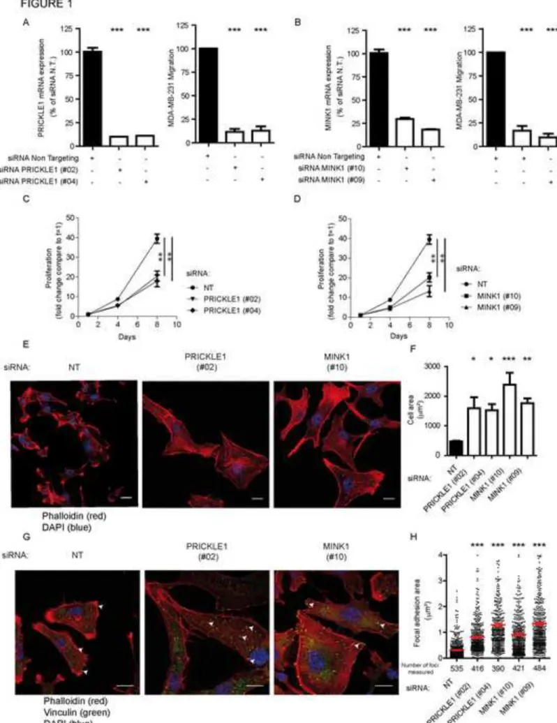
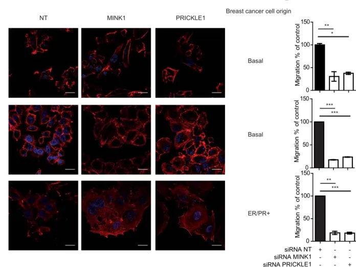
PRICKLE1 and MINK1 are involved in breast cancer cell migration 89
To investigate the role of PRICKLE1 in cancer cell motility, we knocked-down PRICKLE1 90
expression by two siRNAs in MDA-MB-231, a highly invasive basal breast cancer cell line 91
(Ahmed et al., 2012; Luga et al., 2012; Zhang et al., 2010) (Fig. 1A, left panel). 92
Downregulation was correlated with a strong decrease of cell migration in Boyden chamber 93
assays (Fig. 1A, right panel). Given that MINK1 binds to and phosphorylates PRICKLE1 94
(Daulat et al., 2012), we downregulated MINK1 in MDA-MB-231 cells (Fig. 1B, left panel) 95
and measured a strong decrease of cell motility (Fig. 1B, right panel). Downregulation of 96
PRICKLE1 and MINK1 also led to reduced cell proliferation (Fig. 1C, 1D), to increased cell 97
spreading (Fig. 1E) and to the formation of thick actin bundles (Fig. S1A). We also measured 98
the relative size of the cells using flow cytometry analysis based on forward scatter and we 99
observed an increase of the size of cells downregulated for PRICKLE1 and MINK1 compared 100
to cells treated with non-targeting siRNA (Fig. S1B). Both PRICKLE1- and MINK1-depleted 101
cells presented a flattened phenotype as judged by the larger diameter of the nucleus, an 102
increased cell surface (quantified in Fig. 1F), and the absence of lamellipodia, suggesting a 103
lack of cellular polarization. Using the same assays, we obtained similar results with two 104
other basal breast cancer cell lines (SUM159 and SKBR7) and one ER/PR-positive breast 105
cancer cell line (MCF7) (Fig. S2). We also stained MDA-MB-231 cells with anti-VINCULIN 106
antibody, a marker of focal adhesions (FAs), and phalloidin (Fig. 1G), and found larger FAs 107
in PRICKLE1- or MINK1- depleted cells which mainly localized at the tips of actin bundles 108
as compared to control cells (Fig. 1G). We measured the area of more than 390 FAs observed 109
in one experiment, which is representative of three independent experiments using two 110
independent siRNAs per gene (Fig. 1H). We found that downregulation of either PRICKLE1 111
or MINK1 increased the presence of active β1-integrin, a major component of FAs, at the cell 112
6
surface (Fig. S3A), which correlates with the presence of more mature FAs in Fig. 1G. 113
Biotinylation assays of cell surface proteins showed a decreased internalization of β1-integrin 114
in PRICKLE1- or MINK1-depleted cells (Fig. S3B and S3C). Our findings indicate that 115
PRICKLE1 and MINK1 regulate FA stabilization, integrin recycling and therefore actin 116
cytoskeleton organization and cell migration. 117
118
Interaction of PRICKLE1 with MINK1 and phosphorylation of PRICKLE1 are 119
required for cell migration 120
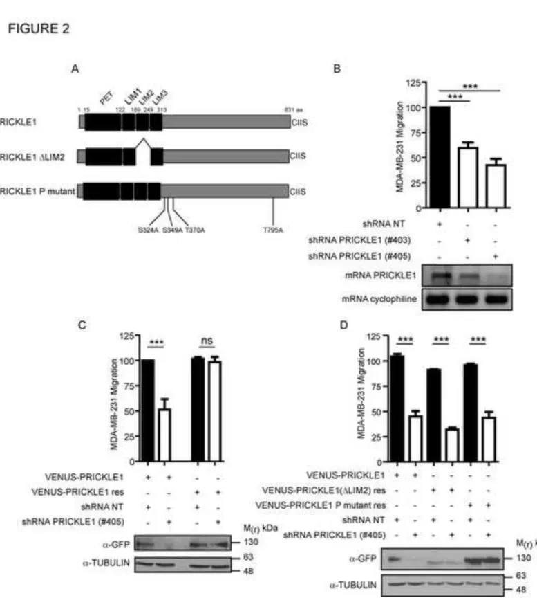
We previously showed that the second LIM domain of PRICKLE1 (LIM2) is required for the 121
interaction with MINK1 (Fig. 2A) (Daulat et al., 2012). We wondered whether the interaction 122
of PRICKLE1 with MINK1, and PRICKLE1 phosphorylation are important for cell 123
migration. We engineered MDA-MB-231 cells stably depleted for PRICKLE1 using two 124
independent shRNAs (Fig. 2B) and observed as with siRNAs (Fig. 1A) a decreased cell 125
migration proportional to knockdown efficiency (Fig. 2B). Cell migration was rescued by 126
expression of a VENUS-PRICKLE1 construct resistant to shRNA-PRICKLE1 (VENUS-127
PRICKLE1 res) (Fig. 2C). To determine the contribution of the interaction between 128
PRICKLE1 and MINK1 in cell migration, we used a similar rescue strategy and expressed a 129
version of PRICKLE1 (VENUS-PRICKLE1 ΔLIM2) unable to bind MINK1 or a version of 130
PRICKLE1 (VENUS-PRICKLE1 P mutant) resistant to MINK1 phosphorylation in MDA-131
MB-231 cells expressing shRNA-PRICKLE1 (Daulat et al., 2012). These two constructs were 132
unable to rescue cell motility of PRICKLE1-deficient MDA-MB-231 cells (Fig. 2D), 133
suggesting that both the interaction between PRICKLE1 and MINK1, and PRICKLE1 134
phosphorylation are required for cell motility. Expression of all constructs was confirmed by 135
western blot analysis (Fig. 2 B-D). 136
7
PRICKLE1 forms a complex with mTORC2 and MINK1 138
To characterize the signaling pathway associated with the MINK1-PRICKLE1 complex, we 139
generated a HEK293T cell expressing FLAG-PRICKLE1 and performed anti-FLAG 140
immunoprecipitation followed by mass spectrometry analysis. Among the identified proteins, 141
in addition to MINK1, we found that RICTOR, SIN1 and LST8, three members of the 142
mTORC2 complex, co-purified with PRICKLE1 (Fig. 3A). As RICTOR, SIN1 and LST8 are 143
associated with mTOR, a serine-threonine kinase responsible for the phosphorylation of AKT 144
(Sarbassov et al., 2005), we assessed its interaction with FLAG-PRICKLE1. Anti-FLAG 145
immunoprecipitation confirmed the presence of endogenous RICTOR and mTOR in the 146
PRICKLE1 complex (Fig. 3B). We also confirmed this interaction in MDA-MB-231 cells 147
stably expressing VENUS-PRICKLE1 (Fig. 3C). mTORC1 and mTORC2 are protein 148
complexes with distinct substrate specificities due to the presence of the RAPTOR and 149
RICTOR subunits, respectively. In HEK293T cells, FLAG-PRICKLE1 was able to co-150
immunoprecipitate with Myc-RICTOR, but not with Myc-RAPTOR, confirming the specific 151
interaction of PRICKLE1 with the mTORC2 complex. We then asked whether MINK1 152
belongs to the PRICKLE1-RICTOR complex. We immunopurified FLAG-MINK1 from 153
HEK293T cells and observed a weak but reproducible MINK1-RICTOR co-purification (Fig. 154
3E, lane 1), which was increased by overexpression of VENUS-PRICKLE1 (Fig. 3E, lane 2), 155
suggesting that the MINK1-RICTOR interaction is dependent on the presence of PRICKLE1. 156
To prove this, we downregulated PRICKLE1 with two independent siRNAs and looked for 157
the presence of RICTOR in immunoprecipitated FLAG-MINK1 (Fig. 3F). The interaction 158
between MINK1 and RICTOR (Fig. 3F, lanes 1 and 2) was decreased upon PRICKLE1 159
downregulation (Fig. 3F, lanes 3 and 4). We next asked if MINK1 modulates the interaction 160
between PRICKLE1 and RICTOR. We immunopurified a mutant form of PRICKLE1 lacking 161
its MINK1 interaction domain (PRICKLE1ΔLIM2) and observed a weaker RICTOR 162
8
interaction (Fig. 3G, compare lanes 2 and 3). Accordingly, overexpression of FLAG-MINK1 163
increased the interaction between VENUS-PRICKLE1 and endogenous RICTOR (Fig. 3H, 164
compare lanes 1 and 3). Expression of a kinase deleted MINK1 reduced this interaction (Fig. 165
3H, compare lanes 2 and 3). Finally, expression of VENUS-PRICKLE1 T370D, a 166
phosphomimetic mutant of PRICKLE1, in MDA-MB-231 cells led to an increased interaction 167
between PRICKLE1 and RICTOR compared to expression of VENUS-PRICKLE1 (Fig. 3I, 168
compare lanes 2 and 3). PRICKLE1 phosphorylation by MINK1 thus positively regulates the 169
interaction between PRICKLE1 and RICTOR. Like mTOR and RICTOR (McDonald et al., 170
2008), FLAG-MINK1 localized at the lamellipodia (Fig. 3J). Moreover, we observed 171
colocalization of RICTOR and VENUS-PRICKLE1 at the lamellipodia of MDA-MB-231 172
cells (Fig. 3K, upper panels). Downregulation of MINK1 led to delocalization of PRICKLE1 173
from the cell cortex as previously shown in Xenopus cells (Daulat et al., 2012), as well as that 174
of RICTOR (Fig. 3K, lower panels). We quantified the decreased PRICKLE1/RICTOR 175
colocalization in the absence of MINK1 by counting the cells harboring cytoskeleton 176
modification seen in Fig. 1G as a marker of MINK1 downregulation (Fig. 3L and Fig. 3M). 177
Altogether these data suggest evidence that MINK1 controls PRICKLE1-dependent 178
recruitment of the mTORC2 complex at the leading edge of migratory cells where it regulates 179
actin cytoskeleton organization (Fig. 3N). 180
181
Stability of focal adhesions is increased by targeting the PRICKLE1-mTORC2 complex 182
Previous works had shown the involvement of MINK1 and mTORC2 in the modulation of 183
actin cytoskeleton organization (Hu et al., 2004; Lamouille et al., 2012; Sarbassov et al., 184
2004). Consistent with these studies, we found that downregulation of RICTOR in MDA-MB-185
231 cells led to actin cytoskeleton reorganization (Fig. S1A), decreased cell migration and 186
increased of cell spreading, and FA area (Fig. S4A-F), increase of relative cell size using flow 187
9
cytometry (Fig. S1B), and alteration of integrin endocytosis (Fig. S3), as observed for 188
PRICKLE1 and MINK1 downregulation (Fig. 1 and S3). Migratory cells have two major 189
populations of FAs, nascent and mature, when adhering to extracellular matrices. Nascent 190
FAs present in the lamellipodia are highly dynamic and short lived. Some of these nascent 191
FAs mature following binding to actin filaments and ROCK activation, and appear as stable 192
patches at the plasma membrane (Burridge and Chrzanowska-Wodnicka, 1996; Parsons et al., 193
2010). As depletion of PRICKLE1 or MINK1 increases the proportion of large, potentially 194
mature, FAs in MDA-MB-231 cells (Fig. 1G), we monitored the stability of FAs by Total 195
Internal Resonance Fluorescence (TIRF) microscopy, a well-suited method for the analysis of 196
dynamic events at the plasma membrane. MDA-MB-231 cells stably expressing VENUS-197
PAXILLIN, a marker of FAs (Ahmed et al., 2012), seeded on collagen were downregulated 198
for PRICKLE1, MINK1 or RICTOR and the FA patches were followed over a period of 11 199
minutes. Fig. 4A (upper panels) shows a typical image of migratory MDA-MB-231 cells in 200
which FAs were localized every minute from 1 minute (red) to 11 minutes (purple) and each 201
FA was tracked and processed by a Matlab-based tracking software (Salles et al., 2013) (Fig. 202
4A, lower panels). Stable (mature) FAs were circled in white and were present throughout the 203
11 min acquisition. Transient FAs were considered unstable (nascent, circled in black) since 204
they appear or disappear within 11 min. Cells transfected with a control siRNA (siRNA NT) 205
had 20±3% of stable FAs whereas the proportion of stable FAs rose in MINK1- (34± 2%), 206
PRICKLE1- (38± 4%) and RICTOR- (32± 4%) depleted cells (Fig. 4B). We also monitored 207
the disassembly of FAs by determining the half-life of these structures: downregulation of 208
PRICKLE1, MINK1 and RICTOR led to stabilization of FAs whose half-life was increased 209
from 6.9 min (siRNA NT) to 8.2 min (siRNA PRICKLE1 or siRNA MINK1) and to 7.6 min 210
(siRNA RICTOR) (Fig. 4C). We also assessed PAXILLIN phosphorylation at Tyrosine 118, 211
a marker of mature FAs (Schaller and Parsons, 1995). Increased phosphorylation was 212
10
observed in PRICKLE1, MINK1 or RICTOR depleted cells confirming the conclusion of the 213
TIRF experiments (Fig. 4D). Our results reveal that the PRICKLE1-MINK1-RICTOR 214
complex controls cell migration of cancer cells by interfering with FA dynamics. 215
216
The PRICKLE1 complex regulates mTORC2 activity 217
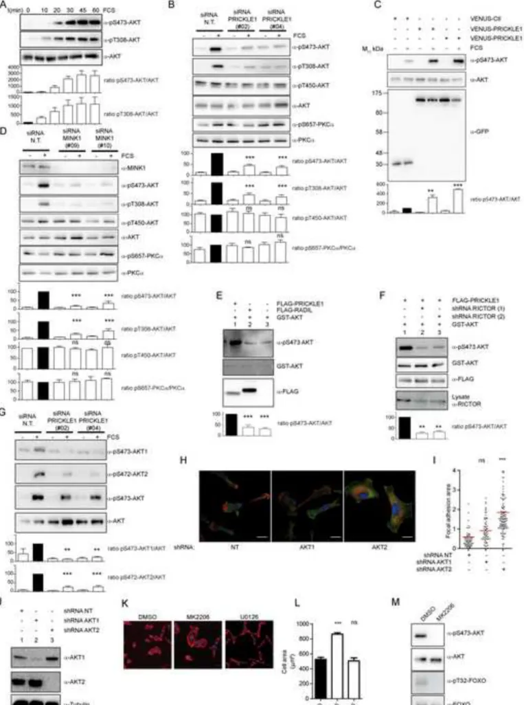
As AKT is a substrate for mTORC2, we asked whether PRICKLE1 contributes to AKT 218
phosphorylation. In MDA-MB-231 cells, AKT phosphorylation monitored using specific anti-219
phosphoSerine 473 (pS473-AKT) and anti-Threonine 308 (pT308-AKT) antibodies was 220
maximal after 30 min of serum treatment (Fig. 5A). AKT phosphorylation at both sites was 221
decreased upon PRICKLE1 downregulation (Fig. 5B). We did not observe an obvious 222
modification of phosphorylation of AKT at Threonine 450 or PKCα at Serine 657 suggesting 223
site- and substrate-specificities of the PRICKLE1-mTORC2 complex. Because the 224
phosphomimetic mutant of PRICKLE1 (T370D) improved PRICKLE1-RICTOR association 225
(Fig. 3I), we examined its effect on AKT phosphorylation. Although overexpression of 226
PRICKLE1 increased AKT phosphorylation at Serine 473 compared to the control, 227
overexpression of PRICKLE1 T370D was slightly more potent (Fig. 5C). We next examined 228
to what extent MINK1 contributes to AKT phosphorylation and found a strong reduction of 229
AKT phosphorylation in the absence of MINK1 (Fig. 5D). As for PRICKLE1 knockdown 230
(Fig. 5B), phosphorylation of AKT on Threonine 450 or of PKCα at Serine 657 was not 231
altered by MINK1 depletion (Fig. 5D). Cell motility of the highly invasive MDA-MB-231 232
cells was not increased by overexpressing either PRICKLE1 or PRICKLE1 T370D (data not 233
shown). We thus used SKBR7, another basal breast cancer cell line expressing lower levels of 234
PRICKLE1 (2.75 fold less PRICKLE1 mRNA levels than MDA-MB-231 cells as measured 235
by Q-PCR) and presenting lower invasive properties. Downregulation of PRICKLE1 and 236
MINK1 decreased SKBR7 cell motility and led to actin skeleton reorganization (Fig. S2), and 237
11
decreased AKT phosphorylation (Fig. S5A). Conversely, overexpression of PRICKLE1 or 238
PRICKLE1 T370D increased AKT phosphorylation, cell motility and cell proliferation of 239
SKBR7 cells (Fig. S5B-D). Obviously, PRICKLE1 T370D was more potent than wild type 240
PRICKLE1 in these assays. These results provide evidence of the importance of PRICKLE1 241
in mediating mTORC2 activity and function. 242
We next purified FLAG-PRICKLE1 from HEK293T cells and performed in vitro kinase 243
assays using recombinant GST-AKT as a substrate. The PRICKLE1 complex promoted AKT 244
phosphorylation (Fig. 5E, lane 1) in comparison with a control RADIL complex (Fig. 5E, 245
lane 2). To test whether this event was due to the presence of the mTORC2 complex 246
associated with PRICKLE1, we downregulated RICTOR expression using two independent 247
shRNAs (Fig. 5F). Purification of the PRICKLE1 complex from these cells and incubation 248
with recombinant GST-AKT reduced AKT phosphorylation (Fig. 5F, lanes 2 and 3). The 249
PRICKLE1 complex thus contributes to AKT phosphorylation in living cells and in vitro. 250
MDA-MB-231 cells express two AKT forms (AKT1 and AKT2) whose phosphorylation was 251
decreased by PRICKLE1 downregulation (Fig. 5G). However, only AKT2 knockdown could 252
recapitulate the phenotype observed in PRICKLE1-depleted cells (actin cytoskeleton 253
reorganization, cell spreading and stabilization of FAs as revealed by vinculin staining) (Fig. 254
5H, Fig. 5I) although both AKT1 and AKT2 were efficiently depleted by specific shRNAs 255
(Fig. 5J). We also looked at the effects of specific inhibitors of AKT (MK2206) and MEK 256
(U0126) on MDA-MB-231 cells. With MK2206 (Fig. 5K, 5L), we observed increased cell 257
spreading and a remodeling of the cytoskeleton with the formation of thick actin bundles (Fig. 258
S1D) which phenocopied the results obtained upon PRICKLE1 or MINK1 downregulation 259
(Fig. 1G, Fig. 1H, Fig. S1). No such effects were found with U0126. However, in contrast to 260
the observation made with siRNAs directed against PRICKLE1, MINK1 and RICTOR (Fig. 261
S1B), we did not observe alteration of cell size under MK2206 treatment compare to control 262
12
by flow cytometry (Fig. S1C). The efficiency of the chemical treatments was assessed by 263
western blot using phosphorylation of AKT and of FOXO, a known AKT substrate, as 264
readouts (Fig. 5M). We thus conclude that PRICKLE1 can promote AKT phosphorylation 265
through its interaction with MINK1 and mTORC2. 266
267
Interaction between PRICKLE1 and mTORC2 is required for AKT phosphorylation 268
and cell migration 269
We next determined the role of the PRICKLE1-mTORC2 interaction in AKT phosphorylation 270
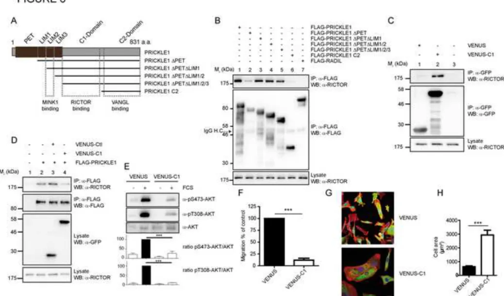
and cell migration. We generated deleted versions of PRICKLE1 lacking the PET and/or the 271
LIM domains and a construct encompassing the PRICKLE1 C-terminus region only 272
(PRICKLE C2) (Fig. 6A). In HEK293T cells, we found that the PET and LIM domains were 273
dispensable for the interaction, and that the PRICKLE C2 construct was not able to bind 274
RICTOR (Fig. 6B). The region between the LIM3 domain and the PRICKLE C2 region is 275
thus likely involved in the interaction with RICTOR. We fused this region (hereafter named 276
C1 domain) to VENUS (VENUS-C1) and transiently expressed the construct in HEK293T 277
cells. VENUS-C1, but not VENUS, was able to co-immunoprecipitate with endogenous 278
RICTOR (Fig. 6C). We next expressed FLAG-PRICKLE1 together with VENUS or VENUS-279
C1 in HEK293T cells and found that expression of VENUS-C1 led to a strong decrease of 280
PRICKLE1-RICTOR interaction, highlighting its dominant-negative effect (Fig. 6D). 281
Overexpression of VENUS-C1 in MDA-MB-231 cells led to a robust inhibition of AKT 282
phosphorylation (Fig. 6E) and cell migration (Fig. 6F). In addition, expression of VENUS-C1 283
phenocopied the increase of cell spreading (Fig. 6G, 6H) and actin cytoskeleton 284
reorganization (Fig. S1E) previously observed by downregulating PRICKLE1 (Fig. 1E, 285
S1A). In conclusion, disruption of the PRICKLE1-RICTOR interaction decreases AKT 286
13
phosphorylation and cell migration similarly to depletion of the PRICKLE1 complex 287
components. 288
289
The MINK1-PRICKLE1-RICTOR complex plays a role in tumor growth and metastatic 290
dissemination 291
We next aimed to assess the in vivo contribution of the PRICKLE1 complex in cancer cell 292
spreading. PRICKLE1 and RICTOR have been linked to tumor progression and dissemination 293
using MDA-MB-231 cells in xenograft experiments (Luga et al., 2012) (Zhang et al., 2010). 294
No such data were available for MINK1. We generated luciferase-positive MDA-MB-231 295
cells stably downregulated for MINK1 expression (Fig. S6A) and observed a decrease of cell 296
motility (Fig. S6B) and cell proliferation (Fig. S6C) as was observed with siRNAs (Fig. 1B, 297
1D). In orthotopic transplantation assays into the mammary fat pad of NOD/SCID/γc mice, 298
we found, after 33 days, a decrease of the primary tumor volume with MINK1-deficient 299
MDA-MB-231 cells compare to control cells (Fig. S6D). Whereas MDA-MB-231 control 300
cells (shRNA NT) invaded the lungs (Fig. S6E) and the livers (Fig. S6G), we observed a 301
lower invasion rate (fewer metastases or no metastases in the lungs and in the livers, see 302
quantification of bioluminescence in Fig. S6F and Fig. S6H) with MINK1-deficient MDA-303
MB-231 cells. Taken together, these in vivo data demonstrate that, like PRICKLE1 (Luga et 304
al., 2012) and RICTOR (Zhang et al., 2010), MINK1 is involved in the metastatic 305
dissemination of MDA-MB-231 cells. 306
We next addressed the role of the PRICKLE1-RICTOR interaction in cancer dissemination in 307
vivo using VENUS or VENUS-C1 in MDA-MB-231 cells stably expressing firefly luciferase. 308
VENUS-C1 expression decreased cell proliferation (Fig. 7A) and AKT phosphorylation (data 309
not shown), and led to compromised in vivo tumor growth (Fig. 7B). Metastasis formation 310
was observed in the lungs of all mice of the control group (7 out of 7 mice), whereas only 3 311
14
out of 8 mice developed lung metastases when VENUS-C1 was expressed (Fig. 7C, 7E). 312
Comparable results were obtained when considering metastasis in the liver (7 out of 7 mice 313
for VENUS as compared to 2 out of 8 mice for VENUS-C1) (Fig. 7D, Fig. 7F). We also 314
performed tail vein injections of luciferase-positive MDA-MB-231 cells expressing dominant-315
negative PRICKLE1 VENUS-C1 (vs VENUS alone) or shRNA MINK1 (vs shRNA control), 316
and monitored the dissemination of cancer cells in the mice. We observed that, upon 317
intravenous injection, MDA-MB-231 cells populate the lungs and that, after 2 or 3 weeks, 318
total luminescence (mostly in lungs) was statistically lower in mice injected with cells 319
transfected with VENUS-C1 (Fig. 7G, Fig. 7H) or shMINK1 (Fig. S6I, Fig. S6J) compared 320
to controls. This phenotype is likely the result of a combinatory decrease of tumor growth and 321
metastatic potential of MDA-MB-231 cells. In conclusion, we have demonstrated the 322
importance of the PRICKLE1-RICTOR association in tumor growth and metastatic 323
dissemination in vivo. 324
325
PRICKLE1 upregulation correlates with poor prognosis in basal breast cancer 326
Breast cancer is classified in five major intrinsic molecular subtypes (luminal A and B, basal, 327
ERBB2-enriched, normal-like) (Perou et al., 2000; Sorlie et al., 2001). Breast cancer cell lines 328
are classified in luminal, basal or mesenchymal subtypes. We analyzed PRICKLE1 mRNA 329
expression in publicly-available datasets of 45 breast cancer cell lines and 5,410 primary 330
invasive breast carcinomas. Basal/mesenchymal cell lines (N=19) showed higher PRICKLE1 331
expression level than luminal cell lines (N=17; p=1.78E-02, Student t-test; Fig. 7I), the 332
expression levels being similar between basal and mesenchymal cell lines (p=0.95, Student t-333
test; data not shown). In clinical samples, PRICKLE1 expression level, measured as 334
continuous value, was also higher in basal breast cancers than in luminal A, luminal B and 335
ERBB2-enriched breast cancers (Fig. S7A, Fig. 7J; Supplementary Table 1). We then 336
15
searched for correlation between PRICKLE1 expression (as binary variable) and 337
clinicopathological features of breast cancers, including metastasis-free survival (MFS). 338
Within the 5,410 breast cancer samples analyzed, 860 tumors (16%) showed PRICKLE1 339
upregulation when compared to normal breast (ratio T/NB ≥2; “PRICKLE1-up” group), and 340
4,550 (84%) did not show any upregulation (ratio <2; “PRICKLE1-no up” group). 341
Correlations were found between “PRICKLE1” status and age of the patient (p<0.001), 342
pathological type (p<0.001), axillary lymph node status (p=0.049), and grade (p=0.007), ER 343
status (p<0.001), PR status (p<0.001), ERBB2 status (p=0.009), and molecular subtypes 344
(p<0.001) (Supplementary Table 2). MFS data were available for 1,037 patients, including 345
613 who remained metastasis-free during a median follow-up of 83 months and 424 who 346
displayed metastatic relapse. The 5-year MFS rate was 61% [95CI, 58-65]. In the whole 347
population, PRICKLE1 upregulation was not associated with MFS (p=0.49, log-rank test). 348
The same analysis was done in each molecular subtype separately. As shown in Fig. 7K, in 349
the basal subtype, PRICKLE1 upregulation was associated with shorter 5-year MFS (41% 350
[95CI, 31-55] when compared with the “PRICKLE1-no up” group (61%, [95CI, 54-68]; 351
p=0.008; log-rank test). By contrast, no correlation with MFS was seen in the luminal A, 352
luminal B, ERBB2-enriched, and normal-like subtypes (data not shown). We next used 353
publicly available data sets to look for a possible correlation between PRICKLE1 mRNA 354
expression and AKT activation. Among the 534 breast cancer samples from the TCGA 355
dataset (https://tcga-data.nci.nih.gov/tcga/), 368 had available data from reverse-phase protein 356
array (RPPA), allowing us to assess the expression of both AKT and activated AKT (pS473 357
and pT308 phosphorylated forms) and their correlation with PRICKLE1 mRNA expression. 358
In the whole series of 368 samples, AKT expression was not associated with PRICKLE1 359
expression, whereas expression of each phosphorylated form was (pS473, p<0.001; pT308, 360
p<0.001; Fig. S7B). The same analysis limited to the basal subtype (N=85) showed similar 361
16
correlations, with higher expression of phosphorylated AKT in the PRICKLE1-up samples 362
than in the “PRICKLE1-no up” (pS473, p=0.015; pT308, p<0.001; Fig. 7L), suggesting a 363
positive correlation between PRICKLE1 expression and AKT activation. Interestingly, in 364
basal breast cancers, PRICKLE1 expression was also correlated to levels of phosphorylated 365
forms of FOXO3A and PRAS40, two downstream components of AKT signaling (Fig. 7L), 366
but not to phosphorylated MAPK and JNK (Fig. S7C). Together these data show 367
overexpression of PRICKLE1 in basal breast cancer, its correlation with poor prognosis and 368
AKT signaling in this molecular subtype. 369
17 Discussion
370
Components of the evolutionary conserved group of developmental PCP genes play a pivotal 371
role in several aspects of cancer progression including cell proliferation, epithelial-372
mesenchymal transition, cell migration and metastatic development. Overexpressed WNT5A 373
in breast cancer and melanoma contributes to enhanced cell migration and resistance to 374
treatment (Anastas et al., 2014; Dissanayake et al., 2007; Pukrop et al., 2006). Deregulation of 375
SCRIBBLE was observed in breast (Anastas et al., 2012) and prostate cancers (Pearson et al., 376
2011), and found associated with increased cell proliferation and cell migration (Anastas et 377
al., 2012; Belotti et al., 2013). VANGL1 and VANGL2 are involved in cell motility, 378
proliferation and dissemination in breast cancer (Anastas et al., 2012; Belotti et al., 2013; 379
Luga et al., 2012; Puvirajesinghe et al., 2016). In addition, overexpression of SFRP1, an 380
inhibitor of the PCP pathway, limits cancer cell motility (Matsuda et al., 2009). Recently, high 381
levels of FRIZZLED2 and its ligand WNT5A were found in metastatic liver, lung, colon and 382
breast cancers (Gujral et al., 2014). This upregulation was correlated with epithelial-383
mesenchymal transition and poor survival, and it was suggested that targeting this pathway 384
may benefit to cancer patients. At the molecular level, alterations of PCP components in 385
cancer lead to increased activation of important cell signaling pathways such as the RAS-386
MAPK and Hippo pathways in the case of SCRIBBLE defects (Cordenonsi et al., 2011; Dow 387
et al., 2008) or the FYN-STAT3 pathways for WNT5A-FRIZZLED up-regulation (Gujral et 388
al., 2014). 389
Here we have studied PRICKLE1, a PCP component first described in Drosophila (Gubb and 390
Garcia-Bellido, 1982), known for its involvement in the metastatic potential of MDA-MB-231 391
breast cancer cell line (Luga et al., 2012). We have shown that PRICKLE1 and its partner 392
MINK1, as well as their interaction, are involved in cancer cell proliferation, migration and 393
actin cytoskeleton organization (Fig. 1, Fig. 2). In agreement with a previous report (Luga et 394
18
al., 2012), we observed that PRICKLE1 localizes at the leading edge of MDA-MB-231 cells 395
and provided evidence that this is a MINK1-dependent process (Fig. 3K). Previous studies 396
have shown that VANGL1 and VANGL2, two binding partners of PRICKLE1, are also 397
located at the leading edge of migratory cancer cells (Anastas et al., 2012; Belotti et al., 2013; 398
Luga et al., 2012). 399
We next purified PRICKLE1-associated partners by a proteomic approach and identified 400
RICTOR as an interactor (Fig. 3). RICTOR belongs to the mTORC2 complex that contains 401
mTOR (Jacinto et al., 2004; Sarbassov et al., 2004), which we found associated with 402
PRICKLE1. This finding highlights a connection between PRICKLE1, MINK1 and the 403
mTORC2 complex, which is important for AKT phosphorylation (Sarbassov et al., 2005), 404
actin cytoskeleton remodeling and cell migration (Jacinto et al., 2004; Sarbassov et al., 2004). 405
Two recent studies have demonstrated a connection between PCP core components, mTORC2 406
pathway, and AKT activation in melanoma and in muscles (Anastas et al., 2014; von 407
Maltzahn et al., 2012). We show that association of the mTORC2 complex with PRICKLE1 is 408
required for phosphorylation of AKT on Serine S473 in cells and in vitro (Fig. 5). 409
Phosphorylation of AKT at Serine 473 and Threonine 450 are required for signaling and 410
stability of AKT, respectively (Facchinetti et al., 2008). We did not found alteration of 411
phosphorylation of AKT at Threonine 450 and PKCα at Serine 657 when the expression of 412
PRICKLE1 or MINK1 was lost (Fig. 5B and 5D) suggesting a role of the PRICKLE1 413
complex in AKT-mediated signaling. The involvement of MINK1 and mTORC2 in actin 414
cytoskeleton remodeling (Hu et al., 2004; Jacinto et al., 2004; Sarbassov et al., 2004) fits well 415
with our observation that these components, as well as PRICKLE1, play a role on FA 416
dynamics (Fig. 1, Fig. 4). We cannot exclude a role for RICTOR in cell migration 417
independently of mTORC2 as shown by others (Agarwal et al., 2013; Zhang et al., 2010). 418
However, downregulation of AKT2 and treatment with MK2206 recapitulated partially the 419
19
phenotypes we observed with loss of PRICKLE1, MINK1 and RICTOR expression (Fig. 5H, 420
Fig. 5K) suggesting a prominent role of AKT in the PRICKLE1-dependent pathway. 421
However, the fact that chemical inhibition of AKT (MK2206 treatment) did not change the 422
cell size by flow cytometry (Fig. S1C) and was less efficient than the absence of MINK1, 423
PRICKLE1, and RICTOR on cell spreading (Fig. 1E, F and Fig. 5K, L) cannot rule out the 424
additional implication of AKT-independent signaling events. It is also intriguing that whereas 425
both AKT1 and AKT2 phosphorylation was decreased by PRICKLE1 downregulation (Fig. 426
5G), only AKT2 loss gave rise to a PRICKLE1-like phenotype (Fig. 5H). Our study supports 427
the conclusion drawn by others about non-overlapping functions of AKT1 and AKT2 (Chin 428
and Toker, 2009). As RICTOR localized at the leading edge of MDA-MB-231 cells (Fig. 3K) 429
(McDonald et al., 2008), together with PRICKLE1 in a MINK1-dependent manner, we 430
propose a model whereby MINK1 controls PRICKLE1-mTORC2 localization at the leading 431
edge of migrating cells, a step necessary for local AKT phosphorylation by mTORC2 (Fig. 432
3N). We were not able to rescue loss of cell migration and cell proliferation of MINK1-433
depleted cells by overexpression of PRICKLE1 or PRICKLE1 T370D. MINK1 is thus 434
required for PRICKLE1 functions, a result in agreement with our conclusion that interaction 435
between MINK1 and PRICKLE1 (and its phosphorylation by MINK1) represents a key step 436
for PRICKLE1 functions. Because MINK1 has probably other substrates than PRICKLE1, it 437
remains important to further study the mode of action of this protein kinase. 438
Novel AKT substrates such as Cortactin, a protein implicated in cell migration and invasion 439
(Wu et al., 2014), could be implicated in the control of actin cytoskeleton by the PRICKLE1-440
mTORC2 complex. Indeed, Cortactin binds to the ARP2/3 complex and regulates actin 441
branching, polymerization during cell motility and FA dynamics (MacGrath and Koleske, 442
2012; Tomar et al., 2012). Previous work has shown that downregulation of mTOR and 443
RICTOR leads to reorganization of actin cytoskeleton (Sarbassov et al., 2004) and FAs 444
20
(Lamouille et al., 2012) in migrating cells. We confirmed these findings using an AKT 445
inhibitor and obtained similar effects by downregulation of the PRICKLE1 complex 446
components (Fig. 1, Fig. 5). To further determine the involvement of PRICKLE1 and its 447
binding partners in FA dynamics, we used TIRF experiments and observed that PRICKLE1, 448
MINK1 and RICTOR play a prominent role in FA stability (Fig. 4). Previous work has found 449
that RICTOR is associated with integrins and regulates AKT phosphorylation (McDonald et 450
al., 2008). Downregulation of PRICKLE1, MINK1 or RICTOR led to a defect of integrin 451
internalization and polarization (Fig. S3) linked to altered FA dynamics and decreased cell 452
migration (Fig. 1, Fig. 4) Interestingly, the Drosophila MINK1 ortholog, Misshapen (Msn), 453
similarly regulates integrin level at the cell surface which probably explains its promigratory 454
function (Lewellyn et al., 2013). A connection exists between the PCP pathway and FA 455
dynamics (Cui et al., 2013; Matsumoto et al., 2010; Wei et al., 2013). Indeed, WNT5A 456
promotes localization of FRIZZLED2 at the leading edge of migrating cells and triggers FAK 457
and PAXILLIN phosphorylation (Matsumoto et al., 2010). Furthermore, SYNDECAN4, a 458
membrane protein with PCP functions associated with FRIZZLED7 (Munoz et al., 2006) and 459
VANGL2 (Escobedo et al., 2013), regulates the formation of FAs through engagement of 460
α5β1 integrin (Saoncella et al., 1999) and, in endothelial cells, regulates mTORC2 461
localization and AKT (Partovian et al., 2008). As PRICKLE1 and MINK1 control 462
internalization and recycling of PCP receptors by a RAB5-dependent mechanism (Daulat et 463
al., 2012), we thus hypothesize that the PRICKLE1 complex could, directly or indirectly, play 464
such a role on integrins or other FA-associated molecules. Future studies will have to 465
determine how the PRICKLE1 complex regulates the functions of integrins. 466
We identified a region (C1) in PRICKLE1 required for its interaction with RICTOR and 467
showed that it behaves as a dominant-negative construct in vitro and in vivo (Fig. 6 and Fig. 468
7) (Tao et al., 2009). Our (Fig. S6) and previously published (Luga et al., 2012) (Zhang et al., 469
21
2010) data show that knockdown of MINK1, RICTOR or PRICKLE1 reduces the capacity of 470
breast cancer cells to invade secondary organs in xenograft experiments. In our previous 471
report, we concluded that only MINK1, and not its paralogs TNIK or MAP4K4, was 472
associated with PRICKLE1 (Daulat et al., 2012). MINK1 has been previously shown to 473
regulate HT1080 cell motility (Hu et al., 2004), a role apparently conserved during evolution 474
(Houalla et al., 2005). We propose a molecular mechanism whereby PRICKLE1 specifically 475
associates with MINK1, and contributes to cell motility in vitro and in vivo. 476
Using a large dataset of clinical primary invasive breast cancers, we found that PRICKLE1 is 477
overexpressed in basal breast cancers where its up-regulation is correlated with poor 478
prognosis (Fig. 7I-L, Fig. S7). We also found that PRICKLE1 mRNA upregulation in basal 479
breast cancers correlates with increased phosphorylation of AKT and its downstream 480
components (FOXO and PRAS40), but not with phosphorylated MAPK and JNK, which 481
nicely confirms our results obtained in cultured cells. No alteration of MINK1 mRNA 482
expression was found in this series (data not shown). Targeted therapy has allowed a 483
significant improvement of patient outcome in breast cancers expressing ERBB2 or hormone 484
receptors. This is not the case for the basal subtype characterized by a high degree of cell 485
proliferation, metastatic development, and frequent relapse. Major efforts are thus required to 486
uncover novel markers and therapeutic targets for this poor prognosis cancer to improve 487
patients care. Targeting the AKT-mTOR pathway is a potential option to treat basal breast 488
cancers (Tao et al., 2014; Yunokawa et al., 2012). In our study, we have shown that inhibition 489
of PRICKLE1, MINK1 or RICTOR expression as well as disruption of the PRICKLE1-490
RICTOR interaction decrease cell migration and tumor growth. Our findings may pave the 491
way to strategies able to inhibit or disrupt this complex that may benefit cancer patients. 492
22 Acknowledgements
493
The authors wish to thank Valérie Ferrier, Michael Sebbagh and Eric Bailly for critical review 494
of the manuscript and people from the cell imaging, cell sorting and animal house facilities. 495
This work was funded by La Ligue Nationale Contre le Cancer (Label Ligue JPB and DB, and 496
post-doctoral fellowship to AMD), Fondation de France (post-doctoral fellowship to AMD), 497
Fondation ARC pour la Recherche sur le Cancer (grant to JPB and AS), and SIRIC (INCa-498
DGOS-Inserm 6038, fellowship to AMD). The Marseille Proteomics (IBiSA) and TrGET 499
platforms are supported by Institut Paoli-Calmettes (IPC) and Canceropôle PACA. Samples 500
of human origin and associated data were obtained from the IPC/CRCM Tumor Bank that 501
operates under authorization # AC-2013-1905 granted by the French Ministry of Research. 502
Prior to scientific use of samples and data, patients were appropriately informed and asked to 503
express their consent in writing, in compliance with French and European regulations. The 504
project was approved by the IPC Institutional Review Board. Jean-Paul Borg is a scholar of 505
Institut Universitaire de France. 506
23 Figure Legends:
508
Figure 1: PRICKLE1 and MINK1 are involved in cell migration and cell proliferation of 509
MDA-MB-231 cells. A. Loss of PRICKLE1 mRNA expression was confirmed by quantitative 510
PCR (left panel). Transfected cells were subjected to a cell migration assay using Boyden 511
chambers (right panel). B. Same as (A) using two independent siRNAs targeting MINK1. C-512
D. PRICKLE1 (C) and MINK1 (D) play a role in cell proliferation. Cell proliferation was 513
measured at the indicated times. E. Cell spreading of MDA-MB-231 treated with the 514
indicated siRNA. F. Quantification in (E). Cell spreading were measured using ImageJ 515
software. G. Same as (E) H. Sizes of focal adhesions were measured using vinculin staining 516
and ImageJ software analysis. One way ANOVA with Tukey post-test statistical analysis. *P 517
<0.05; **P<0.01; ***P<0.001. Data are presented as means ±SEM. Scale bars are 20 µM. See 518
also Figures S1, S2, S3. 519
520
Figure 2: Interaction of PRICKLE1 with MINK1 is required for cell migration. A. 521
Schematic representation of PRICKLE1. Phosphorylation (S324, S349, T370 and T795) and 522
the carboy-terminal farnesylation (CIIS) sites are shown. B. Boyden chamber assays using 523
MDA-MB-231 cells (top panel). Downregulation of PRICKLE mRNA was confirmed by 524
semi-quantitative PCR (bottom panel). C. Rescue of PRICKLE1 expression in MDA-MB-231 525
cells followed by Boyden chamber assay. D. MDA-MB-231 cells stably expressing the 526
indicated PRICKLE1 mutants were subjected to shRNA treatment targeting PRICKLE1 527
(#405) and subjected to Boyden chamber assays. Protein expression was evaluated by western 528
blot with anti-GFP antibody. B-D. One way ANOVA with Tukey post-test statistical analysis. 529
***P<0.001. Data are presented as means ±SEM. 530
24
Figure 3: PRICKLE1 forms a complex with RICTOR and mTOR. A. Schematic 532
representation of the protein complex associated to PRICKLE1. Total Spectral Counts (TSCs) 533
is given for each member of the PRICKLE1 purified protein complex. B. In 293T cells, 534
FLAG-PRICKLE1 is associated with endogenous RICTOR and mTOR. C. MDA-MB-231 535
cells were transfected with VENUS-Ctrl or VENUS-PRICKLE1. After anti-GFP 536
immunoprecipitation, bound proteins were revealed with the indicated antibodies. D. In 293T 537
cells, PRICKLE1 was associated with RICTOR but not RAPTOR. E. In 293T cells, the 538
interaction between MINK1 and RICTOR is enhanced when PRICKLE1 is overexpressed. F. 539
In 293T cells, downregulation of PRICKLE1 leads to a decreased association between 540
MINK1 and RICTOR. G. Lower interaction between RICTOR and PRICKLE1 was observed 541
in the absence of MINK1. H. RICTOR is associated to PRICKLE1 in the presence of MINK1 542
but not MINK1-KD. I. In MDA-MB-231 cells, endogenous RICTOR was more associated 543
with PRICKLE1 T370D than with PRICKLE1. J. FLAG-MINK1 localizes at the cell cortex 544
in MDA-MB-231 cells. K. Colocalization of VENUS-PRICKLE1 and RICTOR at the leading 545
edge of MDA-MB-231 cells (scale bars are 20 µM). Quantification of cortical colocalization 546
of PRICKLE1 and RICTOR and western blot analysis to confirm siRNA MINK1 efficiency 547
are provided in L and M, respectively. N. Model of mTORC2 recruitment at the plasma 548
membrane by PRICKLE1. 549
550
Figure 4: Stability of focal adhesions is increased by targeting of the PRICKLE1-551
mTORC2 complex. A. MDA-MB-231 cells stably expressing VENUS-PAXILLIN were 552
treated with the indicated siRNAs. Top images: rainbow pictures are built by summing up and 553
assigning a different color for every time-point, from red to purple (see color-bar). Stable 554
adhesions appear in white, while dynamic adhesions appear as a pattern only partially 555
depicting the rainbow colors. Bottom images: after tracking, focal adhesion trajectories are 556
25
represented using the same color scheme as for encoding time. Stable adhesions, tracked over 557
the entire time-lapse movie, are circled in white, while unstable adhesions, tracked only over a 558
subset of frames, are circled in black. For each condition and in both panels, representative 559
stable and unstable adhesions are denoted by white and black arrowheads, respectively. B. 560
Statistical analysis representing the percentage of stable and unstable focal adhesions for each 561
siRNA treatment. C. Disassembly rates measured for each focal adhesion under the indicated 562
siRNA treatment. The line inside the box plot represents the median and the + sign represents 563
the mean. Student t test two tailed with 95% confidence of interval. ***P<0.001. Total 564
number (n) of focal adhesion measured is between 741 and 4156, recorded in 18 to 49 565
movies, from at least 3 independent experiments. D. Western blot analysis (anti-phospho-566
PAXILLIN) of MDA-MB-231 cells treated with the indicated siRNAs. See also Figure S4. 567
568
Figure 5: The PRICKLE1 complex phosphorylates AKT through mTORC2. A. Kinetics 569
of AKT phosphorylation in MDA-MB-231 cells stimulated with 5% FCS for the indicated 570
times. B. and D. PRICKLE1 (B) and MINK1 (D) contribute to AKT phosphorylation 571
(treatment: 5% FCS during 30 min). C. Expression of PRICKLE1 and VENUS-572
PRICKLE1 potentiates AKT phosphorylation in MDA-MB-231 cells (treatment: 5% FCS 573
during 10 min). E. In vitro kinase assays were performed on anti-FLAG 574
immunoprecipitations of HEK293T cell extracts. Phosphorylation at Serine 473 of 575
recombinant AKT was evaluated by western blot. F. Same as E. Decreased expression of 576
RICTOR impaired AKT phosphorylation by the PRICKLE1 complex. G. Same as B. 577
PRICKLE1 contributes to AKT1 and AKT2 phosphorylation. H. AKT1 or AKT2 expression 578
was downregulated in MDA-MB-231 cells as confirmed by western blot (J) and focal 579
adhesion area were measured using ImageJ software. Quantification is represented in (I). K. 580
MDA-MB-231 cells seeded on collagen-coated coverslips were treated with the indicated 581
26
inhibitors for 16 hours. Polymerized actin was stained using phalloidin staining. Cell 582
spreading was measured and shown in L. M. Cells treated as in K were analyzed by western 583
blot using indicated antibodies. Statistical analysis was performed using one way ANOVA 584
with Tukey post-test. Statistical analysis was performed against control (black histogram). 585
**P<0.01; ***P<0.001. Data are presented as means ±SEM. See also Figure S1, S5. 586
587
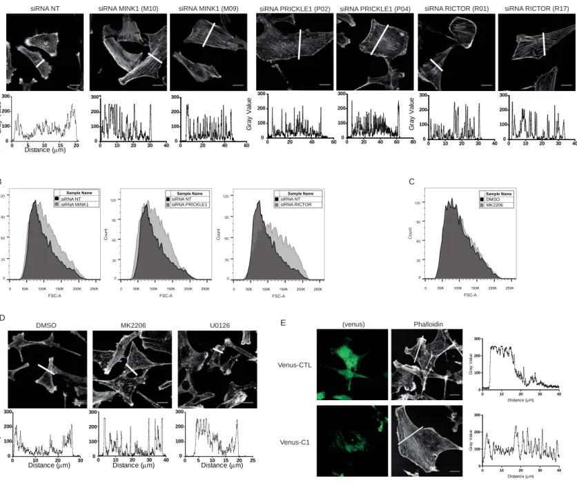
Figure 6: Mapping of the PRICKLE1-mTORC2 complex. A. Schematic of the PRICKLE1 588
mutants. B. Proteins extracted from HEK293T cells expressing the indicated constructs were 589
immunopurified and the presence of endogenous RICTOR was assessed. C. VENUS or 590
VENUS-C1 of PRICKLE1 (see A) were expressed in HEK293T cells and immunopurified to 591
detect the presence RICTOR. D. Overexpression of the C1 domain of PRICKLE was able to 592
decrease the interaction between PRICKLE1 and RICTOR. E. and F. MDA-MB-231 cells 593
were transfected with VENUS or VENUS-C1 and stimulated with FCS for 30 minutes. 594
Phosphorylation of AKT (E) and cell migration (F) were assessed by western blot and 595
Boyden chamber assays, respectively. G and H. same as (E) except that the transfected MDA-596
MB-231 cells were seeded on collagen-coated coverslips. H. Quantification of cell spreading 597
in (G). Scale bar are 20 µM. Statistical analysis was performed against control (black 598
histogram). ***P<0.001. Data are presented as means ±SEM. 599
600 601
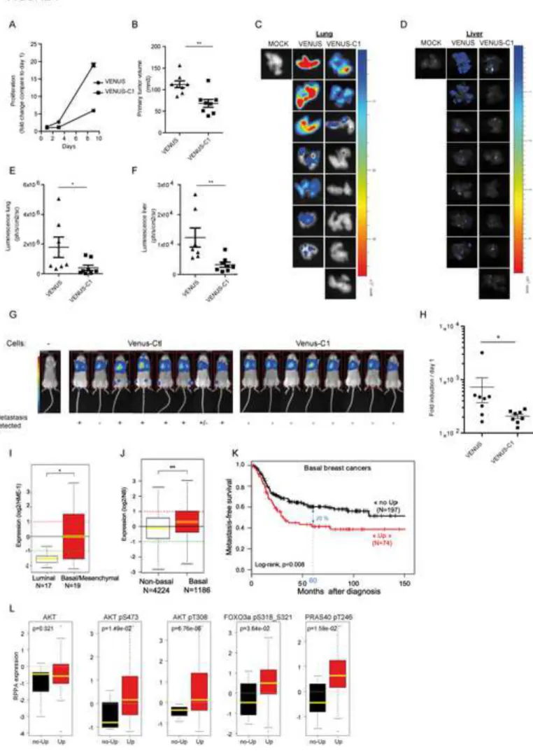
Figure 7: Dominant negative effect of the C1 domain of PRICKLE1 on tumor growth 602
and formation of metastasis. A. MDA-MB-231 cells expressing VENUS or VENUS-C1 of 603
PRICKLE1 were assayed for cell proliferation. B. Mice were xenografted with luciferase-604
positive MDA-MB-231 cells expressing VENUS or VENUS-C1 of PRICKLE1. The volume 605
of primary tumors was measured after 35 days. C-D. Lungs (C) and Liver (D) of sacrificed 606
27
mice were assayed by bioluminescence. Quantification are shown in (E) for lungs and in (F) 607
for livers. G. Mouse tail injection of luciferase-positive MDA-MB-231. After 15 days, the 608
luminescence of the entire mouse was measured and represented in H. ANOVA with 609
Dunnett’s correction for multiple testing was used to assess the significance of differences 610
among the different groups of animals. *P<0.05; **P<0.01; ***P<0.001. I. Box plot of 611
PRICKLE1 expression in breast cancer cell lines. J. Box plot of PRICKLE1 expression across 612
basal versus non-basal breast cancers. K. Kaplan-Meier curves of metastasis-free survival 613
among basal breast cancers patients according to overexpression (Up) versus no 614
overexpression (no Up) of PRICKLE1 mRNA. L. Box plot of standardized protein expression 615
levels (RPPA) of the indicated proteins in TCGA breast basal cancer samples (N=85) between 616
“PRICKLE1-up” and “PRICKLE1-no up” groups. See also Figures S6 and S7, Tables S1 and 617
S2. 618
28 Experimental procedures
619
Cell culture, transfection and antibodies 620
HEK293T, MDA-MB-231, SKBR7, MCF7 cells were obtained from the ATCC. Cells were 621
grown in DMEM containing 10% Fetal Calf Serum. Transfections were performed using 622
Polyethyleimine (Santa Cruz), Lipofectamine 2000 and Lipofectamine LTX (Thermo). siRNA 623
were used as reverse transfection using Lipofectamine RNAimax (Thermo). Antibodies 624
targeting MINK1 and α-β1-INTEGRIN were obtained from Bethyl. α-β1-INTEGRIN (clone 625
9EG7) was obtained from BD Bioscience. The following antibodies were obtained from Cell 626
Signaling: AKT, pS473-AKT, pT308-AKT, RICTOR, mTOR, pS473-AKT1, α-627
pS472-AKT2, α-pT450-AKT, α-PKCα, α-FOXO, α-pT32-FOXO, α-pY118-PAXILLIN and 628
from EMD Millipore: α-pS657-PKCα. 629
Affinity purification, immunoprecipitation and western blot 630
48 hours post-transfection, cells were lysed with the TAP lysis buffer and incubated at 4ºC for 631
1 hour to solubilize proteins. Affinity purification and immunoprecipitations were performed 632
using either streptavidin resin (GE Healthcare) or anti-FLAG-M2 beads (Sigma) for 3 hours at 633
4°C. After extensive washes with lysis buffer, proteins were eluted with 2x Laemmli sample 634
buffer and heated at 95ºC for 5 min in the presence of β-mercaptoethanol (Sigma). Whole cell 635
lysates or purified protein samples were resolved by SDS-polyacrylamide gel electrophoresis 636
(SDS-PAGE) and transferred onto Biotrace NT Nitrocellulose Transfer Membranes (Pall 637
Corporation). Western blotting were performed with antibodies as indicated in figures legend, 638
followed by chemiluminescent detection using appropriate HRP-conjugated antibodies and 639
the SuperSignal West Pico (Thermo Scientific) reagent. 640
Boyden chamber assays 641
50,000 cells were counted and loaded in serum starved condition in the upper chamber of a 642
Boyden chamber. Lower chamber was filled with media supplemented with 5% FCS. After 12 643
29
hours, migrating cells were gently recovered from the bottom side of the chamber by trypsin 644
treatment and counted using a Promega cell titer assay as described elsewhere (Puvirajesinghe 645
et al., 2016). 646
Confocal imaging 647
Cells were seeded on coverslips pretreated with rat tail Collagen (Roche). Cells were fixed 648
using paraformaldehyde followed by permeablization using PBS/Triton X-100 at 0.2%. Cells 649
were treated with the indicated antibody and imaged on confocal LSM 510 META (Zeiss) 650
with a UV laser and × 63 and x40 objectives. Confocal images were analyzed using ImageJ 651
software. 652
TIRF experiments 653
For live-cell TIRF microscopy, engineered MDA-MB-231 stably expressing venus-654
PAXILLIN cells were siRNA transfected with the indicated siRNA and seeded 48 hours later 655
on collagen coated glass bottom Petri dishes. Cells were imaged the next day with a Roper 656
Scientic ILas2 laser illuminator for TIRF Microscopy with 491 nm laser excitation (Cobolt 657
Calyso 100 mW) on a Axio Observer Z1 microscpe (ZEISS) and TIRF objective alpha plan 658
neoFluar x100/1.48 with EM-CCD Evolve 512 Camera (photometrics), and driven by 659
MetaMorph 7 software (Molecular devices). 660
In vitro kinase assay 661
The PRICKLE1 or RADIL complexes were purified from HEK293T cells stably expressing 662
FLAG-PRICKLE1 or FLAG-RADIL. Protein complexes were incubated with soluble 663
recombinant GST-AKT1 protein purified from Escherichia coli. Phosphorylation reactions 664
were performed in kinase buffer (25 mM Hepes, pH 7.4, 25 mM β-glycerophosphate, 25 mM 665
MgCl2, 0.1 mM Na3VO4, 0.5 mM DTT) supplemented with 20 µM ATP at 37°C for 1 hour. 666
Reactions were stopped by addition of 4x Laemmli sample buffer. Proteins were resolved by 667
SDS-PAGE and analyzed by anti-Serine 473-AKT antibody. 668
30 669
Author contributions: A.M.D. and J.P.B. conceived the project. A.M.D. designed and 670
conducted most of the experiments. E.J. and R.C. conducted animal work. S.A. performed 671
mass spectrometry analysis. F.B. and P.F. analyzed the clinical data. A.S. performed the 672
TIRF analysis. D.B. and S.A. provided expertise and feedback. J.P.B. supervised the project. 673
A.M.D. and J.P.B. wrote the manuscript. 674
References 675
Agarwal, N. K., Chen, C. H., Cho, H., Boulbes, D. R., Spooner, E., and Sarbassov, D. D. (2013). 676
Rictor regulates cell migration by suppressing RhoGDI2. Oncogene 32, 2521-2526. 677
Ahmed, S. M., Theriault, B. L., Uppalapati, M., Chiu, C. W., Gallie, B. L., Sidhu, S. S., and Angers, S. 678
(2012). KIF14 negatively regulates Rap1a-Radil signaling during breast cancer progression. J Cell 679
Biol 199, 951-967. 680
Anastas, J. N., Biechele, T. L., Robitaille, M., Muster, J., Allison, K. H., Angers, S., and Moon, R. T. 681
(2012). A protein complex of SCRIB, NOS1AP and VANGL1 regulates cell polarity and migration, 682
and is associated with breast cancer progression. Oncogene 31, 3696-3708. 683
Anastas, J. N., Kulikauskas, R. M., Tamir, T., Rizos, H., Long, G. V., von Euw, E. M., Yang, P. T., 684
Chen, H. W., Haydu, L., Toroni, R. A., et al. (2014). WNT5A enhances resistance of melanoma cells 685
to targeted BRAF inhibitors. J Clin Invest 124, 2877-2890. 686
Belotti, E., Polanowska, J., Daulat, A. M., Audebert, S., Thome, V., Lissitzky, J. C., Lembo, F., 687
Blibek, K., Omi, S., Lenfant, N., et al. (2013). The human PDZome: a gateway to PSD95-Disc large-688
zonula occludens (PDZ)-mediated functions. Mol Cell Proteomics 12, 2587-2603. 689
Burridge, K., and Chrzanowska-Wodnicka, M. (1996). Focal adhesions, contractility, and signaling. 690
Annu Rev Cell Dev Biol 12, 463-518. 691
Chin, Y. R., and Toker, A. (2009). Function of Akt/PKB signaling to cell motility, invasion and the 692
tumor stroma in cancer. Cell Signal 21, 470-476. 693
31
Ciruna, B., Jenny, A., Lee, D., Mlodzik, M., and Schier, A. F. (2006). Planar cell polarity signalling 694
couples cell division and morphogenesis during neurulation. Nature 439, 220-224. 695
Cordenonsi, M., Zanconato, F., Azzolin, L., Forcato, M., Rosato, A., Frasson, C., Inui, M., Montagner, 696
M., Parenti, A. R., Poletti, A., et al. (2011). The Hippo transducer TAZ confers cancer stem cell-697
related traits on breast cancer cells. Cell 147, 759-772. 698
Cui, C., Chatterjee, B., Lozito, T. P., Zhang, Z., Francis, R. J., Yagi, H., Swanhart, L. M., Sanker, S., 699
Francis, D., Yu, Q., et al. (2013). Wdpcp, a PCP protein required for ciliogenesis, regulates directional 700
cell migration and cell polarity by direct modulation of the actin cytoskeleton. PLoS Biol 11, 701
e1001720. 702
Daulat, A. M., Luu, O., Sing, A., Zhang, L., Wrana, J. L., McNeill, H., Winklbauer, R., and Angers, S. 703
(2012). Mink1 regulates beta-catenin-independent Wnt signaling via Prickle phosphorylation. Mol 704
Cell Biol 32, 173-185. 705
Dissanayake, S. K., Wade, M., Johnson, C. E., O'Connell, M. P., Leotlela, P. D., French, A. D., Shah, 706
K. V., Hewitt, K. J., Rosenthal, D. T., Indig, F. E., et al. (2007). The Wnt5A/protein kinase C pathway 707
mediates motility in melanoma cells via the inhibition of metastasis suppressors and initiation of an 708
epithelial to mesenchymal transition. J Biol Chem 282, 17259-17271. 709
Dow, L. E., Elsum, I. A., King, C. L., Kinross, K. M., Richardson, H. E., and Humbert, P. O. (2008). 710
Loss of human Scribble cooperates with H-Ras to promote cell invasion through deregulation of 711
MAPK signalling. Oncogene 27, 5988-6001. 712
Escobedo, N., Contreras, O., Munoz, R., Farias, M., Carrasco, H., Hill, C., Tran, U., Pryor, S. E., 713
Wessely, O., Copp, A. J., and Larrain, J. (2013). Syndecan 4 interacts genetically with Vangl2 to 714
regulate neural tube closure and planar cell polarity. Development 140, 3008-3017. 715
Facchinetti, V., Ouyang, W., Wei, H., Soto, N., Lazorchak, A., Gould, C., Lowry, C., Newton, A. C., 716
Mao, Y., Miao, R. Q., et al. (2008). The mammalian target of rapamycin complex 2 controls folding 717
and stability of Akt and protein kinase C. EMBO J 27, 1932-1943. 718
Gubb, D., and Garcia-Bellido, A. (1982). A genetic analysis of the determination of cuticular polarity 719
during development in Drosophila melanogaster. J Embryol Exp Morphol 68, 37-57. 720