Development of Nanoparticles for Oral
Delivery of Insulin
by
Sunandini Chopra
Bachelor of Mechanical Polymer Engineering, 2012 University of Akron
Submitted to the Department of Mechanical Engineering in partial fulfillment of the requirements for the degree of
Doctor of Philosophy at the
MASSACHUSETTS INSTITUTE OF TECHNOLOGY
MASSACMSI OF TECHNOLOGY
FFB 15?ZI1
U
ARHES
February 2017
Massachusetts Institute of Technology. All rights reserved. The author hereby grants to MIT permission to reproduce and distribute
publicly paper and electronic copies of this thesis document in whole or in part in any medium now known or hereafter created.
A utho r: ... Certified by: ... Accepted by: ... Chai
Signature redacted
Sunandini Chopra Department of Mechanical Engineering Program of Polymer Science and Soft Matter January 15, 2017Signature redacted
Rohit Karnik, Ph.D. Department of Mechanical Engineering Thesis Supervisor .Signature
redacted
RohanAleyaratne,
Ph.D. Department of Mechanical Engineering rman, Department Committee on Graduate StudentsDevelopment of Nanoparticles for Oral Delivery of
Insulin
by
Sunandini Chopra
Submitted to the Department of Mechanical Engineering on January 15, 2017, in partial fulfillment of the
requirements for the degree of Doctor of Philosophy
Abstract
Parenteral administration remains the mainstay of drug administration for protein therapeutics. However, for diseases that require frequent drug dose over long periods of time, injections can result in patient incompliance and poor treatment outcomes. For such diseases, oral drug delivery is the most non-invasive and patient-compliant method of drug administration. Although oral delivery of many small molecule drugs is routine, oral delivery of protein drugs e.g. insulin -presents several challenges including oral bioavailability of the protein
therapeutic because of degradation in the stomach, inactivation and digestion of the therapeutics by the proteolytic enzymes in the luminal cavity, and poor permeability of drugs across the intestinal epithelium.
Polymeric nanoparticle (NP) carriers provide new opportunities for controlled delivery of drugs, and have the potential to address challenges associated with effective oral delivery of insulin. NPs can protect the protein therapeutic from degradation in the GI tract as well as allow targeted transport across the epithelial lining. An efficient NP based oral insulin delivery solution that can enable targeted transport of insulin across the GI tract must have (1) high insulin loading, (2) sub-100 nm size, (3) ability to release insulin before opsonization by macrophages and (4) the ability to be surface-functionalized with ligands that facilitate transport across the epithelium.
This work presents a detailed study on mechanistic understanding of polymeric insulin NP formation with a focus on the effect of synthesis parameters on insulin loading and NP size. We report how buffer conditions, ionic chelation, and NP preparation methods influence insulin loading in poly (lactic-co-glycolic
acid)-b-poly(ethylene glycol) (PLGA-PEG) NPs. We report a 10-fold increase in insulin loading with the use of chelating zinc ions and by the optimization of the pH during nanoprecipitation.
Next, we report the development of novel insulin Eudragit-PLGA-PEG blended NPs (Ins-Eud-NPs) with high insulin loading (13.1%) and sub-100 nm size. These NPs enable rapid release of insulin when triggered by a change in pH that occurs when the NPs cross the duodenal epithelium and go from acidic to neutral pH. The NPs are formed by successfully blending Eudragit S100, a commercially available polymer which dissolves at pH greater than 7 with a non-pH responsive polymer, PLGA-PEG. To enable effective transport of these NPs across the epithelial lining, NPs were designed to use the FcRn transport pathway that mediates IgG antibody transport across epithelial barriers. We report the successful chemical conjugation of the Fc fragment on the surface of Ins-Eud-NPs by overcoming the presence of non-ideal conjugation parameters owing to the pH restrictions of the system.
This dissertation provides mechanistic insights and helps to understand
fundamental concepts about polymeric NP formation and protein encapsulation. The modular NP system developed in this work can be extended to other protein drug delivery systems that are subject to limited drug loading and restricted transport across epithelial barriers.
Thesis supervisor: Rohit Karnik, Ph.D. Title: Associate Professor, MIT
This thesis has been examined by the following Thesis Committee:
Thesis Advisor Rohit Karnik, Ph.D.
Associate Professor of Mechanical Engineering Massachusetts Institute of Technology
Thesis Committee Omid C. Farokhzad, M.D. Associate Professor of Anesthesiology
Harvard Medical School Roger Kamm, Ph.D.
Professor of Mechanical Engineering Massachusetts Institute of Technology
Acknowledgements
I want to dedicate this thesis to my late mother, Dr. Bharti Chopra. She has had the most profound impact on my life, not just as a role model but also as my strongest pillar of strength and my loudest cheerleader. This thesis would not have been possible without her tireless efforts to personally tutor me as a child, the long hours she worked as a gynecologist after which she came back every day and worked even harder to take care of my brother and me, and the lessons of dedication, hard work, and perseverance that she taught me each day growing up. She was the reason I got interested in the sciences at a very young age. It was her belief in my abilities and her desire to provide me with the best
resources and education that brought me to the United States. It was her selfless nature and modern thinking, way ahead of her times, that enabled me to pursue my dreams. My mother was a gifted person with the ability to think the
unthinkable and to execute actions at a level that surpassed everyone. She was a woman of immense dignity, valor, and determination. She was blessed with a sense of eccentricity, which was exhibited in the passion with which she worked, loved, cared for and built relations. Going forward in life, I only hope to be
blessed with a part of her eccentricity, to be able to work like her, to love like her and to value and build relations like she did. She has been my most valuable asset. Although she is not present today, I know that this work will make her proud and her applause would be the loudest. Thank you, Ma.
I also want to take this opportunity to express my sincere gratitude to my advisor, Professor Rohit Karnik, who has provided me unparalleled guidance and
mentorship. I want to thank him for the optimism and positivity that he exudes, which also reflect in his conversations. His belief in me and his ability to probe the fundamental aspects of science were instrumental in helping me through the troughs. The insightful and pertinent questions that he asked were pivotal to this work. I am grateful to him for being so selflessly supportive and so incredibly gracious. I have not just learned how to do good science from him, but also
kindness and compassion. The numerous times when I walked into his office, crestfallen and disappointed he was always successful in reversing that and ensuring that I was pumped with renewed excitement and enthusiasm to solve the problems I face. Professor Karnik's contributions were was paramount to the completion of this work.
I also want to thank my committee members, Professor Omid Farokhzad and Professor Roger Kamm. Professor Farokhzad has been a significant positive influence on my work and my life. He is a true visionary. His futuristic vision and
ability to think ahead of his time have helped me connect my work to the
overarching goal and the big picture. I am thankful for his unconditional support, guidance, and understanding throughout this project.
Professor Kamm has been incredibly kind and generous with his time. He always asked the most pertinent questions, which made me think more about what I was doing and why. It helped me realign my efforts and thoughts to solve the right problems. His role in the completion of this work is invaluable.
I also want to express my profound appreciation for my family - my father, Lt. Col. Tilak Raj Chopra, my brother, Harsh Chopra, my uncle Dr. Vikas Praskash and my aunts Dr. Malini Dusey and Tulika Das. Each one of them has played a vital role in my upbringing and education. My father is my biggest well-wisher and my reality check button. He consistently makes me question and rethink my actions and agendas of life. He is an honest, frank and a doting father (who is more like my brother). My brother (who behaves like my father), is one of the most genuine people I know. I am thankful to him for always being there for me, supporting me unconditionally and setting an example of how to be an ideal human being. My uncle is the reason I came to the United States to pursue higher education. He was one of the reasons I wanted to pursue a doctoral degree. I want to express my heartfelt gratitude to him for recognizing my
potential, and for bringing me to the United States against all the odds. My aunt, Malini Dusey has been instrumental in helping me develop into the person I am today. Her constant questioning of my actions, her honest opinions and her
independent and joyful way of living life have several times made me question my motivation in life. I have had a roller coaster ride with her, but each day I am more in awe of her. Lastly, my aunt, Tulika Das, and I share a special
relationship where we are more friends than anything else. I want to thank her for accepting me the way I am and for being my sounding board. Each one of these people mentioned was crucial to the completion of this thesis.
I also want to take this opportunity to thank all my teachers - from high school to undergrad to grad school. Each one of them has left an indelible mark on my life and helped me evolve my thinking. I want to particularly thank Professor Robert
Langer, for his kind support, guidance, encouragement and access to laboratory equipment. Besides, I want to thank my friends and lab mates for their
unwavering support. I want to thank Dr. Nicolas Bertrand, Dr. Suman Bose, Jaya Singh, Shruti Singh, Sidhant Jena, Kunal Poddar, Sneha Mandhan, Abel
Cortinas, Akhilesh Bakshi, Vidhi Goel, Amy Wang and Trinh Nyguen for their help and support.
I am thankful to the wonderful people in my life who make me a better person each day. I hope you enjoy reading this thesis and I hope this helps to answer fundamental and pertinent questions that can have a positive impact in the area of drug delivery.
CONTENTS
CHAPTER 1 - INTRODUCTION...25
1.1 Current trends in biotechnology...25
1.2 Diabetes and Insulin...28
1.3 Methods of insulin delivery...32
1.4 Physiology of the Gastrointestinal Tract...40
1.5 Barriers to effective Oral Delivery... 43
1.6 Strategies for Oral Delivery...45
1.6.1 Permeation enhancers...45
1.6.2 Protease inhibitors...47
1.6.3 Mucoadhesives...48
1.6.4 Polymeric drug carriers...49
1.7 FcRn Transcytosis pathway... 52
1.8 Desired characteristics in a NP based oral drug delivery solution...54
1.9 Thesis goals...55
CHAPTER 2 - Design of Insulin-Loaded Nanoparticles Enabled by Multi-Step Control of Nanoprecipitation and Zinc Chelation...59
2 .1 A b stra ct... . . .. 5 9 2.2 Introduction... 59
2.3 Experimental Section...62
2 .3 .1 M aterials... . 62
2.3.2 Methods...62
2.3.2.1 Synthesis of Ins-NPs and Ins-Zn-NPs...62
2.3.2.2 Characterization of Ins-NPs and Ins-Zn-NPs...62
2.3.2.3 Measurement of insulin loading...63
2.3.2.4 In vitro insulin release...65
2.3.2.7 Stability of NPs during storage... 66
2.3.2.8 Conformational stability of insulin...66
2.4 Results and Discussion... 67
2.4.1 Effect of washing buffer on insulin loading... 68
2.4.2 Mechanism of NP formation...70
2.4.3 Enhancing insulin loading by zinc chelation...73
2.4.4 Optimizing the size of Ins-Zn-NPs... 75
2.4.5 Effect of pH on formation of Ins-Zn-NPs...76
2.4.6 Optimization of NPs for small and high insulin loading...78
2.5 Conclusions...80
CHAPTER 3 - Design of pH-Responsive Nanoparticles...81
3 .1 A bstra ct... . . 8 1 3.2 Introduction... 81
3.3 Experimental Section... 86
3.3.1 Materials...86
3.3.2 Methods...86
3.3.2.1 Synthesis of Eud-NPs and Eud-Ins-NPs...86
3.3.2.2 Characterization of Eud-NPs and Eud-Ins-NPs...87
3.3.2.3 Measurement of insulin loading...88
3.3.2.4 In vitro insulin release... 89
3.3.2.5 Synthesis of Eud-NP-Fc...91
3.3.2.6 Characterization of Eud-NP-Fc...92
3.3.2.7 In vitro Transcytosis ... 92
3.4 Results and Discussion... 93
3.4.1 Effect incorporating Eudragit in Ins-Zn-NPs...93
3.4.2 Formation of Eud-NPs... 95
3.4.3 Effect of Eudragit on NP size...96
3.4.5 Effect of synthesis buffer on NP size
and insulin loading... 99
3.4.6 Effect of Eudragit on Insulin release...100
3.4.7 pH sensitive in vitro release of insulin from Ins-Eud-NPs...102
3.4.8 Fc conjugation approach and challenges...104
3.4.9 Fc conjugation optimization...106
3.5.10 NP in vitro transcytosis...108
3 .5 C o n clusio ns ... 1 10 CHAPTER 4 -Conclusion and Outlook...113
APPENDIX...119
List of Figures
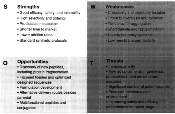
1.1. Strengths, weaknesses, opportunities and threats of the application and development of biopharmaceuticals.
1.2. (A) The rate at which biopharmaceuticals have reached clinical stages has increased over the last decades with 4.6 in 1980s to 128 to 2012. (B) Approval numbers of biopharmaceuticals in each five-year period since 1995 has been fairly constant, with approximately 54 approved biologics for 2010-2014, but approval has significantly increased from early 1990s to now.
1.3. Annual biopharmaceutical sales value (cumulative product sales and sales for the ten top-selling products) for the period 2010 to 2013. This shows the increase in the economic value of protein therapeutics.
1.4. The 20 top-selling biopharmaceutical products in 2013. Humira leads the revenue production in 2013 followed by Enbrel and Remicade.
1.5. (A) The mainstay of drug administration for protein therapeutics via. intramuscular, subcutaneous, intravenous or intradermal injections. (B) Due to the popularity of biopharmaceuticals for various disease treatments in the last decade on an average 12 billion injections have been injected which have caused 20 million infections annually and 100 million adverse reactions. 1.6. Mechanism of how insulin facilitates the uptake of glucose in muscle cells and adipocytes. Insulin binds to the GLUT-4 receptor in the plasma membrane, resulting in phosphorylation of the receptor. Activation of phosphoinositide-3 kinase is a major pathway in the mediation of insulin-stimulated glucose transport and metabolism.
1.7. Structure of insulin. Insulin, a polypeptide hormone that is formed
of two chains A (21 amino acids) and B (30 amino acids) that are connected by two disulfide bridges; an additional disulfide is formed within A chain.
1.8. Alternative methods of insulin delivery. Various routes of delivery have been investigated for insulin delivery such as pulmonary, oral, transdermal, nasal, buccal and by islet transplantation.
injections, subcutaneous infusions, pulmonary delivery and oral delivery. Each of these methods have advantages and disadvantages. However, oral delivery is considered to be the most patient compliant and non-invasive way of
administering insulin.
1.10. Some oral insulin delivery projects that are under development and clinical trial.
1.11. Physiology of the intestinal epithelium. Enterocytes, goblet cells, paneth cells and M-cells are some of the major cell types that make up the intestinal epithelium.
1.12. Barriers to effective delivery of insulin. Various chemical and physical barriers like changes in pH, high protease action and the presence of tight junctions make the problem of oral delivery very challenging.
1.13. Oral insulin delivery Nanoparticle Design criteria. The nanoparticles (NPs) must be able to protect insulin from the harsh chemical environment in the GI tract and from enzymatic degradation. The NPs must be sub-1 00 nm, have the ability to be functionalized and have the capability of delivering majority of insulin before the NPs can opsonized.
1.14. Choice of parameters for understanding the nanoprecipitation method better.
2.1. (a) Insulin standard curve in relation to the BSA standard curve for the BCA assay. (b) Relation between the insulin and BSA standard curves. (c) Effect of DMSO, zinc, and polymeric NPs on the insulin standard curve.
2.2. Ins-NPs and Ins-Zn NPs show the potential of long-term storage as they maintain (a) particle size and (b) zeta potential before and after undergoing the process of freeze-drying. (c) Ins-NPs and Ins-Zn-NPs are stable in water for up to 4 days from synthesis at RT. (d) Conformational stability of insulin in Ins-NPs and Ins-Zn-NPs was confirmed by comparing the circular dichroism spectra of
standard insulin and insulin obtained from Ins-NPs and Ins-Zn-NPs before and after undergoing the process of freeze-drying.
2.3. Insulin loaded PLGA-PEG nanoparticles (Ins-NP). (A) Schematic of Ins-NP
synthesis process by nanoprecipitation, where PLGA-PEG and insulin in dimethyl sulfoxide (DMSO) is added drop-wise to a stirred beaker containing water (or
buffer). TEM images of (B) Ins-NPs and (C) PLGA-PEG NPs without insulin.
Effect of buffer wash on Ins-NP characteristics. (D) Schematic of the synthesis of
Ins-NPs where NPs were either washed with PBS buffer (pH 7.4) or with water only. (E) Insulin loading and encapsulation efficiency show 25-fold higher insulin loading when NPs were washed with water only (8.95% 1.44) as compared to a PBS buffer wash (0.36% 0.19) (n = 3, p = 0.008). (F) NP size data show no significant difference in the size of the Ins-NPs when washed with water or buffer
(n = 3, p = 0.074). Error bars represent s.d.
2.4. Effect of adding insulin at different stages of nanoprecipitation on Ins-NP
characteristics. (A) Schematic showing the synthesis of Ins-NPs by including
insulin mixed with PLGA-PEG in DMSO before nanoprecipitation (top), and by adding insulin in DMSO to empty PLGA-PEG NPs after their formation by
nanoprecipitation (bottom). (B) Insulin loading and encapsulation efficiency show no significant dependence on the stage of adding insulin before (0.36%) or after (0.32%) nanoprecipitation (n = 3, p = 0.79), but (C) NP size shows a significant dependence (n = 3, p = 0.005). Effect of mixing insulin in different phases on
Ins-NP characteristics. (D) Schematic showing the synthesis of Ins-Ins-NPs by adding
DMSO with insulin and PLGA-PEG to water (top), and by adding DMSO with PLGA-PEG to water with insulin dissolved in it (bottom). (E) Insulin loading (and encapsulation efficiency) shows no significant dependence on including insulin in
DMSO (0.36%) or in water (0.28%) (n = 3, p = 0.60), but (F) NP size shows a significant dependence (n = 3, p = 0.0006). Error bars represent s.d. Note: The
NPs with the label 'Insulin added before' in Figure 2.4B and the NPs with the label 'Insulin in organic' in Figure 2.4E are the same nanoparticles.
2.5. Empty PLGA-PEG NPs (-25.44 mV) and Ins-NPs (-27.9 mV) was statistically insignificant. Zeta potential of the Ins-Zn-NPs (-22. 03 mV) is significantly lower
than Ins-NPs and Empty PLGA-PEG NPs (** = p < 0.05).
2.6. Effect of incorporating zinc ions in Ins-NPs. (A) Schematic showing the formation of Ins-Zn hexamers (top) and of Ins-Zn-NPs (bottom). (B) Dark-field TEM/EDX image of unstained Ins-Zn-NPs. The lighter regions in the figure show the presence of NPs, and the red dots correspond to the presence of zinc. The presence of zinc in the same areas where Ins-Zn-NPs were detected confirms the presence of zinc in Ins-Zn-NPs. (C) Insulin loading and encapsulation efficiency show an increase in insulin loading with an increase in zinc ion
concentration. Greater than 10-fold enhancement of insulin loading was obtained for insulin to zinc ratio of 1:9. (n = 3, p = 0.02). (D) The average diameter of empty PLGA-PEG NPs, Ins-NPs, and Ins-Zn-NPs. In case of Ins-Zn-NPs, the
average diameter did not change significantly when different amount of zinc ions are added. The average diameter of Ins-Zn-NPs was larger than that of empty
PLGA-PEG NPs (n = 3, p = 0.0003) and Ins-Zn-NPs and Ins-NPs (n = 3, p =
0.0001). Error bars represent s.d.
2.7. Effect of adding zinc ions at different stages of nanoprecipitation on
Ins-Zn-NPs. (A) Schematic showing the synthesis of Ins-Zn-NPs by adding Ins-Zn
hexamers premixed with PLGA-PEG molecules in DMSO before
nanoprecipitation (top). This forms NPs with high insulin loading (4.07%) and large NP diameter (127.6 nm). (B) Schematic showing the synthesis of Ins-Zn-NPs by adding Ins-Zn hexamers to empty PLGA-PEG Ins-Zn-NPs after
nanoprecipitation (top). This forms NPs with high insulin loading (3.82%) and small NP diameter (78.7 nm). (C) Schematic showing the synthesis of Ins-Zn-NPs by adding zinc ions to preformed Ins-NPs after nanoprecipitation (top). This forms NPs with lower insulin loading (1.55%) and smaller NP diameter (57.9 nm). Dashed lines indicate sizes of empty PLGA-PEG and Ins-NPs for reference.
Error bars represent s.d. For insulin loading n = 3 and PAB = 0.74, pAc = 0.034,
PBc = 0.016. For NP diameter n = 3 and PAB = 0.0004, pAC = 0.0001, PBC = 0.005.
2.8. Effect of pH on Ins-Zn-NP and Ins-NPs. (A) Schematic showing the synthesis of Ins-Zn-NPs in different buffers (pH 4.65, pH 6.1, and pH 6.5). (B) Insulin loading of Ins-Zn-NPs and Ins-NPs that were synthesized in buffers with different pH. Insulin loading increased with increasing pH. (C) Average diameter of empty PLGA-PEG NPs, Ins-NPs, and Ins-Zn-NPs synthesized in different buffers.
2.9. Compilation of the effect of NP synthesis parameters on NP properties. (A)
Compilation of the NPs studied for the purpose of understanding the factors that affect insulin loading and NP size in Ins-NPs and Ins-Zn-NPs. The symbols in blue contain only PLGA-PEG and insulin while the orange symbols represent NPs that contain PLGA-PEG, insulin, and zinc. The size of empty PLGA-PEG NPs has been represented by a grey vertical bar, whose width represents the standard deviation. Ins-NPs (blue) have low loading and small size. Contrary to these NPs, Ins-Zn-NPs formed by including zinc during nanoprecipitation (orange circles) have higher insulin loading, but their diameter is larger. NPs synthesized by adding Zn ions to preformed Ins-NPs (light triangle) show smaller size and moderate loading. The optimal NPs are Ins-Zn-NPs (dark triangle) formed by adding Ins-Zn hexamers to pre-formed NPs combine high loading (3.82 %) and small size (78.7 nm diameter). (B) Qualitative summary of how different synthesis parameters affect insulin loading and size. Insulin release. (C) Release of insulin
from the optimal NPs (Ins-Zn-NPs with insulin loading 3.82%, NP size 78.7 nm) is comparable with the previously reported release of insulin from PLA-PEG NPs'.
3.1. Schematic of the NP synthesis set-up. The Ins-Eud-NPs were synthesized using the nanoprecipitation method. The polymer and the drug was mixed in the organic solvent (DMSO) and the resulting solution was added drop-wise to a beaker with pH 5 buffer that was being constantly stirred at 2000 rpm. Following NP synthesis the NPs were washed multiple times with pH 5 buffer with 150 mM NaCl.
3.2. (a) Design of the dialysis set up to study the in-vitro release of insulin. (b) Absorbance from empty NPs when the dialysis devices were soaked for 4 h and 24 h. (c) In-vitro release of free insulin.
3.3. (A) Addition of Eudragit S100 in the NP formulation for Ins-Zn-NPs (12.8%) did not lead to an improvement in insulin loading when compared with the insulin loading of Ins-Eud-NPs (13.1%). However, presence of Eudragit leads to an improvement in insulin loading. (B) Ins-Zn-Eud-NPs failed to illicit the desired pH response. Insulin released at a faster rate at pH 6 as compared to pH 7.4. This can be attributed to both the pH vales being larger than the pH of the isolelectric point of insulin below which Ins-Zn hexamers are unstable.
3.4. (A) Schematic highlighting the failure of Eudragit S100 to self-assemble to form NPs without the application of an external stabilizer using the process of nanoprecipitation. (B) The volume size distribution of the Eudragit S100 aggregates which confirm the failure of Eudragit to self-assemble to form NPs using the nanoprecipitation method. (C) Schematic of our hypothesis that pH responsive polymer, Eudragit S100 when blended with non-pH responsive polymer can form pH responsive NPs.
3.5. Blending Eudragit S100 and PLGA-PEG in different ratios was successful in forming NPs. The inclusion limit of Eudragit in the system is 50% (w/w) Eudragit as it results in the formation of chunky aggregates. Moreover, increasing amount of Eudragit S100 blended in Emp-Eud-NPs did not affect the size of the NPs. However, on preparing Ins-Eud-NPs a size dependence on the amount of Eudragit was observed. The size of the NPs increased for the compositions that had 20% and 30% (w/w) Eudragit.
3.6. Eudragit does not have a significant effect on insulin loading up to 10% Eudragit in the NPs, but on increasing the Eudragit amount to 20% and 30% we achieve a higher loading.
3.7. NPs with larger diameter and poor insulin loading are formed at low pH buffers (pH 3 and pH 4) while NPs with high insulin loading and smaller size are formed in buffers with pH close to the isoelectric point of insulin ie. pH 5.6.
3.8. Increasing the amount of Eudragit in the NPs slowed doen the release of insulin after 2 h in pH 6 buffer. But no particular trend was observed in the
release of insulin from Ins-Eud-NPs at pH 7.4. Moreover, NPs with 20% Eudragit and 80% PLGA-PEG show the maximum difference in the amount of insulin released in pH 6 and pH 7.4, thus suggesting that this NP composition produces the most pH responsiveness NPs.
3.9. A significant reduction in the rate at which insulin is released from the NPs at pH 6 is observed as compared to pH 7.4. The half-life of insulin in pH 7.4 is between 30-60 min, while the half-life of insulin in pH 6 is 240 min. However, the current measurement is limited by the resolution of the dialysis set-up (1 h). 3.10. On normalizing the data with respect to the release of free insulin it can be seen that almost all of the insulin from the NPs gets released as soon as the NPs are exposed to pH 7.4, while there is a significant reduction in the release of insulin from the NPs in pH 6. The half-life od insulin at pH 6 was 90 min.
3.11. (A) Effect of reaction buffer on Fc conjugation on NP surface. Maximum Fc conjugation was obtained at pH 7.4, followed by pH 5 and pH 6 buffer. (B) Effect of reaction time on Fc conjugation. On incubating Ins-Eud-NPs with Fc-SH in pH 5 buffer for 2 h and 18 h, it was found that the Fc conjugation in 2 h (0.16%) was not much greater than the Fc conjugation in 18 h (0.23%). (C) On increasing the amount of Fc by 5 times Fc conjugation increased to 1.48%. (D) Higher Fc conjugation resulted in longer synthesis time, which resulted in a loss of insulin leading to a reduction in insulin loading in Ins-Eud-NPs-Fc NPs to 6.8%.
3.12. (A) Formation of NPs after 5 min stirring time and 1 mg/mL NP incubation concentration resulted in only 0.7% Fc conjugation. However, when all other synthesis conditions were kept the same and only the stirring time was changed to 15 min and the NP incubation concentration was increased to 10 mg/mL Fc conjugation increased to 1.48%. (B) Conjugation of Fc on the NP surface did not
effect the size of the NPs, size of Ins-Eud-NPs-Fc was 63.9 nm while the size of NPs without Fc was 68 nm.
3.13. Schematic of Ins-Eud-NP-Fc. The Ins-Eud-NP-Fc is prepared from
biodegradable and biocompatible polymers like PLGA-PEG and Eudragit S100. Eudragit imparts the NPs an ability to respond release insulin on being stimulated by pH. The targeting ligand, Fc, has been conjugated to the surface of the NPs to enable them to cross the epithelial barrier by using the FcRn transcytosis
pathway.
3.14 In vitro transepithelial transport data showed that Ins-Eud-NPs-Fc were transported 5 times more across the Caco-2 monolayer relative to non targeted
Ins-Eud-NPs.
Al. Sample and separate and dialysis are the two most commonly used methods for measuring the in vitro drug release profile from polymeric nanoparticles. A2. (A) Insulin release profile at pH 6 and pH 7.4 obtained by analyzing the data using the filtrate. (B) Insulin release profile at pH 6 and pH 7.4 obtained by analyzing the data using the supernatant. Both these curves are counterintuitive relative to the expected insulin release profile. The significant difference in the release curve at pH 6 and pH 7.4 is promising data.
A3. (A) Insulin release after 2 h in different incubation volumes shows that the amount of insulin released by the NPs stabilizes at higher NP incubation volumes suggesting that a non-equilibrium state reaches at high incubation volumes. (B) Insulin release profile at pH 6 and pH 7.4 obtained by analyzing the data using the supernatant and a large incubation volume of 15 mL. Although the insulin release can be quantified at initial time points, at later time points there is a decrease in the amount of insulin released which seems incorrect.
A4. Amount of insulin detected in the filtrate remained constant across the samples that were washed multiple times. However, the amount of insulin detected in the supernatant decreased as the NPs underwent multiple washes. Ins NP w/o any wash represents the case without any loss of insulin.
A5. On washing the NPs with pH 6 buffer, most of the insulin was retained in the supernatant as compared to the filtrate. This suggested that the analyzing the supernatant would give a more accurate estimation of the insulin content at each time point.
A6. (A) After spinning down a 50 pg/mL insulin solution halfway, the
concentration of insulin obtained from the supernatant was 47 pg/mL, similar to the original solution while the concentration of insulin in the filtrate was much lower (28 pg/mL). (B) The sum of insulin detected in the filtrate and the
supernatant do not add up to the total insulin that was centrifuged, confirming the loss of insulin in the centrifugation device.
A7. (A) On centrifuging down the entire volume of insulin, it was found that the concentration of insulin detected in the filtrate was less than the concentration of the original solution, suggesting that measurements done using the filtrate can be inaccurate. (B) The amount of insulin detected in the filtrate was only half of the total amount of insulin in the system. Moreover, the sum of insulin detected in the filtrate and supernatant do not add up to the total amount of insulin in the initial solution, suggesting a loss of insulin.
A8. Insulin left in Ins-Eud-NPs after insulin release at pH 3, pH 6, pH 7.4, and pH 9 initially decreased with time but at longer collection times the amount of insulin left in the NPs increased. However, a pH dependent release is seen from Ins-Eud-NPs as lesser amount of insulin is detected after insulin release in pH great than 7.
A9. (A) Inclusion of a magnetic stir bar in the dialysis device did not help in accurately measuring the insulin release profile. The released insulin failed to diffuse across the membrane fast enough thereby showing an almost constant amount of insulin left in the NPs. (B) Insulin release profile obtained by using the stirred dialysis device set-up could not be used to determine the drug release kinetics as there was inadequate release and variability in the data.
A10. Blue food dye also failed to diffuse out of the dialysis chambers in 60 min. Therefore, the current dialysis set-up cannot be used to measure rapid release of insulin from NPs.
All. Insulin failed to diffuse out of the 10 nm, polycarbonate membrane dialysis chambers in 60 min.
A12. Schematic of the Float A Lyzer dialysis chamber.
Al 3. Food dye failed to rapidly diffuse out of the Float A lyzer dialysis chambers. Control in water is the food dye solution which was not dialyzed at all. Sample 1
is the food dye solution that was allowed to release food dye all night. It showed very little release of food dye. Lastly, Sample 2 is the sample that was allowed to
release the food dye for 6 h with additional stirring inside the chamber.
A14. (A) Schematic of the dialysis set-up using Float A lyzers. To enable
continuous stirring of the buffer outside the dialysis devices, the beaker was put on a magnetic stirring plate, which was further placed on an orbital shaker. (B)
By increasing the convectional flow in the system and by choosing a suitable solvent, majority of the food dye solution managed to escape the dialysis device. Control in PBS is the blue food dye solution that was not exposed to the dialysis set up, while Sample 3 is the sample of blue food dye which was measured after dialysis for 2h 15 min.
Al 5. (A) Schematic of the Float A lyzer with a magnetic stirrer placed inside it. (B) Schematic of the Float A lyzer with an epoxy cylinder placed inside it. Al 6. (A) Dialysis of yellow food dye. For both 50 kDa and 100 kDa MWCO dialysis membrane application of the epoxy cylinder is more effective relative to internal stirring. (B) Dialysis of blue food dye.100 kDa membrane is more effective than the 50 kDa membrane in allowing rapid diffusion of molecules across the membrane for both cases - stirring as well as moving epoxy cylinder. (C) Application of the epoxy cylinder did not seem to have a much greater
advantage over using stirring to produce agitation and movement in the dialysis setup.
A17. On comparing the effectiveness of the application of the stir bar relative to the effectiveness of the epoxy cylinder, 50% of the insulin was diffused from the dialysis chambers with internal stirring and 60% of the insulin was released from chambers with the epoxy cylinder. The difference between the half-life of insulin using these two agitation methods was not drastically different.
Al 8. Release of free insulin solution from Float A lyzer devices (100 kDa
MWCO) with internal stirring at 370C. The half-life of insulin is less than one hour is which is good for measuring rapid release of insulin from polymeric carriers. A19. Insulin release profile from Ins-Eud-NPs. Insulin releases rapidly at pH 7.4 and a slower release was observed at pH 6. The data has been plotted with the diffusion curve of free insulin solution, which superimposes with the insulin
release curve at pH 7.4, indicating that insulin releases instantaneously from NPs at pH 7.4. However, the reduction in insulin release at longer time points
A20. Changing the storage temperature of the NPs collected in between time points did not have a significant effect on the amount of insulin detected form that sample.
A21. On performing a dialysis experiment with only Emp-NPs, the absorbance values increased with time. Here, the absorbance values are plotted for equivalent amount of insulin to emphasize the effect of the data on the insulin release data.
A22. (A) A minor increase in the theoretical value of insulin is detected with an increase in incubation time in samples that were incubated in the Eppendorf tubes but a more prominent increase in the value of insulin was detected in NP samples that were incubated in the Float A lyzer dialysis chambers. (B) The value of absorbance obtained from the buffer solution increased with increasing time points. Here, we have plotted the theoretical amount of insulin calculated from the absorbance data.
A23. On incubating the dialysis chambers overnight in buffer and then using those devices to perform a dialysis experiment on Emp-NPs gives an almost constant value with increasing incubation times.
A24. Design of the dialysis set up to study the in-vitro release of insulin.
A25. In vitro release of Ins-Eud-NPs shows that there is a significant reduction in the rate at which insulin is released from the NPs at pH 6 as compared to pH 7.4. The half-life of insulin in pH 7.4 is between 30-60 min, while the half-life of insulin in pH 6 is 240 min. The release curve of free insulin superimposes on the release curve of insulin at pH 7.4.
A26. On normalized the insulin released in pH 6 and pH 7.4 with respect to the release rate of free insulin solution we can better visualize the difference in the release of insulin at pH 7.4 relative to pH 6.
List of Tables
Table 1. Properties of the semipermeable membrane on which the Caco-2 cells were grown.
CHAPTER 1
INTRODUCTION
1.1 Current trends in biotechnology
The advent of the biotechnology revolution has resulted in the discovery and commercialization of various new biopharmaceuticals2. Biopharmaceuticals
include peptides, antibodies, and nucleic acids that are designed to have a therapeutic value. Due to improved efficacy, better technology and cutting edge
innovation, biopharmaceuticals continue to lead the biotech sector. Figure 1.1 summaries the strengths, weakness, opportunities and threats of the application of biopharmaceuticals 3.
In 1982, after the discovery of the first biopharmaceutical Humulin (recombinant human insulin; Eli Lily, Indianapolis), only eight more biopharmaceuticals were launched in the market in the next decade. Following the approval of the first biopharmaceutical, since 1990s the biotech industry has matured due to a dramatic increase in the number of biologics that have been approved and been launched in the market2. Currently, more than 500 peptides are in preclinical
development, 140 are in clinical trials, and around 60 have been approved by the FDA3. The rate at which these molecules have reached clinical stages has increased over the last decades as seen in Figure 1.2A (4.6 in the 1980s, 9.7 in
the 1990s, 16.8 in 2000s, and 128 in 2012)45. Figure 1.2B shows that the
approval rates of biopharmaceuticals in each five-year period since 1995 has been fairly constant, with approximately 54 approved biologics for 2010-2014. Moreover, since the 1990s till 2013, the biotech industry has reached an estimated total sales value of $140 billion as seen in Figure 1.32. Humira (adalimumab) alone generated global sales of $11 billion in 2013 as seen in Figure 1.4. Figure 1.4 also lists the 20-top biopharmaceutical products of 2013. This economic boom further accelerated and supported the development of several protein-therapeutics. Despite a variety in the kinds of biologics that have been approved since the 1990s, one aspect that remains constant is the way of administering these biologics. The majority of them are designed for parenteral delivery (intramuscular, subcutaneous, intravenous and intradermal, Figure 1.5A). Figure 1.5B shows that approximately 12 billion injections are
administered annually with several unsatisfactory outcomes leading to 20 million infections and 100 million toxic reactions2. Due to these issues, nonparenteral
S Strengths
-Good efficacy, safety, and tolerability
" High selectivity and potency
" Predictable metabolism * Shorter time to market * Lower attrition rates
-Standard synthetic protocols
0 Opportunities
o Discovery of new peptides. including protein fragmentation
* Focused libaries and optimized
designed sequences * Formulation development * Alternative delivery routes besides parental
* Multifunctional peptides and conjugates
W Weaknesses
Cherically and physically instable --Prone to nydrolysis and oxidaiorn 'Tendency for aggregaion
-Short half-'e and fast elimmation
" Usually not oralv availa-le
Figure 1.1. Strengths, weaknesses, opportunities and threats of the application and development of biopharmaceuticals.
A B
128
4.S 91
IWs low* 20DOB 2M1
60-
I60-40-130
Z20 110-2J 0Y: 4 TVM peFigure 1.2. (A) The rate at which biopharmaceuticals have reached clinical stages has increased over the last decades with 4.6 in 1980s to 128 to 2012. (B) Approval
numbers of biopharmaceuticals in each five-year period since 1995 has been fairly constant, with approximately 54 approved biologics for 2010-2014, but approval has significantly increased from early 1990s to now.
M 140 120'
0 10 .0
Total sales Sales (top 10) 125 107 114 140 150 12090 -60 30 0 2010 2011 2012 2013 Year
Figure 1.3. Annual biopharmaceutical sales value (cumulative product sales and sales for the ten top-selling products) for the period 2010 to 2013. This shows the increase in the economic value of protein therapeutics.
R.aning Ptoduct
1 Humira (adaimumab; anti-TNF)
2 EnrAe (ibthrcQOI; anti-TNF) 3 Remicade Onfliximab; anti-TNF) 4 Lanlus (insulin glargina)
5 Rituxan/MabThera (rituximab; anti C020)
6 Avasin (bevacizuwab anti-VEGF) 7 HKrceptin (ant-HER2) 8 Neutasta (petilgrastim)
9 Lucentis (rmnibizumab; anti-VEGF)
10 EpogmnlctiLEpWGxESP (epoetin alfa)
11 NovokgNovorapid (insulin aspart)
12 A ow flFr4 1A)
13 Humalog mix 50:50 (insulin ispro)
14 RabW(tFN.la)
15 AranespiNesp (darbepoetin a)
16 AdateM* cambinate (Octocog a)
17 Lewamir (nsulin demir)
18 ActrapkIONovolin (insulin)
19 ErbRtt(catuximab: anti-ElIF)
20 Eyvsa (aftibarcept; anti-VEGF)
Saes Yarfit
($ ilener appoWed
Palled apby Pawd expiry
(EU) (US)
COMPy
11.00 2002 AbbVi&Eisai
8.76 1998 Amn, Pimr,
Takeda Pnarmaceicafs
8.37 1998 JA., Merck & Mibtubishi
Tanabe Pharma 7.95 2000 Saol 7.91 1997 Siogen-1DEC, Roche 6.97 2004 Rocheienmntech 6.91 1998 RochelGenentech 4.39 2002 Amngen 4.27 2006 RochGGenentech. Novartis 3.35 1989 Amgen, Ji., KHK 3.13 1999 Novo 3.00 1996 BIemn Idec 2.61 1996 LIIy 2.59 1998 Merck Sono 2.42 2001 Awgen, 1HK 2.37 1992 BaxtWr 2.15 2004 Novo 2.02 1991 Novo 1.92 2004 DristOlMyers Squibb, Mck Saono 1.88 2011 RQeneron, Bayer
'FacwaI diaff LatM u BUsin nm It ice . &J, Johnttt a Jahtmn
Figure 1.4. The 20 top-selling biopharmaceutical products in 2013. Humira leads the revenue production in 2013 followed by Enbrel and Remicade.
~0 S to __j 2018 2015 2015 2014 2013 2019 2014 2015 2016 Exp 2015 2015 2015 2015 2016 2016 2028 2018 2014 2016 2017 2019 2014 2016 2013 2015 2015 2014 2013 2024 2014 2016 2021 (ILeVemirl 2017 2014 2020
delivery routes like oral, nasal, pulmonary are preferred because they offer increased patient compliance resulting in improved treatment efficacy. However, some scientists believe that nonparenteral delivery routes will not drastically change the course of treatment and will merely result in better quality of life and
2
treatment experience2
Although the mainstay of drug administration for biologics has been parenteral, some progress has been made on the front of nonparenteral drug delivery. One inhaled insulin product Afrezza (MannKind) had been approved in 2012 which is showing great promise6. This product was approved after a previous inhaled
insulin product, Exubera (Pfizer), failed to deliver in the market and was taken off7. Other than this, some key players in insulin production have promising
products in the pipeline. Novo Nordisk is currently developing an oral insulin candidate using Merrion (Dublin) technology that uses the GIPET® platform to deliver insulin along with a matrix consisting of medium-chain fatty acids that is designed to release insulin in the duodenum8 whereas Oramed Pharmaceuticals (Jerusalem) is also working on an oral insulin formulation, which is based on components that protect insulin in the gastrointestinal tract in combination with absorption enhancers9.
Intercontinental Marketing Statistics (IMS) Health projections suggest that the increasing trend in biologics approval will continue to rise and biologics will continue to increase in terms of overall market share of the pharmaceutical industry (18% in 2012 to 20% in 2017)2. Monoclonal antibodies and insulin therapies are expected to lead the spectrum. However, improved nonparenteral methods of delivery can open access to new frontiers and enable better
treatment outcomes for several biologics. 1.2 Diabetes and Insulin
Scientists predict that the majority of innovations in alternative routes of drug delivery will be in monoclonal antibodies and insulin2. Insulin is a peptide that
was discovered in 1921-1922 by Banting and Best, together with Macleod and Collip at the University of Toronto1 . It is a natural hormone that is produced by the
P-cells
in the pancreas and controls the level of glucose in the blood by facilitating the uptake of glucose, especially in the liver, muscle and adipose tissue.After food uptake, carbohydrates are converted into simple sugars, such as glucose. Glucose is absorbed by the intestines and carried to the liver where
much of it is stored as glycogen. The remainder is carried to the muscles and other tissues where it is either oxidized or stored as glycogen. In some cases the body fails to efficiently metabolize glucose and it circulates in increased
quantities in the blood (hyperglycemia) and is excreted in the urine (glycosuria). This causes glucose to be unavailable to the body as a source of energy, which results in the conversion of glycogen to glucose so that the body can utilize it to fulfill its energy requirements. This results in a loss of protein in the body as it is converted to glucose. The body produces insulin to facilitate the uptake of glucose by the cells and thus reduce the level of glucose in the blood. In some cases there is insufficient production of insulin, which results in uncontrolled level of glucose in the blood. This condition is called Type 2 Diabetes. It is a metabolic disease in which carbohydrates are not sufficiently utilized by the body, thereby
causing a derangement of the normal metabolism of proteins and fats as well as carbohydrates. This derangement of metabolism manifests in increased blood glucose levels, a voracious appetite, hyperglycemia (increase in the percentage of sugar in the blood) and glycosuria (sugar in the urine)".
Another type of diabetes, often confused with Type 2 Diabetes is Type 1 diabetes. Currently 5-10% of the people with diabetes have Type 1 diabetes. Type 1 diabetes, also called Juvenile diabetes, mostly develops in children and teenagers, although it can appear at any age in the life cycle of a human. In Type
1 diabetes, the body's immune system attacks part of its own pancreas, for a reason that is not well understood. This results in complete destruction of the
@-cells leading to absolute insulin deficiency1.
Type 1 and Type 2 Diabetes have shown to have an adverse effect on multiple organs. Type 2 diabetes has shown to induce increased cardiovascular issues that can manifest as coronary artery disease, peripheral artery disease, and carotid artery disease1 4. A study has shown that many people with Type 2
diabetes have the same risk of a cardiovascular event as someone without diabetes who has already had their first heart attack. In addition to the
macrovasular complications of diabetes, several microvascular complications can also occur due to uncontrolled glucose levels in the blood. These are common in patients both with Type 1 and Type 2 diabetes and include eye damage
(sometimes blindness), kidney damage (sometimes requiring dialysis or
transplantation), and nerve damage (resulting in amputation, painful symptoms, erectile dysfunction, and other problems) 6 18.
Diabetes is the world's largest endocrine disease associated with increased morbidity and mortality rate19. The estimated worldwide prevalence of diabetes in
Intramuscular Subcutaneous Intravenous Intradermal
12
billion
injections
Oe
12bi
anuaily
z> 20 Ifmillion
innuaiy
s+
100
million
erseFigure 1.5. (A) The mainstay of drug administration for protein therapeutics via. intramuscular, subcutaneous, intravenous or intradermal injections. (B) Due to the popularity of biopharmaceuticals for various disease treatments in the last decade on an average 12 billion injections have been injected which have caused 20 million infections annually and 100 million adverse reactions.
Stimulation of
glucose transport GLUT-4 cell Inslin
Exercise *,* . fmembrane Insulin
0 r ecemtr-ATP kinase ? rt Tyrosine phosphorylation Translocation to cell Phosphoinos
dR-embrane dependent p IRS
responsive inasos
GLUT-4- Phosphoinositide-3
COnftining kinase SH2
?ii protain domains
Cytoplasm
Figure 1.6. Mechanism of how insulin facilitates the uptake of glucose in muscle cells and adipocytes. Insulin binds to the GLUT-4 receptor in the plasma membrane,
resulting in phosphorylation of the receptor. Activation of phosphoinositide-3 kinase is a major pathway in the mediation of insulin-stimulated glucose transport and
adults (20 to 79 year of age) was 6.6% (285 million people) in 2010 and will increase to 7.8% (438 million people) by the year 203020. More than 471 billions
USD were spent on healthcare for diabetes in 201321. Diabetes is generally controlled with the administration of oral medication (Type 2 diabetes) or by the use of insulin injections (Type 1 and more often Type 2 diabetes)22. Intake of
exogenous insulin is considered as the essential treatment for Type 1 diabetes. Insulin controls the homeostasis of glucose by binding to its receptors on target cells and facilitates the cells to use glucose as energy. On the advent of
increased glucose concentration in the blood (hyperglycemia), the pancreas increase insulin production, because (1) insulin is key in facilitating the cells to intake glucose as use it as an energy source, and (2) insulin also encourages the storage of glucose as glycogen in the liver, muscle and fat cells. Insulin signaling pathways that regulate glucose metabolism in muscle cells and adipocytes make use of the glucose transporter GLUT-4. It is the main insulin responsive glucose transporter and is located primarily in muscle cells and adipocytes. The
mechanism of how insulin facilitates the uptake of glucose in cells can be seen in Figure 1.623. Figure 1.6 shows that the insulin-stimulated movement of GLUT-4 is initiated by the binding of insulin to the alpha subunits of the insulin receptor. This stimulates activation of tyrosine kinase phosphorylation at the beta subunits of the receptor, resulting in a cascade of catalytic activity of the receptor. The activated receptor then phosphorylates a number of intracellular proteins, which in turn alters their activity. Several intracellular proteins have been identified as phosphorylation substrates for the insulin receptor such as IRS-1. Binding of insulin to receptors on adipocytes and skeletal muscle cells leads rapidly to fusion of cell vesicles with the plasma membrane and insertion of the glucose transporter GLUT-4, thereby giving the cell an ability to efficiently take up glucose24. When blood levels of insulin decrease and insulin receptors are no
longer occupied, the glucose transporters are recycled back into the cytoplasm. Unlike the adipocytes and the muscle cells, cells in the brain and liver do not require insulin to facilitate the uptake of glucose in the cells as they do not use
GLUT-4 to transport glucose in the ce-12324.
It has been reported that approximately 20 - 30% of all patients with diabetes take daily insulin injections to maintain their glucose levels in the appropriate range . Depending on the severity of the disease, the treatment using insulin action may require three or more injections per day to meet the required glycemic levels. The need for insulin varies with the kind of diabetes that the patient has, patients with Type 1 fail to produce insulin due to permanent damage to the B-cells and thus require regular insulin to survive, while patients with Type 2
diabetes can possibly manage their condition with diet and exercise along with oral medication to control blood glucose levels. However, if diet and oral
hyperglycemic agents fail to provide the needed glycemic control, patients with Type 2 diabetes have no choice but to start a treatment regime centered on the application of insulin.
The insulin molecule contains 51 amino acids. Although it is active as a
monomer, during its biosynthesis and storage it assembles to dimers and in the presence of zinc, to hexamers. X-ray analysis has revealed the 3-dimensional structure of the insulin molecule in its hexameric, dimeric and monomeric states. Figure 1.7 shows that insulin, a polypeptide hormone is formed of two chains A (21 amino acids) and B (30 amino acids) that are connected by disulfide bridges. Insulin belongs to the group of peptides called IGF (insulin-like growth factors)25.
A.
Cys Al e r GI TLeu
1.3Metodfins
Varou form ofu inin hav benlucentem r sc assotacig
H2N _ COOH
S-S
His Lau Val Glu AaS--S
GySer A i l B. e s miLed C S f o Le ou aAsn GinLe Phe VlC H2N -GCA Phe Tyr Thr Pro Lys -COOH4
Figure 1.7. Structure of insulin. Insulin, a polypeptide hormone that is formed of two chains A (21 amino acids) and B (30 amino acids) that are connected by two disulfide bridges; an additional disulfide is formed within A chain.
1.3 Methods of insulin delivery
Various forms of insulin have been launched in the market such as short-acting, intermediate-acting and long-acting type insulin, which differ in their duration of action. However, as mentioned earlier all forms of exogenous insulin are
currently administered via injections. The frequency of injections for treatment of a metabolic disease like diabetes where frequent doses for long periods of time
need to be taken result in extreme patient incompliance and poor treatment outcomes26. The most common route of insulin administration is subcutaneous
insulin injections. The present practice is the use of one or more doses of insulin, injected subcutaneously. It can be considered that during a lifetime (75 years) a Type 1 diabetic patient receives nearly 80,000 injections27. Despite advances in
devices for the injection of insulin (insulin pens and pumps), several approaches for alternative routes of insulin administration have been developed, such as pulmonary, colonic, nasal, buccal, transdermal and oral insulin delivery2829
These have been summarized in Figures 1.830 and 1.9.
Skin 2 m2 Nasal 10 cn2 Lung 1O cn Mouth 10( cm2 Intestin 300can haled nsun b
Pancreas leet/stem cells Transplantation
Figure 1.8. Alternative methods of insulin delivery. Various routes of delivery have been investigated for insulin delivery such as pulmonary, oral, transdermal, nasal, buccal and by islet transplantation.
Subcutaneous injections
The most common way of administering insulin is via subcutaneous insulin injections. Here the insulin is administered into the layer of skin, which is directly below the dermis and the epidermis. Usually, patients inject themselves in regions with abundant adipose tissue, to allow slow release into the blood and also to reduce pain. Various improvements have been made to insulin injections ranging from thinner and shorter needles to a more stable syringe body3 1
-3 2
Of these modifications, one that stands out is the development of an injection port called i-port Advance. It is a device that combines the injection port and the inserter in one device and eliminates the need for multiple punctures for multiple injections. The i-port Advance@ can be worn on the patient body for up to 72 hours, and patients can inject themselves multiple times without puncturing themselves multiple times32. However, with syringe insulin the main challenges
are the cumbersome preparation of inulin dose and the challenge of accurately dispensing what is needed. Since insulin in the vial or syringe form is cheaper, it is a good option for low-income patients.
Advantages High Bioavallability Disadvantages Physiological stress Weight gain Odema Skin infection Physiological route of insulin is not followed
Advantages No physlological stress No skin Infection Disadvantages Patient Incompliance Lack of standadization of Insulin absorption Price ec~0t~S
I
C, CO 7-4' a Insulin Delivery0
AdvantagesBetter glycernic control Reduced nocturnal hypoglycemia Disadvantages Very expensive Cumbersome Patient education Skin infection Physiological route of insulin Is not followed
Disadvantages Low Bloavalability Advantages
No physiological stress Reduced weight gain No skin Infection Physiological route of insulin Is followed
Figure 1.9. The most well examined methods of insulin delivery are subcutaneous injections, subcutaneous infusions, pulmonary delivery and oral delivery. Each of these methods have advantages and disadvantages. However, oral delivery is considered to be the most patient compliant and non-invasive way of administering insulin.
The most popular improvement in delivery of insulin via subcutaneous injections till date has been the insulin pen. The first commercially available insulin pen, NovoPen® (Novo Nordisk A/S Bagsvaerd, Denmark) was launched in the market in 198533-34. Ever since then a series of NovoPens have been launched like NovoFine®, NovoFine Plus@, and NovoTwist@ with improvements aimed to
3* V-0i
making the process less painful and more accurate. Following the footsteps of NovoPen34, multiple frugal versions of insulin pens have been launched in the market, which has resulted in improved accessibility and usage. The success of insulin pens resulted in several studies regarding improved needle design and injecting experience. Various needle diameters and needle tip shapes have been investigated and it has been reported that shorter and thinner needles have proven to cause less pain to patients33. These achievements in insulin pens have resulted in improved accuracy and a less painful experience for patients.
However, despite these advancements patients still have to inject themselves with a needle each time causing psychological discomfort, especially to those who are needle phobic.
Continuous subcutaneous insulin infusion
In normal physiological conditions, large amounts of insulin is secreted by the pancreas in response to the increased glucose level after food uptake, while a small amount of insulin is secreted to control the hepatic glucose output35. The
exogenous insulin taken by patients via subcutaneous means using insulin pens is unable to sense instantaneous insulin need and provides a predetermined insulin amount that can result in high glycemic variability. In an ideal case, a more physiologic delivery of insulin is desired. This can be achieved with the application of an insulin pump. The first commercially available insulin pump was launched in the market in 1979 in USA3 -37. Since then major improvements and advancements have been made in the technology of insulin pumps. More portable, lighter, accurate and patient friendly pumps have been developed. Recently, insulin pumps that are capable of continuous glucose monitoring
(CGM) have been launched that have open doors for diabetes treatment for Type 1 diabetes patients. Studies have shown that insulin pumps outperform multiple drug injections in achieving high efficacy, better glycemic control and fewer incidents of hypoglycemia37-39. However, insulin pumps fail to provide a
needle-less solution to insulin delivery. Their application currently is limited by their high price, higher chances of subcutaneous infections, patient inconvenience, social stigma and a theoretical higher risk for diabetic ketoacidosis40.
Pulmonary insulin delivery
The pulmonary route has an expansive surface area of over 140 M2, with more
than 95% absorptive surface area29. This promising physiology led to critical research interest in pulmonary delivery of insulin. Attempts to deliver insulin via
the pulmonary route started as early as 1920s41. Pulmonary delivery research
pulmonary route using a nebulizer was successful in producing a hypoglycemic response42 43. Despite the success achieved in insulin delivery via the pulmonary
route, the mechanism of insulin absorption remains unclear. However, some studies have suggested that insulin transport could be dominated by transcytosis
and paracellular mechanisms, but the debate around it still continues.
After decades of research US FDA approved the first inhaled insulin product in 2006. It was called Exubera@. Exubera® was a dry powder inhaler form of
inhaled insulin. It used compressed air to disperse an insulin powder formulation into a spacer reservoir before inhalation. Powdered insulin (60%) was in a mixture of mannitol, glycine and sodium citrate. In a study where patients were given insulin via Exubera® before meals while the control group received subcutaneous injections 2-3 times a day, inhaled insulin intake showed similar
HbA1 c outcomes as subcutaneous insulin intake44-45. The estimated bioavailability of insulin was 5-6% relative to subcutaneous insulin44'
46. However,
studies revealed increased hypoglycemia risk for smokers because more insulin absorption happened relative to non-smokers. All patients on Exubera® were
also required to get biannual pulmonary function tests that added to healthcare
costs47. Despite showing initial promise and providing a noninvasive route for
insulin delivery, Exubera@ was withdrawn from the market by Pfizer in 2007. Many speculate that this can be attributed to increased costs, need for
subcutaneous injections while taking Exuber@, potential pulmonary damage, the
bulky device and design and social stigma44'46'48
After the failure of Exubera@, on June 27, 2014, the US FDA approved another inhaled insulin product, Afrezza@ (MannKind, Danbury, CT)49. It is a ultra-rapid acting inhaled insulin designed to improve postprandial glycemic control in patients with diabetes. This technology is based on drug delivery system developed by MannKind called Technosphere@50. This captures peptides by
using a diketopiperazine derivative. It has been shown that this formulation self-assembles into a lattice array at low pH and dissolves when it is exposed to the neutral pH on the alveolar surface rapidly releasing insulin4 9. This drug is known
to have a more rapid onset of action and a shorter duration of action compared to
rapid insulin analogs (insulin aspart, insulin gluisine, and insulin lispro)5 1-5 2.
Afrezza@ has shown significant promise so far and can potentially be the breakthrough that was long awaited in the alternative insulin delivery space. However, some side effects that came to the forefront in some clinical trials of Afrezza@ were hypoglycemia, cough, and throat pain and irritation4 9. Further
studies need to be done on smokers and patients with asthma to see drug efficacy for those patients. However, long-term effect on pulmonary function and