Actin Dynamics in the Cell Cytoplasm and the Role of Actin
Associated Proteins
by
James L. McGrath
B.S. Mechanical Engineering, Arizona State University, 1991
M.S, Mechanical Engineering, Massachusetts Institute of Technology, 1994
Submitted to the Harvard/MIT Division of Health Sciences and Technology in Partial Fulfillment of the Requirements for the Degree of Doctor of Philosophy in Biological Engineering
at the
Massachusetts Institute of Technology June 1998
MIT LIBRARIES
© Massachusetts Institute of Technology
All rights reserved J l 16
1999
SCHERING
Signature of A uthor... ...
Harvar'/MIT Division of Health Sciences and Technology
Certified by...
7
6/
C.
Forbes Dewey, Jr.
Professor of Mechanical Engineering, MIT Thesis Supervisor Certified by... ... ...
John H. Hartwig Associate Pros of Cell Biology, Harvard Medical School Thesis Supervisor A ccep ted b y ... ... ...
Martha Gray J.W. Kieckhefer Ass ciate Professor of Electrical Engineering
f J 4OGY nces and Technology, Harvard/MIT
Abstract
Cytoplasmic actin distributes between monomeric and filamentous phases. In steady crawling, actin polymerization near the expanding cell periphery is balanced by depolymerization else-where in the cell.Thus the finite lifetime of actin filaments, the diffusivity of actin monomer, and the distribution of actin between its polymerized and unpolymerized phases are predictably key parameters in cell motility. We have developed approaches for measuring these parameters in liv-ing cells and in vitro.
In the techniques of photoactivated fluorescence (PAF) or fluorescence recovery after pho-tobleaching (FRAP), fluorescently derivatized actin is microinjected into a cell where it incorpo-rates into the native cytoskeleton. The evolution of fluorescence following local photoactivation or photobleaching reveals the dynamics of cellular actin. A new mathematical model describes the evolution of fluorescence from a photoactivated band. Using this model to interpret PAF and FRAP experiments in bovine aortic endothelial cells (BAECs), simultaneously measures mono-mer diffusion coefficients, filament turnover rates and the fraction of actin polymono-merized. The results demonstrate that filament turnover is rapid (- 6 minutes) but not diffusion limited in the bulk actin cytoskeleton of BAECs.
BAECs in mechanically wounded monolayers separate into zones with distinct motility and mor-phology, providing a model system to study the role of actin in determining these characteristics. PAF and FRAP experiments reveal that filament turnover and polymer fraction correlate with motility in these monolayers. This is the first demonstration that cells of a single type can alter actin dynamics to increase their speed. The most motile cells turnover actin filaments quickly (- 6 minute lifetimes) and have most of their actin in a diffusive pool (- 60% of total actin). The slow-est cells turnover actin filaments slowly (- 40 minute lifetimes) and have less actin in a diffusive pool (-20% of total actin). A kinetic model of the actin cycle in cells predicts that a simultaneous decrease in filament lifetime and polymerized actin requires accelerating disassembly at 'pointed' filament ends.The loss of gelsolin in fibroblasts, slows motility and filament turnover and
increases the fraction of actin polymerized. This result suggests that the regulation of filament capping and/or severing can produce trends similar to those seen in BAECs. The loss of the actin gelation protein ABP-280 from human melanoma cells slows motility without affecting filament turnover, demonstrating that actin dynamics and cytoskeletal architecture can independently con-trol cell motility.
A new PAF based system improves over existing techniques for measuring actin dynamics in vitro. With minimal invasion in the same actin preparation, the system provides simultaneous esti-mates of filament turnover and the fraction of actin polymerized, and indirectly, filament lengths. The system also provides the first visual evidence of actin filament turnover in vitro.
Acknowledgments
I thank my advisors, Drs. John H. Hartwig and C. Forbes Dewey Jr. for the opportunity to work in an exciting field and in world class laboratories. Thanks to both men for their patience, enthusi-asm and encouragement or, when appropriate of course, impatience, skepticism, and criticism. I am indebted to Dr. Yanik Tardy for his friendship and many scientific contributions to this thesis. I thank my committee members Drs. Doug Lauffenburger and Michael Gimbrone, for their help with the thesis, for their examples in science, and for essential help in my pursuit of an indepen-dent, interdisciplinary curriculum in Biological Engineering. Thanks to Dr. Kurt Barkalow who made many important suggestions and listened closely to every idea no matter how looney. Thanks to the all the students, faculty, and staff at the Experimental Medicine Division at the Brigham and Women's Hospital and the Fluid Mechanics Laboratory at MIT. Most rewarding is the number of remarkable people I have come to know during my Master's and Doctoral studies. I am grateful to Dr. Roger Mark for making possible my independent doctoral program in Biologi-cal Engineering, and to Drs. Lee Gehrke, Joe Bonventre, and Don Ingber for facilitating its com-pletion.
I dedicate this thesis to my loving wife Raymonde ("Nicky") McGrath who has worked as hard as I have and sacrificed more so I could have this dream. Our son Adam, born in October of 1996, taught me that sometimes life is perfect, but that I needed to slow down to see it. I hardly know our beautiful daughter Emily, born a few weeks ago and a few days before my defense (whew!), but I'm sure she too has lessons in store for me. Nicky and I did not pull this off without
consider-able help. Thanks to my mother-in-law, Carmen Violette for coming to Boston at least a half-dozen times to help us through labor or out of jams with baby-sitters, and to my parents, Jim and Gerry McGrath, who's support was always close despite the 3000 miles.
Table of Contents
Abstract
Acknowledgements
Table of Contents
List of Figures
List of Tables
CHAPTER 1 Background and Literature Review
An Introduction to Actin 1-2
The Importance of Actin in Cells 1-2 Actin Structure 1-3
Actin self-assembly 1-4
The Actin Cycle is Regulated in Cells 1-6
Section Summary 1-6
Monomer sequestering proteins 1-7
Barbed end capping/filament severing proteins 1-8 Profilin 1-10
ADF/cofilin 1-11
Actin Directed Signalling Pathways 1-12
Actin Dynamics in the Cell Cytoplasm 1-13
Photoactivation offluorescence and fluorescence recovery after photobleaching 1-13 Monomer diffusion 1-14
Filament turnover and retrograde flow 1-15
The Story of this Thesis 1-17 Works Cited 1-19
Table of Contents
CHAPTER 2 Experimental Set-up and Design
Introduction 2-2
Photoactivated fluorescence and fluorescence recovery after photobleaching 2-2 Properties of Derivatized actin 2-3
Synthesis of caged resorufin-iodoacetamide 2-4
The Synthesis of Caged-Actin 2-4
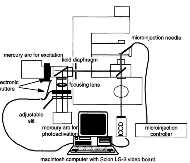
Measurement apparatus 2-6
System characterization 2-11
Lamp stability 2-11
Dynamic response and linearity of the imaging system 2-11
Discussion 2-12 Works Cited 2-13
CHAPTER 3 Interpreting PAF/FRAP
Measurements of Steady-State
Actin Dynamics
Introduction 3-2
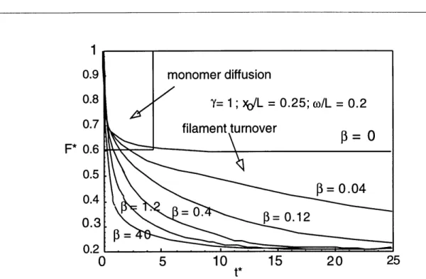
Mathematical model 3-3
Assumptions/idealization/simplifications: 3-3 Derivation of the dimensional problem: 3-4 Non-dimensional problem 3-6
Solution 3-7
Results 3-9
Concentration and fluorescence profile 3-9 Center-line decay 3-9
Long time behavior when fl << 1 3-11
Discussion 3-13 Appendix 3-14 Works Cited 3-17
CHAPTER 4 Baseline and control measurements in subconfluent
endothelial cells
Introduction 4-2
Electron Microscoscopy 4-5 Actin preparation and injections 4-6 PAF/FRAP Experiments 4-6
Photobleaching controls and corrections 4-7 Analysis 4-8
Geometry Considerations 4-9
Jasplakinolide and cytochalasin D treatments 4-9
Results 4-11
Both PAF and FRAP experiments exhibit biphasic decay consistent with the presence of two phases of actin 4-11
Estimates of D, FF, and t 4-12
PAF and FRAP experiments are sensitive to jas and cyto D 4-14
Discussion 4-15 Appendix 4-20 Works Cited 4-22
CHAPTER 5 Correlation of actin dynamics with cell motility and with
expression of actin associated proteins
Introduction 5-2
Methods 5-3
Cell culture and motility analysis 5-3 PAF and FRAP analysis 5-3
Results 5-4
Actin dynamics correlate with cell motility in a wounded endothelial monolayer 5-4 PAF and FRAP experiments agree in subconfluent and confluent endothelial cells 5-5
Gelsolin null and positive fibroblasts and ABP+ melanoma cells agree with endothelial cells correlations, but ABP- melanoma cells do not 5-8
ABP+ and ABP- cells migrate with different mechanisms 5-10
Discussion 5-10
Works Cited 5-12
CHAPTER 6 A Mechanistic Model ofActin Dynamics
Introduction 6-2
The Mathematical Model 6-5
Methods 6-10 System solution 6-10
Table of Contents
Pyrene assay 6-11 Results 6-12
Subconfluent cells have more and shorter filaments than confluent cells 6-12 Accelerating pointed end off rate effects dynamic state while accelerating nucleotide
exchange does not 6-13
Exposing filament barbed ends counters the acceleration of pointed end off rates 6-14 Filament severing shortens filaments and increases turnover but has a negligible effect on polymer fraction 6-14
Experiments and simulations agree qualitatively but not quantitatively 6-16
Discussion 6-18 Works Cited 6-19
CHAPTER 7 A System
for
the Assay of Steady-State Actin Dynamics InVitro
Introduction 7-2
Theory 7-3
Distinguishing filament turnoverfrom filament diffusion in PAF experiments 7-3 Adjusting w to visualize filament turnover 7-5
Methods 7-7
Preparation of caged-actin 7-7
Microcapillary preparation and actin polymerization 7-7 PAF experiments 7-8
Results 7-9
The role of an aci-nitro intermediate in fluorescence rise after uncaging 7-9 Demonstration offilament turnover by PAF 7-10
Discussion 7-10
Works Cited 7-14
List of Figures
Background and Literature Review
FIGURE 1.1. FIGURE 1.2. FIGURE 1.3. FIGURE 1.4. FIGURE 1.5. FIGURE 1.6. CHAPTER 2The structure of skeletal-muscle actin monomer. 1-2 Structure of the actin filament. 1-3
Evidence of actin nucleation. 1-4 Steady-state actin dynamics in vitro. 1-5
The actin cycle in cells and the role of actin associated proteins. 1-7 A recent model of actin dynamics in lamellae. 1-16
Experimental Set-up and Design
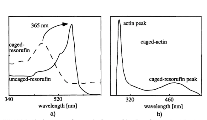
The synthesis of caged-actin. 2-5Absorbance spectra demonstrating the successful synthesis of caged-actin. 2-6
Schematic of the PAF measurement system. 2-7 Filter configurations. 2-8
Computer controlled automation of experiments. 2-9
Dynamic response of CCD and intensifier following sudden exposure to experimental light levels. 2-10
CHAPTER 3 Interpreting PAF/FRAP Measurements of Steady-State Actin Dynamics
FIGURE 3.1 FIGURE 3.2 FIGURE 3.3
Schematic of the model system. 3-4
Evolution of total fluorescence and fluorescence in filaments or monomers. 3-10
Decay of fluorescence in a photoactivated band. 3-11
Actin Dynamics in the Cell Cytoplasm CHAPTER 1 FIGURE 2.1. FIGURE 2.2. FIGURE 2.3. FIGURE 2.4. FIGURE 2.5. FIGURE 2.6. LOF-1
List of Figures FIGURE 4.1 FIGURE 4.2 FIGURE 4.3 FIGURE 4.4 FIGURE 4.5 CHAPTER 5
Photobleaching and background controls in PAF experiments. 4-10 Electron micrographs showing the region of BAECs studied by PAF and FRAP. 4-11
PAF and FRAP experiments in BAECs. 4-12 Corrected PAF and FRAP data. 4-13 Effects ofjasplankinolide. 4-14
Correlation of actin dynamics with cell motility and with
expression of actin associated proteins
Relationship between BAEC morphology and average cell speeds.5-5
PAF images sequence demonstrating different actin dynamics in cells in the a) wound edge and b) confluent regions of a recovering BAEC monolayer. 5-6
Cytoplasmic actin dynamics correlate with BAEC morphology. 5-7 Correlations of motility and actin dynamics. 5-8
Actin dynamics in gelsolin positive and null fibroblasts. 5-9 ABP+ and ABP- melanoma cells migrate with different mechanisms. 5-11
CHAPTER 6
A Mechanistic Model ofActin Dynamics
FIGURE 6.1. FIGURE 6.2. FIGURE 6.3. FIGURE 6.4. FIGURE 6.5. FIGURE 6.6.
Schematic of the cell actin cycle. 6-3
Procedure to identify actin cycle regulatory mechanisms important in endothelial cell motility. 6-4
Effects of pointed end disassembly and nucleotide exchange on the cell cycle parameters. 6-12
Levels of ATP and ADP bound monomer with accelerating filament turnover. 6-13
Effects of increasing filament barbed exposure on the cell cycle parameters. 6-14
Effects of regulating filament number on the actin cell cycle. 6-15
CHAPTER 4
Baseline and control measurements in subconfluent
endothelial cells
FIGURE 5.1. FIGURE 5.2. FIGURE 5.3. FIGURE 5.4. FIGURE 5.5. FIGURE 5.6.Profilin mediated barbed end assembly affects the actin cell cycle. 6-16 Comparison between experimentally determined and kinetic predictions of the actin cycle state in confluent and subconfluent endothelial cells. 6-17
CHAPTER 7
A System for the Assay of Steady-State Actin Dynamics In Vitro
FIGURE 7.1 FIGURE 7.2 FIGURE 7.3 FIGURE 7.4 FIGURE 7.5 FIGURE 7.6
System for the assay of actin dynamics using PAF 7-3
PAF experiment in the presence and of physiological levels of salt. 7-4 Photolysis of 2-nitrobenzyl caged compounds. 7-9
The effects of continuous exposure, pH and DTT on fluorescence rise. 7-11
Visualization of filament turnover. 7-11
Example PAF experiment demonstrating filament turnover. 7-12
Actin Dynamics in the Cell Cytoplasm
FIGURE 6.7. FIGURE 6.8.
List of Tables
CHAPTER 4
CHAPTER 6
Baseline and control measurements in subconfluent
endothelial cells
TABLE 4.1: Parameter estimates from PAF and FRAP in BAECs. 4-19 TABLE 4.2: Dynamic parameters in cells before and after treatment with Cyto
D. 4-19
A Mechanistic Model ofActin Dynamics
TABLE 6.1. Rate constants used in the kinetic model of the actin cycle in cells 6-6 TABLE 6.2. Steady-state rate equations describing the actin cycle in cells 6-9
CHAPTER 1
Background and
Literature Review
Monomeric actin spontaneously polymerizes in physiological buffers to form filaments. In cells, actin filaments are important mechanically. They organize into bundles and networks that define cell shape, strength, and adhesiveness. They are also highly dynamic. In most cells, the entire actin cytoskeleton is remodeled through steady-state filament assembly and disassembly at least once in a half hour. The dynamics of actin are important determinants of remodeling events in cell motility and endocytosis. This chapter introduces the protein actin and reviews what is known about its dynamic behavior in vivo and in vitro. The chapter concludes with an overview of the thesis.
Background and Literature Review
An Introduction to Actin
An Introduction to Actin
FIGURE 1.1. The structure of skeletal-muscle actin monomer. A cleft divides the monomer into two domains and binds a nucleotide and cation.(Sheterline, et aL, 1994).
The Importance of Actin in Cells
Actin is a highly conserved and abundant 42 kD monomeric protein in eukaryotes. Actin spontaneously self-assembles into filaments in the presence of physiological cations and salt. Cells distribute actin filaments into a multifunctional network known as the actin cytoskele-ton. As a organized scaffolding, the actin cytoskeleton determines cell shape and strength and participates in the transport and distribution of organelles. Actin polymerization drives the expansion of the cell membrane during cell spreading, crawling, and activation. Actin filament bundles associate with myosin to apply traction to substrates. Both contraction in the cell body
An Introduction to Actin
and actin dependent membrane extension are required for cell crawling. barbed end
Actin filaments continuously assemble and disassemble throughout the actin
monomer cell cytoplasm. Filament turnover is important in cell motility and other actin dependent processes such as endocytosis.
Actin Structure
poinTed end
FIGURE1.2. Structure
The three dimensional structure of the actin molecule has been solved by x-ray diffraction (Kabsch and Vandekerckhove, 1992;McLaughlin, et al.,
1993;Schutt, et al., 1993) and is shown in Figure 1.1. The molecule is divided into two domains by a cleft which holds a divalent cation (Ca2+ or Mg2+) and a nucleotide (ATP or ADP). Association between multiple residues on neighboring monomers stabilize the double helical actin polymer (Figure 1.2) (Holmes, et al., 1990). The ends of the polar fila-ment are termed "pointed" and "barbed" to reflect the arrowhead
appear-ance of myosin -labeled filaments in electron micrographs.
of the acutnflament. Three isoforms of actin (a, 3, y) are found in higher mammals (Herman,
Actin monomers
assemble into a withble 1993). Most cells express all three isoforms, however a is the primary
barbed and pointed
ends. (Alberts, et al., isoform in muscle and P and y are the primary isoforms in non-muscle
1994)
tissue. The isoforms are very similar in sequence and length indicating that actin has been heavily conserved during mammalian evolution. Small sequence differences concentrated near the N-terminus (Vanderkerchekhove and Weber, 1978;Vanderkerchekhove and Weber, 1979), give the isoforms different activities. The actin
Background and Literature Review
An Introduction to Actin
time (min)
--FIGURE 1.3. Evidence of actin nucleation. A delay in actin polymerization is overcome with the addition of pre-formed nuclei. The result demonstrates that the unfavorable formation of nuclei is the first step in actin polymerization. (Oosawa and Asakura, 1975)
forms distribute into different cell structures (DeNofrio, et al., 1989;Herman and D'Amore, 1989;Hoock, et al., 1991;Peng and Fischman, 1991), and have differing affinities for profilin (Larsson and Lindberg, 1988;Ohshima, et al., 1989), 034 thymosin (Weber, et al., 1992), and myosin (Hennessey, et al., 1993).
Actin self-assembly
Physiological levels of divalent cations and monovalent ions cause actin monomers to sponta-neously polymerize. Consistent with the theory of Oosawa (Oosawa and Asakura, 1962), polymerization begins with the unfavorable formation of a trimeric nucleus (Wegner and Engel, 1975). The slow formation of nuclei produces a lag in the polymerization process,
An Introduction to Actin pointed barbed
I.
net monomer
fluxI
ATP-Actin
0
ADP*Pi-Actin
0
ADP-Actin
FIGURE 1.4. Steady-state actin dynamics in vitro. In the presence of excess ATP net actin assembly at barbed ends and net disassembly at pointed ends drives monomer flux through filaments. The hydrolysis ofATP soon after assembly provides energy driving actin cycle known as treadmilling.
formed, monomers add to barbed ends approximately 10 times faster than to pointed ends (Kondo and Ishiwata, 1976; Bonder, et al. 1986; Korn, et al., 1987;Pollard, 1986).
In the presence of ATP, actin polymerizes until only 0.1 jgM remains unpolymerized (Drenckhanh and Pollard, 1986;Pollard, 1986;Wegner and Isenberg, 1983). This concentration of monomer, the minimum or 'critical concentration' required for filament assembly, exists in a dynamic steady-state with filaments (Figure 1.4). At steady-steady-state, ATP-actin assembles onto filament barbed ends and rapidly hydrolyzes its bound ATP to the intermediate ADP*Pi (Carlier and Pantaloni, 1986). Pi is slowly released from the sides of actin filaments as monomers flux from barbed to pointed ends where they are predominantly bound to ADP (Carlier and Pantaloni, 1986;Korn, et al., 1987). ATP hydrolysis provides the energy required to maintain the biochemical differences between filament ends that permit this monomer flux known as "treadmilling" (Wegner and Isenberg, 1983).
Background and Literature Review The Actin Cycle is Regulated in Cells
Actin depolymerization occurs when fully polymerized actin is diluted below the critical con-centration. In these experiments, ATP-actin capped barbed ends disassemble monomer faster than ADP-actin capped pointed ends by a factor of 5 -10 (Janmey and Stossel, 1986;Pollard, 1986). Slow rates of disassembly at filament pointed ends (0.03 -0.27 sec 1) predict that actin filaments should be relatively stable in cells where access to the majority of barbed ends is blocked by barbed end capping proteins.
The Actin Cycle is Regulated in Cells
Section Summary
In cells actin-associated proteins regulate the dynamics of the ATP-powered actin cycle (Fig-ure 1.5). Some proteins bind to the barbed ends of actin filaments to block filament assembly, others to the pointed ends to accelerate disassembly, and still others to the sides of actin fila-ments to sever them. Monomer sequestering proteins maintain unpolymerized actin in cells at concentrations hundreds of times higher than the critical concentration for filament formation. Monomer affinity for sequestering proteins is higher than for pointed ends but lower than for barbed ends. Consequently, a sudden exposure of barbed ends leads to massive actin polymer-ization. Proteins enhance assembly by catalyzing the exchange of nucleotide on actin mono-mer and escorting the charged monomono-mer to barbed ends. Many of the proteins regulating the actin cycle are themselves regulated by signaling pathways.
The Actin Cycle is Regulated in Cells
filament
severing
acceler
disasser
barbed end
capping
ADP
ac
S
ADP*Pi
ated
ATP ac
nbly
nucleotide
*
(
exchange
"tin
actin
tin
*
monomer
sequestration
FIGURE 1.5. The actin cycle in cells and the role of actin associated proteins. The proteins involved in barbed
end capping, severing, accelerated disassembly, and nucleotide exchange are regulated and represent control points in the actin cycle.
Monomer sequestering proteins
Profilins, ADF/cofilins, and P-thymosins are ubiquitously expressed proteins that bind actin monomers in one-to-one complexes. Of these, only P4 thymosin is present in quantities large
enough to account for the levels of sequestered actin in most cells (Safer, et al., 1990). The others, discussed below, appear to have catalytic roles in the actin cycle.
0-4 thymosin, the most common 3-thymosin isoform, is a 5 kD protein with gLM affinity for
ATP-actin and a 100 fold lower affinity for ADP-ATP-actin (Carlier, et al., 1993). P-4 thymosin hinders
Actin Dynamics in the Cell Cytoplasm 1-7
1444
aw
Background and Literature Review The Actin Cycle is Regulated in Cells
nucleotide exchange on actin (Carlier, et al., 1993). Because it is unregulated, present in cells at similar concentrations to actin, and sequesters actin in 1:1 complexes in vitro, 0-4 thy-mosins were originally thought of as a simple monomer sequestering protein. However, over-expression does not decrease polymerized actin in NIH3T3 cells (Safer and Nachmias,
1994;Sun, et al., 1995) and at high concentrations, 3-4 thymosin (> 20 pM) does not sequester actin efficiently in vitro (Carlier, et al., 1996). No function has been attributed to 0-4 thy-mosin's departure from simple sequestering behavior.
Barbed end capping/ filament severing proteins
Sequestering proteins have a lower affinity for monomer than actin filament barbed ends. To maintain high levels of sequestered monomer, cells express proteins that bind the majority of barbed ends with nM affinity. The most widely expressed are the gelsolin family of proteins and capping protein (CP). Gelsolin requires tpM calcium to bind barbed ends (Yin, et al.,
1981) whereas CP is constitutively active. Both gelsolin (Janmey and Stossel, 1987) and CP (Heiss and Cooper, 1991) can be removed from filament barbed ends by phosphatidylinositol 4,5-bisphosphate (PIP2) and other phosphoinositides. Gelsolin and CP are present in many cells at quantities sufficient to cap all their filaments (Amatruda, et al., 1992;Barkalow, et al.,
1996;Hartwig, 1992). The regulatory features of gelsolin and capping protein allow extracel-lular signals to control actin polymerization by mobilizing intracelextracel-lular Ca2+ and regulating phospholipid metabolism (discussed below).
The importance of the barbed end capping activity of CP in maintaining and remodeling cellu-lar actin has been demonstrated in a number of model systems. The loss of CP in yeast
The Actin Cycle is Regulated in Cells
mutants disrupts the organization and dynamics of the actin cytoskeleton (Amatruda, et al., 1990;Amatruda, et al., 1992). Increased expression of CP in Dictyostelium cell lines decreases the number of free barbed ends, decreases the fraction of actin polymerized, and increases cell motil-ity (Hug, et al., 1995). Regulation of CP activmotil-ity by PIP2 is involved in barbed end exposure dur-ing platelet (Barkalow, et al., 1996) and Dictyostelium activation (Eddy, et al., 1996).
Gelsolin caps, severs and nucleates actin filaments (Burtnick, et al., 1997,Yin et al., 1981, Kwiat-kowski et al., 1985). After severing, gelsolin binds to a barbed end to produce one free and one capped filament. Activation of gelsolin is most effective in vitro at > 10-20 PM Ca2+ (Lamb, et al., 1993), a level -~ 1000 times higher than resting cell levels and 10 times higher than average cell values during Ca2+ mobilization. Gelsolin may be activated by high Ca2+ concentrations near stores or the Ca2+ requirement may be lowered by changes in cellular pH (Lamb, et al., 1993). Regardless of how it occurs, gelsolin is active in cells. Gelsolin-actin form and then dissociate when cells are stimulated to move (Barkalow, et al., 1996;Hartwig, 1992;Howard, et al., 1990; Cunningham, et al., 1991). Cells isolated from gelsolin null mice have phenotypes distinguishable from wild type cells. Most notably, the loss of gelsolin slows motility in fibroblasts (Witke, et al., 1995), neutrophils (Witke, et al., 1995), and neurites (Lu, et al., 1997), enhances stress fiber for-mation in fibroblasts (Witke, et al., 1995), and hinders actin assembly in platelets after thrombin or glass activation (Witke, et al., 1995). Experiments in gelsolin null fibroblasts demonstrate gelsolin's importance in membrane ruffling as these fail to form ruffles or lammelipodia (Azuma, et al., 1998).
Background and Literature Review The Actin Cycle is Regulated in Cells
Profilin
Profilins are small 12-15 kD proteins originally identified as sequestering proteins because of their ability to prevent actin polymerization in vitro (Carlson, et al., 1977). Follow-up studies, however, revealed that profilin actually enhances actin polymerization in many circumstances. At steady-state, and in the presence of ATP, profilin decreases the critical concentration of
actin by a factor of 10 (Pantioni and Carlier, 1993;Pollard and Cooper, 1984) demonstrating that the profilin promotes assembly at barbed ends at steady-state. Profilin-mediated assembly
at steady-state is dramatically demonstrated when profilin is used in combination with a large pool of monomer sequestered by
I34
thymosin (Pantioni and Carlier, 1993). Mammalian profi-lin accelerates the dissociation of ADP from actin monomer 1000 fold (Goldschmidt-Cler-mont, et al., 1991), catalyzing the exchange of ADP for ATP. In the treadmilling model ofactin dynamics (Figure 1.4) ADP actin is released from pointed ends into the monomer pool. Very high rates of filament turnover may overwhelm actin's intrinsic ability to exchange its bound nucleotide and deplete ATP-actin for barbed end assembly. Under these conditions pro-filin can accelerate nucleotide exchange to maintain filament turnover (Goldschmidt-Cler-mont, et al., 1992;Mitchison, 1992;Theriot and Mitchison, 1993). Plant profilins enhance barbed end assembly but do not promote nucleotide exchange indicating that these functions
are uncoupled (Perelroizen, et al., 1996).
In vivo data also argue for profilin's role in promoting polymerization. Yeast lacking profilin are unable to repolymerize actin following temperature elevation (Yeh and Haarer, 1996). An expression study in CHO cells demonstrates that enhanced profilin expression correlates with longer filament lifetimes and an increased polymer fraction (Finkel, et al., 1994).
The Actin Cycle is Regulated in Cells
Like gelsolin, gelsolin-actin complexes and CP, the profilin-actin complex binds PIP2 (Lassing and Lindberg, 1985). Once bound to PIP2, actin disassociates from the complex (Lassing and Lindberg, 1988) and the profilin-PIP2 complex resists degradation by PLC-y (Goldschmidt-Cler-mont, et al., 1990). There is also evidence that profilin is a downstream component of cytoskele-ton targeted pathways of the Ras family of small GTPases (Tanaka and Takai, 1998).
ADF/cofilin
The stability of actin filament pointed ends in vitro (Janmey and Stossel, 1986;Pollard, 1986) could not be resolved with the high rates of turnover measured in vivo until a recent demonstration that members of the ADF/cofilin family bind preferentially to ADP-actin near pointed ends to accelerate disassembly by a factor of 25 (Carlier, et al., 1997). The ability of cofilin to accelerate turnover is supported by shortening of Lysteria tails in cell extracts (Carlier, et al., 1997;Rosenb-latt, et al., 1997) and by experiments in yeasts demonstrating the importance of ADF/cofilin in supporting the surprisingly high rates of filament turnover in these non-motile cells (Lappalainen and Drubin, 1997). However, the yeast and Lysteria data cannot rule out that filament turnover is accelerated by filament severing, an activity originally proposed to explain coflin's reduction of actin gel viscosity (Cooper, et al., 1986). Carlier and colleagues (Carlier, et al., 1997), however demonstrate exhaustively that filament severing cannot explain their kinetic and structural data. ADF/cofilin is regulated by the reversible phosphorylation of a serine residue near the amino ter-minus (Nebl, et al., 1996). Dephosphorylation activates cofilin (Moon and Drubin, 1995). The regulation of the phosphatase is complex as phosphatase inhibitors promote the dephosphorylated state (Okada, et al., 1996;Takuma, et al., 1996) but a recent study in neurites identifies Ca2+and
Background and Literature Review The Actin Cycle is Regulated in Cells
cAMP as second messengers in pathways leading to cofilin deposphorylation (Meberg, et al., 1998).
Actin Directed Signalling Pathways
The regulation of actin associated proteins by phosphoinositides and their breakdown prod-ucts in vitro suggests signaling pathways can control phosphoinositide metabolism to control actin assembly. Phosphoinositides recruit CP and gelsolin from barbed ends to promote actin assembly and bind profilin to block PLC-y mediated hydrolysis. The hydrolysis of phosphoi-nositides liberates gelsolin and capping proteins and produces Ca2+ to activate gelsolin and (in one example) the phosphatase of cofilin (Meberg, et al., 1998). The in vitro data predict that synthesis and hydrolysis of phosphoinositides in vivo should correlate with filament assembly
and filament breakdown respectively. Consistent with this prediction, platelet gelsolin and capping protein are activated during periods of phosphatidylinositol 4, 5 bisphosphate (PIP2) breakdown and lose activity during periods of actin assembly and PIP2 synthesis (Hartwig, et al., 1995;Hartwig, et al., 1996;Kovacsovics, et al., 1995). PIP2 synthesis also correlates with actin assembly in other cell types (Apgar, 1995;Carson, et al., 1992). Examples where PIP2 synthesis does not correlate with actin assembly demonstrate yet unresolved pathways to actin assembly (Dadabay, et al., 1991;Zigmond, et al., 1997).
Members of the Rho family GTPases are essential regulators of signaling pathways directing actin organization (Tapon and Hall, 1997). Constitutively active RhoA promotes filament bun-dling and adhesion complexes in serum starved cells (Hotchin and Hall, 1995;Ridley and Hall, 1992). Active Rac is essential for generating membrane ruffles found at the leading edge of
Actin Dynamics in the Cell Cytoplasm
motile cells (Ridley, et al., 1992). Finally, active Cdc42 elicits narrow membrane extensions known as filopodia (Kozma, et al., 1995).
Links between the Rho family GTPases and actin associated proteins are emerging. Rac, Cdc42, and Rho bind phosphoinositide kinases in vitro and in vivo (Carpenter, et al., 1997;Chong, et al., 1994;Hartwig, et al., 1995;Tolias, et al., 1995), and active Rac induces PIP2 synthesis and barbed end uncapping in permeabilized platelets (Hartwig, et al., 1995). Intermediates in RhoA signaling bind profilin in yeast (Imamura, et al., 1997). Gelsolin is essential for Rac mediated membrane ruffling in cultured fibroblasts (Azuma, et al., 1988).
Actin Dynamics in the Cell Cytoplasm
The term "actin dynamics" includes the kinetics of actin assembly and disassembly, monomer and filament diffusion, and bulk movements of the actin network. Actin is dynamic throughout motile and non-motile cells. The dynamics of actin can vary between cell types and spatially within cells to control cell motility and other actin-dependent processes such as endocytosis. Models and experiments on cell motility focus on the behavior of actin in the protrusive regions of cells. The dynamics of actin in these structures are characterized by net polymerization at the leading mem-brane, retrograde flow from the memmem-brane, and net depolymerization near the cell body.
Photoactivation of fluorescence and fluorescence recovery after photobleaching
The dynamics of actin in living cells has been observed using the techniques of fluorescence recovery after photobleaching (FRAP) and photoactivation of fluorescence (PAF) (Cramer, et al.,
Background and Literature Review Actin Dynamics in the Cell Cytoplasm
1997;Finkel, et al., 1994;Kreis, 1982;Theriot and Mitchison, 1991;Theriot and Mitchison, 1992;Wang, 1985). In these experiments a fluorophore-labeled actin derivative is microin-jected into living cells where it incorporates into the native cytoskeleton. In FRAP, the
fluores-cence derived from the injected actin is quenched locally under intense laser excitation. In PAF, a focused band of UV excitation converts a non-fluorescent fluorophore derivative (a 'caged' fluorophore) back to its fluorescent parent (Mitchison, 1989). In both techniques the evolution of fluorescence in the illuminated region is monitored to infer information about actin dynamics.
Monomer diffusion
The diffusion of ficoll and dextran in cells demonstrates that the mobility of inert particles depend more strongly on their size in cytoplasm than in water (Luby-Phelps, 1987). This non-newtonian behavior is due partly to the sieving effect of cytoskeletal pores and partly to the background of proteins crowding the cytosol (Hou, et al., 1990). The result is that a 28 A actin monomer (Lanni and Ware, 1984) diffusing in cytoplasm experiences a cytoplasmic viscosity which should reduce its mobility 5 fold from aqueous (Luby-Phelps, et al., 1986). However, the consensus values for monomer mobility in cytoplasm are 3-6 x 10⁻⁸ cm2/sec (Giuliano and Taylor, 1994;Luby-Phelps, 1985), = 15 times lower than values in water (Lanni and Ware, 1984). This disparity has not been resolved but may indicate 1) that the turnover of actin fila-ments is so rapid that it hinders the ability of monomers to diffuse; 2) that small filafila-ments dif-fuse through the cytoplasm and contribute to the measured mobility; or 3) that actin monomer binds transiently to sequestering molecules much larger in size than those currently identified (Luby-Phelps, et al., 1988).
Actin Dynamics in the Cell Cytoplasm
Filament turnover and retrograde flow
The first evidence that actin filaments continuously remodel in living cells came from microinjec-tion studies. These studies demonstrated that fluorescently labeled actin incorporates into all existing actin structures in less than a half hour (Amato, 1986;Kreis, et al., 1979). Local pho-tobleaching of fluorescent actin at the leading edge of motile fibroblasts determined a time scale for complete fluorescence recovery of several minutes (Cramer, et al., 1997;Wang, 1985). Kerato-cytes, which can move 10 times faster than fibroblasts appear to turn over actin filaments in lead-ing lamella in tens of seconds (Theriot and Mitchison, 1991). Estimates of filament turnover near or in the cell body of fibroblasts and PtK3 cells are on the order of 5-10 minutes (Cramer, et al., 1997;Theriot and Mitchison, 1991). FRAP studies also demonstrated that actin moves rearward in the leading edge of cells (Theriot and Mitchison, 1992;Wang, 1985), consistent with the tread-milling model of filament turnover seen in vitro (Wegner, 1976). Organelles, surface attached beads, and actin rich membrane protrusions resolved by various optical methods move rearward at similar rates (Fisher, et al., 1988;Waterman-Storer and Salmon, 1997), but are faster than the movement of photoactivated and photobleached actin marks (Condeelis, 1993;Theriot and Mitch-ison, 1992). These results may suggest that PAF and FRAP measure the dynamics of a subset of actin filaments in lamella (Condeelis, 1993;Theriot and Mitchison, 1992).
Before the discovery that ADF/cofilin could accelerate pointed end off rates the treadmilling model for actin dynamics in motile cells could not be resolved with in vitro rates of disassembly, and alternative models were proposed in which lamella were comprised primarily of short fila-ments (Stossel, 1993;Theriot and Mitchison, 1991). Conflicting data on filament lenghts in lamel-lae contributed to this debate (Hartwig, 1992;Hartwig and Shevlin, 1986;Small, et al., 1995). A
Background and Literature Review
Actin Dynamics in the Cell Cytoplasm
a)
very slow turnover
+
cofilin
c)
accelerated turnover + capping protein accelerated turnover+
capping protein
highest turnovertreadmilling funneled assembly
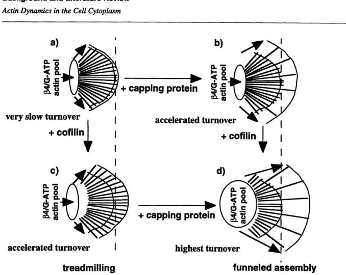
FIGURE 1.6. A recent model of actin dynamics in lamellae (Carlier, 1998). The model describes a synergy
between the actin associated proteins cofilin and capping protein. In the absence of capping protein or cofilin filament turnover is slow and filaments treadmill a). With the addition of only capping protein, disassembling monomers are funneled to a small number offree barbed ends which grow b). With the addition of only cofilin, filament disassembly and barbed end assembly accelerate to give faster treadmilling c) When both cofilin and capping protein exist, the funneled growth at barbed ends is maximized d). Note that as filament turnover accelerates the pool of actin sequestered by f4 thymosin
grows.
current model for lamellar structure and dynamics revises the treadmilling model in light of the ADF/cofilin data (Carlier, 1998;Carlier and Pantaloni, 1997). In this view (Figure 1.6) actin assembly occurs at a small number of free barbed ends beneath the leading edge. The remaining filaments are capped and disassembling quickly due to ADF/cofilin binding, so that
The Story of this Thesis
free barbed ends advances the leading edge at rates higher than would be possible if all barbed ends were free as in treadmilling. The new model also predicts that higher levels of filament turn-over should correlate with a transfer of actin from filaments to sequestered monomer.
The Story of this Thesis
The original ambition of this work was to resolve the in vitro/ in vivo turnover paradox. In review-ing the relevant literature, we noticed that FRAP and PAF experiments were interpreted differ-ently. Specifically, FRAP experiments designed to measure monomer diffusion ignored filament turnover, and PAF experiments assaying filament turnover ignored monomer diffusion. Also resorufin was highly photolabile so that fluorescence decay rates could easily be accelerated by inadvertent photobleaching. I investigated if these issues could explain the surprisingly high rates of filament turnover measured in vivo by Theriot and Mitchison, 1991.
We developed a mathematical model of actin dynamics designed specifically for the interpretation of PAF and FRAP experiments (Chapter 3), and constructed a system similar to the one developed by Theriot and Mitchison, 1991 but with greater temporal resolution (Chapter 2). Endothelial cells provided a model system because of their capacity to dynamically reorganize their actin cytoskel-eton in response to mechanical stimuli. Experiments revealed that filaments turnover in about 7 minutes in the bulk cytoplasm of endothelial cells (Chapter 5). Unlike previous PAF and FRAP studies, our experiments simultaneously produced estimates for the actin monomer diffusion coefficient (3.1 x 10⁻⁸ cm2/sec) and the fraction of actin polymerized (37%) in these cell. This lead to the conclusion that while filament turnover was rapid in the bulk cytoskeleton of
Background and Literature Review The Story of this Thesis
lial cells, it was not diffusion limited. FRAP data obtained in a collaboration demonstrated the general equivalence of the two techniques and that photobleaching had been controlled. The resolution of our imaging system did not allow photoactivation within leading lamellipodia, however the turnover rate in the bulk cytoplasm was still too high to be explained by in vitro turnover rates.
In 1996 the in vitro/ in vivo paradox was apparently resolved with the demonstration that ADF/cofilin accelerated pointed end off rates by a factor of 25 (Carlier, et al., 1997). The
focus of this work then shifted to elucidating the mechanism of cell motility. While previous work demonstrated that filament turnover was fastest in the fastest cell types (Carlier, et al.,
1997;Theriot and Mitchison, 1991;Theriot and Mitchison, 1992;Wang, 1985), there was no evidence that cells of a single type could accelerate turnover as they moved faster. I demon-strated this by applying PAF and FRAP to endothelial cells moving at different rates in wounded monolayers (Chapter 5). Unexpectedly that the fraction of actin polymerized
decreased with increasing speed in these cells. This result confirms the predictions of a recent model of cell motility which attributes the trend to accelerated disassembly with ADF/cofilin. However, experiments in gelsolin positive and null fibroblasts exhibited similar trends, raising the prospect that filament severing may be the mechanism involved. In preliminary experi-ments, subconfluent endothelial cells were found to have three times as many filaments than confluent cells providing further evidence of filament severing (Chapter 6).
To elucidate the mechanisms controlling cell motility in endothelial cells I constructed a kinetic model of the actin cycle that includes filament severing and accelerated pointed end disassembly as mechanisms (Chapter 6). Using protein levels and kinetic rate constants from
Works Cited
the literature, simulations demonstrate that accelerated filament disassembly is the most effective mechanism producing a simultaneous decrease in the polymer fraction and average lifetime of fil-aments. In combination with the experimental data, this kinetic model suggests that both filament severing and accelerated disassembly occur as endothelial cells move faster, but that accelerated disassembly has more influence on the dynamic state of the cytoskeleton.
I have developed a PAF based in vitro system for examining the effects of actin associated
pro-teins on steady-state actin dynamics (Chapter 7). The system improves over existing techniques in providing a non-invasive assay of filament turnover, polymer fraction, and filament length in a single preparation of actin. The system has provided the first visualization of actin filament turn-over.
Works Cited
1. Alberts, B., D. Bray, J. Lewis, M. Raff, K. Roberts, and J. Watson. 1994. Molecular Biology of
the Cell. Garland Publishing, Inc., New York.
2. Amato, P. A. T., D.L. 1986. Probing the mechanism of incorporation of fluorescently labeled actin into stress fibers. J. Cell Biol. 102:1074-1084.
3. Amatruda, J., J. Cannor, K. Tatchell, C. Hug, and J. Cooper. 1990. Disruption of the actin
cytoskeleton in yeast capping protein mutants. Nature. 344:352-354.
Background and Literature Review Works Cited
4. Amatruda, J., D. Gattermeir, T. Karpova, and J. Cooper. 1992. Effects of null mutations and overexpression of capping protein on morhpogenesis, actin distribution and polarized secre-tion in yeast. J Cell Biol. 119:1151-62.
5. Apgar, J. 1995. Activation of protein kinase C in rat basophilic leukemia cells stimulates increased productionof phosphatidylinositol 4-phosphate and phsphatidylinositol 4,5-bisphos-phate: correlation with actin polymerization. Mol Biol Cell. 6:97-108.
6. Azuma, T., W. Witke, T. Stossel, J. Hartwig, and D. Kwiatkowski. 1988. Gelsolin is a down-stream effector of rac for fibroblast motility. Embo J. 17:1362-1370.
7. Azuma, T., W. Witke, T. Stossel, J. Hartwig, and D. Kwitkowski. 1998. Gelsolin is a down-stream effector of rac for fibroblast motility. Embo J. 17:1362-1370.
8. Barkalow, K., W. Witke, D. Kwiatkowski, and J. Hartwig. 1996. Coordinated regulation of platelet actin filament barbed ends by gelsolin and capping protein. J Cell Biol. 134:389-399.
9. Burtnick, L., E. Koepf, J. Grimes, E. Jones, D. Stuart, P. McLaughlin, and R. Robinson. 1997. The crystal structure of plasma gelsolin: implications for actin severing, capping, and nucleation. Cell. 90:661-670.
10. Carlier, M. 1998. Control of actin dynamics. Curr Opin Cell Biol. 10:45-51.
11. Carlier, M., D. Didry, I. Erk, J. Lepault, M. Van Troys, J. Vanderkerckhove, I. Perelroizen, H. Yin, Y. Doi, and D. Pantaloni. 1996. Tb4 is not a simple G-actin sequestering protein and
Works Cited
12. Carlier, M., and D. Pantaloni. 1986. Direct evidence for ADP-Pi-F-actin as the major interme-diate in ATP-actin polymerization. Rate of dissociation of Pi from actin filaments. Biochemistry. 25:7789-7792.
13. Carlier, M., and D. Pantaloni. 1997. Control of actin dynamics in cell motility. Mol Biol Cell. 269:459-467.
14. Carlier, M.-F., C. Jean, K. Rieger, M. Lenfant, and D. Pantaloni. 1993. Modulation of the interaction between G-actin and thymosin f34 by the ATP/ADP ratio: possible implication in the regulation of actin dynamics. Proc. Natl. Acad. Sci., U.S.A. 90:5034-5038.
15. Carlier, M.-F., V. Laurent, J. Santolini, R. Melki, D. Didry, G.-X. Xia, Y. Hong, N.-H. Chua, and D. Pantaloni. 1997. Actin depolymerizing factor (ADF/cofilin) enhances the rate of filament turnover: implication in actin based motility. J. Cell Biol. 136:1307-1322.
16. Carlson, M., P. Babcock, P. Rubenstein, and Y. Wang. 1977. Actin polymerizability is influ-enced by profilin, a low molecular weight protein in non-muscle cells. J Mol Biol. 115:465-483. 17. Carpenter, C., K. Tolias, A. Couvillon, and J. Hartwig. 1997. Signal transduction pathways involving the small G proteins rac and Cdc42 and phosphoinositide kinases. Adv Enzyme Regul. 37:377-390.
18. Carson, M., S. Shasby, S. Lind, and D. Shasby. 1992. Histamine, actin-gelsolin binding, and polyphsphoinositides in human umbilical vein endothelial cells. Am J PHysiol. 263:L664-L669.
Background and Literature Review Works Cited
19. Chong, L., A. Traynor-Kaplan, G. Bokoch, and M. Schwartz. 1994. The small GTP-bind-ing protein Rho regulates a phosphatidylinositol 4-phosphate 5-kinase in mammalian cells.
Cell. 79:507-513.
20. Condeelis, J. 1993. Life at the leading edge: the formation of cell protrusions. Annu Rev Cell Biol. 9:411-441.
21. Cooper, J., J. Blum, R. Williams, and T. Pollard. 1986. Purification and characterization of Actophorin, a new 15,000 dalton actin -binding protein from Acanthamoeba castellanii. J Biol Chem. 261:477-485.
22. Cramer, L. P., M. Siebert, and T. J. Mitchison. 1997. Identification of novel graded polarity actin filament bundles in locomoting heart fibroblasts: implications for the generation of motile force. J. Cell Biol. 136:1287-1305.
23. Cunningham, C., T. Stossel, and D. Kwiatkowski. 1991. Enhanced motility in NIH 3T3 fibroblasts that overexpress gelsolin. Science. 251:1233-1236.
24. Dadabay, C., E. Patton, J. Cooper, and L. Pike. 1991. Lack of a correlation between changes in polyphosphoinositide levels and actin/gelsolin complexes in A431 cells treated with epidermal growth factor. J Cell Biol. 112:1151-1156.
25. DeNofrio, D., T. Hoock, and I. Herman. 1989. Functional sorting of actin isoforms in microvascular pericytes. J Cell Biol. 109:191-202.
Works Cited
26. Drenckhanh, D., and T. Pollard. 1986. Elongation of actin filaments is a diffusion-limited reaction at the barbed end and is accelerated by inert macromolecules. J Biol Chem. 261:12754-12758.
27. Eddy, R., R. Sauterer, and J. Condeelis. 1996. A major agonist-related capping activity in Dic-tyostelium is due to the capping protein, cap32/34. Biochim Biophys Acta. 1314:247-259.
28. Finkel, T., J. A. Theriot, K. D. Dise, G. E Tomaselli, and P. J. Goldschmidt-Clermont. 1994. Dynamic actin structures stabilized by profilin. Proc. Natl. Acad. Sci. USA. 91:1510-1514.
29. Fisher, G., P. Conrad, R. DeBiasio, and D. Taylor. 1988. Centripetal transport of cytoplasm, actin and the cell surface in lamellipodia of fibroblasts. Cell Motil Cytoskeleton. 11:235-47. 30. Giuliano, K. A., and D. L. Taylor. 1994. Fluorescent actin analogs with a high affinity for pro-filin in vitro exhibit an enhanced gradient of assembly in living cell. J. Cell Biol. 124:971-983.
31. Goldschmidt-Clermont, P., M. Furman, D. Wachsstock, D. Safer, V. Nachmias, and T. Pollard. 1992. The control of actin nucleotide exchange by thymosin34 and profilin. A potential regulatory mechanism for actin polymerization in cells. Mol. Biol. Cell. 3:1015-1024.
32. Goldschmidt-Clermont, P., L. Machesky, J. Baldassare, and T. Pollard. 1990. The actin-bind-ing protein profilin binds to PIP2 and inhibits its hydrolysis by phospholipase C. Science.
251:1231-1233.
33. Goldschmidt-Clermont, P., L. Machesky, S. Doberstein, and T. Pollard. 1991. Mechanism of the interaction of human platelet profilin with actin. J Cell Biol. 113:1081-1089.
Background and Literature Review
Works Cited
34. Hartwig, J. 1992. Mechanisms of actin rearrangements mediating platelet activation. J. Cell Biol. 1992:1421-1442.
35. Hartwig, J., G. Bokoch, C. Carpenter, P. Janmey, L. Taylor, and T. Stossel. 1995. Throm-bin receptor ligation and activated rac uncap actin filament barbed ends through
phosphoi-nositide synthesis in permeabilized human platelets. Cell. 82:643-653.
36. Hartwig, J., S. Kung, T. Kovacsovics, P. Janmey, L. Cantley, T. Stossel, and A. Toker. 1996. D3 phosphoinositides and outside-in signaling by glycoprotein IIb-IIIa mediate platelet actin assembly and filpodial extension induced by phorbos 12-myristate 13-acetate. J Biol Chem. 271:32986-32993.
37. Hartwig, J. H., and P. Shevlin. 1986. The architecture of actin filaments and the ultrastruc-tural location of actin-binding protein in the periphery of lung macrophages. J. Cell. Biol.
103:1007-1020.
38. Heiss, S., and J. Cooper. 1991. Regulation of CapZ, an actin capping protein of chicken muscle, by anionic phspholipids. Biochemistry. 30:8753-8758.
39. Hennessey, E., D. Drummond, and J. Sparrow. 1993. Molecular genetics of actin function. Biochem J. 291:657-671.
40. Herman, I. 1993. Actin Isoforms. Curr Opin Cell Biol. 5:48-55.
Works Cited
42. Holmes, K., D. Popp, W. Gebhard, and W. Kabsch. 1990. Atomic model of the actin filament. Nature. 347:44-49.
43. Hoock, T., P. Newcomb, and I. Herman. 1991. 3 actin and its mRNA are localized at the plasma membrane and the regions of moving cytoplasm during the cellular response to injury. J Cell Biol. 112:653-664.
44. Hotchin, N., and A. Hall. 1995. The assembly of integrin adhesion complexes requires both extracellular matrix and intracellular Rho/Rac GTPases. J Cell Biol. 131:1857-1865.
45. Hou, L., E Lanni, and K. Luby-Phelps. 1990. Tracer diffusion in F-actin and Ficoll mixtures
-towards a model for cytoplasm. Biophys J. 58:31-43.
46. Howard, T., C. Chaponnier, H. Yin, and T. Stossel. 1990. Gelsolin-actin interaction in actin polymerization in human neutrophils. J Cell Biol. 110:1983-1992.
47. Hug, C., P. Jay, I. Reddy, J. McNally, P. Bridgeman, E. Elson, and J. Cooper. 1995. Capping protein levels influnce actin assembly and cell motility in dictyostelium. Cell. 81:591-600. 48. Imamura, H., K. Tanaka, T. Hihara, M. Umikawa, T. Kamei, K. Takahashi, T. Sasaki, and Y.
Takai. 1997. Bnilp and Bnrlp: Downstream targets of the Rho family small G-proteins which interact with profilin and regulate actin cytoskeleton in Saccharomyces cerevisiae. EMBO J. 16:2745-2755.
49. Janmey, P. 1994. Phosphoinositides and calcium as regulators of cellular actin assembly and disassembly. Annu Rev Physiol. 56:169-91.
Background and Literature Review Works Cited
50. Janmey, P., and T. Stossel. 1987. Modulation of gelsolin function by phosphatidylinositol 4,5-bisphosphate. Nature. 325:362-364.
51. Janmey, P. A., and T. P. Stossel. 1986. Kinetics of actin monomer exchange at the slow
growing ends of actin filaments and their relation to the elongation of filaments shortened by gelsolin. J. Muscle Res. Cell Motility. 7:446-454.
52. Kabsch, W., and J. Vandekerckhove. 1992. Structure and function of actin. Annu. Rev. Bio-phys. Biomol. Struct. 21:49-76.
53. Korn, E. D., M.-F. Carlier, and D. Pantaloni. 1987. Actin polymerization and ATP
hydrol-ysis. Science. 238:638-644.
54. Kovacsovics, T., C. Bachelot, A. Toker, C. Vlahos, B. Duckworth, L. Cantley, and J. Hartwig. 1995. Phosphoinositide 3-kinase inhibition spares actin assembly in activating plate-lets but reverses platelet aggregation. The Journal of Biological Chemistry. 270:11358-11366.
55. Kozma, R., S. Ahmed, A. Best, and L. Lim. 1995. The Ras-related protein, Cdc42Hs and
bradykinin promote formation of peripheral actin micrspikes in filopodia in Swiss 3T3 fibro-blasts. Mol Cell Biol. 15:1942-1952.
56. Kreis, T., Geiger, B. , Schlessinger, J. 1982. Mobility of microinjected rhodamine actin within living chicken gizzard cells determined by fluorescence photobleaching recovery. Cell. 29:835-845.
Works Cited
57. Kreis, T., K. Winterhalter, and W. Birchmeier. 1979. In vivo distribution and turnover of fluo-rescently labeled actin microinjected into human fibroblasts. Proc. Natl. Acad. Sci. 76:3814-3818.
58. Kwiatkowski, D., P. Janmey, J. Mole, and H. Yin. 1985. Isolation and properties of two actin-binding domains in gelsolin. J Biol Chem. 260:15232-15238.
59. Lamb, J., P. Allen, B. Tuan, and P. Janmey. 1993. Modulation of felsolin function: activation at low pH overrides Ca2 + requirement. J Biol Chem. 268:8999-9004.
60. Lanni, F., and B. Ware. 1984. Detection and characterization of actin monomers, oligomers, and filaments in solution by measurement of fluorescence photobleaching recovery. Biophys J. 46:97-110.
61. Lappalainen, P., and D. Drubin. 1997. Cofilin promotes rapid actin filament turnover in vivo. Nature. 388:1165-1168.
62. Larsson, H., and U. Lindberg. 1988. The effect of divalent cations on the interaction between spleen profilin and different actins. Biochim Biophys Acta. 953:95-105.
63. Lassing, I., and U. Lindberg. 1985. Specific interaction between phosphatidyl-inositol 4,5-bis-phosphate and profilactin. Nature. 314:472-474.
64. Lassing, I., and U. Lindberg. 1988. Spcificity of the interaction between phosphatidylinositol 4,5 bisphosphate and the profilin:actin complex. J Cell Biochem. 37:255-267.
Background and Literature Review Works Cited
65. Lu, M., W. Witke, D. Kwiatkowski, and K. Kosik. 1997. Delayed retraction of filopodia in gelsolin null mice. J Cell Biol. 138:1279-1287.
66. Luby-Phelps, K., Lanni, F., Taylor, D.L. 1985. Behavior of a Fluorescent Analogue of Calmodulin in Living 3T3 Cells. J. Cell Biol. 101:1245-1256.
67. Luby-Phelps, K., Castle, P.E., Taylor, D., and Lanni, F. 1987. Hindered diffusion of inert tracer particles in the cytoplasm of mouse 3T3 cells. Proc. Natl. Acad. Sci., USA. 84:4910-4913.
68. Luby-Phelps, K., F. Lanni, and D. L. Taylor. 1988. The submicroscopic properties of cyto-plasm as a determinant of cellular function. Ann. Rev. Biophys. Biophys. Chem. 17:369-396. 69. Luby-Phelps, K., D. Taylor, and F. Lanni. 1986. Probing the structure of cytoplasm. J Cell Biol. 102:2015-2022.
70. McLaughlin, P., J. Gooch, H. Mannherz, and A. Weeds. 1993. Structure of gelsolin seg-ment 1-actin complex and the mechanism of filaseg-ment severing. Nature. 364:685-692.
71. Meberg, P., S. Ono, L. Minamide, M. Takahashi, and J. Bamburg. 1998. Actin depolymer-izing factor and cofilin phosphorylation dynamics: response to signals that regulate neurite extension. Cell Motil Cytoskeleton. 39:172-190.
72. Mitchison, T. 1989. Polewards microtubule flux in the mitotic spindle: evidence from pho-toactivation of fluorescence. J. Cell Biol. 109:637-652.
Works Cited
73. Mitchison, T. 1992. Compare and contract actin filaments and microtubules. Mol Biol Cell. 3:1309-1315.
74. Moon, A., and D. Drubin. 1995. The ADF/cofilin proteins: stimulus responsiveness modlua-tors of actin dynamics. Mol Biol Cell. 6:1423-1431.
75. Nebl, G., S. Meuer, and Y. Samstag. 1996. Dephosphorylation of serine 3 regulates nuclear translocation of cofilin. J Biol Chem. 271:26276-26280.
76. Ohshima, S., H. Abe, and T. Obinata. 1989. Isolation of profilin from embryonic chicken skel-etal muscle and evaluation of its interaction with different actin isoforms. J Biochem. 105:855-857.
77. Okada, K., H. Takano-Ohmuro, T. Obinata, and H. Abe. 1996. Dephosphorylation of cofilin in polymorphonuclear leukocytes derived from peripheral blood. Exp Cell Res. 227:116-122. 78. Oosawa, F., and S. Asakura. 1962. A theory of linear and helical aggregations of macromole-cules. J Mol Biol. 4:10-21.
79. Oosawa, F., and S. Asakura. 1975. Thermodynamics of the Polymerization of Protein. Aca-demic Press, London.
80. Pantioni, D., and M. Carlier. 1993. How profilin promotes actin filament assembly in the pres-ence of thymosin b4. Cell. 75:1007-1014.
Background and Literature Review
Works Cited
81. Peng, I., and D. Fischman. 1991. Post-translational incorporation of actin into myofibrils in vitro: evidence for isoform specificity. Cell Motil Cytoskel. 20:151-168.
82. Perelroizen, I., D. Didry, H. Christensen, N. Chua, and M. Carlier. 1996. Role of nucle-otide exchange and hydrolysis in the function of profilin in actin assembly. J. Biol. Chem. 271:12302-12309.
83. Pollard, T. 1986. Rate constants for the reactions of ATP-and ADP-actin with the ends of actin filaments. J Cell Biol. 103:2747-2754.
84. Pollard, T., and J. Cooper. 1984. Quantitative analysis of the effect of Acanthameoba pro-filin on actin filament nucleation and elongation. Biochemistry. 23:6631-6641.
85. Ridley, A., and A. Hall. 1992. The small GTP-binding protein rho regulates the assembly of focal adhesions and actin stress fibers in response to growth factors. Cell. 70:389-399.
86. Ridley, A., H. Paterson, C. Johnston, D. Diekmann, and A. Hall. 1992. The small GTP-binding protein rac regulates growth factor-induced membrane ruffling. Cell. 70:401-410.
87. Rosenblatt, J., B. Agnew, H. Abe, J. Bamburg, and T. Mitchison. 1997. Xenopus actin depolymerizing factor/cofilin (XAC) is responsible for the urnover of actin filaments in Liste-ria monocytogenes tails. J. Cell Biol.: 1323-1332.
88. Safer, D., R. Golla, and V. Nachmias. 1990. Isolation of a 5-kilodalton actin-sequesturing peptide from human blood platelets. Proc Natl Acad Sci USA. 87:2536-2540.
Works Cited
89. Safer, D., and V. Nachmias. 1994. Beta thymosins as actin binding peptides. Bioessays. 16:473-479.
90. Schafer, D., and J. Cooper. 1995. Control of actin assembly at filament ends. Ann Rev Dev Biol. 11:497-518.
91. Schutt, C., J. Myslik, M. Rozycki, N. Goonesekere, and U. Lindberg. 1993. The structure of crystalline profilin-~-actin. Nature. 365:810-816.
92. Sheterline, P., J. Clayton, and J. Sparrow. 1994. Actins. Protein Profile. 1:1-121.
93. Small, J., M. Herzog, and K. Anderson. 1995. Actin filament organization in the fish kerato-cyte lamellipodium. J Cell Biol. 129:1275-1286.
94. Stossel, T. 1993. On the crawling of animal cells. Science. 260:1086-1094.
95. Sun, H., K. Katarzyna, and H. Yin. 1995. P-thymosins are not simple actin monomer buffering proteins. J Biol Chem. 271:9223-9230.
96. Takuma, T., T. Ichida, N. Yokoyama, S. Tamura, and T. Obinata. 1996. Dephosphorylation of cofilin in paratid acinar cells. J Biochem (Tokyo). 120:35-41.
97. Tanaka, K., and Y. Takai. 1998. Control of reorganization of the actin cytoskeleton by Rho
family small GTP-binding proteins in yeast. Curr Opin Cell Biol. 10:112-116.
98. Tapon, N., and A. Hall. 1997. Rho, Rac and Cdc42 GTPases regulate the organization of the
actin cytoskeleton. Cur Opin Cell Biol. 9:86-92.
Background and Literature Review
Works Cited
99. Theriot, J., and T. Mitchison. 1991. Actin microfilament dynamics in locomoting cells.
Nature. 352:126-131.
100. Theriot, J., and T. Mitchison. 1992. Comparison of actin and cell surface dynamics in motile fibroblasts. J. Cell Biol. 118:367-377.
101. Theriot, J., and T. Mitchison. 1993. The three faces of profilin. Cell. 75:835-838.
102. Tolias, K., L. Cantley, and C. Carpenter. 1995. Rho family GTPases bind to phosphoi-nositide kinases. J Biol Chem. 270:17656-17659.
103. Vanderkerchekhove, J., and K. Weber. 1978. At least six different actins are expressed in a higher mammal: an analysis based on the amino acid sequence of the amino-terminal tryptic peptide. J Mol Biol. 122:783-802.
104. Vanderkerchekhove, J., and K. Weber. 1979. The complete amino acid sequence of actins from bovine aorta, bovine heart, bovine fast skeletal muscle and rabbit slow skeletal muscle. Differentiation. 14:123-133.
105. Wang, Y. 1985. Exchange of actin subunits at the leading edge of living fibroblasts: possi-ble role of treadmilling. J. Cell. Biol. 101:597-602.
106. Waterman-Storer, C., and E. Salmon. 1997. Actomyosin-based retrograde flow of micro-tubules in the lamella of migrating epithelial cells influences microtubule dynamic instability and turnover and is associated with microtubule breakage and treadmilling. J Cell Biol.
Works Cited
107. Weber, A., V. Nachmias, C. Pennise, M. Pring, and D. Safer. 1992. Interaction of thymosin 34 with muscle and platelet actin: implications for actin sequestration in resting platelets. Bio-chemistry. 31:6179-6185.
108. Wegner, A. 1976. Head to tail polymerization of actin. J. Mol. Biol. 108:139-150.
109. Wegner, A., and J. Engel. 1975. Kinetics of cooperative association of actin to actin fila-ments. Biophys. Chem. 3:215-225.
110. Wegner, A., and G. Isenberg. 1983. 12-fold difference between the critical monomer concen-trations of the two ends of actin filaments in physiological salt conditions. Proc. Natl. Acad. Sci. USA. 1983:4922-4925.
111. Witke, W., A. Sharpe, J. Hartwig, T. Azuma, T. Stossel, D. Kwiatkowski. 1995. Hemostatic, inflammatory, and fibroblast responses are blunted in mice lacking gelsolin. Cell. 81:41-51
112. Yeh, J., and B. Haarer. 1996. Profilin is required for the normal timing of actin polymeriza-tion in response to thermal-stess. FEBS Lett. 398:303-307.
113. Yin, H., J. Hartwig, K. Marayama, and T. Stossel. 1981. Ca2+ control of actin filament length. Effect of macrophage gelsolin on actin polymerization. J Biol Chem. 256:9693-9697.
114. Zigmond, S., M. Joyce, J. Borleis, G. Bokoch, and P. Devreotes. 1997. Regulation of actin polymerization in cell-free systems by GTPys and Cdc42. J Cell Biol. 138:363-374.
Background and Literature Review