HAL Id: hal-00979843
https://hal.archives-ouvertes.fr/hal-00979843
Submitted on 16 Apr 2014
HAL is a multi-disciplinary open access
archive for the deposit and dissemination of
sci-entific research documents, whether they are
pub-lished or not. The documents may come from
teaching and research institutions in France or
abroad, or from public or private research centers.
L’archive ouverte pluridisciplinaire HAL, est
destinée au dépôt et à la diffusion de documents
scientifiques de niveau recherche, publiés ou non,
émanant des établissements d’enseignement et de
recherche français ou étrangers, des laboratoires
publics ou privés.
Distributed under a Creative Commons Attribution - NonCommercial - NoDerivatives| 4.0
International License
Evidence for synonymy between Tetranychus urticae
and Tetranychus cinnabarinus (Acari, Prostigmata,
Tetranychidae): Review and new data
Philippe Auger, Alain Migeon, Edward A. Ueckermann, Louwrens Tiedt,
Maria Navajas Navarro
To cite this version:
Philippe Auger, Alain Migeon, Edward A. Ueckermann, Louwrens Tiedt, Maria Navajas Navarro.
Ev-idence for synonymy between Tetranychus urticae and Tetranychus cinnabarinus (Acari, Prostigmata,
Tetranychidae): Review and new data. Acarologia, Acarologia, 2013, 53 (4), pp.383-415.
�10.1051/ac-arologia/20132102�. �hal-00979843�
DOI: 10.1051/acarologia/2013XXXX
EVIDENCE FOR SYNONYMY BETWEEN TETRANYCHUS URTICAE AND
TETRANYCHUS CINNABARINUS (ACARI, PROSTIGMATA, TETRANYCHIDAE):
REVIEW AND NEW DATA
Philippe A
UGER1,*, Alain M
IGEON1, Edward A. U
ECKERMANN2, 3, Louwrens T
IEDT3and
Maria N
AVAJAS1(Received 19 April 2013; accepted 02 June 2013; published online 19 December 2013)
1Institut National de la Recherche Agronomique, UMR CBGP (INRA / IRD / CIRAD / Montpellier SupAgro), Campus international de Baillarguet, CS 30016, F-34988 Montferrier-sur-Lez cedex, France. (*Corresponding author) auger@supagro.inra.fr, migeon@supagro.inra.fr, navajas@supagro.inra.fr 2ARC-Plant Protection Research Institute, Private bag X134, Queenswood, Pretoria, 0121, South Africa. UeckermannE@arc.agric.za 3North-West University, Potchefstroom Campus, Laboratory for Electron Microscopy, CRB, Potchefstroom, 2520 South Africa. Louwrens.Tiedt@nwu.ac.za
ABSTRACT— The species status of Tetranychus cinnabarinus remains uncertain for some acarologists. In the present paper, we propose to examine the taxonomic status of this tetranychid mite through a review of studies that aimed to clarify its taxonomical position. We present and discuss the main results concerning the principal aspects investigated literature published since the description of Boisduval in 1867. These studies concern morphological, biological and molecular data which have been used to separate or to synonymise T. cinnabarinus and Tetranychus urticae. Additional new morphological and biological data are also included. In light of the data presented, the authors conclude that T. cinnabarinus should be considered as a synonym of the polymorphic species T. urticae to which it constitutes the red form.
KEYWORDS— synonymy; Tetranychidae; green form; red form; breeding; morphology; molecular data
I
NTRODUCTIONIn the genus Tetranychus Dufour the taxonomic sta-tus of the tetranychid mites known as Tetranychus urticae Koch, 1836 complex [or Tetranychus telarius (L., 1758) sensu Pritchard and Baker, 1955] is a long standing issue among acarologists. In the last cata-logue of the spider mite family (Bolland et al., 1998), 44 species names have been synonymised with T. urticae and 47 in the Spider Mites Web database (Mi-geon and Dorkeld, 2006-2013). Among them, the case of the systematic status of Tetranychus
cinnabar-inus Boisduval, 1867 has caused a lot of ink to flow. One of the main reasons is that its original descrip-tion is succinct and mainly based on the colour of the mite: "une belle couleur rouge aurore" meaning a beautiful aurora red colour. Since the second part of the last century, the validity of the taxonomical status of T. cinnabarinus has been often questioned. Numerous authors have addressed this complex is-sue without reaching a consensus on the validity or synonymy of this taxon with T. urticae. In addition to the body colour of the mite, several other criteria have been used either in support of the separation
Auger P. et al.
TABLE1: Criteria used to separate and synonymize Tetranychus urticae and Tetranychus cinnabarinus in the literature dealing with the taxonomic status of the red form of T. urticae.
Literature supporting the validity of
T. cinnabarinus
Literature supporting the synonymy of
T. cinnabarinus with T. urticae
Morphological criteria
Dorsal integumentary lobe shape (1, 2, 3, 4, 5); Aedeagus shape (4, 6); Foreleg chaetotaxy (4, 5);
Combination of characters (5); Taxonomical monography (30, 31)
Dorsal integumentary lobe shape (24); Taxonomical monography (32, 33, 34, 35)
Biological criteria
Reproductive isolation (1, 2, 6, 7, 8, 9, 10, 11, 12, 13, 14, 15, 16, 17); Ability to enter in diapause (6, 8, 10, 12, 18, 19, 20,);
Host plant adaptation (8, 10, 21); Physiological traits (4, 22, 23); Population genetics (23)
Reproductive isolation defective (22, 25, 26, 27, 36); Ability to enter in diapause (22, 25, 28, 29)
Literature cited
(1) Boudreaux (1956), (2) Monroe (1963), (3) Brandenburg and Kennedy (1981), (4) Kuang and Cheng (1990), (5) Zhang and Jacobson (2000), (6) Dillon (1958), (7) Taylor and Smith (1956), (8) Hussey and Parr (1958), (9) Parr and Hussey (1960), (10) Van de Bund and Helle (1960), (11) Boudreaux (1963), (12) Boudreaux and Dosse (1963a), (13) Dosse and Boudreaux (1963), (14) Dosse and Nuber (1963), (15) Saba (1975), (16) Smith (1975), (17) Jordaan (1977), (18) Boudreaux (1958a), (19) Helle and Van de Bund (1962), (20) Hazan et al. (1971), (21) Dosse (1952), (22) Gotoh and Tokioka (1996), (23) Goka et al. (1996), (24) Mollet and Sevacherian (1984), (25) Dupont (1979), (26) Boer (1982), (27) Gotoh et al. (1993), (28) Helle and Overmeer (1973), (29) Vaz Nunes (1986), (30) Zhang (2003), (31) Meyer (1974), (32) Meyer (1987), (33) Baker and Tuttle (1994), (34) Bolland et al. (1998), (35) Ehara (1999), (36) Attwa et al. (2011).
or on the contrary to synonymize T. urticae and T. cinnabarinus. Table 1 summarizes the main relevant literature on this topic.
More recently, molecular approaches have been added to morphological criteria to address the con-troversy on the taxonomical status of T. cinnabari-nus. Unexpectedly, molecular analyses have partly revived this controversy. While most of the molec-ular studies conclude that the two forms are syn-onymous (Navajas, 1998; Hinomoto et al., 2001; de Mendonça et al., 2011; Sun et al., 2012) or do not achieve to separate them (Xie et al., 2006b; Xie et al., 2008), a recent work wrapped up to the valid-ity of this species (Li et al., 2009). While molecular studies have proved to be of high value for system-atics studies of many groups including the Acari, DNA sequences information needs to be manipu-lated with much precaution. In particular, the relia-bility of the DNA data available in public sequence DNA databases, i.e. GenBank, might lack proper taxonomy. In the case of difficult taxonomic issues as T. cinnabarinus, wrongly attributed sequences to species is responsible for erroneous inferences con-cerning the taxonomic position of this species (Hi-nomoto et al., 2007; Ros and Breeuwer, 2007; de Mendonça et al., 2011). Thus, rather than solving the taxonomic position of T. cinnabarinus, molecular
data have increased the confusion regarding valid-ity of this taxon.
The aim of the present paper is to review the lit-erature used to separate or synonymize T. urticae and T. cinnabarinus. The criteria examined include: colour of mites, morphological features like the dor-sal integumentary lobe pattern, aedeagus shape and female leg I chaetotaxy; biology concerning breed-ing, diapause and host plant adaptation and molec-ular approach with phylogenetical relationships be-tween the two taxa. The ability to develop resis-tance to pesticides in the two colour forms was not investigated. In our opinion, data do not allow to know if the resistance development in a colour form is more due to its innate capacity to develop resis-tance than to variable selection pressures as a result of differences in amounts and types of pesticides applied.
In addition, for two of the criteria above mentioned we performed biological experiments (crosses) and morphological measurements (shape of the aedeagus) on five red and green populations. By analysing together published information and new data, this paper sheds new light on the taxo-nomical status of T. cinnabarinus and allows to con-clude on its synonymy with T. urticae.
M
ATERIALS AND METHODS SEM photographsMites were fixed in 70 % ethanol for 24 h and dehydrated in an ethanol series of 80 %, 90 %, and 2X 100 % for 15 min each. The dehydrated samples were critical point dried using liquid car-bon dioxide as transitional fluid. After drying the mites were mounted on SEM stubs with double sided carbon tape and coated with a layer of 20 nm gold/palladium using a sputter coater. The mites were viewed in a FEI Quanta 250 ESEM operating under high vacuum mode at 5 – 10 kV.
Shape of the aedeagus study
Dimensions of aedeagi of males belonging to three strains of green form (GF) (collected in France, Canada and Scotland) and of two strains of red form (RF) (collected in Spain and in Southern France) (Ta-ble 2) have been compared.
L N
H
Shaft axis
Dorsal margin axis α1 α 2
FIGURE 1: Parameters measured for the comparison of the
aedeagus of the two colour forms of Tetranychus urticae: length of the knob (L), width of the neck (N), high of the hook (H), angle between the axis of the knob and the axis of the shaft (α1) and angle between the axis of the knob and the axis of the dorsal margin of the shaft (α2).
Approximately 15 males per strain (13 to 16, de-pending on the character measured), mounted in Hoyer medium (kept in alcohol and cleared in lactic acid for one day before mounting), were examined
using a Leica DM LB 2 phase contrast microscope (10 x 25 HC Plan, Fluotar 100) at a magnification of 2000. Measured parameters are shown in Fig-ure 1. MeasFig-urements were obtained using the imag-ing software Perfect Image® (Clara Vision) coupled with Progres® Capture Pro 2.6 software for image acquisition. Data were analysed with an ANOVA completed by the Newman-Keuls test (α = 5 %).
Breeding experiments
To assess the reproductive compatibility between the two colour forms, crosses and reciprocal crosses with two populations of the GF (from Canada and Scotland) and two populations of the RF [collected in Spain and in Southern France, populations stud-ied in the previous section (Table 2)] were per-formed. Thirty deutonymphs of each stock cultures were isolated on bean leaves (placed on wet cot-ton at 22 °C and 16L:8D) with an equal number of mature males from the required population. All the populations were tested for the endosymbionts Wolbachia presence (Perrot-Minnot et al., 2002). No
Wolbachia were detected. Females were allowed
to laid eggs and they were removed (with males) when the offspring of the cross reached the deu-tonymph stage. The number of emerging F1 fe-males and fe-males were recorded. Newly emerged F1 females were allowed to mate with their brothers, transferred to new leaves and their adult progeny was also scored. In some cases, when available, the F2 females and those of the next generations were allowed to mate with their brothers and cousins and so on.
TABLE2: Collection locations and host plants of green from (GF) and red form (RF) populations of Tetranychus urticae used for aedeagus measurements and breeding experiments.
T. urticae
populations Locality
Geographical
coordinates Host plant GF France 30 years rearing - Phaseolus vulgaris
GF Canada London (ON) 43.00°N 81.28°W Phaseolus vulgaris
GF Scotland Longforgan (P & K) 56.42°N 3.12°W Fragaria x ananasa
RF Spain Banyoles (GI) 42.12°N 2.77°E Solanum lycopersicum
Auger P. et al.
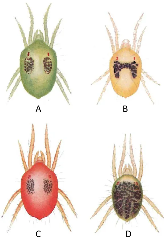
FIGURE2: A, B, C – Colours variations in active green form (GF) females of Tetranychus urticae: A – yellowish green, B – yellowish; C – old dark female; D, E, F – Colours variations in active red form (RF) females of T. urticae: D – carmine red, E – dark red, F – old dark female; Young females of GF (G) and RF (H, I); Starved female of T. urticae showing reduced side spots: J – orange like GF, K – brightly orange RF; Hivernating females of T. urticae: L – GF bright pale orange, M – RF brightly orange, N – RF brightly orange red; Male of the GF of T. urticae (O) and males of the RF (P); Colour variations in eggs: Q – eggs of GF, R – eggs of RF.
R
ESULTS ON REVIEW AND NEW DATA For each criteria investigated, data found in the lit-erature are presented first followed by new data when available.Morphological characters Colour of mites
While the colour of mites is basically physiologi-cally determined, it has been used as morphologi-cal trait to separate RF and GF, and it is included in this paragraph. This parameter is the only criterion available to distinguish between T. urticae and T. cinnabarinus when referring to the original descrip-tion of T. cinnabarinus by Boisduval (1867). Several aspects of the colour have been investigated in rela-tion with the RF and GF.
Adults’ body colour
Depending on the author, the two colour forms of T. urticae may vary in name (Table 3). According to Boudreaux (1956), who reinstated T. cinnabarinus, the colour of the two forms are the following: ac-tive females of the green form (GF) are yellowish or green to dark green, whereas the red form (RF) is carmine red in colour (=carmine spider mite), the carmine colour appearing only in apart of the adult stage (Ewing, 1914; Van de Bund and Helle, 1960; Boudreaux and Dosse, 1963a), a few days after moulting, colour darkens with advancing age (Van de Bund and Helle, 1960) and old females being uniformly dark ruby (Dosse, 1952) (Figure 2 A-F). In addition to the overall body colour of the mite, Boudreaux (1956) also separated the two colour forms by the shape and the number of dark food spots on the dorsal part of the female hysterosoma. Their characteristics are given in Figure 2 A, D, Fig-ure 3 and in Table 3. Blauvelt (1945) has shown that the outer surface of the ventriculus (mid-gut) is in-dented at several intervals by dorsoventral muscles that divide the ventriculus in several broad caeca. The amount and the place where food accumulates into these caeca are responsible for the colour, the shape and the number of dark food spots on the body of the mite. Other female colour data that de-pend on their feeding activity and those of males,
juveniles and eggs of the two forms are also re-ported in Table 3 and in Figures 2 G-R.
Figure 3: A-Drawing of a GF female of T. urticae with one pair of trifid feeding spots; B- Drawing of a RF female of T. urticae with the typical pair of trifid feeding spots and the caudal pair of spots (original drawings from H. Bruce Boudreaux, Annals of the Entomological
Society of America, 1956, 49:43-48)
A
B
FIGURE3: A – Drawing of a GF female of Tetranychus urticae with one pair of trifid feeding spots; B – Drawing of a RF female of T. urticae with the typical pair of trifid feeding spots and the caudal pair of spots (original drawings from H. Bruce Boudreaux — 1956 — Ann. Entomol. Soc. of Ame.,, 49: 43– 48)
Physiological basis of mite colour
Colouration in the two forms of T. urticae is mainly caused by internal pigments because cuticle is not coloured (Metcalf and Newell, 1962; Veerman, 1974;
Geest, 1985). Metcalf and Newell (1962) were
the first to compare the nature of the pigments of GF and RF concluding that tetranychid mite pig-ments are derived from chlorophyll and carotenoids present in the mite diet. A detailed pigment analy-sis of the two colour forms has been done by Veer-man (1974). He found that the same carotenoids are present in the two forms but with differences in the number of keto-carotenoids which were 5 vs. 9 in the GF and the RF respectively. However the author later questioned these results attributing the differences to the technical protocol used (Veerman, 1974). A pigment analysis of the RF and of another green/yellow tetranychid, Tetranychus pacificus Mc-Gregor, 1919, revealed that despite an obvious dif-ference in their body colour these two mites had the same range of carotenoids which were quanti-tatively very close (Veerman, 1972). Moreover, in deeply orange coloured diapausing females of the GF, the RF and of T. pacificus, the number of keto-carotenoids is identical in the three mites and
two-Auger P. et al.
TABLE3: Comparison of several morphological and biological features in the two colour forms of Tetranychus urticae.
Green form Red form Figures
Active Female body colour
brownish or nearly black (1); yellowish-green (5, 9, 25); amber varying to dark green (5); yellowish or green to dark green (7)
yellowish-red (2); orange red (3); brownish-red/reddish-brown (4, 9, 13); dark brownish red (5); dark red (6, 25); carmine red (7); deep red (8)
(Figs. 2 A-F)
Young female body colour
amber coloured (3) amber coloured (3); pale-amber in colour with orange tint (5) (Fig. 2 G-I)
Starved female body colour
amber tint with a frequent orange-like glow (5) to brick-red colour (4) brightly orange in colour (5); brick-red pigment (4) (Fig. 2 J, K)
Hivernating female body colour
bright pale orange (e.g. 1, 5, 14, 15); orange yellow (16); deep orange (20); bright orange (24); orange red (13, 17, 23)
no differences with the GF when exposed to low temperatures (1, 5, 13, 14);
(Fig. 2 L-N)
Feeding dark spots in active females
a pair of large black spots often trifid (10, 11) or divided into 2 or 3 smaller ones (5); a second pair may also be present (3, 12)
2 pairs, the anteriors larger and bifid (11) the second pair in the caudal area (2, 11); second pair of spots sometimes lacking (7)
(Figs. 2 B, E)
Feeding dark spots in young females
(Fig. 2 G-I)
Feeding dark spots in starved/diapausing females
(Fig. 2 J-M)
pale yellowish green to light amber with sometimes a slight orange-like tint (5)
amber to bright orange-like red with fine dark pigmental spots (5)
teleiochrysalis green (21) teleiochrysalis pale-brownish-orange (21); nymphs on carnation (Dianthus caryophyllus L.) orange-like green coloured (5)
Eggs colour clear white, translucent and shining (16, 3, 5) to pale creamy, become yellowish as they develop (16, 19, 20); yellowish-orange before hatching (20); never reddish (11)
amber with pink-reddish tint darkening with development (5), pale to rosy-amber or slightly smoky (3), straw coloured + reddish tinge as hatching time approaches (21), brownish (4), always with a trace of red (11), colour more intense when laid by unmated females (11 some eggs may be clear (12)
(Fig. 2 Q, R)
Literature cited
(1) Pritchard and Baker (1952), (2) Dosse (1952), (3) Dillon (1958), (4) Hussey and Parr (1958), (5) Van de Bund and Helle (1960), (6) Boudreaux (1963), (7) Boudreaux and Dosse (1963a), (8) Veerman (1970), (9) Kuang and Cheng (1990), (10) Pritchard and Baker (1955), (11) Boudreaux (1956), (12) Boudreaux and Dosse (1963b), (13) Dosse (1966), (14) Dosse and Musa (1967), (15) Veerman (1977), (16) Gasser (1951), (17) Parr and Hussey (1966), (18) Parr and Hussey (1960), (19) Hatzinikolis (1970), (20) Veerman (1974), (21) Davis (1961), (22) Ewing (1914), (23) Gotoh (1986), (24) Goka and Takafuji (1990), (25) Gotoh and Tokioka (1996).
Juveniles body colour similar in colour, green (7); Pale amber larvae become green coloured when feeding starts, nymphs are yellowish-green in colour (8, 9); flesh coloured (21); never orange/red in colour (22)
both bear pale-brown side-spots (5) enlarging with age (7)
side spots reduced and often subdivided with starvation (5); lacking in diapausing female or when exposed to low temperatures (e.g. 1, 5, 13, 14, 15, 20)
Male body colour males of the two forms are indistinguishable by body’s colour (4, 18); colour may vary from yellowish to pale green (4, 8, 18), from from light yellow to rust-coloured (19) to yellowish reddish (2)
(Fig. 2 O, P)
fold higher than in active females. Thus, variation in colour between species and between active and diapausing females would be due to variation in keto-carotenoid quantities rather than to differences in their composition. A difference in the expression level of some genes implicated in the carotenoids biosynthesis would be responsible for these varia-tions (Altincicek et al., 2012).
Genetic determinism of mite’s colour
Several authors reported that reciprocal crosses be-tween GF and RF produced hybrids of different nu-ances of red colour: F1 active females can be pale pinkish red (Boudreaux, 1956), carmine (Keh, 1952; Dillon, 1958; Dosse, 1963), orange-red (Hussey and Parr, 1958; Monroe, 1963), reddish, bright red to dark red (Sugasawa et al., 2002), but rarely carmine red as in females of the RF (Jordaan, 1977) (Figure 4 A, B). The obtained F1 females were never green, suggesting that red is a dominant trait ( Keh, 1952;
Taylor and Smith, 1956; Hussey and Parr, 1958; Dosse, 1963; Dosse and Boudreaux, 1963; Monroe, 1963; Jordaan, 1977).
Fertile hybrid females produced in the succes-sive generations may vary from yellow to red (Jor-daan, 1977). F2 females can be green, brownish red, orange red (Hussey and Parr, 1958; Monroe, 1963) or dark red, carmine, pink to yellowish (Monroe, 1963). Another type of F2 females called "inter-mediate" are reddish brown turning green with a faint red tinge with age (Hussey and Parr, 1958) or yellow becoming pinkish after a few days, orange turning green, yellowish turning dark red (Monroe, 1963) and green turning reddish (Keh, 1952; Mon-roe, 1963). Because of the various patterns of F2 female body colours, Hussey and Parr (1958) con-cluded that the redness colour cannot be explained by a single red gene dominant over green. They hypothesised that mite colour would be under the 388
FIGURE4: A – Typical red hybrid F1 females of Tetranychus urticae obtained when crossing GF and RF ; B – rare red with pinkish glow F1 females.
influence of two pairs of non-allelic genes, which was however regarded as an oversimplification of the situation (Monroe, 1963).
In addition to the whole body colour of the mite, the number of dark food spots and the colour of eggs can also be a mark of hybridization. F1 females might lack dark spots and a pair of atypical sup-plementary caudal dark spot is often present in F2 and F3 green females (Keh, 1952; Taylor and Smith, 1956) (Figure 5).
FIGURE5: Additional caudal dark feeding spots present in a F3 hybrid female having a body colour close to that of the GF of Tetranychus urticae.
The eggs laid by hybrid females and their off-spring are variously pigmented (Hussey and Parr, 1958; Monroe, 1963), their colour being frequently
linked to the body colour of the female (Monroe, 1963).
Mite colour for species diagnostics
A few years after the publication of Boisduval (1867) proposing the body colour to distinguish the GF (T. cinnabarinus) from other tetranychid species, Murray (1877) and Harvey (1892) considered that colour was not a reliable criterion to separate mites of the T. urticae species-group. Several authors re-port that the colouration of individuals may vary with the age of mites (Ewing, 1914; Monroe, 1963), with the feeding activity (Ewing, 1914; Pritchard and Baker, 1952) and with host plant (Gasser, 1951; Pritchard and Baker, 1952; Van de Bund and Helle, 1960). Some additional support of the inadequacy of colour to separate the two taxa is provided by colour variations found in the female progeny of a single female (Figure 6) (Ewing, 1914). This au-thor concluded that large variations in the body colour among individuals of species belonging to the genus Tetranychus were partly responsible for numerous species confusion and that T. cinnabar-inus would be probably a synonym of T. urticae. This opinion was shared by Dupont (1979) who concluded that colour variations alone are not suf-ficient for describing a new species because the carotenoid metabolism in the two forms of T. ur-ticae are very similar (Veerman, 1970; 1972; 1974). As body colour is poorly correlated with other mor-phological characters considered to be far essential
Auger P. et al.
Figure 6: Colour variations found among the first and second generation descendants of a
single female of T. telarius L. (= red form of T. urticae). A- active feeding female ; B- female
after a fasting of two days ; C- reddish orange female; D- a black form following vigourous
feeding (original drawings from Ewing H. E., Oregon Agricultural College Experimental
Station Bulletin, 1914, 121: 1-95, permission to reprint these illustrations granted by Oregon
State University and the Oregon Agricultural Experiment Station, 2011).
A
B
C
D
FIGURE6: Colour variations found among the first and second generation descendants of a single female of T. telarius L. (= red form of Tetranychus urticae). A – active feeding female; B – female after a fasting of two days; C – reddish orange female; D – a black form following vigourous feeding (original drawings from Ewing H. E. — 1914 — Oregon Agricultural College Experimental Station Bulletin, 121: 1–95, permission to reprint these illustrations granted by Oregon State University and the Oregon Agricultural Experiment Station, 2011).
FIGURE7: Colour variations in hybrid F2 and F3 females of Tetranychus urticae. A – yellowish-green female; B – pale orange female; C – yellowish female with pinkish tint; D – brownish red, E – pale pinkish, F –yellowish, orange and bronze coloured females.
for species separation, Zhang and Jacobson (2000) estimated that this trait was not a reliable charac-ter to distinguish between the two forms of T. ur-ticae (see "Leg chaetotaxy" and "Other morphologi-cal characters" sections below).
Conversely, (Boudreaux, 1956; Boudreaux and Dosse, 1963a) reported that body colour is a re-liable trait when used alone or when considered with morphological ones (Boudreaux and Dosse, 1963a). Saba (1975) went further in considering that colour was important data for certain tetranychid mite identifications.
New data
Hybrids’ colour. A sample of body colours from F2-F3 hybrid females obtained by our crossings (see "Breeding" section for results) is given in Figure 7 and several "intermediate" F8 hybrid females are figured in Figure 8. The additional posterior pair of maculae occurring in the RF but not in the GF (Dosse, 1952; Boudreaux, 1956) has been consid-ered as an additional criterion to separate the two colour forms. However we have observed these maculae on individuals from several populations of the GF and not only in older females as re-ported by Boudreaux and Dosse (1963b) (Figure 9). Conversely, like Boudreaux and Dosse (1963a) we
also found many specimens of the red form lacking these caudal black spots. Thus, this trait is not dis-tinctive of the two colour forms.
Actually most of the tetranychid mite tax-onomists consider that T. cinnabarinus is a synonym of T. urticae and body’s colour is not given as a character to distinguish between these two species (e.g. Meyer, 1987; Baker and Tuttle, 1994; Bolland et al., 1998; Ehara, 1999). In their opinion, T. urticae presents two colour forms, green and red. We will see in the "Breeding" section, that colour cannot be considered as a specific marker.
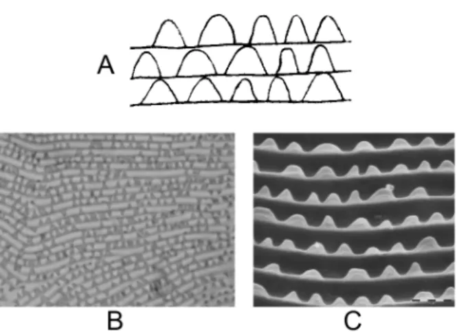
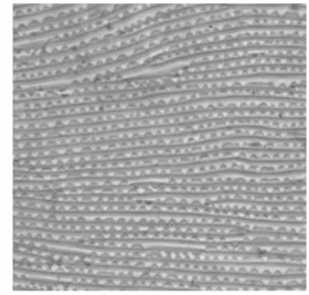
Lobes on dorsal striation Lobe shape and density
Lobes are small structures of various shapes present on the upper edge of integumentary folds of the body of mites (Figure 10 A). On adult GF summer females, the dorsal striation bears semi-oblong lobes (Boudreaux, 1956), some of them wide or rather oblong, rounded, sometimes nar-rower and occasionally triangular (Boudreaux and Dosse, 1963b; Monroe, 1963; Carbonnelle and Hance, 2004) but never apically pointed (Branden-burg and Kennedy, 1981) (Figure 10 B, C and D). On RF adults, lobes are semi-circular or triangularly
Auger P. et al.
FIGURE8: Examples of colour changing in hybrid F8 females of Tetranychus urticae: yellowish female (A) that turned pink-red (B); pale pinkish female (C) that turned pink-red (D); yellowish green female (E) that turned dark-green (F); amber female (G) that turned pink-red (H).
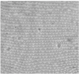
rounded (Boudreaux, 1956), generally more sepa-rated at their base, narrow and relatively pointed (Boudreaux and Dosse, 1963b; Monroe, 1963; Car-bonnelle and Hance, 2004) (Figure 11 A, B and C).
FIGURE9: GF females of Tetranychus urticae with an additional pair of spots in the caudal area.
FIGURE10: Cuticular lobes on the dorsal striation of Tetranychus species. A – general aspect and location of lobes on the dor-sal integumentary folds; B – cuticular lobes in GF females T. urticae (original drawings from H. Bruce Boudreaux — 1956 — Ann. Entomol. Soc. Am., 49: 43–48); C – aspect of GF fe-males of T. urticae lobes using phase-constrast microscope; D – aspect of GF females of T. urticae lobes using SEM.
They are smaller and more triangular than in the GF. However, sometimes broad rounded lobes can be found mixed with other type of lobes (Monroe, 1963; Dosse and Langenscheidt, 1964; Hatziniko-lis, 1970; Meyer, 1974; Brandenburg and Kennedy, 1981).
FIGURE11: Cuticular lobes on the dorsal striation of RF females of Tetranychus urticae. A – original drawing by H. Bruce Boudreaux — 1956 —- Ann. Entomol. Soc. Am. 1956, 49: 43–48); B – cuticular lobes aspect using phase-constrast mi-croscope; C – aspect of lobes using SEM.
Using SEM, Hance et al. (1998) have measured different parameters to characterize the shape of lobes (Figure 12), and only one allowed them to dis-tinguish between the two forms: the ratio between the base of the lobe (B) and its height (H) which was greater in the GF than in the RF (2.75 vs. 2.11).
Fig. 12: Parameters measured on the dorsal integumentary lobes: base (B), distance between the top of two lobes (D), height (H), surface of a lobe (S) (redrawn after Hance et al., 1998).
FIGURE12: Parameters measured on the dorsal integumentary
lobes: base (B), distance between the top of two lobes (D), height (H), surface of a lobe (S) (redrawn after Hance et al., 1998).
However the authors estimated that the lobe morphology is a difficult criterion to distinguish be-tween the two species because they found some-times more variations between individuals than be-tween the two forms.
For long time, taxonomists regarded integumen-tary lobes as qualitative data, until Monroe (1963) first assessed lobe density and used this criterion
Auger P. et al.
to separate the two forms of T. urticae. This au-thor found that females of the GF had 5 to 6 lobes per 10 µm in average, and observed a greater lobe density in the RF than in the GF. While no mention of the value of the RF lobe density is clearly done by Monroe (1963), a new analysis of the data al-lows to estimate this density at about 7 lobes per 10 µm. Using SEM, comparable densities per 10 µm were calculated in GF (6.4 ± 0.8 lobes; range: 5 to 8) and RF (7.5 ± 0.8; range: 6 to 9.5) by Brandenburg and Kennedy (1981), or 5.8 ± 0.2 lobes in the GF and 7.0 ± 0.4 lobes in the RF by Kuang and Cheng (1990). However Brandenburg and Kennedy’s re-sults (1981) show lobe densities confidence intervals overlapping among the two colour forms making this criterion invalid for species diagnostics (Mol-let and Sevacherian, 1984). By contrast, Kuang and Cheng (1990) did not observed such overlap.
Lobe shape variations
While the lobe shape has been used to discrimi-nate between RF and GF, intraspecific variation of the dorsal lobe shape pattern, the lobe density and the lobe development have been widely reported. When strongly developed, lobes might be semi-oblong to semi-circular on the GF but when a slight development is present, lobes are more oblong with more flattened upper side (Van de Bund and Helle, 1960) (Figure 13 A, B and C).
Figure 13: Variations in aspect and development of dorsal lobes in GF females of T. urticae (original drawings from C.F. van de Bund & W. Helle , Entomologia Experimentalis et
applicata, 1960, 3: 142-156). A- less developed lobes; B- normally developed lobes; C-
strongly developed lobes.
Figure 14: Variations in the aspect and development of dorsal lobes in RF females of T.
urticae (original drawings from C.F. van de Bund & W. Helle , Entomologia Experimentalis et applicata, 1960, 3: 142-156). A- less developed lobes; B- normally developed lobes.
Figure 15: Appearance of less developed female dorsal lobes using phase-contrast microscope. A- in GF females of T. urticae; B- in RF females of T. urticae.
A
B
C
A
B
A
B
FIGURE13: Variations in aspect and development of dorsal lobes in GF females of Tetranychus urticae (original drawings from C.F. van de Bund and W. Helle — 1960 — Entomol. Exp. et appl., 3: 142–156). A – less developed lobes; B – normally developed lobes; C – strongly developed lobes.
On RF, slightly developed lobes are mostly semi-circular to rather semi-oblong but they are often tri-angular in case of strong development (Figure 14 A-B). Van de Bund and Helle (1960) reported a slight development in many specimens, concluding that lobes do not allow to distinguish between the two forms (Figures 13 A, 14 A, 15).
Figure 13: Variations in aspect and development of dorsal lobes in GF females of T. urticae (original drawings from C.F. van de Bund & W. Helle , Entomologia Experimentalis et
applicata, 1960, 3: 142-156). A- less developed lobes; B- normally developed lobes; C-
strongly developed lobes.
Figure 14: Variations in the aspect and development of dorsal lobes in RF females of T.
urticae (original drawings from C.F. van de Bund & W. Helle , Entomologia Experimentalis et applicata, 1960, 3: 142-156). A- less developed lobes; B- normally developed lobes.
Figure 15: Appearance of less developed female dorsal lobes using phase-contrast microscope. A- in GF females of T. urticae; B- in RF females of T. urticae.
A
B
C
A
B
A
B
FIGURE14: Variations in the aspect and development of dorsal lobes in RF females of Tetranychus urticae (original drawings from C.F. van de Bund and W. Helle — 1960 — Entomol. Exp. et appl., 3: 142–156). A – less developed lobes; B – normally developed lobes.
Figure 13: Variations in aspect and development of dorsal lobes in GF females of T. urticae (original drawings from C.F. van de Bund & W. Helle , Entomologia Experimentalis et applicata, 1960, 3: 142-156). A- less developed lobes; B- normally developed lobes; C- strongly developed lobes.
Figure 14: Variations in the aspect and development of dorsal lobes in RF females of T. urticae (original drawings from C.F. van de Bund & W. Helle , Entomologia Experimentalis et applicata, 1960, 3: 142-156). A- less developed lobes; B- normally developed lobes.
Figure 15: Appearance of less developed female dorsal lobes using phase-contrast microscope. A- in GF females of T. urticae; B- in RF females of T. urticae.
A
B
C
A
B
A
B
FIGURE15: Appearance of less developed female dorsal lobes
using phase-contrast microscope. A – in GF females of T. ur-ticae; B – in RF females of T. urticae.
While confirmed by Monroe (1963) who also faced this problem, Dosse and Boudreaux (1963) concluded that these variations would only appear in improperly mounted specimens. However, even in well mounted specimens, variations are known to occur among populations from each of the two T. urticae forms (Boudreaux and Dosse, 1963b; Mon-roe, 1963; Dosse and Langenscheidt, 1964; Meyer, 1974; Jordaan, 1977). This leads Monroe (1963) to consider that the differences in lobe are not a consis-tent character of each mite form, as intra- and inter-variation in the lobe characters of each mite colour overlap (Meyer, 1974; Jordaan, 1977).
Environmental factors can also modify dorsal lobe patterns. Mollet and Sevacherian (1984) have shown that the strial lobe density of the GF can change with temperature and moisture (Figure 16) and depending on the season they found specimens with intermediate lobe shape patterns. They con-cluded that lobe density is not a reliable morpho-logical character for separating the two forms. 394
Figure 16: Mean number of dorsal strial lobes/30 micrometer on GF females of T. urticae reared at different temperature and humidity regimes (after Mollet & Sevacherian, 1984). Means sharing a common letter are not significantly different (Duncan's NMRT P<0.05) * Mites collected from cotton leaves and then used for experiments. Their lobes were observed at the start of the experiment to be used as reference.
b a a 16 17 18 19 20 21
Mean number of lobes / 30 µm
20°C- 70 % RH Control * 35°C-15 % RH
FIGURE 16: Mean number of dorsal strial lobes/30
microm-eter on GF females of Tetranychus urticae reared at differ-ent temperature and humidity regimes (after Mollet and Se-vacherian, 1984). Means sharing a common letter are not sig-nificantly different (Duncan’s NMRT P<0.05). * Mites col-lected from cotton leaves and then used for experiments: their lobes were observed at the start of the experiment to be used as reference.
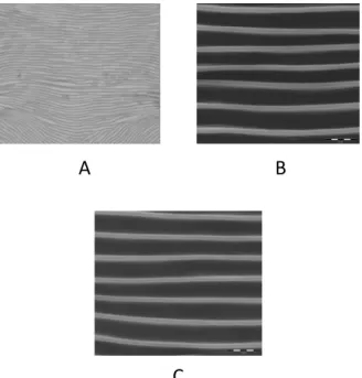
Likewise, the integument of hibernating GF fe-males differs from that of active fefe-males (Pritchard and Baker, 1952) and is characterised by a solid-type striae (smooth striation devoid of lobes) (Figure 17 A).
This striation pattern can also be found in active GF females when during their development a sin-gle instar has been exposed to environmental con-ditions which induced diapause (Parr and Hussey, 1966). During winter months, the lobes of the RF tend to be wider and rounded resembling those of the GF (Boudreaux and Dosse, 1963a; Dosse, 1966; Meyer, 1974; Carbonnelle and Hance, 2004). As in diapausing or hibernating females of the two forms the integumentary striae is smooth (Figure 17 B, C) or with incompletely formed lobes, many authors emphasized that they cannot be separated using this morphological trait (Van de Bund and Helle, 1960; Dosse, 1966; Dosse and Musa, 1967; Meyer, 1974; Jeppson et al., 1975).
Figure 17: Smooth dorsal striation of hibernating females. A- GF females of T. urticae using phase-contrast microscope; B- GF females of T. urticae using SEM; C- RF females of T. urticae using SEM.
A
B
C
FIGURE17: Smooth dorsal striation of hibernating females. A – GF females of Tetranychus urticae using phase-contrast micro-scope; B – GF females of T. urticae using SEM; C – RF females of T. urticae using SEM.
Genetic determinism of the lobe shape
Observations concerning the determinism of the lobe shape are conflicting. According to Boudreaux (1956), the dorsal lobes in F1 hybrids are mostly tri-angular in shape to semi-circular like those found in the RF but occasional oblong lobes typical of the GF can also be observed.
Hybridisation tests between the GF and the RF led Boudreaux (1956) and Dupont (1979) to con-clude on the dominance of the triangular lobes over the oblong ones. Conversely, Monroe (1963) pro-duced F1 hybrids that usually possessed dorsal in-tegument lobes similar to those of the GF, con-cluding that the rounded lobes typical of the GF would usually be dominant over the pointed tri-angular lobes of the RF which would be recessive (Boudreaux, 1963; Monroe, 1963). However, Jor-daan (1977) obtained a different result because most of the F1 females she observed had dorsal lobes of intermediate form. In F2 females Monroe (1963) and Jordaan (1977) found great variations regarding lobe shape and density.
Auger P. et al.
Lobe shape to distinguish between the two colour forms
When Boudreaux (1956) reinstated T. cinnabarinus (the RF of T. urticae) he associated the body colour of the mite with a type of dorsal lobe shape. How-ever, a red Tetranychus (T. urticae forma dianthica Dosse, 1952) found in German greenhouses and similar to the RF exhibited broad integumentary dorsal lobes typical of those usually present in the GF (Boudreaux and Dosse, 1963b; Dosse and Boudreaux, 1963). When crossbreeding this popu-lation with a strain of the RF they freely interbreed, despite a partial reproductive isolation (Dosse, 1963), supporting their synonymy (Pritchard and Baker, 1952; Boudreaux, 1956, 1959).
The hypothesis of hybridization in naturae (vs. laboratory conditions) was put forward to explain these data (Boudreaux and Dosse, 1963b; Dosse and Boudreaux, 1963; Jordaan, 1977) which could ex-plain why several other populations bearing mixed lobe and colour characteristics have been observed (Pritchard and Baker, 1952; Van de Bund and Helle, 1960; Dosse and Boudreaux, 1963; Dosse, 1964; Dosse and Langenscheidt, 1964; Parr and Hussey,
1966). The case of T. urticae forma dianthica
re-veals that the use of lobe shape to separate the two forms of T. urticae might not be valid because the triangular shape of the dorsal lobes is not always linked to the red colour of the mite and as also ob-served in green hybrid females which bear typical lobes of the RF (Jordaan, 1977). Another excep-tion concerning the correlaexcep-tion between the dorsal lobe shape and the body colour of the mite is re-ported from greenhouses where some GF popula-tions with dorsal lobes were closer to RF than to other GF mites (Zhang and Jacobson, 2000). Like-wise, females of these GF populations had a par-ticular foreleg chaetotaxy (see "Leg chaetotaxy" sec-tion below) leading the authors to consider them as T. cinnabarinus (=RF).
New data
The analysis of a collection of mounted T. urticae slides, revealed specimens with poorly developed lobes in the majority of RF but only occasionally in the GF examined, as having been previously re-ported by Van de Bund and Helle (1960). In
partic-ular, the lobes shown in figure 11 B were rarely ob-served on our RF females. The typical lobes shown in this figure were present in only one in four fe-males collected together in the same rearing unit kept in the laboratory. The three other females car-ried poorly developed lobes. It was then impossible to discriminate among RF and GF mites based on this criterion. Among females bearing well devel-oped dorsal lobes, we also found lobe shape vari-ations between populvari-ations of a given form. Like Zhang and Jacobson (2000) we faced the apparent contradiction between the dorsal lobe pattern and the body colour when analysing GF females col-lected in Tunisia. Despite their green colour, they had a dorsal striation with a majority of small quite well separated rounded lobes, sometimes triangu-larly rounded and with rare oblong lobes (Figure 18).
Figure 18: Atypical dorsal lobe pattern present in green T. urticae females of a population bearing additional setae on tarsus and tibia of the front leg.
Figure 19: Dorsal lobe aspect observed on F20-30 hybrid female.
FIGURE18: Atypical dorsal lobe pattern present in green Tetrany-chus urticae females of a population bearing additional setae on tarsus and tibia of the front leg.
Like the specimen collected by Zhang and Jacob-son (2000) these females had a particular leg chaeto-taxy. These data highlight that body colour and lobe shape might be disconnected.
The lobes we observed on dorsal striation of F1 females and of hybrid females of re-established population (F20-30), like reported by Dupont (1979), were closer to those of RF and were mostly triangular to triangularly rounded (Figure 19). 396
Acarologia 53(4): 383–415 (2013) Figure 18: Atypical dorsal lobe pattern present in green T. urticae females of a population
bearing additional setae on tarsus and tibia of the front leg.
Figure 19: Dorsal lobe aspect observed on F20-30 hybrid female. FIGURE19: Dorsal lobe aspect observed on F20-30 hybrid female of Tetranychus urticae.
In conclusion, the dorsal integument lobes is a morphological character often included in descrip-tions of Tetranychus species and in the identification keys made by numerous acarologists. In this sec-tion we have reported different factors that might affect proper observation and distort their shape. As a consequence, both inter- and intra-population variations of the size and/or shape of this trait might occur, sometimes leading to partial overlap-ping among the two forms of T. urticae, making it uneasy to use to distinguish between GF and RF (Pritchard and Baker, 1952; Van de Bund and Helle, 1960; Dosse, 1964; Parr and Hussey, 1966; Jordaan, 1977). Special attention must be given to the global dorsal lobe pattern (i.e. the frequency of the two types of lobes) rather than to the presence or not of one type of dorsal lobe.
The lack of complete link between the type of dorsal lobes and the body colour of the mite also re-duce the reliability of this character to separate the two forms of T. urticae. However, Zhang and Ja-cobson (2000) increased the importance of the lobe shape dorsal striation over the body colour to sepa-rate the GF from the RF, the latter being considered as T. cinnabarinus, because they found a better cor-relation between this criterion and other morpho-logical characters to separate the two forms of T. ur-ticae (see "Other morphological characters" section below).
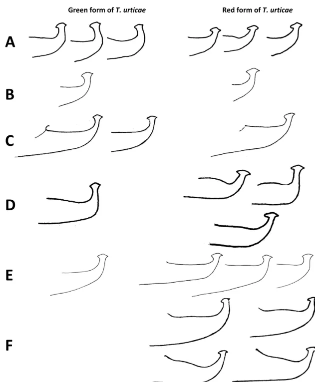
Shape of the aedeagus
According to Boudreaux (1956), who considered the GF and the RF as distinct species and reinstated T. cinnabarinus (RF), the aedeagus of the GF bears a knob with an anterior and a posterior projection, both small and rather acute, a dorsal margin of the knob broadly rounded or somewhat obtusely angu-late and the axis of the knob parallel to the axis of the shaft (Figure 20 A). By contrast, RF has an an-terior projection of the knob slightly rounded and a posterior one acute, a knob dorsal surface obtusely angulate and the axis of the knob that always forms a slight angle with that of the shaft (Figure 20 B).
Figure 20: Aedeagus of the GF male of T. urticae; B- aedeagus of RF male of T. urticae (original drawings from H. Bruce Boudreaux, Annals of the Entomological Society of America, 1956, 49:43-48).
A
B
FIGURE20: A – Aedeagus of the GF male Tetranychus urticae; B – aedeagus of RF male of T. urticae (original drawings from H. Bruce Boudreaux — 1956 — Ann. Entomol. Soc. Am., 49: 43–48).
However, the separation of the two forms might be misleading because several noticeable differ-ences in description of the aedeagi have been re-ported: Saba (1975) found that the axis of the knob of the aedeagus forms an angle with the axis of the shaft which is greater in the GF than in the RF (Fig-ure 21 A). According to Kuang and Cheng (1990) the aedeagus knob of RF is larger than that of GF and their drawings exhibit a knob dorsal margin convex in the two species (Figure 21 B). In addition, aedea-gal variations between and among populations are also reported by several authors (Boudreaux and Dosse, 1963b; Meyer, 1974; Zhang and Jacobson, 2000). The drawings of aedeagi of T. cinnabarinus by many authors clearly illustrate this variability and the difficulty to link the aedeagus shape to one of the colour forms of T. urticae. The shape of the aedeagus knob sometimes overlaps between the two forms: an acute anterior projection can be present on the aedeagi of both colour forms (Figure
Auger P. et al.
Green form of T. urticae Red form of T. urticae
Figure 21: Variations found in the aedeagi of the GF (left) and of the RF (right) of T. urticae. A- drawings from F. Saba, Annals of the Entomological Society of America, 1975, 68: 797-800; B- drawings from H.Y. Kuang and L.S. Cheng, Acta Entomologica Sinica, 1990, 33: 109-116; C- drawings by E.W. Baker and A.E. Pritchard, Hilgardia, 1960, 29: 455-574, copyright 1960 Regents of the University of California; D- drawings from C.F. van de Bund & W. Helle, Entomologia Experimentalis et Applicata, 1960, 3: 142-156; E- drawings from M.K.P. Smith Meyer, 1974; F- drawings from E.A. McGregor, American Midland Naturalist, 44: 257-420 september 1950.
A
B
C
D
E
F
FIGURE21: Variations found in the aedeagi of the GF (left) and of the RF (right) of Tetranychus urticae. A – drawings from F. Saba — 1975 — Ann. Entomol. Soc. Am. 1975, 68: 797–800; B – drawings from H.Y. Kuang and L.S. Cheng — 1990 — Acta Entomol. Sinica, 33: 109–116; C – drawings by E.W. Baker and A.E. Pritchard — 1960 — Hilgardia, 29: 455–574, copyright 1960 Regents of the University of California; D – drawings from C.F. van de Bund and W. Helle — 1960 — Entomol. Exp. et Appl., 3: 142–156; E – drawings from M.K.P. Smith Meyer — 1974 — Entomology Memoir, Department of Agricultural Technical Services, Republic of South Africa: 1-291; F – drawings from E.A. McGregor — 1950 — Am. Midland Natural., 44: 257–420.
Acarologia 53(4): 383–415 (2013)
21 B, D, E) and the aedeagal knob axis can be paral-lel to the axis of the shaft in the two forms (Figure 21 D-E). Moreover, typical knob characteristics of a given colour are sometimes absent in that form but present in the other. This is illustrated by Meyer’s drawings (1974) with an obtuse angulation of the dorsal margin of the aedeagal knob absent on the RF but present on the GF, whereas the rounded dor-sal margin is present on the RF (Figure 21 E). Like-wise, the knobs of the aedeagi of the GF described by Saba (1975) and Kuang and Cheng (1990) clearly form a slight angle with the axis of the shaft but not in most of RF aedeagi (Figure 21 A-B). Sometimes, the heads of the aedeagi of the two colour forms are very similar as observed in Boudreaux and Dosse’s pictures (1963b) (Figure 22 A-B).
Aedeagus shape to distinguish between the two colour forms
Many papers aimed at distinguishing between the two forms of T. urticae do not evoke the shape of the aedeagus. This tend to show that the taxonomi-cal value given to the aedeagus shape to distinguish between the two forms of T. urticae varies among authors and also that for many of them other mor-phological and biological criteria are more efficient and more reliable.
Despite the differences in the aedeagi of the two forms, as emphasized Boudreaux (1956), GF males are almost indistinguishable from RF ones. As dif-ferences are minute between the aedeagi of the two forms (Dosse and Boudreaux, 1963; Saba, 1975) or not consistent (Hatzinikolis, 1970), these authors considered that the shape of the aedeagus is not fully reliable for the separation of the two colour forms of T. urticae. Likewise, female morphological characters such as the body colour and the shape of the dorsal integumentary lobes are the only reliable traits allowing to distinguish between the GF and the RF (Boudreaux and Dosse, 1963b). For other au-thors, however, the aedeagus remains the most use-ful character for species identification (Dillon, 1958; Meyer, 1974; Van de Bund and Helle, 1960; Zhang and Jacobson, 2000).
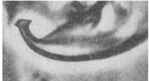
Figure 22: Pictures of the aedeagi of the GF (A) and RF (B) of T. urticae both having acute anterior and posterior projections, angulate dorsal margin and a knob axis forming an angle with the axis of the shaft (original pictures from Boudreaux and Dosse (1963b) with permission from Acarologia).
B
A
FIGURE22: Pictures of the aedeagi of the GF (A) and RF (B) of Tetranychus urticae both having acute anterior and posterior projections, angulate dorsal margin and a knob axis form-ing an angle with the axis of the shaft (original pictures from Boudreaux and Dosse (1963b) with permission from Acarolo-gia).
New data
All the aedeagi from RF mites that we have exam-ined under microscope were close in shape to those described and drawn by Boudreaux (1956) (Figure 23 A). However, in several males of each of the RF samples, the inner projection of the knob was slightly acute (Figure 23 B) and the axis of the knob did not systematically form a slight angle with the shaft (Figure 23 C). The differences are more obvi-ous between the descriptions of the aedeagus of the GF found in the literature cited above in this sec-tion and the aedeagi of the specimens that we have observed. With the exception of 1 or 2 specimens (Figure 23 D), none of these aedeagi bore the typi-cal more or less symmetritypi-cal knob with two compa-rable acute projections and a rounded dorsal mar-gin as figured by Boudreaux (1956). In most of the cases examined they closely resemble those of the
Auger P. et al.
Figure 23: A-Typical aedeagus of the RF of T. urticae found in two populations collected in Spain and
south of France; B- aedeagus of the RF bearing an acute anterior projection; C- aedeagus of the RF
showing no obvious angle between the knob axis and the shaft axis; D- aedeagus of the GF of T.
urticae having the characteristics reported by Boudreaux (1956); E- common aedeagus of the GF
observed in three populations originating from Canada, Scotland and France; F- aedeagus of the GF
bearing a knob with a rounded innate projection and the axis of the knob forming an obvious angle
with the shaft axis.
C
E
A
D
F
B
FIGURE23: A – Typical aedeagus of the RF of Tetranychus urticae found in two populations collected in Spain and south of France; B – aedeagus of the RF bearing an acute anterior projection; C – aedeagus of the RF showing no obvious angle between the knob axis and the shaft axis; D – aedeagus of the GF of T. urticae having the characteristics reported by Boudreaux (1956); E – common aedeagus of the GF observed in three populations originating from Canada, Scotland and France; F – aedeagus of the GF bearing a knob with a rounded innate projection and the axis of the knob forming an obvious angle with the shaft axis.
RF and were closer to the RF than to GF figured by Boudreaux (1956) (Figure 20). They have an asym-metrical knob with a dorsal margin more angulate than rounded. The posterior projection is smaller and more acute than the anterior one which is some-what rounded (Figure 23 E-F). Sometimes the axis of the knob forms a slight angle with the axis of the shaft (Figure 23 F). They are similar to the aedeagi of T. urticae figured by Ehara and Gotoh (1996). A slight difference between the aedeagi of the two colour forms of our samples lies in the inner pro-jection which seems to be slightly more rounded in the RF than in the GF.
The variability of the shape of the aedeagus in the two colour forms detected by several acarol-ogists, in particular the differences in the size of the aedeagi of both forms of T. urticae reported by Kuang and Cheng (1990) and also in the specimens that we examined for this work, prompted us to compare the aedeagi of males of the 5 strains of the two colour forms (Table 2). Among the five parame-ters measured (see Figure 1), significant differences were found between three of them (Table 4): the length of the aedeagus knob (F4,71=11.67, P<0.0001),
the hight of the hook (F4,67=5.27, P<0.0009) and the
angle formed between the axis of the knob and the dorsal margin of the aedeagal shaft (F4,69=3.04,
P=0.023). However, the angle between axis of knob and aedeagal shaft and the width of neck of the aedeagal hook, do not significantly differ between populations (F4,67=2.45, P=0.132; F4,70=0.68, P=0.607
respectively) (Table 4).
The absence of significant differences between populations for the angle formed by the axis of the knob and the aedeagal shaft seems to be due to great intra-population variability, whatever the body colour of the mite is. For example, in males belonging to the RF from France, a quarter of them bears a knob aedeagus whose axis is parallel to that of the shaft. Thus, this criterion which is sometimes used to separate the two colour forms, appears to not to be fully reliable due to intra-population vari-ations. Despite differences found in three morpho-logical characters of the aedeagus, none of them al-low to distinguish all the populations of the RF from all those of GF. Indeed, several populations seem to be morphologically "intermediate". This is the case of the GF Scotland population for the length of the aedeagal knob (Table 4). Individual variabil-ity observed within populations and variabilvariabil-ity be-tween populations of a given colour form demon-strate that there is no systematic and obvious clear-cut difference in the aedeagus shape. Thus, the male aedeagus does not allow separating reliably the two forms of T. urticae.
However, this polymorphism illustrated in Fig-ure 21, which is unique in the genus Tetranychus, is problematic because since the works of Ewing (1913) and Pritchard and Baker (1955) the role of the aedeagus for species diagnostics in the Tetranychus genus has been strongly put forward. It is difficult to know whether this variability is mainly due to
in-TABLE4: Measures of aedeagal features (mean ± SE) in 3 populations of the green form (GF) and 2 of the red form (RF) of Tetranychus urticae.
T. urticae
populations Knob length (µm) Neck width (µm) Hook high (µm)
Angle with shaft axis (°)
Angle with shaft dorsal margin (°) GF France 2.46 ± 0.01 (15)* a 1.37 ± 0.02 (15) a 3.73 ± 0.08 (14) b 3.87 ± 1.02 (15) a 9.22 ± 1.33 (14) ab GF Canada 2.52 ± 0.02 (15) b 1.37 ± 0.01 (14) a 3.13 ± 0.08 (14) a 1.85 ± 0.64 (15) a 7.67 ± 1.00 (15) a GF Scotland 2.57 ± 0.03 (15) bc 1.39 ± 0.02 (15) a 3.79 ± 0.10 (15) b 2.10 ± 0.68 (15) a 8.87 ± 1.11 (15) ab RF Spain 2.59 ± 0.01 (16) c 1.40 ± 0.01 (16) a 3.94 ± 0.07 (16) b 3.14 ± 1.08 (16) a 11.26 ± 1.52 (16) ab RF France 2.63 ± 0.02 (15) c 1.40 ± 0.05 (15) a 3.68 ± 0.27 (13) b 4.88 ± 1.05 (15) a 13.59 ± 1.55 (14) b
* Number of mite observed
Means sharing a different letter within a column are significantly different (ANOVA followed by Student-Newman-Keuls test; P <0.05)
Auger P. et al.
trinsic morphological differences and/or to the ob-servation under microscope of specimens on edge because the aedeagal knob is a roughly lenticular disc and a slight tilt may influence its observation (Dosse and Boudreaux, 1963). In conclusion, the polymorphism observed in the aedeagus of T. ur-ticae could be partly responsible for the denomina-tion of polytypic-species given to the two colour forms by Pritchard and Baker (1955) and Meyer (1987). In addition to other characters, this potential variability has also certainly reinforced the opinion of several taxonomists to consider T. cinnabarinus as a synonym of T. urticae. It is important to note that acarologists such as Pritchard and Baker (1955), Meyer (1987) and Ehara (1999) have described the aedeagal knob of T. urticae (including the two colour forms) as small, forming sometimes a small angle with the axis of the shaft and bearing a dorsal mar-gin as well as the anterior and posterior projections that vary considerably.
Leg chaetotaxy
The Tetranychus sensu stricto females (Tuttle and Baker, 1968) usually bear 14 tactile setae and one solenidion on tarsus I and 9 tactile setae and one solenidion on tibia I. Additional setae observed on leg I tarsus and tibia of Tetranychus specimens have been reported and used to separate GF and RF.
In 1950 McGregor described a new species of red spider mite named T. multisetis. This mite only dif-fers from the RF by its first leg chaetotaxy: one or two extra chemosensory setae (solenidia) proximad to the proximal set of duplex setae are added to the tarsus and tibia (Figure 24 A-B). Later, several cross-ing experiments have shown that this species and the RF freely interbreed (Davis, 1952; Boudreaux, 1956; 1959; Helle and Van de Bund, 1962; Dosse, 1963). Crosses also revealed that the inheritance of the foreleg setation is unique (Table 5): it is trans-mitted from mother to daughter regardless to the
TABLE5: Inheritance of the leg setation on tarsus and tibia of the frontleg in relation to the body colour in crossing experiments between the two colour forms of T. urticae (after Keh, 1952). A and B represent reciprocal crosses.
A Red (R) form Green (G) form ♀ ♂ ♀ Body colour R G R
Setae1 on female leg I:
Tarsus – Tibia 6 – 13 4 – 10 6 – 132 B Red (R) form Green (G) form ♀ ♂ ♀ G4 R4
Setae1 on female leg I:
Tarsus – Tibia 4 – 10 6 – 13 4 – 10
1 Tarsal setae numbers refer to the number of setae proximal to proximal duplex setae.
2 A few females bore 5 to 7 setae on tarsus I and 12 to 14 setae on tibia I.
3 Intermediate body colour.
F1 F1 F2 F2 R I3 Parental Cross Parental Cross Body colour G R
4 Color changes with age: green females turn red.
R G ♀ ♀ R 6 – 132 4 – 10 402
A
B
Proximal
duplex setae
FIGURE24: Chaetotaxy of female tarsus and tibia of the first leg. A – Red form (RF) of Tetranychus urticae (T. cinnabarinus sensu Boudreaux, 1956); B – T. multisetis (=polychaete form of the RF). Additional solenidia are indicated by arrows (A and B original drawings by Baker and Pritchard — 1960 — Hilgardia, 29: 455–574, copyright 1960 Regents of the University of California).
male used in crosses. Breeding experiments be-tween this species and the GF confirmed the
pre-vious results but also showed that foreleg hyper-trichy and the body colour of mites are not linked
Auger P. et al.
(Keh, 1952) (Table 5). Boudreaux (1959) who has studied the genetics of the fore tibia and tarsus hypertrichy concluded that it would be caused by the absence of a "viruslike transovarian substance" present in some populations. Taken together, these results allowed to conclude that T. multisetis was a synonym of the RF and as a consequence that the leg setation was of no value to characterize one of the carmine forms (Pritchard and Baker, 1952; Helle and Van de Bund, 1962). By contrast, popu-lations of the GF whose females’ front leg chaeto-taxy was similar to that of the polychaete form of the RF were also reported (Boudreaux, 1956; Dil-lon, 1958; Helle and Van de Bund, 1962; Rambier, 1965; Hatzinikolis, 1970). Foreleg hypertrichy was also found in T. gloveri Banks, 1900 with samples including polychaete specimens among individuals bearing the usual chaetotaxy, corroborating the tax-onomical invalidity of this morphological criterion and its use to separate the two colour forms of T. urticae (Boudreaux, 1958b; 1959).
Nevertheless, hypertrichy found in the front legs of some specimens of the RF but not in the GF, was retained as an additional tool to separate the GF from the RF (Kuang and Cheng, 1990). In agree-ment with this work, Zhang and Jacobson (2000) went further in considering this morphological trait to be a more reliable character than the colour of the mite to separate the two colour forms of T. urticae. The latters did not separate the GF from the RF by their body colour but by the chaetotaxy of the front leg combined with the dorso-hysterosomal striation lobe shape pattern. They concluded to have ob-tained a better correlation between several morpho-logical characters and the species designation when they separated the 2 colour forms on the basis of this combination of characters (see section "Other mor-phological characters" below). These authors con-cluded that polychaete green and red mites belong to T. cinnabarinus because both bear a similar dor-sal lobe pattern. Only females bearing the usual number of setae on the first leg and dorsal lobes rounded or oblong can then be considered as T. ur-ticae. Moreover, despite the observations concern-ing the matriclinous inheritance of leg hypertrichy (Boudreaux, 1958b; 1959) and leg hypertrichy being
also found in some specimens of T. gloveri, Zhang and Jacobson (2000) concluded that the addition of male solenidia, i.e. hypertrichy, on the female first leg is apomorphic as it is not observed in the GF (green coloured females bearing rounded lobes on their dorsohysterosomal striation, see next section) nor in other related species.
New data
We have found a polychaete population of the GF whose females bear the usual lobe pattern in the dorsohysterosomal striation (Figure 25). This result contrasts with the observations of Zhang and Ja-cobson (2000) who stated that leg hypertrichy was linked to more or less triangularly rounded dorsal lobes. The argument consisting in separating the two taxa by the combination of the foreleg chaeto-taxy and the shape of the dorsal lobes, is invalid be-cause these characters are not linked. This is also the case for the RF described by Dosse (1952) and called T. urticae forma dianthica.
Figure 25: Cuticular lobe pattern present on the dorsal striation of a polychaete french population of the GF of T. urticae showing rounded and oblong dorsal lobes.
FIGURE25: Cuticular lobe pattern present on the dorsal striation of a polychaete french population of the GF of Tetranychus urticae showing rounded and oblong dorsal lobes.
In conclusion, according to the results obtained by Davis (1952), Keh (1952) and Boudreaux (1959) on the heritability of the hypertrichy of the first leg and to the explanation of these results by the study of Boudreaux (1959), who concluded that se-tae transmission does not depend on genetic mech-anism but on cytoplasmic transmission and, as the front leg hypertrichy can be found in both females 404
of the GF (green females bearing the usual dorsal lobe pattern) and of the RF but also in females of T. gloveri, the presence of extra setae on the forelegs is not distinctive of species but more probably re-sults of a cytoplasmic factor. As a consequence, this criterion is of no value to support the taxonomical separation of the GF from the RF.
Other morphological characters
Zhang and Jacobson (2000) have tested the pos-sibility to use several morphological characters to improve and validate the separation between the
two forms of T. urticae. They have examined
seven different characters in specimens from green-house tomato crops in United Kingdom (Figure 26). The values of these characters have been analysed using ANOVA to assess their variations between and within each colour form when species are de-fined according to their body colour (green vs red) and when mites are grouped according to their front leg chaetotaxy (normal chaetotaxy vs hyper-trichy) whatever their body colour. When mites are grouped according to their body colour they observed significant differences between red and green mites for 3 characters and variations are more often significant within colour form than between colour forms (Figure 26 A). When mites are grouped according to their type of leg chaetotaxy (T. urticae bearing the usual number of setae on tarsus I and tibia I and T. cinnabarinus corresponding to green and red individuals with hypertrichy and dorsal lobes more triangular than rounded) they empha-sized that their results showed more significant ferences between the two groups: significant dif-ferences between groups are found in 4 characters and, for these characters, variations among forms are less significant than interspecific ones (Figure 26 B). Within specimens collected in UK the colour of the mite is poorly correlated with the differences found in other morphological characters and thus they questioned the use of this criterion to sepa-rate the two species. They stated that the addi-tional characters studied are better correlated with the differences found in the leg chaetotaxy and in the dorsohysterosomal lobe pattern and as a con-sequence they could be readily used to separate T. urticae from T. cinnabarinus.
In this study, whatever the criterion used to char-acterise the two forms of T. urticae (body colour vs. front leg chaetotaxy), the additional morphologi-cal characters seem to be too variable within each colour form to be used as additional tools to sepa-rate the two colour forms. Indeed, even when mites are separated by leg chaetotaxy, intra-group varia-tions are more important (significant with 5 char-acters) than the interspecific one (significant with 4 characters) (Figure 26 B). These data reveal a lit-tle bit more the difficulty to find obvious morpho-logical clear-cut differences to separate the colour forms because of the great variation found among each character.
Moreover, Zhang and Jacobson (2000) have per-formed their measurements on females collected in greenhouse tomato crop in UK only. As a conse-quence, the potential variations due to host plants and to geographical origins in the two forms were not assessed in this study. Furthermore, we have seen in the previous section (see "Leg chaetotaxy") that the use of the foreleg chaetotaxy to distinguish between the GF and the RF is not valid and that polychaete females can bear lobes on dorsal stri-ation that are similar to those observed on green T. urticae bearing the usual chaetotaxy (Figure 25). Thus, the differences they have observed cannot be used to consider the two colour forms as two sepa-rated species.
Biological Characters Breeding
Another issue, which has been written about ex-tensively, is the ability of crossing between the two forms. It is generally reported that only the first mating is effective, later ones are not (Helle, 1967). In addition, many authors like Smith et al. (1969) and Dieleman and Overmeer (1972) report the pref-erence of females to mate with their one lineage. Helle and van de Bund (1962) and Hussey and Parr (1963) also report the frequent sibmatings. All these comportments lead to rapid isolation of popula-tions, sometimes accelerated by host plant selec-tion (Perrot-Minnot et al., 2002). Thus, inter-strain incompatibility is very frequent and can occur on even short distance, less than 10 km (Helle and
Auger P. et al.
NS for not significant at P=0.05 ; for P<0.05 ; for P<0.01 ; for P<0.001
Figure 26: F -values from ANOVA of six morphological characters showing intraspecific and
interspecific differences between T. urticae and T. cinnabarinus specimens when grouped
according to their body colour (A) or to their front leg chaetotaxy (B) (after Zhang and
Jacobson (2000)). (a) Distance between the insertions of the two solenidia ω’’ and ω’ of the
duplex setae of the first leg; (b) ratio between the length of the outer vertical setae v
2and
the distance between the base of this setae and the inner scapular setae sc
1;(c) length of the
inner scapular setae; (d) distance between the insertions of the pair of the genital setae g
1;
(e) ratio between the length of the subcapitular setae m and the distance between the bases
of the pair of m setae.
A
B
Intra-group variations Inter-group variations
0 2 4 6 8 10 12 14 16 18 20 Ratio m/(distance m-m) Distance g1-g1 Length of sc1 Ratio v2/(v2-sc1) Distance between ω''-ω' (N1) Length of Tibia I
Seatae number on Tibia I
NS NS NS NS NS a b d c e 0 10 20 30 40 50 60 Ratio m/(distance m-m) Distance g1-g1 Length of sc1 Ratio v2/(v2-sc1) Distance between ω''-ω' (N1) Length of Tibia I
Seatae number on Tibia I
NS NS NS NS NS
FIGURE26: F–values from ANOVA of six morphological characters showing intraspecific and interspecific differences between Tetrany-chus urticae and T. cinnabarinus specimens when grouped according to their body colour (A) or to their front leg chaetotaxy (B) (after Zhang and Jacobson (2000)).