1
Commensal-Specific Immune Responses At
The Intestinal Mucosa
By
Djenet Bousbaine
B.Sc. Life Sciences and Technology
Swiss Federal Institute of Technology (EPFL), Lausanne, Switzerland (2010) M.Sc. Life Sciences and Technology
Swiss Federal Institute of Technology (EPFL), Lausanne, Switzerland (2012)
Submitted to the Microbiology Graduate Program in partial fulfillment of the requirements for the degree of
Doctor of Philosophy at the
MASSACHUSETTS INSTITUTE OF TECHNOLOGY
February 2020
© 2020 Massachusetts Institute of Technology. All rights reserved.
Signature of Author………. Djenet Bousbaine Microbiology Graduate Program
January 15, 2020
Certified by………... Hidde Ploegh Senior investigator Boston Children’s Hospital
Thesis supervisor
Accepted by………. Jacquin C. Niles Associate Professor of Biological Engineering Chair of Microbiology Program
3
Commensal-Specific Adaptive Immune
Response At The Intestinal Mucosa
by
Djenet Bousbaine
Submitted to the Microbiology Graduate Program on January 15, 2020, in partial fulfillment of the
requirements for the degree of Doctor of Philosophy
Abstract
The intestinal mucosa harbors a dense community of microbes that breaks down polysaccharides indigestible by the host, synthesizes essential vitamins, stimulates maturation of the immune system, and outcompetes the growth of pathogenic species. In return, the host provides commensals with a habitat rich in energy derived from ingested food. The intestinal immune system faces the daunting task of maintaining homeostasis despite the enormous load and diversity of antigens present at this site. Failure to maintain this balance has dramatic consequences and can cause food allergies, inflammatory bowel disease or invasive infections. Peripherally-induced Foxp3+
-regulatory T cells (pTregs) maintain immune homeostasis at the intestinal mucosa by regulating effector T cell responses against dietary antigens and microbes. Similarly to pTregs, a subset of small intestine intraepithelial lymphocytes CD4+CD8αα+ (CD4
IELs)
exhibit regulatory properties and promote tolerance against dietary antigens. In this thesis, I describe a new commensal-specific CD4+ T cell model obtained by somatic cell
nuclear transfer using, as a donor, a single pTreg from the mesenteric lymph node. In chapter 1, I provide an overview of the interplay between the microbiota and the mucosal immune system. In chapter 2, we describe our newly developed model and use it to assess how the identity of the T cell receptor (TCR) affect the fate of a T cell. In chapter 3, I describe the antigen and epitope recognized by this transnuclear (TN) TCR and show that TN cells can protect against intestinal inflammation in a colitis model. In chapter 4, I describe how TN cells can also differentiate into T follicular helper and promote systemic
4
responses. In chapter 5, we developed a strategy to target antigens to outer membrane vesicles (OMVs) of Bacteroides and thereby assess the antigen-specific responses to OMVs. In chapter 6, I provide concluding remarks and discuss future prospective for our findings. In the appendix, I describe a new mouse model to site-specifically label and track the B cell receptor of primary B cells.
Thesis Supervisor: Hidde Ploegh Title: Senior investigator
5 Acknowledgments
My time in graduate school has been some of the most fulfilling years in my life. MIT is a unique place to learn and contribute to science alongside friendly and talented people. I cannot begin to express my gratitude to all the amazing people who have supported and encouraged me during these past few years and the ones who have helped me get to where I am today.
To my advisor, Hidde Ploegh, thank you for your insatiable curiosity and contagious enthusiasm. I will forever be grateful to you for taking me in your lab and the opportunities you have provided me that I would not have had elsewhere. Thank you for creating such a unique environment, filled with exceptionally talented people, where I could explore my own ideas and for always providing everything I needed. You taught me how to navigate complex problems, scientific and personal, and come up with out-of-the-box solutions. I’m very thankful to the incredible amount of freedom I had in your lab while knowing that your door was always open when I needed guidance or someone to be excited with me about new data. Thank you for always being supportive and providing sincere feedback.
I am indebted to my partner in crime in the lab, Angelina Bilate, for your generous and perpetual support long after you left the lab. For your mentorship, your “we can do it” attitude and pushing me to achieve the highest standards every day, every experiment. For your scientific excellence and essentially serving as a second unofficial advisor to me. For your friendship and making the lab such a wonderfully fun place to be. Your support in the last few years is invaluable to me and I can surely say my journey throughout graduate school would have been very different without you.
To my thesis committee, Mike Laub and Darrell Irvine, I am very thankful for your support and thoughtful advice along the years. To my expert, Jim Moon for taking the time to meet with me, offer your guidance and providing critical reagents.
I also would like to thank my former advisers, John McKinney and Jorge Galán for establishing the basis of my training and helping to pave my path to MIT. Thank you for your continuing support well after I left your labs.
To the wonderful students I had the pleasure to train and learn from. Priscillia Perrin, Preksha Bhagchandani, Paris Pallis and Laura Fisch thank you for your hard work, positive attitude and your contribution to this thesis.
Many of my best ideas and solutions came from conversations with nearby scientists who were generous enough to provide long email responses or brainstorm over a coffee. Thank you especially to Gabriel Victora, Luka Mesin, Dave Vanlnsberghe and Mark Mimee.
To the members of the Ploegh lab past and present. For the people who welcomed me in the lab and made me feel at home- Jasdave Chahal for your positive energy, Ana Avalos for getting me up and running and my buddies Ali Rashidfarrokhi, Andi Kratzert and Kaspar Bresser for all the fun. Florian Schmidt, Nick McCaul, Matthias Truttmann and Jingjing Ling for always helping and teaching others. Ross Cheloha and Ella Li for always being available for my questions. Mohammad Rashidian for distracting me with politics when I needed to be cheered up. To the constant influx of European master students for bringing fun and positivity to the lab.
6
To my terrific collaborators, Mark Mimee, Mathilde Poyet, Scott Olesen, Dave Vanlnsberghe for your contributions to this thesis, our many discussions and your friendship.
To my microbiology class for being absolutely awesome and fun! Ben, Dave, Mary and Isaak, you were always a community I could rely on for basically anything. To my close friends from MIT and across the river, Rasmus Herlo for your upbeat attitude, Liron David for being the sunshine you are, Mark Sieber for your positive attitude and David Angeles for cheering me up when I needed it. To my roommates Vania Crisstiany and Kerline Moncy for creating a home away from home. To the members of the MIT gamelan Galak Tika for keeping me sane. Andreas Liapis for feeding me, Emeric Viani, Leslie Tilley, Evan Ziporyn, Ryan Mbira, Ilya Sukhotin and Mark Stewart for your friendship.
Thank you to the people who keep the lab up and running, Robert Miller and Simona Stella, you provide the necessary foundations for the Ploegh lab research. Thank you for feeding me chocolate and your wisdom in times of trouble.
To my family for sending me back to Boston with enough chocolate and spices to last until the next visit. Thank you for being supportive of my career even though it is difficult to be so far apart. Last but not least thank you to Conor for taking care of me over the past 6 years and making sure I was never hangry. For making my life full of puns and cat videos and the fun adventures we took along the years. Finally, thank you for troubleshooting all my cloning, reading and editing this document and reminding me that not everyone is an immunologist.
7
Abstract 3
CHAPTER 1 - ADAPTIVE IMMUNE RESPONSES AT THE INTESTINAL MUCOSA 16
1.1 Introduction 17
1.2 The Immune system 18
The different layers of the immune system 18
Adaptive immune responses 20
Central tolerance 23
BCR and TCR repertoires 23
1.3 Microbiome 24
1.4 Mucosal immune system 27
Organization of the gut mucosal system 27
Antigen uptake and presentation in GALT 28
Intraepithelial lymphocytes (IELs). 28
1.5 Development of the immune system in the gut and commensals 30
1.6 Homeostasis at the gut mucosa is mainly maintained by T cells 31
Regulatory T cells (Tregs) 31
CD4IELS 32
Tr1 cells 34
1.7 Mechanism of suppression by Tregs and CD4IELs 34
Tregs 34
CD4IELs 38
1.8 TCR-independent modulation of T cell responses by commensals 38
1.9 Antigen-specific T cell responses to commensals 40
1.10 tools to study immune-microbiota interactions 42
Germ-free and gnotobiotic animals 42
Antibiotic treatment of mice 43
MHC Tetramers 47
1.11 Transnuclear models to study specific immune cell types 48
References 51
CHAPTER 2 - TISSUE-SPECIFIC EMERGENCE OF REGULATORY AND
INTRAEPITHELIAL T CELLS FROM A CLONAL T CELL PRECURSOR 65
Abstract 66
Introduction 67
8
Discussion 92
Materials and methods 94
References 100
CHAPTER 3 - ANTIGEN-SPECIFIC INDUCTION OF CD4+CD8ΑΑ+
INTRAEPITHELIAL T LYMPHOCYTES BY BACTEROIDETES SPECIES 102
Abstract 103
Main text 104
Materials and methods 135
References 145
CHAPTER 4 - INDUCTION OF COMMENSAL-SPECIFIC IGG1 BY PTREG
TRANSNUCLEAR T CELLS 148
Abstract 149
Introduction 150
Results 152
Discussion 156
Materials and methods 159
References 162
CHAPTER 5 - ENGINEERING BACTEROIDES OUTER MEMBRANE VESICLES FOR
IMMUNE MODULATION 164
Abstract 165
Introduction 166
Results 168
Discussion 177
Materials and methods 178
References 182
9
Microbial niche and immune modulation 186
What is the role of β-hex for P. goldsteinii? 186
Is the function of β-hex required for its recognition by the immune system? 188
How is β-hex acquired by antigen-presenting cells? 189
Do CD4IELs recognize abundant antigens? 189
Does P. goldsteinii, B. vulgatus or B. fragilis also activate the aryl hydrocarbon receptor? 190
How do CD4IELs protect mice against colitis? 192
References 193
CHAPTER 7 - APPENDIX: SITE-SPECIFIC LABELING OF OVALBUMIN-SPECIFIC B
CELL RECEPTOR 196
Abstract 197
Introduction 198
Results 200
Discussion 212
Materials and methods 214
10
List of figures
Figure 1.1 Cell types of the immune system. 19
Figure 1.2 CD4+ T helper cell subsets. 22
Figure 1.3 Localization of commensals at the intestinal mucosa 26 Figure 1.4 Mechanisms of suppression by regulatory T cells. 37 Figure 1.5 Antigen-specific and non-specific CD4+ T cell responses to commensals in the intestine. 41
Figure 2.1 Generation of the pTreg TN mouse. 69 Figure 2.2 Low clonal frequency of pTreg precursors allows development of Tregs. 71 Figure 2.3 pTreg TN precursors do not facilitate development of Tregs in the thymus. 73 Figure 2.4 pTreg TCR allows for the development of CD4IEL. 75 Figure 2.5 Age-dependent accumulation of CD4+CD8α+ T cells in the small intestine of pTreg TN/RKO
mice. 77
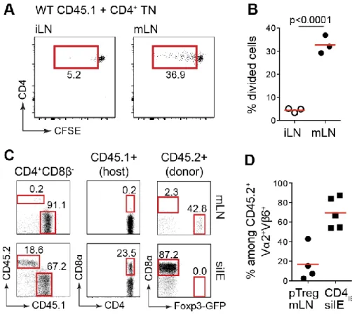
Figure 2.6 Naïve CD4+ TN T cells from pTreg TN/RKO expand and differentiate into pTregs and CD4 IELs
in immunodeficient hosts. 79
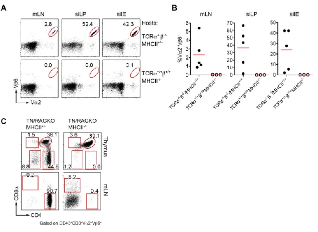
Figure 2.7 Expansion and differentiation of pTreg TN T cells in WT polyclonal hosts. 80 Figure 2.8 CD4+ T cells from pTreg TN/RKO mice are MHCII-restricted. 81
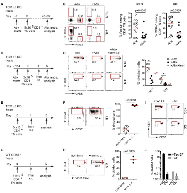
Figure 2.9 Antibiotic treatment depleted the microbiota. 82 Figure 2.10 pTreg and CD4IEL development and expansion are dependent on the microbiota. 83
Figure 2.11 Intestinal bacterial antigens induce proliferation of CD4+ T cells from pTreg TN/RKO mice. 85
Figure 2.12 CD4+ T cells from pTreg TN/RKO expand and convert into CD4
IEL in lymphopenic hosts
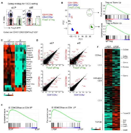
obtained from Taconic but not from Jackson Laboratory. 86 Figure 2.13 Gene expression analysis of pTreg and CD4IEL TN cells. 87
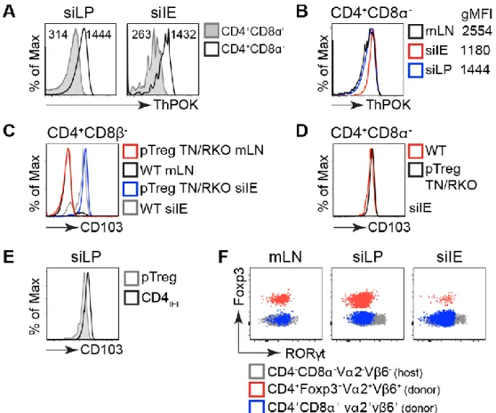
Figure 2.14 Low levels of ThPOK and high levels of CD103 in TN CD4IEL. 88
Figure 2.15 Mice from the WIBR animal facility are colonized by SFB. 89 Figure 2.16 Foxp3 deficiency does not confer pathogenicity to CD4IEL. 91
Figure 3.1 The microbiome influences the development and maintenance of CD4IELs. 105
Figure 3.2 The pTreg TN TCR recognizes the predominant member of the Altered Schaedler Flora. 108 Figure 3.3 The pTreg TN TCR recognizes P. goldsteinii. 110 Figure 3.4 P. goldsteinii induces CD4IELs in both monoclonal and polyclonal SPF mice. 112
Figure 3.5 P. goldsteinii is abundant in SPF mice but does not rescue CD4IEL accumulation in germ-free
mice. 114
Figure 3.6 The TN TCR specifically recognizes epitopes from Bacteroidetes β-N-acetylhexosaminidase in
complex with I-Ab. 116
Figure 3.7 Mass spectrometry of P. goldsteinii antigen-enriched fraction. 118 Figure 3.8 Truncations identify the segment of β-hexosaminidase recognized by the TN TCR. 120 Figure 3.9 A broad range of Bacteroidetes species encode epitopes recognized by the TN TCR. 123 Figure 3.10 TN T cells differentiate into CD4IELs upon colonization of immunodeficient hosts. 125 Figure 3.11 Oral delivery of cognate peptide is sufficient to induce proliferation and conversion of TN cells
into CD4IELs. 127
Figure 3.12 β-hexosaminidase MHCII tetramers identify antigen-specific CD4+ T cells in WT SPF mice. 129 Figure 3.13 Migration and expansion TN and WT CD4+ T cells in lymphocyte-induced colitis. 131 Figure 3.14 The β-hexosaminidase TN epitope is recognized by WT CD4IELs and mediates protection
against intestinal inflammation. 133
Figure 4.1 Transfer of TN cells into TCRαβ-/- mice promotes IgG1 class switching 152
Figure 4.2 Depletion of microbiota abrogates expansion and differentiation of TN cells, and abolishes
IgG1 response. 153
Figure 4.3 The serum of TCRαβ-/- mice that received TN T cells binds to commensal bacteria. 154
11
Figure 4.5 Serum IgG1 from TN-recipient mice does not always bind to commensal bacteria. 156 Figure 4.6 Models to explain diverse TN-induced IgG1 responses 158
Figure 5.1 Outer membrane vesicles. 167
Figure 5.2 Bacteroides Vesicle Incorporation Tag (BVIT) fusion targets proteins to OMVs. 168 Figure 5.3 BVIT Design Rules Are Applicable to Lipoproteins Across the Bacteroides genus. 170 Figure 5.4 P. goldsteinii OMVs do not activate TN cells in vitro. 171 Figure 5.5 Incorporation of Ovalbumin (OVA) and β-hex into Bacteroides OMVs. 172 Figure 5.6 Presentation of OVA-Loaded OMVs to Naive OTII CD4+ T Cells. 173
Figure 5.7 Cytokine Profile of OTII in vitro proliferation Assay. 174 Figure 5.8 TN cells respond to β-hex loaded OMVs in vitro. 175 Figure 5.9 β-hex loaded OMVs can activate TN cells in vivo. 176
Figure 7.1 The κ-LPETG mouse model 200
Figure 7.2 Specific labeling of Igκ LPETG-derived serum immunoglobulins and purified B cells using SrtA 202 Figure 7.3 Organization of the heavy and light chain of the OB1-LPETG mouse model 204 Figure 7.4 Igκ-LPETG mice have near normal B cell development 206 Figure 7.5 SrtA labeling does not activate B cells 207 Figure 7.6 Altered BCR organization in IgG1 B cells 209 Figure 7.7 Quantification of the number of BCRs on the surface of naive B cells 210
12
List of tables
Table 1-1 Characteristics of the different populations of intraepithelial lymphocytes found in the small
intestine. 29
Table 1-2 Commensal-specific CD4+ T cell models 47
13 List of abbreviations
A2A Adenosine receptor 2A
Ag Antigen
AHR Aryl hydrocarbon receptor AHR Aryl hydrocarbon receptor ALDH Aldehyde dehydrogenase APC Antigen-presenting cell
APRIL A proliferation inducing ligand BAFF B-cell activating factor
BCR B cell receptor
cAMP Cyclic Adenosine Monophosphate CD4IELs CD4 intraepithelial cells
CDR3 Complementarity determining region 3 CFSE Carboxyfluorescein succinimidyl ester cLP Colonic lamina propria
CR Charles River
CTLA-4 Cytotoxic T-lymphocyte-associated gene 4
DC Dendritic cell
DP Double-positive
DT Diphtheria toxin
EAE Experimental autoimmune encephalomyelitis EDTA Ethylenediaminetetraacetic acid
EF Excluded flora
ESC Embryonic stem cell
FACS Fluorescence activated cell sorting Flt3L FMS-like tyrosine kinase 3 ligand GALT Gut-associated lymphoid tissue
GF Germ-free
GFP Green fluorescent protein
Gzm Granzyme
HDAC Histone deacetylase
IBD Inflammatory bowel disease ICOS Inducible T-cell costimulator IDO Indolamine 2,3-dioxygenase
IE Intraepithelial
IEC Intestinal epithelial cell IEL Intraepithelial lymphocyte
IFN Interferon
Ig Immunoglobulin
IgA Immunoglobulin A
IL Interleukin
ILCs Innate lymphoid cells ILF Isolated lymphoid follicles
14
iNK Invariant natural killer
IPEX immune dysregulation, polyendocrinopathy, enteropathy, X-linked
Jax Jackson
KO Knock-out
LAG-3 Lymphocyte-activation gene 3 LCN2 Lipocalin-2
LiLP Large intestine lamina propria
LN lymph node
LP Lamina propria
MAIT Mucosal associated invariant T MHC Major histocompatibility complex MsLN Mesenteric LN
NF-κB Nuclear factor-kappaB
NK Natural killer
nTreg natural regulatory T cells OMV Outer membrane vesicle OTU Operational taxonomic unit
OVA Ovalbumin
PBS Phosphate buffered saline
PP Peyer’s Patches
PSA capsular polysaccharide A pTreg Peripheral regulatory T cell
qPCR Quantitative PCR
RA Retinoic acid
RNA-seq RNA sequencing
RORγT retinoid-related orphan receptor gamma T rRNA Ribosomal ribonucleic acid
SCFAs Short-chain fatty acids SCNT Somatic cell nuclear transfer SFB Segmented filamentous bacteria SFB Segmented filamentous bacteria SiLP Small intestine lamina propria
SP Single-positive
SPF Specific pathogen free SPF Specific pathogen free
Tac Taconic
Tconvs Conventional CD4+ T cell
TCR T cell receptor Tfh T follicular helper
TGFβ Tumor growth factor beta
Th CD4+ T helper
TL Thymus-leukemia antigen
TN Transnuclear
Tr1 Type 1 regulatory cells Treg Regulatory T cell
15
ZPS Zwitterionic capsular polysaccharides β -hex β-N-acetylhexosaminidase
16
Chapter 1 - Adaptive immune responses at the
intestinal mucosa
17 1.1 Introduction
The human body is home to a wide range of microbes, belonging to all three domains of life (1). Some of these microbes are beneficial to their host. They are often referred to as commensals, while other microbes can cause disease and are called pathogens. Commensals play an important role in human metabolism, provide essential nutrients and protect against invading pathogens(2). They colonize our skin and mucosal surfaces with high efficiency and comprise at least as many cells as the human body(3) . Perhaps even more impressive is the diversity of the species present (estimated to be between 500-1000 species), and their massive gene content (100 times greater than the human genome)(4). Changes in microbiome composition have been associated with pathology, ranging from inflammatory bowel disease, cardiometabolic disorders, cancer and neuropsychiatric disorders(5-12). The highest concentration of microbes is present in the intestinal mucosa. This location harbors trillions of microbes and represents one of the densest and most diverse ecosystems known (2, 13). The close proximity of commensals, the continuous exposure to dietary antigens and pathogens represents a unique challenge to the immune system. To achieve homeostasis, the immune system has developed a highly sophisticated network of cells that promotes tolerance towards beneficial antigens while protecting the body against invading pathogens. As a result, gut-associated tissues contain the highest number of immune cells in our body which display a fascinating complexity of immune phenotypes. Dysregulation of this network can cause disease: tolerance to pathogens may lead to infection, while an overactive immune response directed towards commensals or food antigens can lead to inflammatory bowel disease and food allergies, respectively (14, 15). In this chapter, I discuss the role of the microbiome in the development, maintenance and function of the immune system. I provide a deeper look at the gastrointestinal tract and mechanism of immune tolerance to commensal antigens.
18 1.2 The Immune system
The different layers of the immune system
Our bodies have evolved a set of defense strategies that protect us from external attacks by pathogenic invaders. These strategies are mediated by components that comprise anatomic and physiologic barriers, inflammatory mediators and cells, all collaborate to maintain health. The first line of defense of the immune system are physical barriers such as the skin and epithelia. The importance of this barrier is highlighted by the increased risk of infection after burns or wounds. Mucus is another type of physical barrier, the significance of which is exemplified by the pathologies of cystic fibrosis patients who cannot efficiently secrete mucus. These mechanical lines of defense are complemented by chemical protective layers such as the low pH present in the stomach or the secretion of antimicrobial peptides in the gut. Finally, these two barriers are supported by the presence of microbes that prevent the invasion of our body by pathogens (16).
The cellular compartment (i.e the ensemble of cells) of the immune system is composed of a network of specialized subsets of cells, all of which derive from pluripotent hematopoietic stem cells. These different subsets belong to either the innate immune system, which provides immediate responses, or the adaptive immune system, which supports long-term specific responses to a particular pathogen. The main cell types of the immune system are summarized in Fig. 1.1. For the purpose of this thesis I will focus mainly on the adaptive immune response. I direct the readers to an excellent review by Iwasaki and Medzhitov on the innate immune system and its cross talk with adaptive immunity(17).
19 Figure 1.1 Cell types of the immune system.
The immune system is composed of many different cell types and subtypes. Bone marrow hematopoietic stem cell can differentiate into lymphoid and myeloid progenitor cells. Lymphoid stem cells yield T and B-lymphocytes and natural killer (NK) T cells. Myeloid stem cells give rise to granulocytes (neutrophils, eosinophils, mast cells and basophils), NK cells and phagocytic cells (dendritic cells and macrophages).
20
Adaptive immune responses
The adaptive immune system, can recognize virtually any molecule or antigen (an antigen is defined as any substance that can evoke an immune response). The molecules involved in this process are the antigen receptors: the B cell receptor (BCR) expressed exclusively on B cells and the T cell receptor (TCR) on T cells. Both BCRs and TCRs possess a variable region, important for interaction with antigen, and a constant region. The diversity of the variable regions available to the TCR and BCR is specified in the genome: variable regions are assembled by somatic gene rearrangement of diverse genetic elements, a process known as V(D)J recombination (18). All antigen receptors of the adaptive immune system use two subunits, each equipped with a variable region, one of which is constructed from V, D and J elements, while its partner uses V and J elements. The variable region is assembled from randomly chosen V (variable), D (diversity) and a J (joining) segments. This rearrangement is mediated by complex enzymatic machinery that includes the Rag1 and 2 recombinase (18). The importance of these receptors is evident from the phenotype of RagKO animals (Rag1-/- or Rag2-/-), which do not possess
either mature B or T cells and are thus severely immunocompromised (19). In fact, conditional ablation of the BCR leads to B cell death (20). Therefore, these receptors are essential for both the development and survival of B and T cells.
An important difference between these receptors is the type of antigens they recognize. BCRs can recognize nearly any type of intact antigen such as protein, carbohydrate, DNA or even small molecules, to name a few. In contrast, the TCR only recognizes antigens displayed on the surface of other cells, bound to major histocompatibility complex (MHC) molecules. These complexes of proteins bind to pre-processed peptides and present them to T cells (21, 22). Another important difference is the absence of secreted form of the TCR, in contrast to B cells, which secrete antibodies, the secreted version of the BCR. Secretion of antibodies is important to neutralize extracellular pathogens and is an essential function of B cells.
T cells can be subdivided into two main classes, CD4+ or CD8+ T cells, based on the
21
stabilize the interaction between the TCR and the MHC molecules with which they interact. Indeed, the CD4 co-receptor recognizes MHC class II molecules (MHCII), while CD8 interacts with MHC class I molecules (MHCI). These two classes of T cells have very different effector functions. CD8+ T cells are cytotoxic and kill cells that display their
cognate antigens on MHCI, while CD4+ T cells orchestrate the immune response by
cell-cell contacts and through release of cytokines. CD4+ T cells can activate other cells, for
example to induce them to secrete antibodies. Importantly, MHCI and MHCII have different distributions among cells, which reflects the function of the T cells that interact with them. CD8+ T cells recognize predominantly intracellular pathogens present in the
cytosol such as viruses. Because any cell can be infected by viruses, all nucleated cells express MHCI. In contrast, only a subset of cells expresses MHCII. These cells are called antigen-presenting cells and include B cells, dendritic cells and macrophages. Antigens destined for presentation by MHCII molecules are picked up from extracellular space, proteolytically processed and presented to CD4+ T cells. When a CD4+ T cell interacts
with a B cell that presents the cognate antigen, it activates the B cell to differentiate and secrete antibodies. Similarly, CD4+ T cells can activate infected macrophages to destroy
intracellular pathogens when the appropriate antigens are presented to CD4+ T cells.
Upon activation by antigen-presenting cells, CD4+ T cells can differentiate into a variety
of effector subsets including Th1, Th2, Th17, T follicular helper (Tfh) and peripheral T regulatory (pTreg) (Fig. 1.2). Decision to differentiate into each of these lineages is governed primarily by the cytokines present in the microenvironment and the affinity of the TCR for its antigen(23). In addition, the presence of certain microbes and pathogens can influence the fate decision of CD4+ T cells in both antigen-dependent and
independent manners. Each of these subsets produces a defined set of cytokines and chemokines largely driven by transcription factors which act as “master regulators”. Long considered irreversible, these fates appear to be more plastic than initially thought, especially in the intestine(24). For example, a majority of colonic Tregs express both Foxp3 and RORγT, the characteristic master regulators of Tregs and Th17, respectively (25). During homeostasis, these cells selectively restrict pro-inflammatory Th17 but under inflammation, they lose Foxp3 expression and promote intestinal inflammation (26). This
22
example illustrates how plasticity of CD4+ T cells in the intestine can influence tissue
homeostasis.
Figure 1.2 CD4+ T helper cell subsets.
CD4+ T cells can differentiate into a diverse set of effector T cell lineages with distinct functions. Activation
of naïve CD4+ T cells by antigen-presenting cells in the presence of the appropriate cytokine environment
leads to commitment into one of these fates (or others not represented here). Each subset expressed a defined set of cytokines and surface markers, mostly controlled by transcription factors which act as master regulators such as Foxp3 for Tregs.
23
Central tolerance
An essential feature of the adaptive immune system is its ability to recognize almost any antigen. This comes in handy when dealing with rapidly evolving pathogens, but it poses a risk- our immune cells can recognize and destroy our own tissues. In fact, it is estimated that close to 75% of human immature B cells show some measure of self-reactivity(27). Therefore, the immune system must have mechanisms in place to ignore and tolerate “self” antigens, while actively responding to pathogenic or “non-self” antigens.
Central tolerance prevents the development of auto-reactive T or B cells. After rearrangement of their TCR genes, T cells go through a selection process to test the functionality of their properly assembled TCR. This process takes place in the thymus. Newly generated T cells are tested for their ability to interact with self-peptide, loaded onto MHC molecules. T cells that express a TCR unable to properly interact will not survive, while T cells that interact too strongly with MHC products complexed with self-peptides will be deleted. These positive and negative selection steps ensure that only functional T cells contribute to the pool of mature T cells. Because negative selection is imperfect, additional mechanisms are in place in the periphery to ensure that self-reactive T cells become non-responsive or anergic. Similar to T cells, B cells can be deleted during development or rendered anergic in the periphery when they recognize self-antigens (16). BCR and TCR repertoires
The ensemble of antigen receptors found in a tissue is referred to as the repertoire. These repertoires are affected by the antigens present in a given tissue. When lymphocytes are activated by their cognate antigens, they proliferate and their representation within the repertoire increases. Studies of repertoires are usually done by sequencing the TCR or BCR either at the population level, or after single-cell sorting. The use of animal models in which one of the two receptor chains is “fixed” reduces diversity and facilitates repertoire analysis (28-33). Analysis of the TCR repertoire of T cells in the colonic lamina propria showed that the majority of colonic Tregs were induced locally in response to microbiota-derived antigens(34). Analysis of the TCR repertoire can also inform on the
24
development of a particular T cell subset. Colonic Th17 and RORγt+Foxp3+ Tregs were
found to share a subset of TCRs, suggesting that RORγt+Foxp3+ Tregs could be the
precursors of some Th17 cells. This hypothesis was subsequently validated using a commensal-specific transgenic line (see below) (35, 36). A major question remains the extent to which receptor repertoires are linked to the function of the cells that express particular antigen receptors.
1.3 Microbiome
The microbiome is the ensemble of microbes living in and on our bodies. This dense community of microbes helps digest our food, provides essential nutrients, regulates and supports maturation and continued education of our immune system and protects us against invading pathogens (2, 37-39). Over the past decade, the number of studies on the microbiome has exploded. We now appreciate its contribution to human health. Imbalance in the composition of these microbial communities, called dysbiosis, is associated with a range of chronic diseases, including atherosclerosis, metabolic disorders, asthma, and autism spectrum disorder (40-49). Early work established that transfer of the microbes associated with these phenotypes can transfer disease from a sick donor to a healthy recipient in cases such as obesity, and conversely, that a microbiota from a healthy donor, when transferred into a sick recipient, can ameliorate disease. This raises the possibility of using microbes as therapies(50). As often is the case in a new field of research, many of these early findings report associations. Much effort is currently concentrated on moving from correlation to causation and identification of the molecular mechanisms responsible for the phenotypes observed(8, 51). The lack of suitable tools complicates the study of the molecular mechanisms at play in these complex communities. Contrary to common belief, the majority of gut microbes can be cultured, although many require specialized growth conditions not easily accessible to most laboratories (2, 52-56). As of today, most of these microbes are genetically intractable, making mechanistic studies a challenge(2).
The microbiome is established at birth, evolves considerably over the first 3 years of life and then remains mostly constant throughout adulthood (37, 57-59). Each individual
25
harbors a unique set of microbes, often similar between family members(60). Within an individual, different anatomical sites carry distinct populations of microbes. Variations in microbial composition are affected by both genetics and the environment in ways still poorly understood (2). Within the intestine, the type and abundance of microbes varies along the gastrointestinal tract. For instance, aerobic bacteria colonize the upper GI tract while anaerobes are found mostly in the large intestine. The load of bacteria also varies along the intestine, ranging from 103-104 bacteria/ml in the upper part of the GI tract, 108
bacteria/ml in the ileum and up to 1011 in the colon (61, 62). The emergence of
high-throughput sequencing of the 16s ribosomal RNA (rRNA) gene has enabled identification of the bacterial composition in the intestine, irrespective of our ability to culture them in vitro. All bacterial species and archea carry a 16s rRNA gene, which can be specifically amplified and sequenced to identify the relative abundance of operational taxonomic units (OTUs) in a given sample(63). Gram-positive Firmicutes such as Clostridia and gram-negative Bacteroidetes (primarily Bacteroides, Parabacteroides, Alistipes, and Prevotella) are the two most abundant phyla found in the mammalian gut (64). The remaining bacteria make up less than ten percent and include mainly Actinobacteria, Verrucomicrobia, Proteobacteria and Fusobacteria (2, 64).
While most microbes reside in the lumen of the intestine, others establish their niche in the crypts, the mucus, attached to the epithelium or even beyond the epithelium (65) (Fig. 1.3). These distinct localizations reflect the different microbial lifestyles and allow unique interactions with the immune system. A notable example is segmented filamentous bacteria (SFB) which intimately attaches to epithelial cells in the small intestine and promotes the specific induction of pro-inflammatory Th17 cells (66).
The mucus is a thick layer of glycoproteins produced by goblet cells that serves as a physical barrier to the microbiome. Microbes found in the mucus are highly specialized to survive in this environment (2). Only a limited number of bacteria have an adequate repertoire of genes that encode catabolic glyosidic enzymes capable of digesting mucus-derived glycoproteins. They include members of the genus Akkermansia such as A. muciniphila, the genus Bacteroides like B. thetaiotaomicron and Bifidobacterium, such as B. bifidum. Some of these species establish a stable relationship with mucus while others,
26
like B. thetaiotaomicron, digest host mucus when preferred food sources become scarce (67, 68).
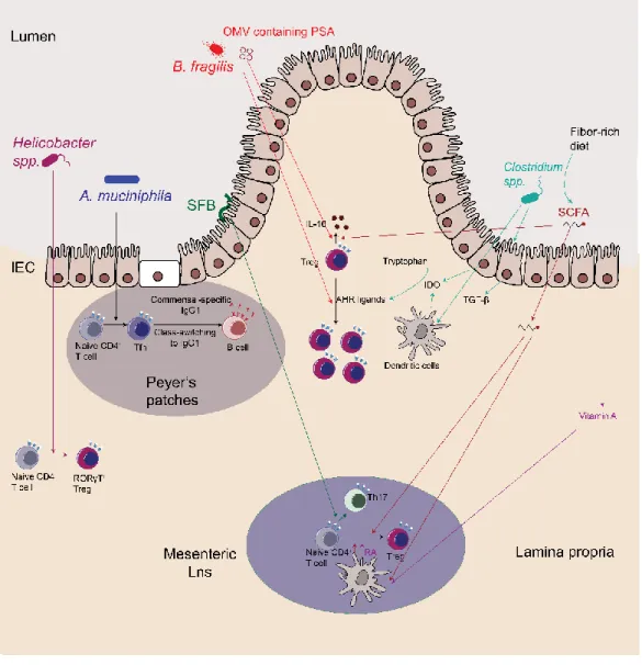
Figure 1.3 Localization of commensals at the intestinal mucosa
Commensal bacteria in the intestine can be found in the lumen, the mucus or within gut-associated lymphoid tissues.
Intestinal microbes are largely confined to the luminal side of the gut, but a few bacterial species can breach through the epithelial barrier and stably colonize gut-associated lymphoid tissues(69, 70). Inside Peyer’s patches (PPs) in mice, monkeys and humans, these bacteria can promote the secretion of commensal-specific IgA (69). The most dominant species found in PPs belong to the Alcaligenes genus, with less abundant
27
members derived from Ochrobactrum spp., Serratia spp. and Burkholderia spp. The anatomical containment of these species is mediated by Innate lymphoid cells (ILCs) and is disrupted in patients with HIV, cancer or cystic fibrosis, causing peripheral dissemination of these bacteria (70). More studies are required to better understand how these species establish a stable niche within gut-associated lymphoid tissues.
1.4 Mucosal immune system
Most of what is known about our immune system derives from studies that examine the responses to pathogens. However, the majority of interactions between microbes and the immune system occurs at barrier sites and are mostly symbiotic in nature. The particularities and properties of this type of immunity remain mostly obscure. As described above, a dense population of microbes lives closely and even within lymphoid organs in our intestine. This population of microbes constantly interacts, communicates with and educates our immune system(71-73). In turn, the immune system maintains homeostasis and prevents dissemination of these microbes via a sophisticated network of cells and structures at mucosal surfaces. The intestinal mucosal immune system contains the largest number of immune cells in our body, most of which express complex effector phenotypes not observed anywhere else in the body.
Organization of the gut mucosal system
The barrier that physically separates the luminal microbes of the gut from our body is made of a single layer of epithelial cells connected by tight junctions. The epithelium itself is home to a set of specialized cells that support the barrier function of intestinal epithelial cells (IECs). These include the mucus-secreting goblet cells, anti-microbial peptide- producing cells named Paneth cells and a complex set of lymphocytes, the intraepithelial lymphocytes (IELs). Below the epithelium, another set of specialized structures is found. The Peyer’s patches (PPs) of the small intestine and the numerous isolated lymphoid follicles are both present on the submucosal layer of the intestinal wall and are important for the induction of IgA. Beneath the epithelium, separated by a loose layer of connective tissue, is the lamina propria, which contains a high number of activated lymphocytes. All
28
of these sites are connected to afferent lymphatics, which drain cells and antigens to the mesenteric lymph nodes(16).
The microbes present in the lumen of the gut are also separated from our body by a mucus layer, which is approximately 400µM thick in humans and 150µM in mice and prevent most microbes from entering in direct contact with the epithelium (74, 75). In the colon, the mucus forms two distinct layers, the innermost of which is mostly sterile (74). The mucus itself contains lysozyme and other antimicrobial peptides (AMPs) (76). Antimicrobial peptides are short 10-50 amino acid peptides with an overall positive charge and broad specificity against most pathogenic but also commensal bacteria (77). Paneth cells, present in the crypts of the small intestine, are a major source of such AMPs (77). Antigen uptake and presentation in GALT
Also present in the epithelium are microfold or “M” cells, which are continually picking up antigens from the lumen of the intestine and transferring them to gut-associated lymphoid tissue (GALT). This route of entry is highjacked by pathogens such as Salmonella enterica (78). On the basal side of M cells, antigens are released into the extracellular space by transcytosis and are then in turn picked up by antigen-presenting cells (APCs). Antigen-loaded APCs can then move to the Peyer’s patches (PPs) for antigen presentation to T cells or the APCs migrate to the mesenteric lymph nodes where they activate cognate T cells (16). Yet another mode of entry for antigens is via CX3CR1-positive macrophages, which possess long dendrites that extend between epithelial cells, project into the gut lumen and sample luminal antigens (79). Remarkably, these dendrites can engulf whole bacteria before delivering them to the lamina propria.
Intraepithelial lymphocytes (IELs).
The other class of cells present in the epithelium are called intraepithelial lymphocytes (IELs). This compartment contains the largest number of T cells in our body as well as a diverse range of other immune cells such as eosinophils and Innate Lymphoid Cells (ILCs) (80, 81). IELs represent the front line of defense against invading pathogens due to their immediate proximity to antigens present in the gut lumen. As a result, IELs express
29
activation markers such as CD69 but also inhibitory types of receptors. They are therefore often referred to as “activated yet resting” (82). IELs are further characterized by the expression of gut-homing markers such as CD103, cytotoxic markers such as granzyme B and anti-inflammatory molecules such as LAG-3 (83, 84). Another typical characteristic of IELs is the expression of the CD8αα homodimer, which can recognize both classical major histocompatibility complex I (MHC-I) and non-conventional MHC-I molecules such as the thymus leukemia (TL)-antigen on epithelial cells (85, 86). Interaction between CD8αα and TL-antigen attenuates TCR activation and may contribute to keeping IELs in a non-responsive state(87). In fact, mice deficient in TL-antigen are more susceptible to intestinal inflammation in a spontaneous model of colitis (87). A summary of the IELs found in the small intestine is described in Table 1.1.
Table 1-1 Characteristics of the different populations of intraepithelial lymphocytes found in the small intestine.
30 IgA production in the gut
The production of commensal-specific IgA antibodies is yet another strategy used by the immune system to maintain homeostasis at the gut mucosa. IgA is the dominant antibody isotype present at mucosal surfaces. IgA dimers can be transcytosed to the intestinal lumen where they can coat commensals, toxins and pathogens and thus restrict access to mucosal surfaces(88). Class-switching to and secretion of IgA can either require T cell help in the presence of cognate antigen, or be T-independent in the presence of TGF-β, BAFF and APRIL.
T-cell deficient mice have starkly reduced amounts of free IgA, suggesting that most IgA production is T-dependent (89). Sequence analyses showed that most of the IgA produced sustained somatic hypermutation consistent with T cell involvement (88, 90). Sorting of IgA-coated bacteria in T-deficient mice (91) has yielded conflicting results concerning the relative contribution of T cells in IgA responses in the gut. Therefore, further studies are required to reach a consensus. Beyond providing a physical barrier to antigens in the lumen, IgA can modulate the expression of genes by commensals. For example, coating of B. thetaiotaomicron with IgA prevents the expression of genes that elicit inflammatory responses in the host(92).
1.5 Development of the immune system in the gut and commensals
Studies in germ-free and mice harboring a defined minimal flora (gnotobiotic) mice have shown that interactions between the microbiome and the immune system are important for the development and normal physiology of the immune system (1). Germ-free or antibiotic-treated mice have impaired immune responses to pathogens(66, 93-95). This is not surprising, since germ-free mice possess abnormal anatomical immune structures and immune cell numbers in the gut: they lack mucosal-associated invariant T cells (MAIT) cells(96), display poorly formed Peyer’s patches, have small mesenteric lymph nodes (1), show impaired development of isolated lymphoid follicles and present with altered composition of CD4+ T cells, including a lower frequency of Tregs (97, 98) and
IgA-producing B cells (96, 99). Antibiotic-treated mice likewise show altered gut structures, such as a reduced number of Peyer’s patches, smaller lymph nodes as well
31
as an altered myeloid and lymphoid compartment (100). Both single isolates and consortia can repair some of these abnormalities (97, 98). The importance of the microbiome is also observed at weaning in Standard Pathogen-Free (SPF) mice. The transition from a milk-based diet to solid food is associated with a drastic shift in microbial composition and a marked increase in pTregs specific for commensal antigens in the colon (34, 97, 98, 101, 102).
1.6 Homeostasis at the gut mucosa is mainly maintained by T cells
Although homeostasis at the gut mucosa is maintained by many arms of the immune system, T cell responses appear to be central to this regulation. Lack of T cells is associated with spontaneous development of colitis in SPF but not in germ-free mice (103, 104). Similarly, MHCII-deficient mice develop colitis, suggesting that CD4+ T cells
play an important role in the prevention of spontaneous colitis (103). In humans, a decrease in the number of CD4+ T cells in HIV patients is often associated with intestinal
inflammation, even in the absence of pathogens(105, 106). Conversely, inappropriate T cell responses are also associated with disease. TCRα-/- mice develop spontaneous
colitis, mediated by pathogenic IL-4 -producing CD4+TCRββ+ cells (107). Overall, these
experiments suggest that T cell responses at the gut mucosa must be tightly regulated to avoid the development of colitis.
Regulatory T cells (Tregs)
Tolerance to commensal and dietary antigens is mediated by Foxp3+ peripheral T
regulatory cells (pTregs). In contrast to natural Tregs (nTregs), which develop in the thymus and recognize self-antigens, pTregs develop from naïve CD4+ T cells and are
specific for antigens found within tissues. Foxp3 is a transcription factor essential for Treg development, maintenance and function and its expression serves as the canonical marker of Tregs (108, 109). Tregs are present in every organ of the body, but it is in the intestine that they are the most abundant, accounting for up to 30% of CD4+ T cells in the
colonic lamina propria (97-99, 110, 111). Development of intestinal Tregs is heavily dependent on the microbiota. Treg numbers are decreased in both germ-free and
32
antibiotic-treated mice. Their functional maturation is also affected by the presence of the microbiota. The few Tregs that remain in the absence of commensals exhibit a severe reduction in the expression of ICOS, CTLA-4 and IL-10, all key markers for the function of Tregs (97, 98, 110-112).
The essential suppressive role of Tregs was originally demonstrated by depletion of CD4+CD25+ Treg cells, which led to the development of multi-organ autoimmunity,
including intestinal inflammation (113). These findings received support from results in the colitis model developed by Powrie and colleagues. Transfer of naïve CD4+CD45RBhigh
lymphocytes into a syngeneic lymphopenic host causes a progressive and chronic wasting disease similar to Inflammatory bowel disease (IBD) (114, 115). Co-transfer of pathogenic cells (CD4+CD45RBhigh cells) and CD4+CD25+ Tregs efficiently prevented and
even reversed the development of colitis in this model (116, 117). Conclusions from these experiments were further complemented by the study of genetic deficiencies. Null mutations in Foxp3 in both mice and humans are associated with diarrhea, colitis and a multitude of other severe autoimmune phenotypes (118-121). More recently, administration of antibodies directed against cytotoxic T lymphocyte antigen-4 (CTLA-4) to cancer patients, which targets a central suppressive marker on Tregs, caused development of colitis as a side effect(122). The regulatory function of Tregs is therefore essential throughout the body and in particular in the intestine.
CD4IELS
While Tregs are key players in the establishment and maintenance of intestinal tolerance, they are not solely responsible for this regulation. Deficiencies in IL-10R in humans is associated with the development of colitis, with an earlier onset and higher penetrance than patients with Foxp3 deficiencies (123). In humans, intestinal inflammation is not associated with a decreased frequency of Foxp3+ Tregs (124, 125). This supports the
notion that other cell types could play a role complementary to that of Tregs, for example via the (immunosuppressive) IL-10 pathway.
CD4+CD8αα+ intraepithelial lymphocytes (CD4IELS) are a subset of lymphocytes that
germ-33
free mice, and in IBD and celiac disease patients (127-129). Similarly to Tregs, CD4IELS
display regulatory functions and promote tolerance to dietary antigens(128). The function of CD4IELS appears to be complementary to that of Tregs. Depletion of CD4IELs in mice
that lack Tregs causes disease (128). Forced differentiation of CD4+ cells into CD4 IELs
prevents intestinal inflammation. Blocking the development of CD4IELs can accelerate
intestinal damage in a model of colitis(130). Therefore, CD4IELs is a distinct subset of T
cells that works in concert with Tregs to maintain intestinal homeostasis.
The development of CD4IELs is initiated when a naïve CD4+ T cell encounters its cognate
tissue-derived antigen in the draining lymph nodes. Migration to the intestine requires acquisition of gut-homing markers such as CD103, possibly mediated by migratory DC in the mesenteric lymph nodes, which produce TGFβ and retinoic acid (RA) (131-137). The activated cells then travel to the lamina propria of the small intestine and finally the epithelium (130, 137-140). The final step in the acquisition of the IEL phenotype likely occurs once the cells have reached the epithelium(128). There, the transcription factors T-bet and Runx-3 are induced, while the master regulator of the T-helper lineage, Thpok is lost (130, 139, 141). In vitro, naïve CD4+ T cells can be converted to CD4+CD8αα+ IELs
in the presence of RA, TGFβ, IFN and their cognate antigen (141). The exact mechanism responsible for this remarkable imprinting in vivo remains to be defined.
IFN is another factor that is essential for the development of CD4IELs, as IFNR-deficient
mice show a sharp decrease in the frequency of CD4IELs(141). In vitro, the T-bet inducing
cytokine IL-27 promotes the differentiation of CD4IELs, similarly to IFN. While IL-27 is not
required in vivo for development of CD4IELs, localized production of IL-27 by the
engineered commensal L. lactis promotes the production of IL-10 by CD4IELs (141, 142).
CD4IELs can also develop from pTregs through loss of Foxp3 expression after having
settled in the epithelium (128, 143).
The nature of the antigens recognized by CD4IELs is currently unknown. Germ-free mice
show few CD4IELs, while their frequency increases with age in SPF mice. Microbial
antigens and/or other microbial components may therefore be important for their development (139, 144-148). In contrast to peripheral T cells, IELs, including CD4IELs,
34
appear to have a restricted TCR repertoire (149-157). This suggests that a limited number of antigens may be sufficient to shape this compartment. Regardless of antigen specificity, secretion of indole derivates by L. reuteri promotes CD4IELs development in a
TCR-independent manner, via the aryl-hydrocarbon receptor (AHR) (151). However, this process only occurs in the presence of a normal microbiota, suggesting that these small molecules are not sufficient for the development of CD4IELs.
Tr1 cells
T regulatory type 1 (Tr1) cells are a T cell subset also important for maintenance of homeostasis in the gut. While their suppressive capability is established both in vitro and in vivo, their phenotype is still poorly characterized. Like Tregs, these cells require activation by their cognate antigen to exert their function(158). Defects in the frequency or function of Tr1 cells was shown in different models of autoimmune and inflammatory diseases (159). Tr1 cells can be generated in vitro under conditions of chronic activation in the presence of Interleukin-10 (IL-10) (160). In vivo, these cells produce high levels of IL-10 and appear short-lived. I address the reader to an excellent review for further information (161).
1.7 Mechanism of suppression by Tregs and CD4IELs
Tregs
Despite the importance of Tregs in imposition of peripheral tolerance, the molecular mechanism(s) of suppression involved remain incompletely understood and somewhat controversial(162). Tregs can suppress the function of multiple cell types, including B cells, NKT cells, NK cells, CD4+ and CD8+ T cells, monocytes and dendritic cells. Several
mechanisms have been described including cell-to-cell- (i.e in the absence of APC), contact-dependent- or independent-suppression, as well as through the intermediate of APCs. Instead of relying on a single, universal strategy to suppress cells, Tregs seem to leverage a variety of mechanisms, alone or in combination, depending on the context (Fig. 1.4). Regardless of the mechanism used, Tregs need to be activated by their
35
cognate antigen to become suppressive(163-167). Once activated, Tregs can suppress T cells independently of their antigen leading to “bystander-suppression”(168, 169). Cytolysis
One strategy used by Tregs to suppress immune responses is via cell killing of effector T cells using granzyme-B (Gzm-B) or granzyme A (Gzm-A) (170, 171), important, for example, to maintain transplant tolerance (172).
Metabolic disruption
Tregs can also create an environment that disfavors T cell proliferation. Cyclic AMP (cAMP) is a potent repressor of T cell proliferation. Tregs produce high levels of intracellular cAMP, which can be delivered to the cytoplasm of target cells via gap junctions (173). Additionally, Tregs can repress T cell functions by generating high levels of extracellular adenosine. CD39/CD73 present on the surface of Tregs can convert extracellular ATP into adenosine. Adenosine in turn binds to adenosine receptor 2A (A2a)
on effector T cells, leading to repression of their function (174-176). Tumors maintain high levels of adenosine in their microenvironment (177, 178). Mice deficient for A2A show
enhanced elimination of tumors compared to WT mice (178). Tregs maintain surface expression of CD25, the subunit of the high-affinity IL-2 receptor. Surrounding Tregs are deprived of 2, a cytokine essential for T cell survival. This scavenging of IL-2 leads to apoptosis of the target cell (179).
Inhibitory cytokines
Tregs also express cytokines, either membrane-bound or secreted, that can suppress other immune cells. TGF-β can be produced in large amounts by Tregs and in so doing suppress T cell proliferation in vitro(180). Mice deficient in TGF-β develop T cell-mediated autoimmunity (181, 182). IL-10 is another cytokine produced by Tregs, important for their suppressive activity. Indeed, IL-10-deficient Tregs fail to maintain homeostasis in a T cell transfer-induced colitis models (183). IL-35 is a cytokine central to Treg cell function,
36
including the control of T cell proliferation(184). This cytokine is preferentially expressed by Tregs, upregulated on cells with suppressive activity and required for maximal suppression by Tregs (185, 186). Deficiencies in either of the IL-35 chains reduces Treg function in vitro and in IBD models in vivo(187).
Suppression by targeting APCs
An alternative strategy used by Tregs to exert suppression is by their action on antigen presenting cells (APCs). Intravital microscopy showed that Tregs form long-lasting interactions with DCs upon entry into lymph nodes. This in turn impairs the ability of effector T cells to be activated by DCs (188). Tregs could thus affect T cell responses by acting directly on APCs.
Expression of key checkpoint molecules such as CTLA-4 on Tregs could account for this property. Treg-specific deficiencies in expression of CTLA-4 leads to spontaneous fatal autoimmune diseases, underlining the importance of CTLA-4 for Treg function (189). Interaction between CTLA-4 and DCs stimulates them to produce indoleamine 2,3-dioxygenase (IDO), an enzyme that catabolizes tryptophan. Starvation of T cells in the absence of tryptophan prevents T cell proliferation and may eventually lead to T cell apoptosis (190). The presence of the tryptophan metabolite kynurenine also favors expansion of Tregs, presumably by activation of AHR(191). CTLA-4 similarly promotes downregulation of costimulatory molecules CD80 and CD86 on DCs, negatively affecting their ability to activate T cells(192).
LAG-3 is a checkpoint molecule involved in Treg function. LAG-3 is expressed on the surface of Tregs and binds to MHCII with high affinity, affecting the maturation and ability of DCs to activate T cells(193). LAG-3 deficiency or blockade reduces Treg functions, while ectopic expression of LAG-3 confers regulatory properties to CD4+ T cells (194).
Other checkpoint molecules present on Tregs, such as induced costimulatory molecule (ICOS), have been investigated for their suppressive function. Their role is less apparent than the examples cited (195, 196).
37 Figure 1.4 Mechanisms of suppression by regulatory T cells.
Tregs can suppress T conventional cells using a range of strategies. Tregs can induce cell death in effector cell via secretion of granzymes. Alternatively, Tregs can secrete immunosuppressive cytokines (IL-10, TGF-β and IL-35), starve effector T cells by scavenging IL-2, produce high levels of adenosine and cAMP, which suppress effector T cells. Finally, Tregs can act indirectly on effector cells by promoting downregulation of co-stimulatory molecules on dendritic cells (DCs). Tregs also promote production of indoleamine 2,3-dioxygenase (IDO) by DCs, which catabolizes tryptophan into AHR ligands, which in turn promote Tregs expansion.
38
CD4IELs
The mechanisms used by Tregs to prevent inflammation have been extensively studied, but the strategies used by CD4IELs are less clear. This is hardly surprising, given that the
role of CD4IELs in intestinal homeostasis has been appreciated only recently. CD4IELs
display a regulatory cytokine signature, including the expression of IL-10, TGF-β and IFN-(138, 142). RNAseq analysis showed that CD4IELs also express high levels of Gzm-A
and Gzm-B (143). Upon reaching the epithelium, precursors of CD4IELs increase
production of IL-10(138). IL-10 is not required for development of CD4IELs but seems
essential for their anti-inflammatory role in the intestine (138). Remarkably, IL-10 produced by CD4IELs and other cells in the small intestine can protect against inflammation
in the colon (138, 197, 198).
1.8 TCR-independent modulation of T cell responses by commensals
The action of the immune system at mucosal surfaces is achieved through coordination of a complex set of cells and signaling networks. T cells can adopt different effector fates. The differentiation into each of these fates depends on environment, context and type of antigen(s) recognized (23). Specific microbes and pathogens likewise can skew the differentiation of immune cells towards specific fates (23, 66, 97, 199). However, the signals, molecules and pathways used by microbes in these processes are still poorly understood. In the following I will summarize current knowledge about TCR-dependent and independent mechanisms.
Clostridium species promote the development of both mucosal and systemic Tregs via induction of TGF-β by epithelial cells (97, 112, 200). Similarly, mice colonized with the Altered Schaedler Flora (ASF) show de novo generation and activation of pTregs, which in turn prevent immune deviation towards more inflammatory responses (98). It is unclear whether these responses are antigen-specific or not. Colonization with the probiotic Bifidobacterium breve promotes the development of IL-10 producing Tr1 cells and improves intestinal pathology in a colitis model while B. longum and B.bifidum promote Tregs development (201-203). Monocolonization of germ-free mice with Bacteroides
39
species boosts the frequencies of both tolerogenic pDC and pTregs (204). Tolerogenic pDcs and intestinal epithelial cells produce the enzyme Indoleamine 2,3-dioxygenase-1 (IDO) which promotes the development of pTregs in response to the gut microbiota (97, 205, 206). In turn, Tregs promote IDO expression in DCs via CTLA-4-B7-1 and -2 interaction thus essentially acting as a tolerogenic positive feedback loop(190, 207). Microbes can shape the immune system by secretion of metabolites. Natural products produced by the microbiota are abundant and their concentrations can be equivalent or even higher than a typical drug dose (10uM-1mM) (208). Short chain fatty acids (SCFAs) produced by commensal microbes are some of the most abundant compounds secreted by microbes(209). SCFAs are released by anaerobic commensals when they ferment dietary carbohydrates that their host cannot digest. Typical SCFAs produced are acetate, propionate, and butyrate. Acetate and propionate are usually produced by Bacteroidetes, whereas Firmicutes generate most of the butyrate (210, 211). Besides their use as an energy source for the host, these molecules can be sensed by host receptors and modulate the immune system. A number of receptors specific for SCFAs have been described, including PPARγ, GPR109a, GPR41 and GPR43 (72, 212-216). PPARγ is a receptor essential for the protective effect of Tregs against T-cell induced colitis (217). Activation of PPARγ by a synthetic ligand is also protective against intestinal inflammation, in the dextran sulphate induced-colitis model (218). SCFA can also bind to the butyrate receptor GPR109a on macrophages and DCs to promote expression of IL-10 and aldehyde dehydrogenase (ALDH), which catabolizes vitamin A into retinoic acid (RA) (72).Secretion of RA by APC upregulates gut homing markers such as CD103 by pTregs(134, 219). GPR109a-deficient mice are prone to develop colonic inflammation and colon cancer(213). SCFA can also act directly on Foxp3 expression in T cells by repression of histone deacetylase (HDAC), thereby inducing Tregs differentiation and accumulation(220, 221).
Secretion of polysaccharide A (PSA) by B. fragilis via outer membrane vesicles (OMVs) expands Tregs and enhances their function in a TLR2-dependent manner (222-224). PSA is part of a larger family of molecules known as capsular zwitterionic polysaccharides (ZPSs), which can activate CD4+ T cells in complex ways (225-227). Screening for
40
predicted ZPS operons in bacterial genomes identified a long list of commensals, some already associated with anti-inflammatory responses (228). Crude lysates from P. distasonis, and in particular membrane fractions, reduced intestinal inflammation in dextran sulphate sodium (DSS) colitis model and increased the frequency of Tregs in the mesenteric lymph nodes (229). P. distasonis is also recognized by a colonic Treg TCR, suggesting that its anti-inflammatory capacity might be both TCR-dependent and independent (34). Similarly, the most abundant member of the ASF (see above), P. goldsteinii (also referred to as ASF519), is predicted to produce ZPS, suggesting that the increased frequency of Tregs observed in ASF-colonized mice could be due to ZPS molecules from P. goldsteinii (98, 228). Neff and colleagues also identified C. ramosum and C. symbiosum as two potent producers of ZPS, both of which belong to the consortium of Clostriums shown to induce Tregs (112)(228).
In addition to the mechanisms described above, A number of isolated or consortiums of commensals can influence T cell fate in the gut. I direct the readers to excellent reviews for more information (221, 230, 231).
1.9 Antigen-specific T cell responses to commensals
Although the intestinal microbiota contains a vast diversity of species, only a handful of microbes have been shown to modulate the immune system. What is probably even more remarkable is the specificity with which these microbes modulate the immune system. Littman and colleagues pioneered this work by showing that Segmented Filamentous Bacteria (SFB) induced quasi-clonal Th17 cells in an antigen-specific manner (232). Helicobacter spp. induce antigen-specific colonic RORγt+ Tregs, further extending this
concept (26, 35). K. pneumoniae from the oral cavity induces antigen-specific Th1 response in the colon after antibiotics treatment (233). Strains of Staphylococcus epidermidis induce specific and long-lasting CD8+ T cell response in the skin restricted
by non-classical Class I MHC products (234) . The mucus-degrading bacterium A. muciniphila promotes the differentiation of Tfh and commensal-specific IgG1 (235). Therefore, specific commensal bacteria can reprogram a population of immune cells and thereby induce long-lasting and specific modulation of the immune response.