HAL Id: tel-02003420
https://tel.archives-ouvertes.fr/tel-02003420
Submitted on 1 Feb 2019HAL is a multi-disciplinary open access archive for the deposit and dissemination of sci-entific research documents, whether they are pub-lished or not. The documents may come from teaching and research institutions in France or
L’archive ouverte pluridisciplinaire HAL, est destinée au dépôt et à la diffusion de documents scientifiques de niveau recherche, publiés ou non, émanant des établissements d’enseignement et de recherche français ou étrangers, des laboratoires
molecular dysregulation in cerebellum in a mouse model
for ARCA2, a recessive ataxia with coenzyme Q10
deficiency
Tiphaine Jaeg
To cite this version:
Tiphaine Jaeg. Exploring the mitochondrial function in muscle and molecular dysregulation in cere-bellum in a mouse model for ARCA2, a recessive ataxia with coenzyme Q10 deficiency. Neurobiology. Université de Strasbourg, 2017. English. �NNT : 2017STRAJ082�. �tel-02003420�
ÉCOLE DOCTORALE DES SCIENCES DE LA VIE ET DE LA SANTE
Institut de Génétique et Biologie Moléculaire et Cellulaire
THÈSE
présentée par :Tiphaine JAEG
soutenue le : 03 octobre 2017pour obtenir le grade de :
Docteur de l’université de Strasbourg
Discipline/ Spécialité: Aspects moléculaires et cellulaires de la biologie
Exploring the mitochondrial function in muscle
and molecular dysregulation in cerebellum in a
mouse model for ARCA2, a recessive ataxia with
coenzyme Q10 deficiency
THÈSE dirigée par :Dr PUCCIO Hélène Directrice de Recherche, IGBMC, Inserm U964, UMR 7104 CNRS, Université de Strasbourg
RAPPORTEURS :
Dr RÖTIG Agnès Directrice de Recherche, Institut Imagine, Inserm U1163
Dr QUINZII Catarina MD PhD, Assistant Professor, Columbia University Medical Center
AUTRES MEMBRES DU JURY :
Dr DUPUIS Luc Directeur de recherche, Inserm U1118, Université de Strasbourg
MEMBRE INVITE
Try to leave this world a little better than you found it and, when your turn comes to die, you can die happy in feeling that at any rate you have not wasted your time but have done your best Robert Baden‐Powell
Acknowledgements
I would like to dedicate these first line to Hélène Puccio, my PhD supervisor who accepted to host me in her lab starting from September 2013. Thanks for the freedom you gave me and all what I learned during these four years. Thanks to the Dr. Luc Dupuis, Philippe Isope and Yvon Trottier who helped us decide what should be the best path to pursue with. Also, a big thank for having evaluated my work at key moments of my thesis which helped me go further. I would like to thank the Dr. Agnès Rötig, Catarina Quinzii, Luc Dupuis and Philippe Isope for having accepted to evaluate my work, and taken part of their time to read through this manuscript. I thank Laurence, for the help she has provided me for experiments. Thank for having explained me patiently some assays, how to handle mice, how to deal with mice colonies and how to build cohorts. Thanks also for having accepted to generate and genotype so many mice for the purpose of my experiments. I’m really grateful.Thanks to Floriana, Leïla and Pankaj with whom I worked with, trying to unravel the cause of ARCA2 and who have cheered me up and shared doubts, questions, ideas about this hard but fascinating project.
Thanks to all the lab members, for the nice discussions we had and all what I learned from you! For sharing protocols, benches, and brainstorming for new experiments and troubleshooting.
Thanks to Nadège and Aurélie, for their kindness and benevolence. You laugh and smiles each day made my everyday life so much nicer! I also greatly appreciated all the little things you did to make my life easier and to help me take decisions for certain experiments.
I now have to dedicate an entire paragraph to Charline, so here you are ;‐p! Thanks for having shared the back of the lab with me. I will never forget how we cheered each other up, and for our numerous discussions. Thanks for having been there, with me, late in the lab and for all our debates and brainstorming about protocols, new experimental design, publications and so on. Thanks for having wondered about me and make me realize I needed to slow down at some points to be able to achieve this PhD. Thanks for the numerous giggles that made life in the lab so much nicer and gave me energy to work more and persevere. And a simple big thank for your smiles.
Samantha and Arielle, this PhD would not have been the same without you! We shared our doubts, rejoice, questions… You were always around, even late in the cell culture room or the lab, sharing chocolate or motivation to cheer up. I will never forget these precious moments so thanks!
Thanks also to Angélique, Francesca, Florent, Aurore, Léa, Mélanie, with whom I sometimes ate, and often met in the corridors, thanks for the long discussions, your advices and tips! And thanks for your friendship.
Thanks to the IGBMC platforms and services for all the help provided. Thanks to Nadia, Jean‐Luc and Josiane for the electron microscopy. Thanks to Betty, Marion and all the girls from the cell culture facility. A big thanks to Marc, Marcel and Yves who helped me with microscopy. A huge thanks to Pascal for his patience and time, explaining microscopy, building macros to help the little PhD student I was.
Thanks also to Frédéric Doussau, for teaching me how to perform organotypic cultures, immunofluorescence with them, and all the nice ideas he shared.
Thanks to Paulo and Francine, who simplified our PhD lifes. A special thanks to Paulo for having supervised the organization of the PhD retreat. And thanks to Arielle, Samantha, Damola, Hichem, Federica, Tajith and Tiago for the nice PhD retreat we have built up. Merci à tous les Openlabistes, avec qui j’ai partagé cette belle aventure. Je vous suis reconnaissante pour les parfois longues discussions dans la voiture, direction les interventions, à refaire le monde et surtout la biologie, à essayer de tourner et retourner les problèmes sur lesquels nous bloquions pour nos sujets de thèse. Merci pour vos remarques et idées ! Et merci pour les bons moments passés à expliquer la science simplement. Merci à ma famille et tous mes amis et qui m’ont encouragé et soutenu tout au long de cette thèse. Merci à Maëlyne, Soléane, Auxence et Isaure, de m’avoir fait voir la beauté de la vie, d’avoir échangé autant de tendresse, de sourires, d’avoir joué et rit. Pour tous ces moments hors du temps avec vous qui m’ont permis de prendre du recul, de réaliser que la vie est un magnifique cadeau, et m’ont permis de persévérer : MERCI. A Fanny et Lowik, sans vous la vie aurait beaucoup moins de saveur. Si je réussi cette thèse, c’est en partie grâce à vous. Merci pour les questions et remarques qui m’ont déstabilisé mais qui m’ont permis de me remettre en question et d’avancer. Fanny, merci de comprendre la petite biologiste que je suis et d’avoir partagé tous ces trucs et astuces. Ton soutien a été précieux, tout comme ton sourire et ta joie qui m’ont fait garder le sourire et souvent reprendre des forces. Lowik, tu as le don pour amuser la galerie, semer une bonne ambiance, lancer des phrases cinglantes qui titillent mais permettent de repenser les choses autrement. Merci d’être toi, de rester toujours disponible pour moi et de m’avoir aidé à surmonter ce beau défi, notamment en nous proposant de belles sorties, pique‐nique… pour que je puisse faire des pauses et reprendre des forces. A mes parents, sans qui je ne serais pas là aujourd’hui. Papa et Maman, merci pour votre patience et votre amour. Merci d’être toujours là pour moi tout en me laissant libre. Merci pour vos encouragements, et d’avoir réussi à trouver les mots pour m’aider à surmonter les épreuves et les découragements. Merci de m’avoir forcé à réfléchir, à voir les choses différemment, à sortir de ma zone de confort, à me dépasser et surtout à me poser les bonnes questions au bon moment. Merci d’avoir accepté mes choix, et de m’aider à aller jusqu’au bout. Vous m’avez aidé à murir et vous continuez inlassablement de la faire. Vous avez grandement participé à la réussite de ma thèse (et de beaucoup d’autres belles réussites !). Enfin je dédie ces dernières lignes à Guillaume, mon mari. Merci de m’accompagner tous les jours sur les chemins de la vie. Merci d’avoir accepté de partager ma vie, et de m’avoir encouragé à faire cette thèse. Merci pour ta patience, surtout lorsque je te disais, j’essaye de ne pas rentrer trop tard et que je n’y arrivais pas. Merci de m’avoir demandé en mariage et d’avoir accepté de m’épouser dans une période où je n’étais pas très présente. Tu es resté là discrètement à mes côtés, sans faillir et j’espère pouvoir en faire autant pour toi. Merci pour tes sourires, tes blagues qui ont toujours su dissiper les nuages et me permettre d’entrevoir la vie et cette thèse beaucoup plus positivement. Merci de m’avoir encouragé tout au long de ces 4 années et de m’avoir sans cesse motivé à aller jusqu’au bout. Tu as ta part dans l’accomplissement de cette thèse !
TABLE OF CONTENT
Acknowledgements ‐‐‐‐‐‐‐‐‐‐‐‐‐‐‐‐‐‐‐‐‐‐‐‐‐‐‐‐‐‐‐‐‐‐‐‐‐‐‐‐‐‐‐‐‐‐‐‐‐‐‐‐‐‐‐‐‐‐‐‐‐‐‐‐‐‐‐‐‐‐‐‐‐‐‐‐‐‐‐‐‐‐‐‐‐‐‐‐‐‐‐‐‐‐‐‐‐‐‐‐‐‐‐‐‐‐ 1 LIST OF FIGURES ‐‐‐‐‐‐‐‐‐‐‐‐‐‐‐‐‐‐‐‐‐‐‐‐‐‐‐‐‐‐‐‐‐‐‐‐‐‐‐‐‐‐‐‐‐‐‐‐‐‐‐‐‐‐‐‐‐‐‐‐‐‐‐‐‐‐‐‐‐‐‐‐‐‐‐‐‐‐‐‐‐‐‐‐‐‐‐‐‐‐‐‐‐‐‐‐‐‐‐‐‐‐‐‐‐‐‐ 6 LIST OF TABLES ‐‐‐‐‐‐‐‐‐‐‐‐‐‐‐‐‐‐‐‐‐‐‐‐‐‐‐‐‐‐‐‐‐‐‐‐‐‐‐‐‐‐‐‐‐‐‐‐‐‐‐‐‐‐‐‐‐‐‐‐‐‐‐‐‐‐‐‐‐‐‐‐‐‐‐‐‐‐‐‐‐‐‐‐‐‐‐‐‐‐‐‐‐‐‐‐‐‐‐‐‐‐‐‐‐‐‐‐‐ 8 ABBREVIATIONS ‐‐‐‐‐‐‐‐‐‐‐‐‐‐‐‐‐‐‐‐‐‐‐‐‐‐‐‐‐‐‐‐‐‐‐‐‐‐‐‐‐‐‐‐‐‐‐‐‐‐‐‐‐‐‐‐‐‐‐‐‐‐‐‐‐‐‐‐‐‐‐‐‐‐‐‐‐‐‐‐‐‐‐‐‐‐‐‐‐‐‐‐‐‐‐‐‐‐‐‐‐‐‐‐‐‐‐ 9 INTRODUCTION ‐‐‐‐‐‐‐‐‐‐‐‐‐‐‐‐‐‐‐‐‐‐‐‐‐‐‐‐‐‐‐‐‐‐‐‐‐‐‐‐‐‐‐‐‐‐‐‐‐‐‐‐‐‐‐‐‐‐‐‐‐‐‐‐‐‐‐‐‐‐‐‐‐‐‐‐‐‐‐‐‐‐‐‐‐‐‐‐‐‐‐‐‐‐‐‐‐‐‐‐‐‐‐‐‐‐‐‐‐ 12 I. Ataxia ‐‐‐‐‐‐‐‐‐‐‐‐‐‐‐‐‐‐‐‐‐‐‐‐‐‐‐‐‐‐‐‐‐‐‐‐‐‐‐‐‐‐‐‐‐‐‐‐‐‐‐‐‐‐‐‐‐‐‐‐‐‐‐‐‐‐‐‐‐‐‐‐‐‐‐‐‐‐‐‐‐‐‐‐‐‐‐‐‐‐‐‐‐‐‐‐‐‐‐‐‐‐‐‐‐‐‐‐‐‐‐ 14 1. Autosomal dominant ataxia ‐‐‐‐‐‐‐‐‐‐‐‐‐‐‐‐‐‐‐‐‐‐‐‐‐‐‐‐‐‐‐‐‐‐‐‐‐‐‐‐‐‐‐‐‐‐‐‐‐‐‐‐‐‐‐‐‐‐‐‐‐‐‐‐‐‐‐‐‐‐‐‐‐‐‐‐‐‐‐ 15 2. X‐linked ataxias ‐‐‐‐‐‐‐‐‐‐‐‐‐‐‐‐‐‐‐‐‐‐‐‐‐‐‐‐‐‐‐‐‐‐‐‐‐‐‐‐‐‐‐‐‐‐‐‐‐‐‐‐‐‐‐‐‐‐‐‐‐‐‐‐‐‐‐‐‐‐‐‐‐‐‐‐‐‐‐‐‐‐‐‐‐‐‐‐‐‐‐‐‐‐‐ 16 3. Autosomal recessive ataxias ‐‐‐‐‐‐‐‐‐‐‐‐‐‐‐‐‐‐‐‐‐‐‐‐‐‐‐‐‐‐‐‐‐‐‐‐‐‐‐‐‐‐‐‐‐‐‐‐‐‐‐‐‐‐‐‐‐‐‐‐‐‐‐‐‐‐‐‐‐‐‐‐‐‐‐‐‐‐‐ 16 4. Treatable ataxias ‐‐‐‐‐‐‐‐‐‐‐‐‐‐‐‐‐‐‐‐‐‐‐‐‐‐‐‐‐‐‐‐‐‐‐‐‐‐‐‐‐‐‐‐‐‐‐‐‐‐‐‐‐‐‐‐‐‐‐‐‐‐‐‐‐‐‐‐‐‐‐‐‐‐‐‐‐‐‐‐‐‐‐‐‐‐‐‐‐‐‐‐‐ 23 II. Cerebellum ‐‐‐‐‐‐‐‐‐‐‐‐‐‐‐‐‐‐‐‐‐‐‐‐‐‐‐‐‐‐‐‐‐‐‐‐‐‐‐‐‐‐‐‐‐‐‐‐‐‐‐‐‐‐‐‐‐‐‐‐‐‐‐‐‐‐‐‐‐‐‐‐‐‐‐‐‐‐‐‐‐‐‐‐‐‐‐‐‐‐‐‐‐‐‐‐‐‐‐‐‐‐‐‐ 24 1. Structure ‐‐‐‐‐‐‐‐‐‐‐‐‐‐‐‐‐‐‐‐‐‐‐‐‐‐‐‐‐‐‐‐‐‐‐‐‐‐‐‐‐‐‐‐‐‐‐‐‐‐‐‐‐‐‐‐‐‐‐‐‐‐‐‐‐‐‐‐‐‐‐‐‐‐‐‐‐‐‐‐‐‐‐‐‐‐‐‐‐‐‐‐‐‐‐‐‐‐‐‐‐‐‐‐ 24 a. Topographical anatomy ‐‐‐‐‐‐‐‐‐‐‐‐‐‐‐‐‐‐‐‐‐‐‐‐‐‐‐‐‐‐‐‐‐‐‐‐‐‐‐‐‐‐‐‐‐‐‐‐‐‐‐‐‐‐‐‐‐‐‐‐‐‐‐‐‐‐‐‐‐‐‐‐‐‐‐‐‐‐‐‐‐ 24 b. Cellular level ‐‐‐‐‐‐‐‐‐‐‐‐‐‐‐‐‐‐‐‐‐‐‐‐‐‐‐‐‐‐‐‐‐‐‐‐‐‐‐‐‐‐‐‐‐‐‐‐‐‐‐‐‐‐‐‐‐‐‐‐‐‐‐‐‐‐‐‐‐‐‐‐‐‐‐‐‐‐‐‐‐‐‐‐‐‐‐‐‐‐‐‐‐‐‐‐ 25 2. Functions and implications ‐‐‐‐‐‐‐‐‐‐‐‐‐‐‐‐‐‐‐‐‐‐‐‐‐‐‐‐‐‐‐‐‐‐‐‐‐‐‐‐‐‐‐‐‐‐‐‐‐‐‐‐‐‐‐‐‐‐‐‐‐‐‐‐‐‐‐‐‐‐‐‐‐‐‐‐‐‐‐‐‐ 28 3. Affections ‐‐‐‐‐‐‐‐‐‐‐‐‐‐‐‐‐‐‐‐‐‐‐‐‐‐‐‐‐‐‐‐‐‐‐‐‐‐‐‐‐‐‐‐‐‐‐‐‐‐‐‐‐‐‐‐‐‐‐‐‐‐‐‐‐‐‐‐‐‐‐‐‐‐‐‐‐‐‐‐‐‐‐‐‐‐‐‐‐‐‐‐‐‐‐‐‐‐‐‐‐‐‐ 29 III. Coenzyme Q ‐‐‐‐‐‐‐‐‐‐‐‐‐‐‐‐‐‐‐‐‐‐‐‐‐‐‐‐‐‐‐‐‐‐‐‐‐‐‐‐‐‐‐‐‐‐‐‐‐‐‐‐‐‐‐‐‐‐‐‐‐‐‐‐‐‐‐‐‐‐‐‐‐‐‐‐‐‐‐‐‐‐‐‐‐‐‐‐‐‐‐‐‐‐‐‐‐‐‐‐‐‐‐ 31 1. Distribution ‐‐‐‐‐‐‐‐‐‐‐‐‐‐‐‐‐‐‐‐‐‐‐‐‐‐‐‐‐‐‐‐‐‐‐‐‐‐‐‐‐‐‐‐‐‐‐‐‐‐‐‐‐‐‐‐‐‐‐‐‐‐‐‐‐‐‐‐‐‐‐‐‐‐‐‐‐‐‐‐‐‐‐‐‐‐‐‐‐‐‐‐‐‐‐‐‐‐‐‐ 32 2. Functions ‐‐‐‐‐‐‐‐‐‐‐‐‐‐‐‐‐‐‐‐‐‐‐‐‐‐‐‐‐‐‐‐‐‐‐‐‐‐‐‐‐‐‐‐‐‐‐‐‐‐‐‐‐‐‐‐‐‐‐‐‐‐‐‐‐‐‐‐‐‐‐‐‐‐‐‐‐‐‐‐‐‐‐‐‐‐‐‐‐‐‐‐‐‐‐‐‐‐‐‐‐‐‐ 33 a. Mitochondrial respiration ‐‐‐‐‐‐‐‐‐‐‐‐‐‐‐‐‐‐‐‐‐‐‐‐‐‐‐‐‐‐‐‐‐‐‐‐‐‐‐‐‐‐‐‐‐‐‐‐‐‐‐‐‐‐‐‐‐‐‐‐‐‐‐‐‐‐‐‐‐‐‐‐‐‐‐‐‐‐ 33 b. Electron transport ‐‐‐‐‐‐‐‐‐‐‐‐‐‐‐‐‐‐‐‐‐‐‐‐‐‐‐‐‐‐‐‐‐‐‐‐‐‐‐‐‐‐‐‐‐‐‐‐‐‐‐‐‐‐‐‐‐‐‐‐‐‐‐‐‐‐‐‐‐‐‐‐‐‐‐‐‐‐‐‐‐‐‐‐‐‐‐‐ 34 c. Antioxidant ‐‐‐‐‐‐‐‐‐‐‐‐‐‐‐‐‐‐‐‐‐‐‐‐‐‐‐‐‐‐‐‐‐‐‐‐‐‐‐‐‐‐‐‐‐‐‐‐‐‐‐‐‐‐‐‐‐‐‐‐‐‐‐‐‐‐‐‐‐‐‐‐‐‐‐‐‐‐‐‐‐‐‐‐‐‐‐‐‐‐‐‐‐‐‐‐‐ 35 d. Other functions ‐‐‐‐‐‐‐‐‐‐‐‐‐‐‐‐‐‐‐‐‐‐‐‐‐‐‐‐‐‐‐‐‐‐‐‐‐‐‐‐‐‐‐‐‐‐‐‐‐‐‐‐‐‐‐‐‐‐‐‐‐‐‐‐‐‐‐‐‐‐‐‐‐‐‐‐‐‐‐‐‐‐‐‐‐‐‐‐‐‐‐‐ 36 3. Biosynthesis ‐‐‐‐‐‐‐‐‐‐‐‐‐‐‐‐‐‐‐‐‐‐‐‐‐‐‐‐‐‐‐‐‐‐‐‐‐‐‐‐‐‐‐‐‐‐‐‐‐‐‐‐‐‐‐‐‐‐‐‐‐‐‐‐‐‐‐‐‐‐‐‐‐‐‐‐‐‐‐‐‐‐‐‐‐‐‐‐‐‐‐‐‐‐‐‐‐‐‐‐ 36 a. Mevalonate pathway ‐‐‐‐‐‐‐‐‐‐‐‐‐‐‐‐‐‐‐‐‐‐‐‐‐‐‐‐‐‐‐‐‐‐‐‐‐‐‐‐‐‐‐‐‐‐‐‐‐‐‐‐‐‐‐‐‐‐‐‐‐‐‐‐‐‐‐‐‐‐‐‐‐‐‐‐‐‐‐‐‐‐‐‐ 37 b. Complex Q ‐‐‐‐‐‐‐‐‐‐‐‐‐‐‐‐‐‐‐‐‐‐‐‐‐‐‐‐‐‐‐‐‐‐‐‐‐‐‐‐‐‐‐‐‐‐‐‐‐‐‐‐‐‐‐‐‐‐‐‐‐‐‐‐‐‐‐‐‐‐‐‐‐‐‐‐‐‐‐‐‐‐‐‐‐‐‐‐‐‐‐‐‐‐‐‐‐‐ 38 4. Deficiencies ‐‐‐‐‐‐‐‐‐‐‐‐‐‐‐‐‐‐‐‐‐‐‐‐‐‐‐‐‐‐‐‐‐‐‐‐‐‐‐‐‐‐‐‐‐‐‐‐‐‐‐‐‐‐‐‐‐‐‐‐‐‐‐‐‐‐‐‐‐‐‐‐‐‐‐‐‐‐‐‐‐‐‐‐‐‐‐‐‐‐‐‐‐‐‐‐‐‐‐‐ 40 a. Primary deficiencies ‐‐‐‐‐‐‐‐‐‐‐‐‐‐‐‐‐‐‐‐‐‐‐‐‐‐‐‐‐‐‐‐‐‐‐‐‐‐‐‐‐‐‐‐‐‐‐‐‐‐‐‐‐‐‐‐‐‐‐‐‐‐‐‐‐‐‐‐‐‐‐‐‐‐‐‐‐‐‐‐‐‐‐‐‐‐ 41 b. Secondary deficiencies ‐‐‐‐‐‐‐‐‐‐‐‐‐‐‐‐‐‐‐‐‐‐‐‐‐‐‐‐‐‐‐‐‐‐‐‐‐‐‐‐‐‐‐‐‐‐‐‐‐‐‐‐‐‐‐‐‐‐‐‐‐‐‐‐‐‐‐‐‐‐‐‐‐‐‐‐‐‐‐‐‐‐ 42 c. Treatments ‐‐‐‐‐‐‐‐‐‐‐‐‐‐‐‐‐‐‐‐‐‐‐‐‐‐‐‐‐‐‐‐‐‐‐‐‐‐‐‐‐‐‐‐‐‐‐‐‐‐‐‐‐‐‐‐‐‐‐‐‐‐‐‐‐‐‐‐‐‐‐‐‐‐‐‐‐‐‐‐‐‐‐‐‐‐‐‐‐‐‐‐‐‐‐‐‐ 43 IV. ARCA2 ‐‐‐‐‐‐‐‐‐‐‐‐‐‐‐‐‐‐‐‐‐‐‐‐‐‐‐‐‐‐‐‐‐‐‐‐‐‐‐‐‐‐‐‐‐‐‐‐‐‐‐‐‐‐‐‐‐‐‐‐‐‐‐‐‐‐‐‐‐‐‐‐‐‐‐‐‐‐‐‐‐‐‐‐‐‐‐‐‐‐‐‐‐‐‐‐‐‐‐‐‐‐‐‐‐‐‐‐‐‐‐ 44 1. Clinical manifestation ‐‐‐‐‐‐‐‐‐‐‐‐‐‐‐‐‐‐‐‐‐‐‐‐‐‐‐‐‐‐‐‐‐‐‐‐‐‐‐‐‐‐‐‐‐‐‐‐‐‐‐‐‐‐‐‐‐‐‐‐‐‐‐‐‐‐‐‐‐‐‐‐‐‐‐‐‐‐‐‐‐‐‐‐‐‐‐ 442. Genetic cause ‐‐‐‐‐‐‐‐‐‐‐‐‐‐‐‐‐‐‐‐‐‐‐‐‐‐‐‐‐‐‐‐‐‐‐‐‐‐‐‐‐‐‐‐‐‐‐‐‐‐‐‐‐‐‐‐‐‐‐‐‐‐‐‐‐‐‐‐‐‐‐‐‐‐‐‐‐‐‐‐‐‐‐‐‐‐‐‐‐‐‐‐‐‐‐‐‐‐ 45 3. COQ8A ‐‐‐‐‐‐‐‐‐‐‐‐‐‐‐‐‐‐‐‐‐‐‐‐‐‐‐‐‐‐‐‐‐‐‐‐‐‐‐‐‐‐‐‐‐‐‐‐‐‐‐‐‐‐‐‐‐‐‐‐‐‐‐‐‐‐‐‐‐‐‐‐‐‐‐‐‐‐‐‐‐‐‐‐‐‐‐‐‐‐‐‐‐‐‐‐‐‐‐‐‐‐‐‐‐‐ 49 a. Functions ‐‐‐‐‐‐‐‐‐‐‐‐‐‐‐‐‐‐‐‐‐‐‐‐‐‐‐‐‐‐‐‐‐‐‐‐‐‐‐‐‐‐‐‐‐‐‐‐‐‐‐‐‐‐‐‐‐‐‐‐‐‐‐‐‐‐‐‐‐‐‐‐‐‐‐‐‐‐‐‐‐‐‐‐‐‐‐‐‐‐‐‐‐‐‐‐‐‐‐‐ 49 b. Localization ‐‐‐‐‐‐‐‐‐‐‐‐‐‐‐‐‐‐‐‐‐‐‐‐‐‐‐‐‐‐‐‐‐‐‐‐‐‐‐‐‐‐‐‐‐‐‐‐‐‐‐‐‐‐‐‐‐‐‐‐‐‐‐‐‐‐‐‐‐‐‐‐‐‐‐‐‐‐‐‐‐‐‐‐‐‐‐‐‐‐‐‐‐‐‐‐‐ 51 4. Mouse model ‐‐‐‐‐‐‐‐‐‐‐‐‐‐‐‐‐‐‐‐‐‐‐‐‐‐‐‐‐‐‐‐‐‐‐‐‐‐‐‐‐‐‐‐‐‐‐‐‐‐‐‐‐‐‐‐‐‐‐‐‐‐‐‐‐‐‐‐‐‐‐‐‐‐‐‐‐‐‐‐‐‐‐‐‐‐‐‐‐‐‐‐‐‐‐‐‐‐ 52 a. Cerebellar phenotype ‐‐‐‐‐‐‐‐‐‐‐‐‐‐‐‐‐‐‐‐‐‐‐‐‐‐‐‐‐‐‐‐‐‐‐‐‐‐‐‐‐‐‐‐‐‐‐‐‐‐‐‐‐‐‐‐‐‐‐‐‐‐‐‐‐‐‐‐‐‐‐‐‐‐‐‐‐‐‐‐‐‐‐‐ 52 b. Epilepsy and intellectual disability ‐‐‐‐‐‐‐‐‐‐‐‐‐‐‐‐‐‐‐‐‐‐‐‐‐‐‐‐‐‐‐‐‐‐‐‐‐‐‐‐‐‐‐‐‐‐‐‐‐‐‐‐‐‐‐‐‐‐‐‐‐‐‐‐‐‐‐‐ 52 c. Exercise intolerance ‐‐‐‐‐‐‐‐‐‐‐‐‐‐‐‐‐‐‐‐‐‐‐‐‐‐‐‐‐‐‐‐‐‐‐‐‐‐‐‐‐‐‐‐‐‐‐‐‐‐‐‐‐‐‐‐‐‐‐‐‐‐‐‐‐‐‐‐‐‐‐‐‐‐‐‐‐‐‐‐‐‐‐‐‐‐ 53 d. Dyslipidemia in mouse ‐‐‐‐‐‐‐‐‐‐‐‐‐‐‐‐‐‐‐‐‐‐‐‐‐‐‐‐‐‐‐‐‐‐‐‐‐‐‐‐‐‐‐‐‐‐‐‐‐‐‐‐‐‐‐‐‐‐‐‐‐‐‐‐‐‐‐‐‐‐‐‐‐‐‐‐‐‐‐‐‐‐‐ 53 RESULTS ‐‐‐‐‐‐‐‐‐‐‐‐‐‐‐‐‐‐‐‐‐‐‐‐‐‐‐‐‐‐‐‐‐‐‐‐‐‐‐‐‐‐‐‐‐‐‐‐‐‐‐‐‐‐‐‐‐‐‐‐‐‐‐‐‐‐‐‐‐‐‐‐‐‐‐‐‐‐‐‐‐‐‐‐‐‐‐‐‐‐‐‐‐‐‐‐‐‐‐‐‐‐‐‐‐‐‐‐‐‐‐‐‐‐‐‐‐‐‐ 54 I. Goals of my PhD project ‐‐‐‐‐‐‐‐‐‐‐‐‐‐‐‐‐‐‐‐‐‐‐‐‐‐‐‐‐‐‐‐‐‐‐‐‐‐‐‐‐‐‐‐‐‐‐‐‐‐‐‐‐‐‐‐‐‐‐‐‐‐‐‐‐‐‐‐‐‐‐‐‐‐‐‐‐‐‐‐‐‐‐‐‐‐‐‐ 55 II. Manuscript ‐‐‐‐‐‐‐‐‐‐‐‐‐‐‐‐‐‐‐‐‐‐‐‐‐‐‐‐‐‐‐‐‐‐‐‐‐‐‐‐‐‐‐‐‐‐‐‐‐‐‐‐‐‐‐‐‐‐‐‐‐‐‐‐‐‐‐‐‐‐‐‐‐‐‐‐‐‐‐‐‐‐‐‐‐‐‐‐‐‐‐‐‐‐‐‐‐‐‐‐‐‐‐‐‐ 57 III. Interactions ‐‐‐‐‐‐‐‐‐‐‐‐‐‐‐‐‐‐‐‐‐‐‐‐‐‐‐‐‐‐‐‐‐‐‐‐‐‐‐‐‐‐‐‐‐‐‐‐‐‐‐‐‐‐‐‐‐‐‐‐‐‐‐‐‐‐‐‐‐‐‐‐‐‐‐‐‐‐‐‐‐‐‐‐‐‐‐‐‐‐‐‐‐‐‐‐‐‐‐‐‐‐‐‐ 58 1. COQ proteins downregulations ‐‐‐‐‐‐‐‐‐‐‐‐‐‐‐‐‐‐‐‐‐‐‐‐‐‐‐‐‐‐‐‐‐‐‐‐‐‐‐‐‐‐‐‐‐‐‐‐‐‐‐‐‐‐‐‐‐‐‐‐‐‐‐‐‐‐‐‐‐‐‐‐‐‐‐ 59 2. COQ8A interactors ‐‐‐‐‐‐‐‐‐‐‐‐‐‐‐‐‐‐‐‐‐‐‐‐‐‐‐‐‐‐‐‐‐‐‐‐‐‐‐‐‐‐‐‐‐‐‐‐‐‐‐‐‐‐‐‐‐‐‐‐‐‐‐‐‐‐‐‐‐‐‐‐‐‐‐‐‐‐‐‐‐‐‐‐‐‐‐‐‐‐‐ 60 IV. Mitochondrial respiration ‐‐‐‐‐‐‐‐‐‐‐‐‐‐‐‐‐‐‐‐‐‐‐‐‐‐‐‐‐‐‐‐‐‐‐‐‐‐‐‐‐‐‐‐‐‐‐‐‐‐‐‐‐‐‐‐‐‐‐‐‐‐‐‐‐‐‐‐‐‐‐‐‐‐‐‐‐‐‐‐‐‐‐‐‐‐ 63 1. Establishment of myoblast primary cultures ‐‐‐‐‐‐‐‐‐‐‐‐‐‐‐‐‐‐‐‐‐‐‐‐‐‐‐‐‐‐‐‐‐‐‐‐‐‐‐‐‐‐‐‐‐‐‐‐‐‐‐‐‐‐‐‐‐‐ 63 2. Measurements of mitochondrial respiration ‐‐‐‐‐‐‐‐‐‐‐‐‐‐‐‐‐‐‐‐‐‐‐‐‐‐‐‐‐‐‐‐‐‐‐‐‐‐‐‐‐‐‐‐‐‐‐‐‐‐‐‐‐‐‐‐‐‐ 66 V. Epilepsy ‐‐‐‐‐‐‐‐‐‐‐‐‐‐‐‐‐‐‐‐‐‐‐‐‐‐‐‐‐‐‐‐‐‐‐‐‐‐‐‐‐‐‐‐‐‐‐‐‐‐‐‐‐‐‐‐‐‐‐‐‐‐‐‐‐‐‐‐‐‐‐‐‐‐‐‐‐‐‐‐‐‐‐‐‐‐‐‐‐‐‐‐‐‐‐‐‐‐‐‐‐‐‐‐‐‐‐‐‐ 74 1. Morphometric analysis ‐‐‐‐‐‐‐‐‐‐‐‐‐‐‐‐‐‐‐‐‐‐‐‐‐‐‐‐‐‐‐‐‐‐‐‐‐‐‐‐‐‐‐‐‐‐‐‐‐‐‐‐‐‐‐‐‐‐‐‐‐‐‐‐‐‐‐‐‐‐‐‐‐‐‐‐‐‐‐‐‐‐‐‐‐‐ 74 2. Ultrastructural analysis ‐‐‐‐‐‐‐‐‐‐‐‐‐‐‐‐‐‐‐‐‐‐‐‐‐‐‐‐‐‐‐‐‐‐‐‐‐‐‐‐‐‐‐‐‐‐‐‐‐‐‐‐‐‐‐‐‐‐‐‐‐‐‐‐‐‐‐‐‐‐‐‐‐‐‐‐‐‐‐‐‐‐‐‐‐ 77 3. Hippocampal primary cultures ‐‐‐‐‐‐‐‐‐‐‐‐‐‐‐‐‐‐‐‐‐‐‐‐‐‐‐‐‐‐‐‐‐‐‐‐‐‐‐‐‐‐‐‐‐‐‐‐‐‐‐‐‐‐‐‐‐‐‐‐‐‐‐‐‐‐‐‐‐‐‐‐‐‐‐‐ 80 4. Excitotoxicity ‐‐‐‐‐‐‐‐‐‐‐‐‐‐‐‐‐‐‐‐‐‐‐‐‐‐‐‐‐‐‐‐‐‐‐‐‐‐‐‐‐‐‐‐‐‐‐‐‐‐‐‐‐‐‐‐‐‐‐‐‐‐‐‐‐‐‐‐‐‐‐‐‐‐‐‐‐‐‐‐‐‐‐‐‐‐‐‐‐‐‐‐‐‐‐‐‐‐ 81 VI. Ataxia ‐‐‐‐‐‐‐‐‐‐‐‐‐‐‐‐‐‐‐‐‐‐‐‐‐‐‐‐‐‐‐‐‐‐‐‐‐‐‐‐‐‐‐‐‐‐‐‐‐‐‐‐‐‐‐‐‐‐‐‐‐‐‐‐‐‐‐‐‐‐‐‐‐‐‐‐‐‐‐‐‐‐‐‐‐‐‐‐‐‐‐‐‐‐‐‐‐‐‐‐‐‐‐‐‐‐‐‐‐‐‐ 84 1. Deregulated ions channels? ‐‐‐‐‐‐‐‐‐‐‐‐‐‐‐‐‐‐‐‐‐‐‐‐‐‐‐‐‐‐‐‐‐‐‐‐‐‐‐‐‐‐‐‐‐‐‐‐‐‐‐‐‐‐‐‐‐‐‐‐‐‐‐‐‐‐‐‐‐‐‐‐‐‐‐‐‐‐‐ 84 2. Cellular models of cerebellum ‐‐‐‐‐‐‐‐‐‐‐‐‐‐‐‐‐‐‐‐‐‐‐‐‐‐‐‐‐‐‐‐‐‐‐‐‐‐‐‐‐‐‐‐‐‐‐‐‐‐‐‐‐‐‐‐‐‐‐‐‐‐‐‐‐‐‐‐‐‐‐‐‐‐‐‐ 86 a. Cerebellar organotypic cultures ‐‐‐‐‐‐‐‐‐‐‐‐‐‐‐‐‐‐‐‐‐‐‐‐‐‐‐‐‐‐‐‐‐‐‐‐‐‐‐‐‐‐‐‐‐‐‐‐‐‐‐‐‐‐‐‐‐‐‐‐‐‐‐‐‐‐‐‐‐‐‐ 86 Characterization ‐‐‐‐‐‐‐‐‐‐‐‐‐‐‐‐‐‐‐‐‐‐‐‐‐‐‐‐‐‐‐‐‐‐‐‐‐‐‐‐‐‐‐‐‐‐‐‐‐‐‐‐‐‐‐‐‐‐‐‐‐‐‐‐‐‐‐‐‐‐‐‐‐‐‐‐‐‐‐‐‐‐‐‐‐‐‐‐‐‐‐‐‐‐ 86 Ion channels ‐‐‐‐‐‐‐‐‐‐‐‐‐‐‐‐‐‐‐‐‐‐‐‐‐‐‐‐‐‐‐‐‐‐‐‐‐‐‐‐‐‐‐‐‐‐‐‐‐‐‐‐‐‐‐‐‐‐‐‐‐‐‐‐‐‐‐‐‐‐‐‐‐‐‐‐‐‐‐‐‐‐‐‐‐‐‐‐‐‐‐‐‐‐‐‐‐‐‐ 93 b. Cerebellar primary cultures ‐‐‐‐‐‐‐‐‐‐‐‐‐‐‐‐‐‐‐‐‐‐‐‐‐‐‐‐‐‐‐‐‐‐‐‐‐‐‐‐‐‐‐‐‐‐‐‐‐‐‐‐‐‐‐‐‐‐‐‐‐‐‐‐‐‐‐‐‐‐‐‐‐‐‐‐ 98 Characterization ‐‐‐‐‐‐‐‐‐‐‐‐‐‐‐‐‐‐‐‐‐‐‐‐‐‐‐‐‐‐‐‐‐‐‐‐‐‐‐‐‐‐‐‐‐‐‐‐‐‐‐‐‐‐‐‐‐‐‐‐‐‐‐‐‐‐‐‐‐‐‐‐‐‐‐‐‐‐‐‐‐‐‐‐‐‐‐‐‐‐‐‐‐‐ 98 3. CoQ levels in cerebellum ‐‐‐‐‐‐‐‐‐‐‐‐‐‐‐‐‐‐‐‐‐‐‐‐‐‐‐‐‐‐‐‐‐‐‐‐‐‐‐‐‐‐‐‐‐‐‐‐‐‐‐‐‐‐‐‐‐‐‐‐‐‐‐‐‐‐‐‐‐‐‐‐‐‐‐‐‐‐‐‐‐ 103 4. Pure genetic background ‐‐‐‐‐‐‐‐‐‐‐‐‐‐‐‐‐‐‐‐‐‐‐‐‐‐‐‐‐‐‐‐‐‐‐‐‐‐‐‐‐‐‐‐‐‐‐‐‐‐‐‐‐‐‐‐‐‐‐‐‐‐‐‐‐‐‐‐‐‐‐‐‐‐‐‐‐‐‐‐‐ 105 5. Deregulated pathways ‐‐‐‐‐‐‐‐‐‐‐‐‐‐‐‐‐‐‐‐‐‐‐‐‐‐‐‐‐‐‐‐‐‐‐‐‐‐‐‐‐‐‐‐‐‐‐‐‐‐‐‐‐‐‐‐‐‐‐‐‐‐‐‐‐‐‐‐‐‐‐‐‐‐‐‐‐‐‐‐‐‐‐‐ 108
a. Transcriptional level ‐‐‐‐‐‐‐‐‐‐‐‐‐‐‐‐‐‐‐‐‐‐‐‐‐‐‐‐‐‐‐‐‐‐‐‐‐‐‐‐‐‐‐‐‐‐‐‐‐‐‐‐‐‐‐‐‐‐‐‐‐‐‐‐‐‐‐‐‐‐‐‐‐‐‐‐‐‐‐‐‐‐‐‐ 108 b. Translational level ‐‐‐‐‐‐‐‐‐‐‐‐‐‐‐‐‐‐‐‐‐‐‐‐‐‐‐‐‐‐‐‐‐‐‐‐‐‐‐‐‐‐‐‐‐‐‐‐‐‐‐‐‐‐‐‐‐‐‐‐‐‐‐‐‐‐‐‐‐‐‐‐‐‐‐‐‐‐‐‐‐‐‐‐‐‐ 116 c. Hydrogen sulfide metabolism ‐‐‐‐‐‐‐‐‐‐‐‐‐‐‐‐‐‐‐‐‐‐‐‐‐‐‐‐‐‐‐‐‐‐‐‐‐‐‐‐‐‐‐‐‐‐‐‐‐‐‐‐‐‐‐‐‐‐‐‐‐‐‐‐‐‐‐‐‐‐‐ 119 VII. Dyslipidemia ‐‐‐‐‐‐‐‐‐‐‐‐‐‐‐‐‐‐‐‐‐‐‐‐‐‐‐‐‐‐‐‐‐‐‐‐‐‐‐‐‐‐‐‐‐‐‐‐‐‐‐‐‐‐‐‐‐‐‐‐‐‐‐‐‐‐‐‐‐‐‐‐‐‐‐‐‐‐‐‐‐‐‐‐‐‐‐‐‐‐‐‐‐‐‐‐‐ 120 DISCUSSION AND PERSPECTIVES ‐‐‐‐‐‐‐‐‐‐‐‐‐‐‐‐‐‐‐‐‐‐‐‐‐‐‐‐‐‐‐‐‐‐‐‐‐‐‐‐‐‐‐‐‐‐‐‐‐‐‐‐‐‐‐‐‐‐‐‐‐‐‐‐‐‐‐‐‐‐‐‐‐‐‐‐‐‐‐‐‐‐‐‐‐ 125 I. Dyslipidemia ‐‐‐‐‐‐‐‐‐‐‐‐‐‐‐‐‐‐‐‐‐‐‐‐‐‐‐‐‐‐‐‐‐‐‐‐‐‐‐‐‐‐‐‐‐‐‐‐‐‐‐‐‐‐‐‐‐‐‐‐‐‐‐‐‐‐‐‐‐‐‐‐‐‐‐‐‐‐‐‐‐‐‐‐‐‐‐‐‐‐‐‐‐‐‐‐‐‐‐‐‐ 126 II. Epilepsy ‐‐‐‐‐‐‐‐‐‐‐‐‐‐‐‐‐‐‐‐‐‐‐‐‐‐‐‐‐‐‐‐‐‐‐‐‐‐‐‐‐‐‐‐‐‐‐‐‐‐‐‐‐‐‐‐‐‐‐‐‐‐‐‐‐‐‐‐‐‐‐‐‐‐‐‐‐‐‐‐‐‐‐‐‐‐‐‐‐‐‐‐‐‐‐‐‐‐‐‐‐‐‐‐‐‐‐ 128 III. Ataxia ‐‐‐‐‐‐‐‐‐‐‐‐‐‐‐‐‐‐‐‐‐‐‐‐‐‐‐‐‐‐‐‐‐‐‐‐‐‐‐‐‐‐‐‐‐‐‐‐‐‐‐‐‐‐‐‐‐‐‐‐‐‐‐‐‐‐‐‐‐‐‐‐‐‐‐‐‐‐‐‐‐‐‐‐‐‐‐‐‐‐‐‐‐‐‐‐‐‐‐‐‐‐‐‐‐‐‐‐‐ 129 IV. Mitochondrial respiration ‐‐‐‐‐‐‐‐‐‐‐‐‐‐‐‐‐‐‐‐‐‐‐‐‐‐‐‐‐‐‐‐‐‐‐‐‐‐‐‐‐‐‐‐‐‐‐‐‐‐‐‐‐‐‐‐‐‐‐‐‐‐‐‐‐‐‐‐‐‐‐‐‐‐‐‐‐‐‐‐‐‐‐‐ 136 V. Long term perspectives ‐‐‐‐‐‐‐‐‐‐‐‐‐‐‐‐‐‐‐‐‐‐‐‐‐‐‐‐‐‐‐‐‐‐‐‐‐‐‐‐‐‐‐‐‐‐‐‐‐‐‐‐‐‐‐‐‐‐‐‐‐‐‐‐‐‐‐‐‐‐‐‐‐‐‐‐‐‐‐‐‐‐‐‐‐‐‐ 138 1. Redundancy ‐‐‐‐‐‐‐‐‐‐‐‐‐‐‐‐‐‐‐‐‐‐‐‐‐‐‐‐‐‐‐‐‐‐‐‐‐‐‐‐‐‐‐‐‐‐‐‐‐‐‐‐‐‐‐‐‐‐‐‐‐‐‐‐‐‐‐‐‐‐‐‐‐‐‐‐‐‐‐‐‐‐‐‐‐‐‐‐‐‐‐‐‐‐‐‐‐‐ 138 2. Tissue specificity ‐‐‐‐‐‐‐‐‐‐‐‐‐‐‐‐‐‐‐‐‐‐‐‐‐‐‐‐‐‐‐‐‐‐‐‐‐‐‐‐‐‐‐‐‐‐‐‐‐‐‐‐‐‐‐‐‐‐‐‐‐‐‐‐‐‐‐‐‐‐‐‐‐‐‐‐‐‐‐‐‐‐‐‐‐‐‐‐‐‐‐‐ 139 3. Localization ‐‐‐‐‐‐‐‐‐‐‐‐‐‐‐‐‐‐‐‐‐‐‐‐‐‐‐‐‐‐‐‐‐‐‐‐‐‐‐‐‐‐‐‐‐‐‐‐‐‐‐‐‐‐‐‐‐‐‐‐‐‐‐‐‐‐‐‐‐‐‐‐‐‐‐‐‐‐‐‐‐‐‐‐‐‐‐‐‐‐‐‐‐‐‐‐‐‐ 140 4. Function‐‐‐‐‐‐‐‐‐‐‐‐‐‐‐‐‐‐‐‐‐‐‐‐‐‐‐‐‐‐‐‐‐‐‐‐‐‐‐‐‐‐‐‐‐‐‐‐‐‐‐‐‐‐‐‐‐‐‐‐‐‐‐‐‐‐‐‐‐‐‐‐‐‐‐‐‐‐‐‐‐‐‐‐‐‐‐‐‐‐‐‐‐‐‐‐‐‐‐‐‐‐‐ 141 5. Final conclusion ‐‐‐‐‐‐‐‐‐‐‐‐‐‐‐‐‐‐‐‐‐‐‐‐‐‐‐‐‐‐‐‐‐‐‐‐‐‐‐‐‐‐‐‐‐‐‐‐‐‐‐‐‐‐‐‐‐‐‐‐‐‐‐‐‐‐‐‐‐‐‐‐‐‐‐‐‐‐‐‐‐‐‐‐‐‐‐‐‐‐‐‐‐ 141 ANNEX ‐‐‐‐‐‐‐‐‐‐‐‐‐‐‐‐‐‐‐‐‐‐‐‐‐‐‐‐‐‐‐‐‐‐‐‐‐‐‐‐‐‐‐‐‐‐‐‐‐‐‐‐‐‐‐‐‐‐‐‐‐‐‐‐‐‐‐‐‐‐‐‐‐‐‐‐‐‐‐‐‐‐‐‐‐‐‐‐‐‐‐‐‐‐‐‐‐‐‐‐‐‐‐‐‐‐‐‐‐‐‐‐‐‐‐‐‐‐‐ 142 I. Material and Methods ‐‐‐‐‐‐‐‐‐‐‐‐‐‐‐‐‐‐‐‐‐‐‐‐‐‐‐‐‐‐‐‐‐‐‐‐‐‐‐‐‐‐‐‐‐‐‐‐‐‐‐‐‐‐‐‐‐‐‐‐‐‐‐‐‐‐‐‐‐‐‐‐‐‐‐‐‐‐‐‐‐‐‐‐‐‐‐‐ 143 II. Additional data ‐‐‐‐‐‐‐‐‐‐‐‐‐‐‐‐‐‐‐‐‐‐‐‐‐‐‐‐‐‐‐‐‐‐‐‐‐‐‐‐‐‐‐‐‐‐‐‐‐‐‐‐‐‐‐‐‐‐‐‐‐‐‐‐‐‐‐‐‐‐‐‐‐‐‐‐‐‐‐‐‐‐‐‐‐‐‐‐‐‐‐‐‐‐‐‐‐ 154 1. Skeletal muscle ‐‐‐‐‐‐‐‐‐‐‐‐‐‐‐‐‐‐‐‐‐‐‐‐‐‐‐‐‐‐‐‐‐‐‐‐‐‐‐‐‐‐‐‐‐‐‐‐‐‐‐‐‐‐‐‐‐‐‐‐‐‐‐‐‐‐‐‐‐‐‐‐‐‐‐‐‐‐‐‐‐‐‐‐‐‐‐‐‐‐‐‐‐‐ 154 2. Cerebellum ‐‐‐‐‐‐‐‐‐‐‐‐‐‐‐‐‐‐‐‐‐‐‐‐‐‐‐‐‐‐‐‐‐‐‐‐‐‐‐‐‐‐‐‐‐‐‐‐‐‐‐‐‐‐‐‐‐‐‐‐‐‐‐‐‐‐‐‐‐‐‐‐‐‐‐‐‐‐‐‐‐‐‐‐‐‐‐‐‐‐‐‐‐‐‐‐‐‐‐ 156 3. Hydrogen sulfide ‐‐‐‐‐‐‐‐‐‐‐‐‐‐‐‐‐‐‐‐‐‐‐‐‐‐‐‐‐‐‐‐‐‐‐‐‐‐‐‐‐‐‐‐‐‐‐‐‐‐‐‐‐‐‐‐‐‐‐‐‐‐‐‐‐‐‐‐‐‐‐‐‐‐‐‐‐‐‐‐‐‐‐‐‐‐‐‐‐‐‐‐ 158 BIBLIOGRAPHY ‐‐‐‐‐‐‐‐‐‐‐‐‐‐‐‐‐‐‐‐‐‐‐‐‐‐‐‐‐‐‐‐‐‐‐‐‐‐‐‐‐‐‐‐‐‐‐‐‐‐‐‐‐‐‐‐‐‐‐‐‐‐‐‐‐‐‐‐‐‐‐‐‐‐‐‐‐‐‐‐‐‐‐‐‐‐‐‐‐‐‐‐‐‐‐‐‐‐‐‐‐‐‐‐‐‐‐‐ 160 FRENCH SUMMARY ‐‐‐‐‐‐‐‐‐‐‐‐‐‐‐‐‐‐‐‐‐‐‐‐‐‐‐‐‐‐‐‐‐‐‐‐‐‐‐‐‐‐‐‐‐‐‐‐‐‐‐‐‐‐‐‐‐‐‐‐‐‐‐‐‐‐‐‐‐‐‐‐‐‐‐‐‐‐‐‐‐‐‐‐‐‐‐‐‐‐‐‐‐‐‐‐‐‐‐‐‐‐ 185 I. Introduction ‐‐‐‐‐‐‐‐‐‐‐‐‐‐‐‐‐‐‐‐‐‐‐‐‐‐‐‐‐‐‐‐‐‐‐‐‐‐‐‐‐‐‐‐‐‐‐‐‐‐‐‐‐‐‐‐‐‐‐‐‐‐‐‐‐‐‐‐‐‐‐‐‐‐‐‐‐‐‐‐‐‐‐‐‐‐‐‐‐‐‐‐‐‐‐‐‐‐‐‐‐ 185 II. Résultats‐‐‐‐‐‐‐‐‐‐‐‐‐‐‐‐‐‐‐‐‐‐‐‐‐‐‐‐‐‐‐‐‐‐‐‐‐‐‐‐‐‐‐‐‐‐‐‐‐‐‐‐‐‐‐‐‐‐‐‐‐‐‐‐‐‐‐‐‐‐‐‐‐‐‐‐‐‐‐‐‐‐‐‐‐‐‐‐‐‐‐‐‐‐‐‐‐‐‐‐‐‐‐‐‐‐ 187 1. Dyslipidémie ‐‐‐‐‐‐‐‐‐‐‐‐‐‐‐‐‐‐‐‐‐‐‐‐‐‐‐‐‐‐‐‐‐‐‐‐‐‐‐‐‐‐‐‐‐‐‐‐‐‐‐‐‐‐‐‐‐‐‐‐‐‐‐‐‐‐‐‐‐‐‐‐‐‐‐‐‐‐‐‐‐‐‐‐‐‐‐‐‐‐‐‐‐‐‐‐‐ 187 2. Interactions ‐‐‐‐‐‐‐‐‐‐‐‐‐‐‐‐‐‐‐‐‐‐‐‐‐‐‐‐‐‐‐‐‐‐‐‐‐‐‐‐‐‐‐‐‐‐‐‐‐‐‐‐‐‐‐‐‐‐‐‐‐‐‐‐‐‐‐‐‐‐‐‐‐‐‐‐‐‐‐‐‐‐‐‐‐‐‐‐‐‐‐‐‐‐‐‐‐‐ 187 3. Respiration mitochondriale ‐‐‐‐‐‐‐‐‐‐‐‐‐‐‐‐‐‐‐‐‐‐‐‐‐‐‐‐‐‐‐‐‐‐‐‐‐‐‐‐‐‐‐‐‐‐‐‐‐‐‐‐‐‐‐‐‐‐‐‐‐‐‐‐‐‐‐‐‐‐‐‐‐‐‐‐‐‐ 188 4. Atteinte neuronale ‐‐‐‐‐‐‐‐‐‐‐‐‐‐‐‐‐‐‐‐‐‐‐‐‐‐‐‐‐‐‐‐‐‐‐‐‐‐‐‐‐‐‐‐‐‐‐‐‐‐‐‐‐‐‐‐‐‐‐‐‐‐‐‐‐‐‐‐‐‐‐‐‐‐‐‐‐‐‐‐‐‐‐‐‐‐‐‐‐ 190 a. Epilepsie ‐‐‐‐‐‐‐‐‐‐‐‐‐‐‐‐‐‐‐‐‐‐‐‐‐‐‐‐‐‐‐‐‐‐‐‐‐‐‐‐‐‐‐‐‐‐‐‐‐‐‐‐‐‐‐‐‐‐‐‐‐‐‐‐‐‐‐‐‐‐‐‐‐‐‐‐‐‐‐‐‐‐‐‐‐‐‐‐‐‐‐‐‐‐‐‐‐‐‐ 190 b. Ataxie‐dégénérescence des cellules de Purkinje ‐‐‐‐‐‐‐‐‐‐‐‐‐‐‐‐‐‐‐‐‐‐‐‐‐‐‐‐‐‐‐‐‐‐‐‐‐‐‐‐‐‐‐‐‐‐‐‐ 191 III. Conclusions‐perspectives ‐‐‐‐‐‐‐‐‐‐‐‐‐‐‐‐‐‐‐‐‐‐‐‐‐‐‐‐‐‐‐‐‐‐‐‐‐‐‐‐‐‐‐‐‐‐‐‐‐‐‐‐‐‐‐‐‐‐‐‐‐‐‐‐‐‐‐‐‐‐‐‐‐‐‐‐‐‐‐‐‐‐‐‐ 193
LIST OF FIGURES
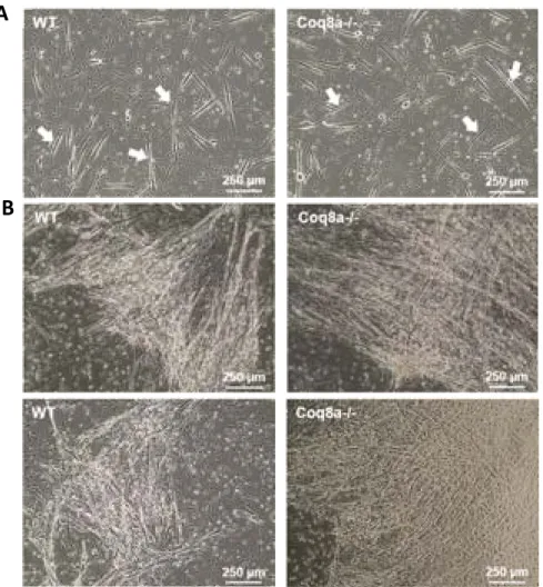
Figure 1 : Cerebellar structure in mice, adapted from (White and Sillitoe, 2013). 24 Figure 2 : Cerebellar cortex structure, adapted from (Apps and Garwicz, 2005). 25 Figure 3 : Coenzyme Q (6 to 10) chemical structure. 31 Figure 4 : Two possible coenzyme Q localization in lipid bilayers, adapted from (Turunen et al., 2004). 31 Figure 5 : Redox states of coenzyme Q. 33 Figure 6 : Mitochondrial respiratory chain with coenzyme Q. 34 Figure 7 : Mevalonate pathway, adapted from (Bentinger et al., 2010). 38 Figure 8 : Coenzyme Q biosynthesis in mitochondria of S.cerevisiae, adapted from (Laredj et al., 2014). 39 Figure 9 : Mutations found in ARCA patients, adapted from (Lagier‐Tourenne et al., 2008). 46 Figure 10 : Scheme of the COQ8A/ADCK3 human protein, adapted from (Lagier‐Tourenne et al., 2008). 49 Figure 11 : Complex Q in mammalian cells, adapted from (Floyd et al., 2016). 51 Figure 12 : Loss of COQ8A induces a deficiency in COQ7 in embryos. 59 Figure 13 : COQ8A interacts with COQ5 and COQ4, and the interaction with COQ5 increases in presence of ADP. 61 Figure 14 : Loss of COQ8A induces a deficiency in COQ7 and COQ5 in myoblasts. 63 Figure 15 : Coq8a‐/‐ myotubes seem to form bigger networks. 64 Figure 16 : Coq8a‐/‐ myoblasts do not proliferate more rapidly. 64 Figure 17 : Myogenic transcription factors expression are unchanged in Coq8a‐/‐ primary muscle cells. 65 Figure 18 : Scheme of the measurements performed with a Seahorse apparatus. 67 Figure 19 : Mitochondrial respiration is not affected in Coq8a‐/‐ myoblasts. 68 Figure 20 : Mitochondrial respiration is not affected in Coq8a‐/‐ myoblasts under low glucose conditions. 69 Figure 21 : Mitochondrial respiration is not affected in Coq8a‐/‐ myotubes. 70 Figure 22 : Glycolysis is not affected in Coq8a‐/‐ myoblast. 71 Figure 23 : Mitochondrial basal respiration is not affected in Coq8a‐/‐ cerebellar cultures. 72 Figure 24 : Areas (A) and lengths (B) of brain structures measured for morphometric analysis. 75 Figure 25 : Coq8a‐/‐ brain morphology is similar to WT. 75 Figure 26 : Similar brain morphologies of Coq8a‐/‐ mice compared to WT, observed by morphometric analysis of brain critical sections at the level of the hippocampus. 76Figure 27 : Invagination of neuronal nuclei in hippocampus observed by electron microscopy in 30 weeks old Coq8a‐/‐ mice. 78 Figure 28 : Normal axons observed by electron microscopy in hippocampus of 30 weeks old Coq8a‐/‐ mice. 79 Figure 29 : Coq8a‐/‐ hippocampal primary cultures are as healthy as heterozygous. 80 Figure 30 : Loss of COQ8A induces a deficiency in COQ7 and COQ5 in hippocampus. 81 Figure 31 : Loss of COQ8A deregulates NR2B in the hippocampus 82 Figure 32 : Loss of COQ8A deregulates GAD65/67 in the hippocampus. 82 Figure 33 : Loss of COQ8A does not deregulate ion channels tested in the cerebellum. 85 Figure 34 : Coq8a‐/‐ and WT cerebellar organotypic cultures have preserved lobules. 87 Figure 35 : Coq8a‐/‐ cerebellar organotypic cultures have preserved Purkinje neurons. 88 Figure 36 : Coq8a‐/‐ cerebellar organotypic cultures have preserved Purkinje neurons and Golgi organization. 89 Figure 37 : Swollen Golgi and degenerating Purkinje neurons are observed in Coq8a‐/‐ and WT cerebellar organotypic cultures. 91 Figure 38 : Abnormal lipid structures and healthy axons and synapses are observed in Coq8a‐/‐ and WT cerebellar organotypic cultures. 92 Figure 39 : Similar pattern for CACNA1A in Coq8a‐/‐ and WT cerebellar organotypic cultures. 94 Figure 40 : Similar pattern for ITPR1 in Coq8a‐/‐ and WT cerebellar organotypic cultures. 95 Figure 41 : Similar pattern for KCNC3 in Coq8a‐/‐ and WT cerebellar organotypic cultures. 96 Figure 42 : Similar pattern for SLC7A5 in Coq8a‐/‐ and WT cerebellar organotypic cultures. 97 Figure 43 : Coq8a‐/‐ cerebellar primary cultures have preserved neurons which form networks. 99 Figure 44 : Coq8a‐/‐ cerebellar primary cultures have preserved neural arborization. 100 Figure 45 : Coq8a‐/‐ cerebellar primary cultures have preserved Purkinje neurons and glial cells. 101 Figure 46 : Golgi apparatus is similar in Coq8a‐/‐ and WT cerebellar primary cultures. 102 Figure 47 : Loss of COQ8A induces a deficiency of COQ7 in cerebellum. 103 Figure 48 : Loss of COQ8A does not induce a deficiency in COQ7 in cerebellum of 1 week old mice. 104 Figure 49 : Coq8a‐/‐ mice still present motor coordination deficiency on accelerating rotarod. 105 Figure 50 : Degenerating Purkinje neurons observed in Coq8a‐/‐ cerebellum of 30 weeks. 106 Figure 51 : Swollen Golgi observed in Coq8a‐/‐ and WT cerebellum of 30 weeks. 107 Figure 52 : Coq8a‐/‐ mice have reduced levels of ApoAII from 1 to 30 weeks in cerebellum. 110 Figure 53 : Coq8a‐/‐ mice have reduced levels of COPA from 1 to 30 weeks in cerebellum. 110 Figure 54 : Kcnab3 and Kcnj10 are upregulated in cerebellum of 5 weeks old Coq8a‐/‐ mice. 111 Figure 55 : Some ionic transporters are upregulated in cerebellum of 5 weeks old Coq8a‐/‐ mice. 112 Figure 56 : Genes implicated in neuronal functions are upregulated in cerebellum of 5 weeks old Coq8a‐/‐ mice. 113
Figure 57 : Genes implicated in metabolism are deregulated in cerebellum of 5 weeks old Coq8a‐/‐ mice. 115 Figure 58 : Genes implicated in lipid metabolism are deregulated in cerebellum of 5 weeks old Coq8a‐/‐ mice. 116 Figure 59 : SYT17 was the only deregulated transcript also found to be deregulated at the protein level in Coq8a‐/‐ cerebellum at 5 weeks. 117 Figure 60 : KCNAB3 levels and localization look similar in Coq8a‐/‐ and WT 5 weeks old cerebellum.118 Figure 61 : Loss of COQ8A induces an increase in TST levels specifically in female’s cerebellum at 5 weeks. 119 Figure 62 : Scheme of the mevalonate pathway as well as the fatty acid synthesis and oxidation pathway. 121 Figure 63 : Coq8a‐/‐ mice do not show any difference in the expression of genes implicated in lipid biosynthesis in the liver. 122 Figure 64 : Coq8a‐/‐ mice show similar structures in the liver at 25 weeks. 124 Figure 65 : COQ8A is expressed in brown adipose tissue and its expression increases upon cold exposure in mice. 127
LIST OF TABLES
Table 1: Autosomal recessive spinocerebellar ataxia according to the mutated gene functions (based on Beaudin et al., 2017). 18Table 2: CoQ9 and CoQ10 content in rats and humans tissues, adapted from (Åberg et al., 1992). 32
Table 3 : CoQ9 amounts in fractions extracted from rat liver, adapted from (Kalén et al., 1987). 32 Table 4 : Reported ARCA2 patients with their clinical symptoms and mutations. 47 Table 5 : Results of proteomic analysis of using 30 weeks old Coq8a‐/‐ and WT mice. 58 Table 6 : Optimizations for Seahorse assays on primary myoblasts 67 Table 7 : Ingenuity Pathway Analysis, diseases and functions probably affected in cerebellum according to RNA‐sequencing data. 109 Table 8 : Ingenuity Pathway Analysis of RNA‐sequencing of quadriceps. 154 Table 9 : Gene coming up in quadriceps by RNA‐sequencing of 30 weeks old Coq8a‐/‐ mice and assessed by qRT‐PCR. 155 Table 10 : RNA‐sequencing and qRT‐PCR results from Coq8a‐/‐ and WT mice cerebellum. 157 Table 11 : Results of proteomic analysis of 30 weeks old Coq8a‐/‐ and WT mice cerebellum obtained for proteins for which deregulated transcripts levels were observed. 158 Table 12 : Results of proteomic analysis of 30 weeks old Coq8a‐/‐ and WT mice cerebellum obtained for proteins implicated in hydrogen sulfide catabolism 158
ABBREVIATIONS
2‐DG 2‐Deoxy‐D‐glucose 4‐HB 4‐hydroxybenzoate ABL Abetalipoproteinemia ADCK aarF domain‐containing protein kinases ADP Adenosine diphosphate AM Amygdala nuclei AOA1 and AOA2 Ataxia‐oculomotor apraxia 1 and 2 ARCA1 and 3 Autosomal recessive cerebellar ataxia type 1 and 3 ARCA2 Autosomal Recessive Cerebellar Ataxia type 2 ARSACS Autosomal recessive spastic ataxia of Charlevoix‐Saguenay AT Ataxia‐telangiectasia ATLD1 Ataxia‐telangiectasia‐like disorder 1 ATP Adenosine triphosphate AVED Ataxia with vitamin E deficiency BAT Brown adipose tissue BNHS Boucher‐Neuhauser syndrome cc Corpus callosum CoQ Coenzyme Q CTX Cerebrotendinous xanthomatosis D3V Dorsal third ventricle DAPI 4',6‐diamidino‐2‐phenylindole del Deletion DG Dentate gyrus dhc Dorsal hippocampal commissure DMQ demethoxyubiquinone E17.5 Embryonic day 17.5 EAOH Early‐onset ataxia with oculomotor apraxia and hypoalbuminemia ECAR Extracellular acidification rate ER Endoplamic reticulum FAD Flavin adenine dinucleotide FCCP Carbonyl cyanide‐p‐trifluoromethoxyphenylhydrazone fi Fimbria hippocampus FRDA Friedreich’s ataxia fs Frameshift mutations FXTAS Fragile X tremor‐ataxia syndrome GABA Gama‐aminobutyric acid GFP Green fluorescent protein HAB 3‐hexaprenyl‐4‐aminobenzoic acid Hb Habenular nucleus HBB 3‐hexaprenyl‐4‐hydroxybenzoic acid HDL High density lipoproteinHMG‐CoA 3‐hydroxy‐3‐methylglutaryl‐coenzyme A HP Hippocampus ic Internal capsule ins Insertion IOSCA Infantile onset spinocerebellar ataxia KO Knock‐out LDL Low density lipoprotein LV Lateral ventricles M1 Motor cortex MIRAS Mitochondrial recessive ataxia syndrome Mol Molecular layer of the hippocampus MSS Marinesco‐Sjogren syndrome mt Mammillothalamic tract NAD Nicotinamide adenine dinucleotide NGS Normal goat serum NMDA N‐methyl‐D‐aspartate NOX NADH‐oxidase NPC Niemann‐Pick type C OCR Oxygen consumption rate Oligo Oligomycin opt Optic tract Or Hippocampal oriens layer OXPHOS Oxidative phosphorylation P0 Post‐natal day 0 pABA para‐aminobenzoic acid PHARC Polyneuropathy, hearing loss, ataxia, retinitis pigmentosa, and cataract PHB Polyprenyl‐4‐OH‐benzoate PHYH phytanoyl‐CoA hydroxylase Pir Piriform cortex PP Pyrophosphate PTP permeability transition pore PTZ Pentylenetetrazole qRT‐PCR Real‐time quantitative reverse transcription PCR Rad Hippocampal stratum radiatum layer ROS Reactive oxygen species Rot/Ant Rotenone / Antimycine A RSGc Retrosplenial granular cortex S2 Secondary somatosensory cortex SANDO Sensory ataxic neuropathy, dysarthria, and ophthalmoparesis SCA autosomal dominant spinocerebllar ataxia SCAN1 Spinocerebellar ataxia, autosomal recessive with axonal neuropathy SCAR autosomal recessive spinocerebllar ataxia SD Standard deviation
SESAME syndrome Syndrome with seizures, sensorineural deafness, ataxia, mental retardation, and electrolyte imbalance shRNA Short hairpin RNA SQRDL Sulfide‐quinone oxidoreductase TBA Total brain area TILpy Total internal hippocampal pyramidal cell layer UCP Uncoupling protein WAT White adipose tissue WT Wild‐type XLSA X‐linked sideroblastic anemia and ataxia
INTRODUCTION
Introduction
This PhD work has been in the continuity of the ARCA2 (Autosomal Recessive Cerebellar Ataxia type 2) mouse model characterization which was previously performed by Floriana Licitra (Licitra, 2013). Her results, and more recent data that we obtained, led to a publication (Stefely et al., 2016). The genetic causes for ARCA2 were discovered in 2008 (Lagier‐Tourenne et al., 2008; Mollet et al., 2008) and a mouse model was generated shortly afterwards. It is a constitutive knock‐out mouse, deleted in the ARCA2 causing gene: COQ8A (previously named ADCK3 and CABC1). As the ARCA2 mouse model recapitulated most of the patient’s symptoms, the aim of my project was to gain insight into the pathophysiological mechanisms leading to the different phenotypes observed in this model organism.ARCA2 is also part of the Coenzyme Q (CoQ) deficiencies. Therefore, ataxias are first going to be introduced, followed by cerebellar structures and functions, coenzyme Q functions and biosynthesis, and finally the ARCA2 disease with the observed phenotypes and COQ8A function.
I. Ataxia
The name ataxia comes from ancient Greek, “α”: without /negative and “τάξις”: order, and means the absence of order. It is a syndrome defined by the loss of coordination and balance observed in patients. Various types of ataxia exist, and they are usually classified according to their causes. These causes are broad and range from genetic mutations to infections, including nutritional deficiencies or additional symptoms of other diseases (Van Gaalen et al., 2014; Manto and Marmolino, 2009). Therefore, different classes are defined to differentiate them. A common classification divides ataxias in three categories: acquired, inherited and sporadic.
Sporadic ataxias are mainly defined as ataxias for which genetic or acquired etiology are lacking and with a late onset (Klockgether, 2010; Klockgether et al., 1990).
Acquired ataxias conversely distinguished by the fact that they ensue from toxicity, environmental factors or effects of another disease. It is a heterogeneous group with broad causes. One of the well‐ known cause of acquired ataxia is chronic alcoholism (Jaatinen and Rintala, 2008). Alcohol, like heavy metals, chemotherapy or anti‐epileptic drugs are part of the molecules responsible for toxic ataxias (Nachbauer et al., 2015). Infections, by human immunodeficiency virus, or in case of encephalitis or Creutzfeldt‐Jakob diseases are other causes of acquired ataxias (Akbar and Ashizawa, 2015). Furthermore, acquired ataxias can also arise from autoimmune diseases (e.g.: anti glutamic acid decarboxylase antibodies or gluten), focal lesions (e.g.: strokes, anoxia, tumors, trauma), amino acid or vitamin deficiency (e.g.: vitamin B1, B12 or E) (Akbar and Ashizawa, 2015). This shows that ataxia can be induced by a large variety of threats and defects.
Finally, the hereditary ataxias are due to genetic causes and are also multiple. This category of ataxia is further subdivided in three major classes depending on their mode of inheritance: autosomal dominant (SCA), autosomal recessive (SCAR), and the X‐linked forms of ataxia. To these types, the episodic (EA which are periodic) and the spastic (SPAX) ataxia are added within the classification of the inherited ataxias (Bird, 2016).
Additional symptoms, like dysarthria and dysphagia (respectively speech and swallowing difficulties) are often found is ataxic patients when the disease progresses. However, what invariably characterizes ataxia is the implication of cerebellum or its afferent or efferent connections (Klockgether, 2007). These neurodegenerative diseases, induced by a defect in cerebellar structures in combination or not
with extra‐cerebellar lesions, are therefore named spinocerebellar ataxias (Manto and Marmolino, 2009).
Autosomal recessive ataxias are more frequent than the dominants forms, with a mean prevalence estimated around 3.5:100,000 for the recessive ataxias and around 2.5:100,000 for the autosomal dominant forms (Ruano et al., 2014). These prevalences vary depending on the continents, and even on the countries, probably due to founder mutations localization. The prevalence is calculated as a mean of the prevalence for each disease but specific ataxias, like SCA3 for the dominant forms, and FRDA for the recessive forms, are more frequent. In the case of a dominant ataxia one of the parents is affected but might not be aware or having declared the symptoms when this person has a child, or even at the onset of the child disease, whereas for recessive forms both parents are asymptomatic carriers of a mutation.
1. Autosomal dominant ataxia
The autosomal dominant forms of ataxias mostly have an insidious onset in adulthood (around 35 years old) and the diseases are slowly progressive (Paulson, 2009). Up to date forty‐three autosomal dominant ataxias were discovered and they have already been extensively classified (Akbar and Ashizawa, 2015; Bird, 2016). Some of these spinocerebellar ataxias (SCA) have specific symptoms, enabling to discriminate them. This is the case for SCA7 characterized by retinal degeneration (Sailer and Houlden, 2012). Unfortunately, for most of the SCA, the associated phenotypes are common, so genetic testing is needed for determination of the specific gene responsible for the disease. Autosomal dominant ataxias can be further sub‐classified according to the type of mutations inducing the diseases. Part of the SCA are due to nucleotide repeat expansions and the others, to conventional mutations (Akbar and Ashizawa, 2015). Regarding repeat expansions, two different cases emerged: repeats in exons (CAG repeat), leading to polyglutamine expansion, so protein gain of function (for SCA1‐3, 6, 7, 17 and DRPLA) or repeats in introns, resulting in RNA gain of function or chromatin modification (SCA 8, 10, 12, 31, 36) (Loureiro et al., 2016; Sailer and Houlden, 2012). For polyglutamine expansions, genetic anticipation is observed: the longer the repeat is, the earlier the disease declares and the more severe it is (Durr, 2010).
Numbering of the SCA was performed according to the chronology of discovery but diseases are gradually being grouped when the same gene was described and assigned for different SCA. This is the case for SCA19 and 22 which are already considered as the same ataxia, as they both arise from
mutations in KCND3. It could also be performed for SCA15, 16 and 29 which are induced by mutations in ITPR1 although the resulting additional symptoms are different (Bird, 2016). All episodic ataxias described so far and one spastic ataxia (SPAX1) are also due to autosomal dominant mutations (Bird, 2016). At later course, episodic ataxias might be constant but they are first periodic, with transient episodes of incoordination and imbalance (Akbar and Ashizawa, 2015). This periodicity differentiates them from the spinocerebellar ataxias for which the symptoms are uninterrupted and slightly progressive.
2. X‐linked ataxias
Few ataxias occur with a X‐linked inheritance. Some X‐linked ataxias had previously been described but the precise genetic affection could not be determined (Farlow et al., 1987; Schmidley et al., 1987). More recently, six genes have been implicated in these X‐linked cerebellar ataxias with the most frequent disease being Fragile X tremor‐ataxia syndrome (FXTAS) (Bird, 2016). FXTAS, like some SCA, is due to a repeat expansion in a non‐coding DNA region (5’ UTR). This disease has the particularity of being due to a FMR1 premutation. This premutation is an expansion comprising between 55 and 200 repeats and induces a RNA gain of function. In contrast, larger expansions (>200 CGG repeats) induce silencing of the FMR1 gene, so a loss of function and cause the Fragile X syndrome (Amiri et al., 2008; Oostra and Willemsen, 2009). Two other X‐linked ataxias, XLSA (X‐linked sideroblastic anemia and ataxia) and SCAX1 are due to mutations in transporters. XLSA is due to mutations in Abcb7, coding for a mitochondrial ATP‐binding cassette transporter implicated in iron homeostasis, and SCAX1 results from mutations in Atp2b3, coding for a plasma membrane calcium ATPase (Bekri et al., 2000; Zanni et al., 2012).3. Autosomal recessive ataxias
Autosomal recessive ataxias typically begin during childhood or early adulthood, with fewer cases of later onset. They are characterized by a gait and balance defect persistent and progressively worsening (Anheim et al., 2012). Unlike for the SCA, a number has not been assigned each time a new recessive ataxia was discovered. Recessive ataxias are usually classified according to the prevalence of the disease, the other clinical features, or the type of nervous system dysfunction. In an attempt to homogenize classification, a similar system as the SCA is gradually being used, with the acronym SCAR for “SpinoCerebellar Ataxia autosomal Recessive”. Yet, not all recessive ataxias have been assigned aSCAR number. In particular, the most frequent recessive ataxias do not have a SCAR number, whereas the rare ones, with only a few reported families, do. Likewise another acronym: ARCA for “Autosomal Recessive Cerebellar Ataxia” was employed to name three ataxias with pure cerebellar affection, including ARCA2, also named SCAR9 (Sailer and Houlden, 2012).
Like for the SCA, genetic testing is needed to determine which gene is mutated and define the precise disease. Indeed, different mutations, in the same gene, can lead to different age of onset, progression severity and different additional symptoms (Anheim et al., 2012). Furthermore, different recessive ataxias display common clinical features, and some other diseases have ataxia has one of their prominent or secondary feature, which makes it difficult for clinicians to rapidly determine the specific ataxia without a multiple‐gene panel test.
As mentioned previously different types of classification are used for recessive ataxias. One of them is based on the affected part of the nervous system and results in three main groups (Anheim et al., 2012). These three groups are listed here and examples of such ataxias are given (and in gray: examples of recessive disorders with ataxia as a prominent feature). Pure cerebellar ataxia (without neuropathy) Autosomal recessive cerebellar ataxia type 1, 2 and 3 (ARCA1 to ARCA3) Niemann‐Pick type C disease Cerebellar ataxia with pure sensory neuropathy (proprioceptive affection) Friedreich’s ataxia (FRDA) Ataxia with vitamin E deficiency (AVED) Sensory ataxic neuropathy, dysarthria, and ophthalmoparesis (SANDO) Infantile onset spinocerebellar ataxia (IOSCA) Abetalipoproteinemia (ABL) Cerebellar ataxia with sensorimotor axonal neuropathy (proprioceptive and motor affection) Ataxia‐telangiectasia (AT) Ataxia and oculomotor apraxia (AOA 1 and 2) Autosomal recessive spastic ataxia of Charlevoix‐Saguenay (ARSACS) Spinocerebellar ataxia with neuropathy (SCAN1) Refsum disease (RD) Cerebrotendinous xanthomatosis (CTX)
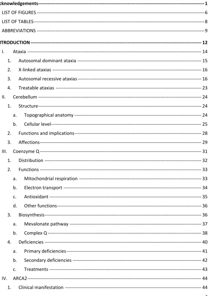
Most of the ataxic patients present cerebellar atrophy, except FRDA and some other pure sensory neuropathy ataxic patients for which an atrophy of the spinal cord is usually observed (Anheim et al., 2012). The most common recessive ataxias are FRDA, AOA1, AOA2 and AT (Bird, 2016). ARCA2, also known as SCAR9, is a rare form of recessive degenerative ataxia, with approximately 35 reported patients up to date. This disease will be further described later, in a separate section (IV). Like other types of ataxias, recessive ataxias are heterogeneous, complex and comprise a wide range of deregulated pathways. These pathways can also be used to further sub‐classify recessive ataxias in six groups: DNA repair defects, abnormal protein folding, metabolic defect, mitochondrial dysfunction, trafficking and other functions (Manto and Marmolino, 2009; Vermeer et al., 2011). This classification is not redundant with the classification according to nervous system affection, as ataxias with the same type of pathway involved do not lead to the same type of degeneration.
Ataxia Gene Protein Function
DNA repair defect
AT ATM ATM DNA repair and cell cycle
checkpoint controller
ATLD1 MRE11A MRE11 DNA repair
AOA1 (or EAOH) APTX Aprataxin single‐stranded DNA repair
AOA2 SETX Senataxin RNA/DNA helicase
SCAN1 TDP1 Tyrosyl‐DNA phosphodiesterase 1 DNA repair SCAR23 TDP2 Tyrosyl‐DNA phosphodiesterase 2 DNA repair Mitochondrial dysfunction
FRDA FXN Frataxin Iron‐sulphur biogenesis
IOSCA TWNK (or
C10orf2)
Twinkle Mitochondrial DNA helicase
SANDO (MIRAS) POLG DNA polymerase subunit γ‐1 Mitochondrial DNA replication ARCA2 (SCAR9) COQ8A (or
ADCK3)
COQ8A (or ADCK3) CoQ biosynthesis SCAR2 PMPCA Mitochondrial‐processing
peptidase subunit α Mitochondrial presequences cleavage Table 1: Autosomal recessive spinocerebellar ataxia according to the mutated gene functions (based on Beaudin et al., 2017). AT: ataxia‐telangiectasia; ATLD1: ataxia‐telangiectasia‐like disorder 1; AOA1 and 2: ataxia‐oculomotor apraxia 1 and 2; EAOH: early‐onset ataxia with oculomotor apraxia and hypoalbuminemia; SCAN1: Spinocerebellar ataxia, autosomal recessive with axonal neuropathy; FRDA: Friedreich’s ataxia; IOSCA: infantile onset spinocerebellar ataxia; SANDO: sensory ataxic neuropathy, dysarthria, and ophthalmoparesis; MIRAS: mitochondrial recessive ataxia syndrome, ARCA2: autosomal recessive cerebellar ataxia type 2; SCAR2‐23: spinocerebellar ataxia type 2‐ 23.
Ataxia Gene Protein Function Protein misfolding
ARSACS SACS Sacsin Protein folding and quality
control
MSS SIL1 SIL1 Protein translocation and
folding
SCAR16 STUB1 E3 ubiquitin‐protein ligase CHIP Misfolded protein degradation Metabolic defect
AVED TTPA Alpha‐tocopherol transfer protein Vitamin E transport
CTX CYP27A1 Sterol 26‐hydroxylase Sterol synthesis
Refsum disease PHYH phytanoyl‐CoA dioxygenase Peroxisomal fatty‐acid oxidation
BNHS PNPLA6 Neuropathy target esterase Fatty acid synthesis ABL MTP Microsomal triglyceride transfer protein large subunit Lipid transport Trafficking ARCA1 (SCAR8)
SYNE1 Nesprin‐1 Cytoskeleton‐organelle binding
SCAR15 (Salih ataxia) RUBCN (or KIAA0226) Run domain Beclin‐1‐interacting and cysteine‐rich domain‐ containing protein Endocytosis
SCAR11 SYT14 Synaptotagmin‐14 Putative role in exocytosis and vesicular trafficking
SCAR14 SPTBN2 Spectrin β chain, non‐erythrocytic 2 Membrane cytoskeleton and membrane protein trafficking SCAR20 SNX14 Sorting nexin‐14 Intracellular trafficking SCAR21 SCYL1 N‐terminal kinase‐like protein Regulation of retrograde Golgi‐
ER trafficking Other functions ARCA3 (SCAR10) ANO10 (or TMEM16K) Anoctamin‐10 Calcium signaling SESAME syndrome KCNJ10 ATP‐sensitive inward rectifier potassium channel 10 Potassium buffering SCAR13 GRM1 Metabotropic glutamate receptor 1 Neurotransmitter receptor SCAR18 GRID2 Ionotropic glutamate receptor, δ‐2 Neurotransmitter receptor SCAR19 SLC9A1 Sodium/hydrogen exchanger 1 pH homeostasis
Table 1: Autosomal recessive spinocerebellar ataxia according to the mutated gene functions (continued). ARSACS: autosomal recessive spastic ataxia of Charlevoix‐Saguenay; MSS: Marinesco‐Sjogren syndrome; AVED: ataxia with vitamin E deficiency; CTX: cerebrotendinous xanthomatosis; BNHS: Boucher‐Neuhauser syndrome; ABL: abetalipoproteinemia ; ARCA1 and 3: autosomal recessive cerebellar ataxia type 1 and 3; BNHS: Boucher‐ Neuhauser syndrome; ABL: abetalipoproteinemia; SESAME syndrome: seizures, sensorineural deafness, ataxia, mental retardation, and electrolyte imbalance; SCAR2‐23: spinocerebellar ataxia type 2‐23.
DNA repair defect is one of the pathological mechanism leading to recessive ataxias. This defect comprises mutations in genes necessary to sense, excise and repair DNA damage (Filla and De Michele, 2011). Two categories can be distinguished: single‐ and double‐strand break DNA repair defects. On