HAL Id: hal-01412338
https://hal.archives-ouvertes.fr/hal-01412338
Submitted on 8 Dec 2016
HAL is a multi-disciplinary open access
archive for the deposit and dissemination of
sci-entific research documents, whether they are
pub-lished or not. The documents may come from
teaching and research institutions in France or
abroad, or from public or private research centers.
L’archive ouverte pluridisciplinaire HAL, est
destinée au dépôt et à la diffusion de documents
scientifiques de niveau recherche, publiés ou non,
émanant des établissements d’enseignement et de
recherche français ou étrangers, des laboratoires
publics ou privés.
Public Domain
How Xer-exploiting mobile elements overcome cellular
control
Caroline Midonet, François-Xavier Barre
To cite this version:
Caroline Midonet, François-Xavier Barre. How Xer-exploiting mobile elements overcome cellular
con-trol. Proceedings of the National Academy of Sciences of the United States of America , National
Academy of Sciences, 2016, 113 (30), pp.8343-8345. �10.1073/pnas.1608539113�. �hal-01412338�
How
Xer-exploiting mobile elements overcome
cellular
control
Caroline Midonetand François-Xavier Barre
Most strains of Neisseria gonorrheae (Ng), the causative agent of the sexually transmitted disease gonorrheae, and a few strains of Neisseria meningitidis (Nm), which is responsible for a large number of meningitides, harbor a 57-kb horizontally acquired genetic element, the gon-ococcal genomic island (GGI) (1–3). Certain versions of the GGI are associated with disseminated gonococcal infection (1, 4). In addition, the GGI encodes numerous homologs of type IV secretion system genes, which are necessary for DNA secretion and facilitate natural trans-formation of the Neisseria (1, 2, 4). GGI are found inte-grated at the chromosomal dimer resolution site of their host chromosome, dif, and are flanked by a partial repeat of it, difGGI(Fig. 1A) (1, 5). The dif site is the target of two
highly conserved chromosomally encoded tyrosine recombinases, XerC and XerD, which normally serve to resolve dimers of circular chromosomes through the ad-dition of a crossover between directly repeated dif sites (6). This reaction raises questions on how GGI could be stably maintained (5). The results presented by Fournes et al. (7) in PNAS shed a new light on this apparent paradox.
The Xer machinery is highly conserved in bacteria. The dif sites consist of 11-bp XerC- and XerD-binding motifs, separated by an overlap region at the border of which recombination occurs (Fig. 1B). Recombination is under the control of a hexameric DNA pump, FtsK (Fig. 1C) (8). FtsK is a powerful translocase (9) and strips DNA from most proteins (10). However, a direct interaction between its extreme C-terminal domain, FtsKγ, and the Xer recombinases stops it (Fig. 1C) (11, 12) and activates the exchange of a pair of strands by XerD catalysis when in the presence of a synaptic complex (Fig. 1C) (8, 11, 13). The exchange of a second pair of strands by XerC catalysis converts the resulting Holliday junction into product (Fig. 1C) (8, 13). FtsK belongs to the cell division machinery. It assembles at midcell when most of the chromosomal DNA has been replicated and segregated, which restricts recombination at dif to the time of cell division (14, 15) and to the chromosome replication ter-minus region (16, 17).
Numerous mobile elements have been shown to exploit Xer recombination. Plasmids use it for the
resolution of multimers, the formation of which com-promises vertical transmission from mother to daughters by reducing the number of independently segregating plasmid units (18). Integrating mobile element exploiting Xer (IMEX) use it to insert into the dif site of one of the chromosomes of their host (19). In both cases, the FtsK control imposed on Xer recombination must be over-come, because the replication/segregation cycle of plas-mids and the integration/excision cycle of IMEX should be independent from the cell cycle. Moreover, Xer re-combination leads to the formation of plasmid multimers when they harbor a dif site (17, 20) and to the excision of the intervening DNA between directly repeated dif sites (17, 21). Correspondingly, the central region of plasmid sites seems to prevent FtsK-dependent XerD catalysis (Fig. 1B) (22), and the central region of the attachment sites of most IMEX lacks the necessary homology to sta-bilize XerD-mediated strand exchanges with dif (Fig. 1B) (23, 24). This is not the case for the central region of the different alleles of difGGI(Fig. 1D). The problem was most
striking for the most common of these alleles, difGGI1,
which differs from the neisserial dif by only 4 bp (Fig. 1D). In PNAS, Fournes et al. (7) observe that the Ng Xer recombinases efficiently bound to difGGI1, synapsed it
with difNg, and catalyzed complete recombination
re-actions between the two sites when activated by Ng FtsKγ. However, they noticed that recombination was reduced in the presence of the FtsK translocation module. The authors smartly hypothesize that FtsK translocation inhibited recombination by stripping Ng XerD from difGGI1, which they successfully verified
in vitro.
It was previously suggested that GGI initially harbored true neisserial dif sites and that their stabili-zation resulted from mutations that occurred after their integration (5). Many different types of mutations, including mutations in the central region of the dif sites and mutations abolishing the binding of the recombi-nases to them, could impede Xer recombination. Why, then, should difGGI1 harbor mutations that blocked
FtsK-dependent recombination without affecting XerC and XerD binding and synapse formation? One of the difGGIalleles found in Nm strains, attPGGI2, harbors
Institute for Integrative Biology of the Cell, Université Paris-Saclay, CEA, CNRS, Université Paris Sud, 91198 Gif sur Yvette, France
two out of four of the bases that differentiate difGG1from difNg,
which suggests that these changes were not randomly picked up (Fig. 1D, blue bases of difGGI1and difGGI2). Indeed, it is striking to
note that difGGI2is fully palindromic and carries two
XerC-bind-ing arms (Fig. 1D). In contrast, 8 out 11 of the bases of the XerD-binding arm of difGGI3differentiate it from the XerD arm of dif
sites (Fig. 1D). The attachment site of a V. cholerae IMEX, the toxin-linked cryptic phage (TLCϕ) harbors four of these bases (Fig. 1D, blue bases of difGGI3and attPTLC) (25). We previously
demonstrated that XerD poorly bound to attPTLC, which is
suffi-cient to prevent XerD-mediated FtsK-dependent recombination (25). Thus, it is tempting to propose that GGI are IMEX and difGGI
sites were selected not only to escape but also to overcome the normal cellular control imposed on Xer recombination by FtsK. GGI harboring difGGI3 probably belong to the TLCϕ class of
IMEX, which integrate into and excise from the genome of their host via a XerD-first FtsK-independent recombination pathway (25). GGI harboring difGGI1and difGGI2probably define a new class
of IMEX. Future work will need to address the Xer recombination pathway they exploit and if they can truly integrate independently of FtsK. In addition, it will be interesting to determine which fac-tors encoded in the genome of GGI IMEX and/or in the genome of their host help them overcome the cellular control that is normally imposed on Xer recombination, as observed for plasmids (18) and the CTXϕ class of IMEX (26).
Acknowledgments
Research in the F.-X.B. laboratory is funded by the European Research Council under the European Community’s Seventh Framework Programme (FP7/2007- 2013 Grant Agreement 281590).
1 Dillard JP, Seifert HS (2001) A variable genetic island specific for Neisseria gonorrhoeae is involved in providing DNA for natural transformation and is found more often in disseminated infection isolates. Mol Microbiol 41(1):263–277.
2 Hamilton HL, Dom´ınguez NM, Schwartz KJ, Hackett KT, Dillard JP (2005) Neisseria gonorrhoeae secretes chromosomal DNA via a novel type IV secretion system. Mol Microbiol 55(6):1704–1721.
3 Snyder LAS, Jarvis SA, Saunders NJ (2005) Complete and variant forms of the‘gonococcal genetic island’ in Neisseria meningitidis. Microbiology 151(Pt 12): 4005–4013.
4 Hamilton HL, Schwartz KJ, Dillard JP (2001) Insertion-duplication mutagenesis of Neisseria: Use in characterization of DNA transfer genes in the gonococcal genetic island. J Bacteriol 183(16):4718–4726.
5 Dom´ınguez NM, Hackett KT, Dillard JP (2011) XerCD-mediated site-specific recombination leads to loss of the 57-kilobase gonococcal genetic island. J Bacteriol 193(2):377–388.
6 Midonet C, Barre F-X (2014) Xer site-specific recombination: Promoting vertical and horizontal transmission of genetic information. Microbiol Spectrum 2(6): MDNA3-0056-2014.
7 Fournes F, et al. (2016) FtsK translocation permits discrimination between an endogenous and an imported Xer/dif recombination complex. Proc Natl Acad Sci USA 113(28):7882–7887.
8 Aussel L, et al. (2002) FtsK is a DNA motor protein that activates chromosome dimer resolution by switching the catalytic state of the XerC and XerD recombinases. Cell 108(2):195–205.
9 Saleh OA, P ´erals C, Barre FX, Allemand JF (2004) Fast, DNA-sequence independent translocation by FtsK in a single-molecule experiment. EMBO J 23(12):2430–2439.
D-path C-path XerD XerC FtsK FtsK-mediated Xer recombination difNg difGGI
A
C
B
D
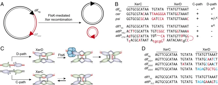
Fig. 1. (A) Schematic of XerCD-mediated excision of the GGI. Black double lines, N. gonorrheae chromosomal DNA; red double lines, GGI DNA; black triangles, difNgand red triangle, difGGI. (B) Sequence alignment of the Xer recombination site of mobile elements and their cognate
partner chromosomal dif site; attPCTX, CTXϕ attachment site; attPVGJ, VGJϕ attachment site; cer, core dimer resolution site of plasmid ColE1;
difEc, E. coli dif; dif1Vc, V. cholerae chr1 dif; and psi, core dimer resolution site of plasmid pSC101. Apart from attPCTX, which is the stem of a
forked hairpin from the single-stranded form of the genome of CTXϕ, a single of the two DNA strands is represented in the 5′ to 3′ orientation from left to right. Bases of cer and psi that differ from difEcand bases from attPCTXand attPVGJthat differ from dif1Vcare indicated in red. Plus (+)
and minus (–) signs indicate whether the sites can engage in recombination pathways initiated by XerC or XerD strand exchanges; +Kdenotes
FtsK-dependent recombination pathways. (C) Schematic of Xer recombination. XerD and XerC are represented in magenta and green, respectively. Following the Cre paradigm, the active pair of recombinases are drawn with their extreme C-terminal domains contacting the partner recombinases in cis. Blue circles represent the hexamer of FtsK. (D) Sequence alignment of difNg, the three different types of difGGI,
dif1Vc, and attPTLC. Bases of the dif-like site of mobile elements that differ from their cognate dif partner are highlighted in color, with blue
highlighting those that are identical in difGGI1and difGGI2and in attPGGI3and attPTLC.
10 Lee JY, Finkelstein IJ, Arciszewska LK, Sherratt DJ, Greene EC (2014) Single-molecule imaging of FtsK translocation reveals mechanistic features of protein-protein collisions on DNA. Mol Cell 54(5):832–843.
11 May PFJ, Zawadzki P, Sherratt DJ, Kapanidis AN, Arciszewska LK (2015) Assembly, translocation, and activation of XerCD-dif recombination by FtsK translocase analyzed in real-time by FRET and two-color tethered fluorophore motion. Proc Natl Acad Sci USA 112(37):E5133–E5141.
12 Graham JE, Sivanathan V, Sherratt DJ, Arciszewska LK (2010) FtsK translocation on DNA stops at XerCD-dif. Nucleic Acids Res 38(1):72–81.
13 Zawadzki P, et al. (2013) Conformational transitions during FtsK translocase activation of individual XerCD−dif recombination complexes. Proc Natl Acad Sci USA 110(43):17302–17307.
14 Kennedy SP, Chevalier F, Barre FX (2008) Delayed activation of Xer recombination at dif by FtsK during septum assembly in Escherichia coli. Mol Microbiol 68(4): 1018–1028.
15 Steiner WW, Kuempel PL (1998) Cell division is required for resolution of dimer chromosomes at the dif locus of Escherichia coli. Mol Microbiol 27(2):257–268. 16 Deghorain M, et al. (2011) A defined terminal region of the E. coli chromosome shows late segregation and high FtsK activity. PLoS One 6(7):e22164. 17 Barre FX, et al. (2000) FtsK functions in the processing of a Holliday junction intermediate during bacterial chromosome segregation. Genes Dev 14(23):
2976–2988.
18 Colloms SD (2013) The topology of plasmid-monomerizing Xer site-specific recombination. Biochem Soc Trans 41(2):589–594. 19 Das B, Mart´ınez E, Midonet C, Barre F-X (2013) Integrative mobile elements exploiting Xer recombination. Trends Microbiol 21(1):23–30.
20 Recchia GD, Aroyo M, Wolf D, Blakely G, Sherratt DJ (1999) FtsK-dependent and -independent pathways of Xer site-specific recombination. EMBO J 18(20): 5724–5734.
21 P ´erals K, Cornet F, Merlet Y, Delon I, Louarn JM (2000) Functional polarization of the Escherichia coli chromosome terminus: The dif site acts in chromosome dimer resolution only when located between long stretches of opposite polarity. Mol Microbiol 36(1):33–43.
22 Capiaux H, Lesterlin C, P ´erals K, Louarn JM, Cornet F (2002) A dual role for the FtsK protein in Escherichia coli chromosome segregation. EMBO Rep 3(6):532–536. 23 Das B, Bischerour J, Val M-E, Barre F-X (2010) Molecular keys of the tropism of integration of the cholera toxin phage. Proc Natl Acad Sci USA 107(9):4377–4382. 24 Das B, Bischerour J, Barre F-X (2011) VGJϕ integration and excision mechanisms contribute to the genetic diversity of Vibrio cholerae epidemic strains. Proc Natl
Acad Sci USA 108(6):2516–2521.
25 Midonet C, Das B, Paly E, Barre F-X (2014) XerD-mediated FtsK-independent integration of TLCϕ into the Vibrio cholerae genome. Proc Natl Acad Sci USA 111(47):16848–16853.
26 Bischerour J, Spangenberg C, Barre F-X (2012) Holliday junction affinity of the base excision repair factor Endo III contributes to cholera toxin phage integration. EMBO J 31(18):3757–3767.