RESEARCH OUTPUTS / RÉSULTATS DE RECHERCHE
Author(s) - Auteur(s) :
Publication date - Date de publication :
Permanent link - Permalien :
Rights / License - Licence de droit d’auteur :
Institutional Repository - Research Portal
Dépôt Institutionnel - Portail de la Recherche
researchportal.unamur.be
University of Namur
The human testes
Hussain, Aatif; Gilloteaux, Jacques
Published in:
Translational Research in Anatomy
DOI:
10.1016/j.tria.2020.100073
Publication date:
2020
Document Version
Publisher's PDF, also known as Version of record
Link to publication
Citation for pulished version (HARVARD):
Hussain, A & Gilloteaux, J 2020, 'The human testes: Estrogen and ageing outlooks', Translational Research in
Anatomy, vol. 20, 100073. https://doi.org/10.1016/j.tria.2020.100073
General rights
Copyright and moral rights for the publications made accessible in the public portal are retained by the authors and/or other copyright owners
and it is a condition of accessing publications that users recognise and abide by the legal requirements associated with these rights.
• Users may download and print one copy of any publication from the public portal for the purpose of private study or research.
• You may not further distribute the material or use it for any profit-making activity or commercial gain
• You may freely distribute the URL identifying the publication in the public portal ?
Take down policy
If you believe that this document breaches copyright please contact us providing details, and we will remove access to the work immediately
and investigate your claim.
Contents lists available at
ScienceDirect
Translational Research in Anatomy
journal homepage:
www.elsevier.com/locate/tria
The human testes: Estrogen and ageing outlooks
Aatif Hussain
a, Jacques Gilloteaux
a,b,∗aDepartment of Anatomical Sciences, St George's University School of Medicine, KBT Global Scholar's Program, Newcastle Upon Tyne, NE1 8ST, United Kingdom bDepartment of Medicine, NARILIS - Unit of Research in Molecular Physiology (URPHyM), Université de Namur, 5000, Namur, Belgium
A R T I C L E I N F O Keywords: Testis Receptors Estrogen Testosterone Leydig cells Sertoli cells Human Aging Gonadotrophins Crystalloids Pathology Xenobiotic Public health Contraception A B S T R A C T
This survey highlights some of the fine structures and functions associated with estrogen in the human testes, ageing and contraception. Clarifications obtained with knockout mice models as well as some clinical in-vestigations showed that estrogen receptors significantly influenced the overall maintenance of the testis functions through aromatase activity, intervening in the testosterone production by the Leydig cells and, in-directly with the Sertoli cells. Other autocrine, paracrine and endocrine fading activities of the seminiferous tubule's interstitium, including vascular supply, curtail the maturation of the male gametes while maintaining the blood-testis barrier in ageing. Do Reinke, Charcot-Böttcher and Lubarsch crystalloids, biopsy markers of specific testis cells, resulted of normal or altered functions and/or accumulated deposits out of ageing? The hypothalamo-pituitary-testis axis and feed-back homeostasis (with pineal influence?) regulating the re-productive tissues and phenotype characteristics, can be progressively changed according to individual health history, encompassing life time accumulated environmental toxicants, pharmaceuticals, and age-reduced car-diovascular fitness. The monitoring of all those long-term effects is needed to be better understood to provide future human public health in the care for the old adult, aging population.
‘Causa latet, vis est notissima’ [The cause is hidden, the result is very
evident], Ovidius, Metamorphoses, IV. 287.
1. Introduction
Biomedical science's courses comprise studies on the reproductive
systems in which gonads in all animals, including humans, are included.
The male testes (or testicles) as in " two together”, considering the slang
uses in the Ancient Greek ‘παραστάται ‘(i.e. parastátai, “colleagues’),
are homologous to the female ovaries. There, the developed and mature
structures and functions classic views are that the developed and
ma-tured structure and functions of the testes are regulated by the episodic
hypothalamus gonadotrophin (GnRH) secretions whose stimuli act via
the hypothalamo-pituitary portal vessels to induce a pulsatile
expres-sion of both secretions of some of the basophil and eosinophil cells of
the anterior pituitary gland: the luteinizing hormone (LH) and/or the
follicle-stimulating hormone (FSH). Thus, on one hand, classic view
notes that LH influences the interstitial cells of Leydig's LH receptors
(LHR) to produce the gonadal testosterone (T) to influence
spermio-genesis of the spermatospermio-genesis and making androgens, including
di-hydrotestosterone (DHT). Levels of T free serum and transported by a
circulation carrier, with that of DHT, also contribute in characterizing
the ‘male’ phenotype, by influencing other accessory male reproductive
organs and tissues. On the other hand, FSH has receptors on Sertoli cells
of the seminiferous tubules and upstream ducts exiting the testes
modulating spermatocytogenesis of the spermatogenesis i.e. taking the
haploid spermatogonia to spermatocytes and producing estrogens and
having receptors for it. These sex steroid secretions of the testes feed
back to that hypothalamo-pituitary axis and are also autocrine to their
tissues of origin [i.e.
1-3
]. (see
Table 1
)
Caracteristically associated with the female gender or sex as ‘female’
sex steroid hormone, discovered to cycle the guinea pig, a century ago
[
4
,
5
], estrogen and estrogen receptors have also been detected in
dif-ferent cells of male's organs about 20 years later [
6
].
Ageing changes can modify the male organs and steroid influences
as hormonal activities linked with the typical testes functions thus,
estrogen changes would reveal a lesser considered aspect of the human
male reproductive system resulting in or accompanying other disorders,
including prostatic cancer. This apparently minor estrogen functions in
the male reproductive system along with some of these aforementioned
topics, have not and are not yet even alluded in textbooks of
micro-scopic anatomy and should get adjusted with pathology and physiology.
https://doi.org/10.1016/j.tria.2020.100073
Received 14 January 2020; Received in revised form 24 March 2020; Accepted 24 March 2020
∗Corresponding author. Department of Anatomical Sciences, Drill Hall 0013 at UNN- School of Life and Health Sciences, St George's University School of Medicine, KBT Global Scholar's Program, Newcastle upon Tyne, NE1 8ST, United Kingdom.
E-mail address:[email protected](J. Gilloteaux).
Available online 24 April 2020
2214-854X/ © 2020 The Authors. Published by Elsevier GmbH. This is an open access article under the CC BY-NC-ND license (http://creativecommons.org/licenses/BY-NC-ND/4.0/).
After more than 40 years in biomedical publications, it is time to reveal
these basic hormonal, homeostatic activities, especially since general
practitioners have to be trained to likely later provide care to an
creased ageing population and stimulate further translational
in-vestigations [
7
]. Perhaps the neglected actions of the pineal, in view of
seasonal or cyclic influences mainly relegated or considered for rodents
[
8
] and those of prolactin, made by the acidophil cells of the pituitary
gland as suggested by some tumors [
7
,
9–13
] could render later on the
topic even more complex, but worth considering, in view of modern
living behaviors and the complex medical problems of the individual.
This can include portrayals brought by unprepared media when
trans-gender and trans-gender changes are brought into public sensationalism.
In this brief review, we have also attempted to highlight the known
functions of estrogen in mature males and considerations about the
antagonistic effects of estrogen-like or xenosteroid disruptors favoring
some of the structural and dysfunctional atrophy bearing functional
similarities of the aged human conditions (decreased sperm counts,
cryptorchidism, hypospadias and testicular cancer) [
14–17
].
2. Materials
This survey has been created out of from literature searches
throughout public access systems, including the public accessed United
States Library of Medicine internet site (
https://www.ncbi.nlm.nih.
gov/pubmed
) as well as Google search access. Illustrations were
ob-tained out of old microscopic preparations available to medical and
biomedical students, at the University of Namur, Namur, Belgium,
made by and for the Laboratory of Cells and Tissues between 1965 and
1980 out of necropsies from human body donations. Those histology
sections were prepared out of at least one human adult (> 40 year old
male) and another sample from specimens from an elderly (> 80 years
of age), after body perfusion embalming, tissues were further fixed in
formalin and processed in paraffin sections, stained with either
hema-toxylin-eosin or trichrome stain and accessible through
www.histology.
be
internet site.
3. Estrogen
3.1. Terminology
Named originally ‘oestrin’ and isolated as ‘an ovarian hormone’ by
Nobelists Allen and Doisy [
18
] and Butenandt [
19
]. The steroid is
in-volved in cyclical changes associated with procreation, or oestrus
[
20
,
21
], a topic reviewed in Medvei [
22
]. The American English
‘es-trogen’ for the English ‘oes‘es-trogen’ is used throughout this survey. It is
derived from ancient Greek ‘οίστρος’ (or oistros, meaning ‘oestrus’ as
‘inspiration’ meaning figuratively ‘sexual passion or desire’) and a suffix
‘γένος’ (or genos as ‘producer of’) i.e. generating the periodic state of
sexual activity of females of most mammals [
23
]. The term ‘estrogen’ as
used in the previous paragraph and in the following text is meant
‘fe-male sex steroids’ and as a ‘general’ term used to encompass, in ‘fe-male
tissues, the following three steroids and their metabolites: estrone (E1)
has one, estradiol (E2) has two, estriol (E3) has three hydroxyl groups.
However, 17β-estradiol (estradiol or E2) is the most prevalent in
ac-tivity of E1, E2 and E3 in human and mammals; another type of
estrogen, called estetrol (E4), produced during female pregnancy, was
evidently not included [
23–25
]. Thus, E2 abbreviation used is for all sex
steroid estrogens throughout the text, including those circulating in
lesser amount or/and active metabolites.
3.2. E2 functional aspects and gender differences
E2 is commonly known as a hormone secreted by the ovaries
throughout the reproductive span of reproductive activity in the female.
Ovary estrogen is responsible for various mechanisms ranging from
forming the female phenotype, promoting the cyclical growth of
folli-cles, and preparing for pregnancy several organs to reducing
in-flammatory effects in females [
26
], and even the level of pain
percep-tion [
27
]. The ovaries are the major source of circulating estrogens in
females as evidenced by oophorectomy, entirely removing estrogen;
this simple amputation has allowed a more clear and early
under-standing of estrogen functions in cases of endocrine dysfunctions. This
is in contrast when compared in males where a diffuse production of
estrogen meant that there are intricate estrogen-deficient productions
and thus, less understood. It has been documented that estrogen plays a
large role in spermatogenesis and in maintaining libido [
28
]. Estriol
[E2] is a most active form of estrogen steroid molecule that has been
found to be a clinically significant; however, compared with the female
gender, E2 level in men is still above that found in postmenopausal
women [
29
,
30
] and is able to carry out the endocrine and autocrine
effects noted in the human male reproductive system [
9
,
30–37
].
3.3. E2 production is through consumption of testosterone by CYP19A1
aromatase
Revisiting biochemistry basics, one recalls that cholesterol is the
precursor molecule modified by the desmolase CYP11A into
pregne-nolone, then by a mitochondrial dehydrogenase into progesterone. This
latter becomes testosterone (T) through CYP17 hydroxylase activity
[
3
,
7
,
28–37
], a member of the cytochrome P450 reductase enzyme
su-perfamily. Finally, another cytochrome P450 reductase enzyme
CYP19A1 (aromatase or EC 1.14.14) modifies T into E2 and related
steroid metabolites. This key enzyme is required for E2 production; it
not only plays a crucial role in testicular maintenance but it consumes T
produced by Leydig cells in the interstitium. T has been shown to
de-crease with age in both humans and rodents [
3
,
8
,
9
,
15
,
30–39
] and, thus,
the plasma level T and some of its metabolites, with inhibin, can
in-fluence back the same hormone's release by gonadotrophs to see an E2
increased in seminal plasma [
39
,
40
].
T actions throughout the male body has anabolic actions and on
specific reproductive organs can be episodically stimulated and mostly
converted as E2, as mentioned above. A small other source of E2 is
androstenedione, originating from the suprarenal zona reticularis.
Recent studies showed that human CYP19A1 gene is located in the 21.2
region of the long arm of the chromosome 15 (i.e. 15q21.2 region [
41
].
In addition to regulatory regions, this gene contains nine exons (exons
II–X) and contains 10 promoters, which are used according to the needs
and the characteristics of the tissue [
42
,
43
]. In comparison, mouse
CYP19 gene is localized on chromosome 9 where three promoters
specifically control the CYP19A1 gene expression [
44
]. Human
ar-omatase is mostly found in adipose tissues and only 10–25% in the
testes [
6
,
31
,
45
]. In final, the proper action of E2 as steroid depends on
two main factors: (i) the T converting enzyme and (ii) its receptors
found in various tissues and organs of the human male human including
the brain, the adipose and bone tissues, the skin (including the
mam-mary glands), the testes and the penis [
3
,
30–37
]. In comparisons,
re-ports suggest that high T concentrations inhibit spermatogonia
pro-liferation in models when spermatogenesis has been damaged by
processes such as irradiation [
46
]. During meiosis, T stimulates the
progression of spermatocytes through this process and inhibits
apop-tosis [
47
], and as seen later. E2 may there be co-operating with it. The
Table 1
Testis and main E2 Receptors in Human Seminiferous Tubule Cells.
Stroma Myoid cells LCs SCs Spgonia Spcytes Spids Spzoa
ER-As ER-Bs ER-As ER-Bs ER-Bs ER-As ER-Bs
Abbreviations: ER-As = E2 alpha receptors; ER-Bs = E2 beta receptors; LCs = Leydig cells; Scs = Sertoli cells; Spgonia = Spermatogonia; Spcytes = Spermatocytes; Spids = Spermatids; Spzoa = Spermatozoa; Stroma: mainly myoid cells.
site of action of androgens in spermatogenesis has been extensively
studied [
48–51
].
The diffuse E2 production in males means that there are potential
complex endocrine dysfunctions in male's E2-deficient productions
compared with females. This diversity of actions due to receptors and
multiplicity of target tissues has probably hindered progress in the
understanding of clinical deficient cases and patterns of the male
human tissues [
6
,
9
,
38
,
40
]. Even though numerous biomedical studies
have used successfully murine models to clarify the many sites of
ar-omatase activities, one may still be careful in validating those data for
humans which may still have differences in functionality or regulations
[
30–40
,
45–52
]. Aromatase, the main enzyme converting androgens to
E2, is also found in Leydig cells, Sertoli cells, germ cells and elongated
spermatids in the male reproductive system [
37
,
38
]. Therefore, if any
of these locations housing aromatase were to decrease or disappear, a
reduction in E2 production would result. Factors known to increase
aromatase activity include age, obesity, insulin, gonadotropins, and
alcohol. Aromatase activity is antagonized or decreased by prolactin,
anti-Müllerian hormone and the common herbicide glyphosate [
15
,
17
]
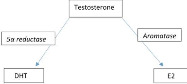
and explained in further paragraphs] and, in elderly, could it be that T
is tapped by 5-α reductase (EC 1.3.99.5) into more dihydrotestosterone
(DHT) and eventually favor benign prostatic hyperplasia (BPH) and
prostate cancer? [
39
] (
Fig. 1
).
In female, aromatase activity appears to be enhanced in certain
E2-dependent local tissue next to breast tissue, endometrial cancer,
en-dometriosis, and uterine fibroids. At this point, one could likely
gen-eralize that E2 (and some of its metabolites) or synthetic E2 (i.e.
die-thylstilbestrol or DES) are known to entice body receptive cells to enter
the cell cycle by stimulating DNA replication and typical or altered
transcripts, especially in cases of excessive unbalanced circulating
le-vels, estrogen would favor carcinogenesis [
53–61
]. E2 was found in
human testes Sertoli cells adenocarcinomas [
48
,
51
]. During
differ-entiation, germ cells are able to synthesize E2, acting as intracrine or
autocrine modulating factors of their own maturation [
48–51
]. E2 and
environmental non-estrogens but with E2-like activities have been
shown to be able to stimulate mammalian sperm capacitation,
acro-some reaction and fertilizing ability [
60
]. Many of these E2 effects were
mediated by cognate receptors, ligand-activated transcription factors,
binding to responsive components of the hormone-responsive genes
[
37
,
38
,
45
,
53
,
54
,
60–65
].
3.4. Aromatase excess syndrome and stress
A rare syndrome where in boys due to mutations in the CYP19A1
gene can induce gynecomastia and precocious puberty accompanied by
a rapid epiphyseal closure that leads to short stature. This condition is
inherited as an autosomal dominant fashion but the long-term
anti-ar-omatase treatment into adulthood and further is unknown [
66
,
67
].
Excess aromatase activity can be caused by medical procedures
invol-ving serious cardiovascular risks surgeries [
68
,
69
].
3.5. Aromatase deficiency syndrome
This syndrome is found in youth, caused by a mutation of gene
CYP19 and inherited in an autosomal recessive way. Accumulations of
androgens during pregnancy may lead to intact males but tallness due
to retarded epiphyseal closure. The aging outcome of this defect is
unclear at this time [
70–72
] while some effect could be preventative of
osteoporosis and can be worth further exciting clinical investigations
[
73
].
3.6. Inhibition of aromatase and environmental toxicology
This inhibition can result in hypoestrogenism and can be caused by
pharmacy and natural products, still quasi exclusively studied in
women [
74–77
]. These compounds are aimed to inhibit the production
of E2 in postmenopausal women; thus, a current clinical usage of
anastrozole, exemestane, and letrozole as they became useful in the
management of patients with breast cancer lesions found to be E2
re-ceptor positive. Aromatase inhibitors are also beginning to be
pre-scribed to men on T replacement therapy as a way to keep E2 levels
from spiking once doses of T are introduced to their systems. Natural
aromatase inhibitors tend to cause fewer serious side effects than
ta-moxifen and related medications in elderly patients which can cause or
favor the heart, venous thromboembolic events (VTE), stroke and
fre-quent osteoporosis in some cases, and has yet to be evaluated more in
male patients [e.g. Ref. [
69
,
73
]. Other common disruptors of aromatase
are widely used in agriculture as herbicide and antifungal and their
long term toxicity measured in vitro and evaluated in cohorts of
pa-tients could harbor a large number of defects that are still to grow in
diagnostic among many farming populations, ignored by industrial
agriculture developments, unawareness or neglect of administrative
policies in public health [e.g. Ref. [
74–88
]. Finally, among traffic
pol-lutants, lead can affect fertility and therefore it is crucial that long-term
clinical surveys would verify this environmental public topic [
78
].
4. E2 production and the testes
The finding of the E2 source in human testes was originally derived
from dog experimentations [
89
] when rare Sertoli cell's tumors were
observed (e.g. Ref. [
90
,
91
]) or in some forms of seminiferous tubule
anomalies [
92
], varicocele [
93
] and cryptorchidism [
37
,
94
,
95
]. These
histopathologic structures induced by exogenous E2 were even more
pronounced when fetal or neonatal animals were exposed
devel-opmentally to high E2 levels though of synthetic diethylstilbestrol
(DES) or other natural or man-made environmental estrogens [
93
].
Those stimuli can produce permanent changes in the structure and
function of postnatal human male reproductive organs, similarly as
those found in females using rodent model offspring through more than
one generation [
49
,
54–62
,
93
]. Most of the DES animal studies have
been especially important because they have predicted some of the
extensive male abnormalities after DES exposure during pregnancy
[
62
,
94
,
95
] caused by the intratesticular sites of aromatization in the
developing tissues of human testes [
36–38
]. Measurement of E2 in
peripheral blood and urine [
49
,
52
] indicated that E2 is ‘very low in
humans’ and that ‘estrogen levels were also found in testis and in
semen’ [
96
]. The primary source in immature testes was first
hy-pothesized to be Sertoli cells, confirmed by the Sertoli cell tumors, and
later with in vitro studies [
90
,
91
,
97
]. Then, direct evidence that normal
Sertoli cells can synthesize E2 under the regulation of FSH and cyclic
AMP. However, in adult and aging testes, studies have always
con-sented
that
the
main
E2
source
was
Leydig
cells
[
6
,
8
,
12
,
21
,
29
,
32–40
,
98–100
] as proposed by Western and Northern
analyses [
41
,
60–62
,
68
,
69
]. Meanwhile, aromatase was also localized
by immunohistochemistry [
74
] in spermatogonia, elongating
sperma-tids and other associated tissues and organs of the male reproductive
system [
9
,
32–40
,
60
,
61
,
70–73
,
100–103
].
Fig. 1. The role of estrogen in the ageing process of the male reproductive
system appears to be triggered by the lack of blood flow to testicular cells. While estrogen does not appear to play a role in initiating testicular atrophy, it demonstrates the effect of promoting degeneration that is already in progress.
4.1. E2 receptors
E2 hormone receptors (ERs) are located intracellularly in a similar
distribution as aromatase. In the testes seminiferous tubules, these
re-ceptors are localized in the cytoplasm and Golgi of the round
sperma-tids [
32–37
] while at least two types of ERs have been identified in
females, ER-alpha or α (ER-A) and ER-beta or β (ER-B); the same ones
have been found in males [
104–107
]. The most abundant ER found in
males is the ER-B form which is ubiquitous to almost every cell in the
interstitium and seminiferous tubules, but the ER-A, located specifically
in that interstitium have been found to play a key role in maintaining a
differentiated epithelium morphology and to regulate fluid
reabsorp-tion in the male reproductive system [
32–37
,
104–109
]. ER-A Knock
Out (ERA KO) studies conducted in mice have found the tall, ciliated
columnar shaped epithelial cells of the efferent ductules with abundant
microvilli to undergo metaplasia towards smaller, cuboidal shaped cells
with sparse microvilli.
E2 receptor makes possible to influence the differentiation of the
efferent ductular epithelium [
105
]. As KO studies indicated,
compro-mising ERs lead to atrophy and other morphological changes of the cells
they reside, thus, a lack and/or any compromised ERs could therefore
potentially be linked with testicular atrophy, also correlated with the
natural ageing process. E2, as any steroid hormone, diffuses through
plasmalemma, acts through nuclear receptors to ultimately regulate
gene transcription and expression in a similar distribution as the
aforementioned aromatase in the testes [e.g. 32–37, 108–109]. Animal
models, such as rabbit, rats and mice (wild and KO) studies with the use
of radiolabeled E2, initiated by Mulder and the group of van
Beurden-Lamers [
105
,
106
,
108
] have been able to make the human physiology
understood about the E2 role in the male reproductive system, along
with the understanding of E2 receptor biology. Out of a study
con-ducted in 1996, knocking out (KO) estrogen receptors in male mice, in
particular the ER-A, exhibited a pattern of progressive testicular tissue
degeneration [
111
]. This further supported by more recent studies
in-dicating that the ER-A is implied to mediate testicular germ cell
dif-ferentiation through a mechanism involving Sertoli cells [
112
,
113
]. It
should be noted that rats ER-A KO showed no significant impairment in
spermatogenesis; this is in stark contrast with aromatase KO studies
[
111
]. These findings exemplify the higher function E2 synthesis and
presence plays in spermatogenesis and overall testicular health. The
ERs are required for the effects of E2 to be observed thus, a destruction
of these receptors can come along with the effects of testicular changes
through ageing, thus can help further progress toward their involution
process. Other studies investigated the opposite end of the spectrum by
artificially increasing the production of ERs in mice which lead to
Leydig cell hyperplasia, cryptorchidism and undescended testis all
re-sulting in lower testosterone levels [
30
,
63
]. These other data could
potentially be due to a shunt-like effect where an increased number of
receptors for E2 calls for an increased production of E2 thus depleting
testosterone levels halting the dissention of the testis, one of the effects
of normal testosterone function. With increased amount of E2 receptors
linked with Leydig cell hyperplasia, it can logically be extended that a
lack of E2 can potentially result in hypoplasia, decreased function and
eventual testicular atrophy in the human male reproductive system.
Reports on human testis tissues [
38
,
40
,
100–108
,
113–122
] showed
most cells with both ER-A and ER-B receptors but with Leydig cells and
spermatogonia with quasi only ER-B. Another large series of other
re-ports are quoted in Ref. [
37
,
38
] while another review is mainly dealing
with the huge number of murine experiments [
100–103
]. One should
note that these conclusions only hold true if mice are assumed to be
adequate models for human males. These animal model studies hold a
strong argument for parallel investigation towards human ageing and
could potentially further aid in the search for a method to slow or
ex-tinguish the natural ageing process of the male reproductive system
where a proper balance between T and E2, through aromatase activity,
is key for an appropriate functioning during reproductive activity. In
the meantime, a large number of studies have verified those receptor's
action in primates, normal human and in pathology [
65–72
,
113–138
]
as well as medication against male osteoporosis [
73
].
5. Tunica vaginalis
The tunica vaginalis, serous membrane lining the human testis,
developed from the peritoneum [
139
,
140
] and, thus, pediatric testes
are considered intraperitoneal because the tunica vaginalis, a
perito-neum derivative, covers the testes. Studies have shown that, with
ageing, this usually liquid lining can adhere to the testis wall and
ob-literates the mesentery present on the testis. Thus, with ageing, the
testes appear behind the tunica vaginalis rather than inside [
140
]. This
loss of lining has been observed in other conditions such as
cryp-torchidism which has been associated with testicular atrophy [
123
]. If
this lining has been shown to be diminished in the elderly and a
pa-thological loss of it has been linked to testicular atrophy; therefore, it
should be reasonable to accept that the tunica vaginalis plays some key
anatomical role in maintaining testicular health.
6. Testis interstitial cells of Leydig or interstitial endocrinocytes
6.1. Ageing and hypogonadism?
The interstitial cells of Leydig (LCs), officially-named ‘interstitial
endocrinocytes’ [
141
], can be detected in the testis stroma, and usually
found adjacent to the seminiferous tubules [
142–163
]. A population of
‘fetal'-type mesenchymal cells are destined to become LCs from the 8th
to the 20th week of gestation, and produce enough testosterone until
birth and at puberty. ‘adult'-type LCs differentiate in the post-natal
testis. Indeed, they are preceded in the testis by masculinization of a
male fetus [
7–10
,
14
,
34–37
,
99
,
156
,
157
]. Leydig cells respond to LH
from some of the basophils of the anterior pituitary gland to produce T
in males with a decreased quality and production according to aging in
rodents [
158
,
159
] and humans [
157
,
160
], thus sustaining the gender
phenotype. LCs markers should express T through aromatase (see above
paragraphs) and Reinke crystal location [
161
,
162
], but also can be
identified through histochemical markers such as calretinin [
162–166
],
more specifically than with inhibin [
167
,
168
].
Mature LCs are polyhedral in shape, and have a large prominent
ovoid nucleus containing a prominent nucleolus, an eosinophilic
cyto-plasm with mitochondria, endocyto-plasmic cisterns and numerous
lipid-filled vesicles, lipofuscin pigment and peculiar, rod-shaped crystal-like
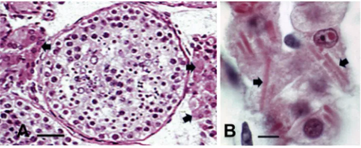
structures (3–20 μm in diam.), as named ‘crystalloids of Reinke’ of no
known function [
142–151
], besides to be eventual marker of LC tumors
[
160
,
161
, see paragraph 5.5 ] and
Fig. 2
A-B versus
Fig. 3
A-B.
LCs undergo obvious cell changes as the person ages
[
99–104
,
156
,
157
,
168
] and the same studies showed with a murine
Fig. 2. A-B Light microscopy preparation out of paraffin sections from a mature
human testis where the seminiferous tubules sections are busy by lining sper-matogonia and other progeny gonocytes and spermatids. In the stroma, many small circulatory vessels are surrounded by large eosinophil aggregates of Leydig cells (arrows). B: enlarged aspects of Leydig cells containing Reinke crystalloids as elongated rod-like shapes (arrows). Scale in A is 20 μm; in B it is 5 μm.
model of accelerated Leydig cell ageing by knocking out the Nrf2 gene
responsible for protection against oxidative stress which led to loss of
cytoplasm and eventual atrophy of the cells as in primary
hypogo-nadism [
168
]. Such deficiency and in excess have been reported by
some of the early studies with histology in mammals, among others, the
equine type and human [
142–151
], (see
Fig. 2
A and B), including
electron microscopy [
146–152
,
154
,
155
], All LCs can produce T in the
presence of LH, as first demonstrated in mouse [
37
,
111
,
152
] which
level decreases with age as noted in rats [
158
,
159
], like those found in
human [
30–37
,
147–162
]. Age-related defects include altered transport
of cholesterol, a hormone precursor, into the mitochondria and
LH-stimulated
cAMP
production
for
intracellular
signaling
[
121
,
124
,
129
,
132
,
134
,
162
]. In light of this evidence, it stands to
reason that ageing can reduce testosterone function by mechanisms
reducing the T production of the LCs as in hypogonadism [
163–165
].
Studies reveal that the age-like effects observed in the testes are not due
to a decrease in LC count, but rather by a sort of cooperation in the
ability of the cells to make T in response to LH that could be resulted by
the ‘fitness’ of the associated, local vascular supply caused by blockade
or lag in gonadotropin response [
148
,
168
] The ER-A type receptors
(ERs-A), are located specifically in the interstitium of the testis, have
been found to play a key role in maintaining a differentiated epithelium
morphology and to regulate fluid reabsorption in the male reproductive
system [
32–38
]. Once T becomes scarce, its progeny, E2, will decrease
resulting in less protective factors and eventual further testicular
atrophy. It has been found that deletions or mutations causing impaired
aromatase enzyme formation in males has been linked with diminished
or even ablated steroidogenesis [
30
,
34–37
]. Additionally, a diminished
or lack of aromatase not only impairs steroidogenesis, but can also lead
to infertility and impaired spermatogenesis [
7–10
,
14
,
34–37
,
151
,
157
].
Since spermatogenesis requires proper LC and SC functioning, the
lowering E2, stemming from a significant altered aromatase, could be
the root of further testicular impairments [
159–168
]. This preservative
role is clearly highlighted in the female reproductive system.
It has been found that the total number of LCs in elderly male, over
the age of 60, can decrease to up to 44% [
151
,
157
,
163
,
168
,
170
].
Cellular changes include dedifferentiation, decreased amount of smooth
endoplasmic reticulum and mitochondria organelles, multinucleation,
and a greater number and larger sized Reinke crystalloid inclusions
[
140–167
]. The high oxygen demand established by the abundant
mi-tochondria in LCs places them at increased ischemic risk; these
orga-nelles demonstrating variable morphology according to metabolic
ac-tivity [
170–172
] and, along with them, the peroxisomes enzymes can
be implicated without increasing their catalase marker [
173
]. These
changes in LCs that come about through the natural ageing process in
the male reproductive system can compromise the functionality of the
cell thus contributing to a decline in estrogen production [
163–170
].
Other previous investigations showed with morphology, including
immunocytochemistry, the presence of clear and dense-core vesicles
and the co-location of several neuropeptides in LCs, making LCs a new
member of the diffuse neuroendocrine system [
174
]. However, studies
are yet to come with age changes in this topic.
In the testes, the LCs can be ectopically located, i.e. in the tunica
albuginea, the rete testis or the interlobular septa as well as within
hyalinized seminiferous tubules or those with advanced atrophy and
marked thickening of the tunica propria, i.e. tubules in adult
cryp-torchid testes, Klinefelter syndrome, and in some other primary
hypo-gonadisms as reported in a most recent contribution [
175
].
6.2. Leydig cells and aging alcoholism
In older men and in those with chronic alcoholism, LCs, as well as
ectopic LCs may show atrophic features [
175
].
6.3. Leydig cells and Werner's syndrome
Patients suffering from Werner's Syndrome, a known form human
progeria, are found to be lacking the gene for the production of WRN, a
protein necessary for preventing early onset senescence of tissue. WRN
appears to be only induced by E2 and not T; this also makes E2 a
protective agent against early senescence of the reproductive system
[
20
]. These findings suggest that a decreased and lack of estrogen can
cause progressive, diminished function of the male reproductive system
[
176
].
6.4. Leydig cell tumors
LCs tumors are usually benign [
177
] even though they involved
gonadotrophin perturbations or are irresponsive to gonadotrophin
[
177–179
], often associated with infertility. Estrogen production rates
and excretion were 5- to 10-fold elevated in 2 patients studied, but
could be entirely accounted for by tumor metabolism of circulating
steroids, notably dehydroepiandrosterone sulfate, and, thus E2 and
xenoestrogen exposure could participate in LC tumorigenesis
[
163
,
177–179
].
6.5. Crystalloids of Reinke
In 1896, Reinke [
179
] reported the rod or corn-shaped CRs
struc-tures measuring up to 20 μm in the LCs of the human testis. Named after
Reinke by others [
143–146
], he found them as rice-shaped with iron
hematoxylin staining preparations. It was then hypothesized accretion
to form larger ones, as suggested through electron microscopy
ob-servations [
1
,
146–150
,
154–170
,
171-189
]. In azoospermia, there are
cases when no crystalloid can be seen but pigments [
190–199
] and
there are said ‘to be inconsistent among individuals and among the cells
of an individual’ [
148
]. The crystalloids are variable in form and size
and are rectilinear. (about 3 μm in width et 20 μm long], with angles
that can be sharp or rounded as they are made of macromolecules 5 nm
in diameter and evenly spaced of 19 nm, present in highly ordered
pattern of internal structure. For other authors [
192–194
] and, as
re-ported in Ref. [
195
], the hexagonal lattice has a 24 nm constant. These
peculiar inclusions likely relate to LC steroidogenesis because a similar
type of crystalloids was detected in the adrenal cortex [
192
], the ovary
LCs [
196
] and even in human SCs [
196
] and swine, including the
cryptorchid ones [
198
]. In cases of azoospermia the same LCs contained
fatty deposits with initially formed electron-pale deposits that appear
like those of cholesterol, as they are electron-lucent [
199
], thus
re-sembled cholesterol unesterified in the human gallbladder and cystic
duct cholesterolosis [
200
,
201
]. Others, such as in Ref. [
202
] have
classified these LC deposits. Authors have also found ectopic LCs and
Reinke crystals relationships, are these deposits formed through normal
functions or metabolic defects or/and ageing deposition of
proteinac-eous residues? [
203–205
]. When observed with the light microscope,
Fig. 3. A-B Light microscopy preparation out of paraffin sections from an
el-derly human testis where the seminiferous tubule sections display a poor lining and luminal content, a thickened basal membrane, when compared to mature testis (Fig. 2A). The stroma appears poorly populated due to a poor stromal cell population, including Leydig cells. They are rare and scattered (arrows) among the stromal spaces with vacuolated content (lipids) and only small Reinke crystals (arrow). Scale in A is 20 μm and in B is 5 μm.