HAL Id: hal-03001479
https://hal.archives-ouvertes.fr/hal-03001479
Submitted on 12 Nov 2020HAL is a multi-disciplinary open access
archive for the deposit and dissemination of sci-entific research documents, whether they are pub-lished or not. The documents may come from teaching and research institutions in France or abroad, or from public or private research centers.
L’archive ouverte pluridisciplinaire HAL, est destinée au dépôt et à la diffusion de documents scientifiques de niveau recherche, publiés ou non, émanant des établissements d’enseignement et de recherche français ou étrangers, des laboratoires publics ou privés.
Palmitate is increased in the cerebrospinal fluid of obese
humans and induces memory impairment in mice via
pro-inflammatory TNF-α
Helen Melo, Gisele da S Seixas da Silva, Marcella Sant'ana, Camila
Vieira, Ligo Teixeira, Julia Clarke, Vivian Miya Coreixas, Bruno de Melo,
Juliana Fortuna, Leticia Forny-Germano, et al.
To cite this version:
Helen Melo, Gisele da S Seixas da Silva, Marcella Sant'ana, Camila Vieira, Ligo Teixeira, et al.. Palmitate is increased in the cerebrospinal fluid of obese humans and induces memory impairment in mice via pro-inflammatory TNF-α. Cell Reports, Elsevier Inc, 2020, �10.1016/j.celrep.2020.01.072�. �hal-03001479�
1
Palmitate is increased in the cerebrospinal fluid of obese humans and induces memory impairment in mice via pro-inflammatory TNF-α
Helen M. Melo1, Gisele da S. Seixas da Silva2, Marcella Ramos Sant'Ana 3, Camila Vieira
Ligo Teixeira4, Julia R. Clarke5, Vivian S. Miya Coreixas1, Bruno C. de Melo1, Juliana T. S.
Fortuna1, Leticia Forny-Germano1,José Henrique Ledo1, Maíra S. Oliveira1, Claudia P.
Figueiredo5,Raphaelle Pardossi-Piquard6, Frédéric Checler6, José María Delgado-García7,
Agnès Gruart7, Licio A. Velloso8, Marcio L. F. Balthazar 4, Dennys E. Cintra3, Sergio T.
Ferreira1,9 and Fernanda G. De Felice1,10,11*
1 Institute of Medical Biochemistry Leopoldo de Meis, Federal University of Rio de Janeiro,
Rio de Janeiro, RJ 21941-902, Brazil.
2 Federal Institute of Education, Science and Technology of Rio de Janeiro, Rio de Janeiro,
RJ 20270-021, Brazil.
3 Laboratory of Nutritional Genomics (LabGeN), School of Applied Sciences); CELN -
Nutrigenomics and Lipids Research Center, School of Applied Sciences, University of Campinas (UNICAMP), Limeira, SP 13484-350, Brazil
4 Brazilian Institute of Neuroscience and Neurotechnology, BRAINN; Department of
Neurology, Neuroimaging Laboratory, University of Campinas (UNICAMP), Campinas, SP 13083-887, Brazil.
5 School of Pharmacy, Federal University of Rio de Janeiro, Rio de Janeiro, RJ 21941-902,
Brazil.
6 Université Côte d’Azur, INSERM, CNRS/UMR7275, IPMC, team labeled “Laboratory of
Excellence (LABEX) Distalz”, 660 route des Lucioles, 06560, Sophia-Antipolis, Valbonne, France.
7 Division of Neuroscience, Pablo de Olavide University, Seville-41013, Spain.
8 Laboratory of Cell Signalling, Obesity and Comorbidities Research Centre, University of
Campinas, Campinas, SP 13084-761, Brazil.
9 Institute of Biophysics Carlos Chagas Filho, Federal University of Rio de Janeiro, Rio de
Janeiro, RJ 21941-902, Brazil.
10 Centre for Neuroscience Studies & Department of Psychiatry, Queen's University,
Kingston, ON K7L 3N6, Canada.
11 Lead Contact
2
Summary
Obesity has been associated with cognitive decline, atrophy of brain regions related to learning and memory, and higher risk of developing dementia. However, the molecular mechanisms underlying these neurological alterations are still largely unknown. Here, we investigate the effects of palmitate, a saturated fatty acid present at high amounts in fat-rich diets, in the brain. Palmitate was increased in the cerebrospinal fluid (CSF) of overweight and obese patients with amnestic mild cognitive impairment. In mice, intracerebroventricular infusion of palmitate impaired synaptic plasticity and memory. Palmitate induced astroglial and microglial activation in the hippocampus of mice, and its deleterious impact was mediated by microglia-derived TNF-α signaling. Results establish that obesity is associated with increases in CSF palmitate. By defining a pro-inflammatory mechanism by which abnormal levels of palmitate in the brain impair memory, results further suggest that anti-inflammatory strategies may constitute an approach to attenuate memory impairment in obesity.
3
Introduction
Worldwide obesity has nearly tripled in the past four decades. In 2016, more than 1.9
billion adults were overweight and over 650 million were obese (WHO, 2018). Overweight
and obesity states are defined by abnormal or excessive fat accumulation that may impair
health (WHO, 2018), and are frequently associated with metabolic and cardiovascular
disorders. Diabetes, dyslipidemia and hypertension often overlap in overweight and obese
humans (Chan et al., 1994; Guh et al., 2009; Hu et al., 2001; Janssen et al., 2002; Mokdad et
al., 2003; Must et al., 1999; Wilson et al., 2002). Obesity and/or increased fat intake are
linked to cognitive decline and a higher risk of developing dementia (Kalmijn et al., 2004;
Kivipelto et al., 2005).
Individuals become overweight and/or obese when there is an imbalance between
calories consumed and calories expended, often when an increased intake of unbalanced diets
high in fat is combined with decreased physical activity (Spiegelman and Flier, 2001). In
obesity-related insulin resistance and type 2 diabetes (T2D), consumption of high-fat meals
results in excess serum levels of saturated fatty acids (SFAs) (Meek et al., 1999; Roust and
Jensen, 1993) which impair metabolism and cause peripheral insulin resistance
(Fernández-Real et al., 2003; Mayer et al., 1993; Parker et al., 1993; Vessby et al., 2001). Palmitate is
the most abundant SFA present in the circulation (Quehenberger et al., 2010), and also in the
cerebrospinal fluid (CSF) (Pilitsis et al., 2001). Increased brain uptake and accumulation of
palmitate was reported in obese patients with metabolic syndrome (Karmi et al., 2010). In
mice, SFAs, including palmitate, have been shown to affect the hypothalamus, a key
regulator of peripheral metabolic homeostasis (Milanski et al., 2009; Thaler et al., 2012;
4
reticulum stress and increased pro-inflammatory response in the hypothalamus of mice
(Benoit et al., 2009; Cheng et al., 2015; Marwarha et al., 2016). However, it remains unclear
whether palmitate is increased in the central nervous system of overweight or obese humans,
and if it interferes with mechanisms of synaptic plasticity and memory.
We hypothesized that palmitate levels in the central nervous system could be altered
in obesity and that palmitate could affect brain regions involved in learning and memory. We
found that palmitate levels were increased in the CSF of overweight and obese humans with
amnestic mild cognitive impairment (aMCI). Elevated CSF palmitate levels were associated
with decreased cognitive performance in overweight individuals with diabetes, dyslipidemia
and/or hypertension. When infused into the lateral brain ventricle of mice, palmitate impaired
synaptic plasticity and memory. Palmitate reduced insulin expression and induced
phosphorylation of neuronal insulin receptor substrate 1 (IRS-1) at multiple inhibitory serine
residues in primary hippocampal cultures. Palmitate further activated astrocytes and
microglia in vitro and in the hippocampi of mice, and caused an increase in hippocampal
TNF-α levels. Minocycline, a tetracycline antibiotic commonly used to inhibit microglial
activation, protected against palmitate-induced cognitive impairment. Palmitate failed
to cause cognitive impairment in mice treated with infliximab, a monoclonal TNF-a-neutralizing antibody, and in TNF-a receptor knock-out (TNFR1−/−) mice, indicating that
TNF-a signaling underlies the deleterious impact of palmitate on memory. Results establish a mechanism leading to memory deficits triggered by palmitate and provide a rational basis
5
Results
Palmitate is increased in the cerebrospinal fluid (CSF) of overweight and obese humans
Because it has not been demonstrated whether levels of palmitate in the CNS are
altered in overweight or obese humans, we initially measured palmitate levels in the CSF
from normal weight, overweight and obese individuals with aMCI (Teixeira et al., 2016,
2018). We found that palmitate levels were increased in the CSF of overweight and obese
humans (Figure 1A; normal weight, N = 14; overweight, N = 26; obese, N = 9; ****p <
0.0001, one-way ANOVA followed by Holm-Sidak’s post hoc test). CSF palmitate levels
further showed positive correlations with body mass index (BMI) and with abdominal
circumference in individuals with aMCI (Figure 1B, C; N = 49 subjects; **** p < 0.0001,
linear regression). This correlation was observed in men and women (Figure S1A-D; N = 16
males/33 females; *p < 0.05 (male); ****p < 0.0001 (female), linear regression). We next
stratified this cohort into patients affected or not by diabetes, dyslipidemia and/or
hypertension, conditions that usually overlap in overweight and obese patients. Only 2 out of
14 normal weight subjects had diabetes, dyslipidemia or hypertension. On the other hand, 6
out of 9 obese subjects had diabetes, dyslipidemia and/or hypertension. Among overweight
humans we found a more equilibrate proportion of individuals with (N = 15) and without (N
= 11) diabetes, dyslipidemia and/or hypertension (Table S1). We found that overweight
individuals with diabetes, dyslipidemia or hypertension exhibited a trend of increase in CSF
palmitate compared to overweight individuals not affected by these co-morbid conditions
(Figure 1D; individuals with diabetes, dyslipidemia and/or hypertension, N = 15; individuals
6
CSF palmitate correlated inversely with cognitive performance in overweight
individuals. We initially found a trend of decrease in mini-mental state examination (MMSE)
scores for overweight subjects with diabetes, dyslipidemia and/or hypertension compared to
individuals without these conditions (Figure S1E; patients with diabetes, dyslipidemia and/or
hypertension, N = 15; patients without diabetes, dyslipidemia and/or hypertension, N = 11;
p = 0.099, unpaired t-test). We next observed that CSF palmitate correlated inversely and
significantly with MMSE score in overweight subjects affected by diabetes, dyslipidemia
and/or hypertension (Figure 1E; N = 14; *p < 0.05, linear regression), but not in overweight
subjects without these conditions (Figure 1F; N = 11, no statistical significance, linear
regression). Overall, these results indicate that obesity is associated with increased CSF
palmitate levels in humans. Results further show that CSF palmitate is elevated in overweight
individuals with diabetes, dyslipidemia and/or hypertension and is associated with decreased
cognitive performance.
Palmitate inhibits synaptic plasticity and impairs memory in mice
Numerous studies in humans and animal models indicate that obesity and a diet rich
in saturated fatty acids are linked to deficits in learning and memory (Arnold et al., 2014;
Knight et al., 2014; Whitmer et al., 2008) and we next sought to investigate possible
mechanisms associated with these effects. We first evaluated the impact of palmitate on
synaptic plasticity by analyzing the induction and maintenance of hippocampal (CA3-CA1
synapses) long-term potentiation (LTP) in alert behaving adult mice. Compared to control
7
mice that received a stereotaxic infusion of palmitate (0.3 nmol) into the dorsal hippocampus
10 days prior to recordings (Figure 2A, B; N = 6 vehicle-infused/N= 8 palmitate-infused
mice; ****p < 0.0001, unpaired t-test). Moreover, recordings on subsequent days revealed
that palmitate persistently impaired the LTP response (Figure 2C; N = 6 vehicle-infused/N=
8 palmitate-infused mice; * p < 0.05, two-way ANOVA followed by Holm-Sidak’s post hoc
test). In control experiments, mice that received an intracerebroventricular (i.c.v.) infusion of
the monounsaturated fatty acid, oleic acid, presented normal LTP response and maintenance
10 days after infusion (Figure S2A-B, N = 6 vehicle-infused/N= 4 oleate-infused mice; no
statistical significance).
We next examined the effects of an i.c.v. infusion of palmitate on memory. Ten days
after i.c.v. infusion of palmitate, mice presented memory deficits in novel object recognition
(NOR), novel object location (NOL) and step-down inhibitory avoidance (IA) tests (Figure
2D-F; NOR: vehicle (N = 7), palmitate (N = 7), ***p < 0.0005, unpaired t-test comparing
exploration times in the novel object; NOL: vehicle (N = 9), palmitate (N = 8), ****p <
0.0001, unpaired t-test comparing exploration times in the NOL; IA: vehicle (N = 11),
palmitate (N = 9), **p < 0.005, Mann-Whitney test).
Palmitate-infused mice also showed memory impairment in the NOR test 24 hours
after infusion (Figure S2C; vehicle (N =10), palmitate (N = 9), ***p < 0.001, unpaired t-test
comparing exploration times in the novel object). In contrast, mice that received an i.c.v.
infusion of oleic acid presented normal performances in the NOR test both 24 hours and 10
days after infusion (Figure S2D, E; NOR 24h: vehicle (N =10), oleate (N = 10); NOR 10
days: vehicle (N = 10), oleate (N = 10); no statistically significant differences comparing
8
Palmitate further impaired memory in the Barnes maze task, as revealed by an
increased primary latency and a trend, albeit not statistically significant, increase in errors by
palmitate-infused mice at the fourth (last) day of training and reduced time spent in the target
zone during the test phase (Figure 2 G-I; latency (training): vehicle (N = 7), palmitate (N =
6), **p < 0.005, two-way ANOVA followed by Sidak’s post hoc test; errors (training):
vehicle (N = 7), palmitate (N = 6), p = 0.09, two-way ANOVA followed by Sidak’s post hoc
test; time in target zone (test): vehicle (N = 7), palmitate (N = 6), **p < 0.005, unpaired
t-test).
No statistically significant differences were observed in average weights between
vehicle- and infused mice (Figure S2F, N = 6 vehicle-infused/N= 5
palmitate-infused mice). In addition, control measurements of total distance travelled (Figure S2G) and
average speed (Figure S2H) during the Barnes maze task training phase, as well as the
number of crossings in an open field arena ten days after i.c.v. infusion (Figure S2I), showed
that palmitate did not affect locomotor activity of mice. Furthermore, ten days after infusion,
no differences in the total amount of time spent exploring the two objects in the NOR test
were detected between vehicle- and palmitate-infused mice, indicating no impact on
motivation or exploratory behavior (Figure S2J). Altogether, assessment of memory
employing various tests indicated that i.c.v. palmitate induced memory impairment in mice.
We next investigated whether palmitate altered synaptic proteins in mice.
Immunoreactivity of the pre-synaptic marker protein, synaptophysin, was reduced in the
hippocampus of mice ten days after i.c.v. infusion of palmitate (Figure S3A; vehicle (N = 9),
post-9
synaptic marker protein, PSD-95 10 days after palmitate infusion (Figure S3B; vehicle (N =
5, palmitate (N = 5), no statistically significant difference).
Palmitate inhibits neuronal IRS-1 signaling via microglial activation in vitro
Both in vitro and in vivo studies have indicated that insulin regulates neuronal survival, acts
as a growth factor, and regulates circuit function and plasticity (Chiu et al., 2008; Lee et al.,
2011; Zhao et al., 2010). These findings prompted us to analyze whether palmitate would
impact neuronal insulin signaling in primary hippocampal cultures (containing ~ 50 %
neurons, 50 % non-neuronal cells, largely astrocytes and microglia). Exposure to palmitate
(200 µM for 24 hours) triggered a marked reduction in insulin expression (Figure 3A; N = 3
independent cultures; **p < 0.005, paired t-test) in the absence of changes in cell viability
for up to 48 hours (Figure S4A, B; N = 5 independent cultures; *p < 0.05, one-way ANOVA
followed by Dunnett’s post-hoc test). Previous studies have used similar concentrations of
palmitate to induce insulin resistance in hypothalamic cells (Mayer and Belsham, 2010) and
increased cytokine levels in purified astrocyte culture (Liu et al., 2013).
Hippocampal neurons exposed to palmitate further showed decreased IRS-1
phosphorylation at tyrosine residue 612 (Figure 3B-C; N = 4 independent cultures; *p < 0.05,
unpaired t-test) and increased phosphorylation at inhibitory serine residues 636
(IRS-1pSer636), 307 (IRS-1pS307) and 312 (IRS-1pS312) (Figure 3D-I; IRS-1pSer636, N = 4
independent cultures; IRS-1pSer307, N = 3 independent cultures; IRS-1pSer312, N = 3
10
in IRS-1pSer636 in hippocampal neurons exposed to oleate (Figure S4 C-D; N = 3
independent cultures).
Palmitate induces hippocampal inflammation
Landmark studies have shown that inflammation is a key mechanism underlying impaired
peripheral insulin signaling in obesity and T2D (Hotamisligil and Spiegelman, 1994;
Hotamisligil et al., 1993, 1996). Specifically, hypothalamic inflammation has been
associated with obesity (Milanski et al., 2009; Thaler et al., 2012). Thus, we next investigated
if palmitate could cause hippocampal inflammation. We found that exposure of hippocampal
cultures to palmitate (200 µM for 4 hours) induced an increase in GFAP immunoreactivity
(Figure 4A-B; N = 3 independent cultures; **p = 0.008, unpaired t-test). Similarly, palmitate
caused hippocampal astrogliosis in mice 10 days after i.c.v infusion (Figure 4 C-D; N= 6 per
group, *p < 0.05, unpaired t-test).
Using purified microglial cultures, we found that exposure to palmitate (200 µM for
2 hours) induced microglial activation, as indicated by increased Iba-1 immunoreactivity and
average cell area (Figure 4 E-G; N = 3 independent cultures, **p < 0.005, *p < 0.05, unpaired
t-test). Similarly, palmitate increased the number of Iba-1 positive cells in the hippocampi of
palmitate-infused mice (Figure 4 H-I; N = 5 vehicle-infused, N = 6 palmitate-infused mice;
** p = 0.0057, unpaired t-test). Control experiments showed that exposure to oleate had no
effect on Iba-1 immunoreactivity or average cell area in microglial cultures and that
11
(Figure S5A-C; N = 3 independent cultures, *p < 0.05, one-way ANOVA followed by
Holm-Sidak’s post hoc test).
Microglia is implicated in palmitate-induced memory impairment
Microglial activation induced by palmitate was fully blocked by the tetracycline antibiotic,
minocycline (Figure 5 A-C; N = 3 independent cultures, *p < 0.05, two-way ANOVA
followed by Holm-Sidak’s post hoc test). To determine whether palmitate-induced activation
of a microglial inflammatory response could lead to increased IRS-1 phosphorylation in
neurons, we exposed hippocampal cultures to conditioned medium from purified microglial
cultures that had been previously exposed to palmitate (200 µM for 2 hours). Microglial
conditioned medium (MCM) induced an increase in neuronal IRS-1pSer636 (Figure 5D, E;
N = 3 independent cultures; *p < 0.05, unpaired t-test), and this increase was fully prevented
by minocycline in hippocampal neurons exposed to palmitate (Figure 5F, G; N = 3
independent cultures, *p < 0.05; **p < 0.005; two-way ANOVA followed by Holm-Sidak’s
post hoc test).
Palmitate failed to cause memory impairment in mice treated with minocycline (50
mg/kg/d, IP, for 3 consecutive days ending on the day of vehicle or palmitate i.c.v. infusion)
(Figure 5H; vehicle (N = 10), palmitate (N = 7), vehicle plus minocycline (N = 9), palmitate
plus minocycline (N = 8), *p < 0.05; **p =0.005; ***p < 0.001 (Two-way ANOVA followed
by Holm-Sidak’s post hoc test comparing exploration times in the novel object). Moreover,
while palmitate increased the number of Iba-1 positive cells in the hippocampi of control
12
minocycline (Figure 5I-J; vehicle (N = 9), palmitate (N = 11), vehicle plus minocycline (N =
4), palmitate plus minocycline (N = 4), ** p < 0.005, two-way ANOVA followed by
Dunnett’s post hoc test).
Pro-inflammatory TNF-a mediates palmitate-induced cognitive impairment in mice
Palmitate has been shown to increase the expression of TNF-α in adipocyte and
muscle cells (Ajuwon and Spurlock, 2005; Jové et al., 2006). We found that exposure of
hippocampal cultures to palmitate (200 µM for 4 hours) induced an increase in TNF-a in the medium (Figure 6A; N = 8 independent cultures, significance of pairing: correlation
coefficient (r) = 0.94, ***p < 0.0001; **p = 0.006, paired t-test).
We further found that palmitate induced an increase in TNF-α mRNA and protein
levels in the hippocampus that could be detected 24 hours after infusion in mice (Figure 6B,
C; mRNA: vehicle (N = 11), palmitate (N = 8); protein: vehicle (N = 10), palmitate (N = 14);
*p < 0.05, unpaired t-test). TNF-α mRNA (but not protein levels) remained elevated 10 days
after infusion of palmitate (Fig. 6D, E; mRNA: vehicle (N = 6), palmitate (N = 4); *p < 0.05
(unpaired t-test); protein: vehicle (N = 9), palmitate (N = 10), no statistically significant
difference, unpaired t-test).
Neuronal IRS-1pSer636 induced by palmitate in hippocampal cultures was prevented
by infliximab (1 µg/ml), a TNF-a neutralizing monoclonal antibody, suggesting that increased TNF-α secreted by microglia mediates the inhibition of neuronal IRS-1 signaling
13
induced by palmitate (Figure 6 F-G; N = 3 independent cultures, **p < 0.005; two-way
ANOVA followed by Holm-Sidak’s post hoc test).
We next asked whether a strategy to attenuate abnormally increased TNF-a signaling could alleviate memory impairment induced by palmitate. To test this hypothesis, we carried
out an i.c.v. injection of the infliximab (10 ng in 1 µl) 1 hour before the infusion of palmitate
in mice. Infliximab completely prevented palmitate-induced NOR memory deficits both 24
hours and 10 days post-infusion, suggesting that TNF-α mediates palmitate-induced
cognitive impairment (Figure 6H-I; 24 h: vehicle (N = 10), palmitate (N = 10), vehicle plus
infliximab (N = 9), palmitate plus infliximab (N = 10), *p < 0.05, **p < 0.005, two-way
ANOVA followed by Holm-Sidak’s post hoc test comparing exploration times in the novel
object); 10 days: vehicle (N = 9), palmitate (N = 9), vehicle plus infliximab (N = 10),
palmitate plus infliximab (N = 10), **p < 0.005, two-way ANOVA followed by
Holm-Sidak’s post hoc test comparing exploration times in the novel object). Palmitate also did not
cause microgliosis in mice treated with infliximab (Figure 6 J-K; vehicle (N = 10), palmitate
(N = 12), vehicle plus infliximab (N = 5), palmitate plus infliximab (N = 6), **p < 0.005,
two-way ANOVA followed by Dunnett’s post hoc test).
We further examined whether infliximab could reverse memory impairment in mice
fed a high fat diet (HFD; 35% fat content by weight). Mice fed an HFD for 12 weeks
exhibited memory impairment in the NOR test (Figure 7A; normal diet (N = 9), HFD (N =
10), *p < 0.05, unpaired t-test comparing exploration times in the novel object). HFD-fed
mice were then divided into two groups. One group continued to receive HFD for 4 additional
weeks, while the other group received HFD for 4 additional weeks and were treated i.c.v.
14
While mice that were kept on a HFD up to 16 weeks continued to exhibit memory impairment
in the NOR test, short-term treatment with infliximab reversed memory impairment induced
by HFD (Figure 7 B; HFD (N = 4), HFD plus infliximab (N = 5), *p < 0.05, unpaired t-test
comparing exploration times in the novel object).
TNFR1 deletion prevents memory impairment in mice
To further investigate the role of TNF-α in memory impairment induced by palmitate,
we infused palmitate (9 nmol, i.c.v.) in TNFR1-/- or wild type C57BL/6 mice. Significantly,
mice lacking TNFR1 were protected from memory impairment induced by palmitate both 24
hours (Figure 7C) and 8 days (Figure 7D) after infusion (24h: vehicle-infused wild type (N
= 9), palmitate-infused wild type (N = 9), vehicle-infused TNFR1-/- (N = 9),
palmitate-infused TNFR1-/- (N = 10); 8 days: vehicle-infused wild type (N = 10), palmitate-infused
wild type (N = 10), vehicle-infused TNFR1-/- (N = 7), palmitate-infused TNFR1-/- (N = 8);
*p < 0.05, **p < 0.005, ***p < 0.0005, ****p < 0.0001, two-way ANOVA followed by
Holm-Sidak’s post hoc test).
Discussion
Obese and T2DM patients show structural brain alterations with reduced cerebral
volume, including hippocampal and cortical atrophies (Gustafson, 2006; Gustafson et al.,
2004; Ho et al., 2010). Moreover, T2D and obese patients, as well as animal models of
15
2014; Takeda et al., 2010), and T2D favors the development of dementia later in life (Ott et
al., 1996).
Consumption of HFDs containing elevated amounts of SFAs induces obesity and
T2D, and the association between obesity and dementia is increasingly rising concerns
regarding the possible deleterious impact of unhealthy diets in the brain (Kalmijn et al., 2004;
Morris et al., 2003). In animal models of obesity and AD, administration of a HFD induces
cognitive impairment and accelerates AD pathology (Jeon et al., 2012; Takeda et al., 2010).
Moreover, damage to blood-brain barrier permeability as well as increased palmitate levels
in the brain have been observed in mice fed a HFD (Spinelli et al., 2017; Takechi et al., 2017).
Our results established that increased body weight is associated with increased CSF
palmitate in humans. Results further showed that CSF palmitate is elevated in overweight
individuals with diabetes, dyslipidemia and/or hypertension and, importantly, is associated
with decreased cognitive performance. These findings suggest that the brain may sense
alterations that occur in obesity.
In peripheral tissues, inflammatory and metabolic stress signaling cascades trigger
disruption of insulin signaling (Hotamisligil et al., 1993, 1996) and palmitate induces insulin
resistance in hypothalamus (Mayer and Belsham, 2010), liver (Nakamura et al., 2009) and
muscle (Yuzefovych et al., 2010) cells. Our findings in hippocampal cultures indicated that
palmitate disrupted insulin expression and signaling, with decreased insulin mRNA levels
and IRS-1pTyr612, and increased IRS-1pSer636, pSer307 and pSer312. In line with current
findings, mice fed a HFD exhibited increased immunoreactivity for IRS-1pSer616 in the
hippocampus and impaired spatial memory (Arnold et al., 2014). Increased palmitate levels
16
humans with diabetes, dyslipidemia and/or hypertension, contributing to decreased cognitive
performance.
Our analysis indicated that palmitate levels in the CSF ranged from 0.8–2.7 µg/ml in
normal weight humans to 1.5–4.1 µg/ml and 3.8–5.9 µg/ml in overweight and obese
individuals, respectively. An early study reported that healthy humans present ~ 0.64 µg
palmitate/ml CSF, similar to the range we have found (Pilitsis et al., 2001). More recently,
lipid profiles were evaluated in brain and plasma of post-mortem T2D elderly subjects and
reported ~ 20% increase in palmitate cholesteryl esters in brain homogenates from T2D in
comparison to controls (Ginneken et al., 2017). However, this study did not include a
quantitative analysis of palmitate levels. Additional studies aiming at investigating levels of
palmitate in the CSF and/or brains in the context of obesity are anticipated in the field.
Palmitate did not induce cognitive impairment in mice that had been treated with
either infliximab or minocycline, strategies to neutralize TNF-α or microglia, respectively.
These results suggest that palmitate acts on microglial cells, which likely secret soluble
factors, including TNF-α, that impair memory. Peripheral inflammation in obesity and T2D involves many components of the classical inflammatory response, including abnormal
production of TNFα, IL-1β and IL-6, and activation of a network of inflammatory signaling
pathways (Gregor and Hotamisligil, 2011; Hotamisligil, 2017). Microglia are centrally
involved in the immune response in the CNS. When activated, microglia release
proinflammatory cytokines and become an antigen presenting cell (Hanisch and Kettenmann,
2007). TNF-α is increased in the serum of obese patients (Hotamisligil et al., 1995), mediates
insulin resistance in obesity and T2D (Hotamisligil, 1999), and has been recently implicated
17
and Parkinson’s disease (Heneka et al., 2014; McCoy and Tansey, 2008; Wyss-Coray and
Mucke, 2002). Elevated TNF-α has further been linked to cognitive deficits (Azevedo et al.,
2013; Lourenco et al., 2013). In peripheral tissues and hypothalamic cells, is suggested that
palmitate activates Toll-like receptors 2/4 leading to increased inflammatory response,
insulin resistance and cellular stress (Holland et al., 2011; Shi et al., 2006).
In conclusion, current findings indicate that palmitate levels are increased in the CSF
of overweight and obese humans. In mice, palmitate induces hippocampal inflammation, and
triggers increased TNF-α levels, inhibition of synaptic plasticity and memory impairment.
Collectively, results suggest a mechanism by which excess palmitate derived from unhealthy
diets affects brain function and contributes to cognitive impairment in obese and T2D
patients. Further, strategies aimed at decreasing brain inflammation in obesity and T2D may
help to alleviate the impact of palmitate in the brain. A healthy lifestyle that prevents
excessive weight gain may prevent the increase in SFAs in the circulation and in the brain,
thus contributing to healthy brain aging.
Acknowledgments
This work was supported by grants from Human Frontiers Science Program (HFSP) and John
Simon Guggenheim Foundation (to F.G.D.F.), National Institute for Translational
Neuroscience (INNT/Brazil), the Brazilian funding agencies Conselho Nacional de
Desenvolvimento Científico e Tecnológico (CNPq) and Fundação de Amparo à Pesquisa do
Estado do Rio de Janeiro (FAPERJ) (to S.T.F. and F.G.D.F). H.M.M, G.S.S.S, B.C.M, J.R.C,
18
postdoctoral fellowships. This work has been partially developed and supported through the
LABEX (excellence laboratory, program investment for the future) DISTALZ (Development
of Innovative Strategies for a Transdisciplinary approach to ALZheimer’s disease) and the
Hospital University Federation (FHU) OncoAge (FC). We thank Ana Claudia Rangel and
Mariângela Viana for administrative and technical support. We thank Dr. Wagner Seixas
(Federal University of Rio de Janeiro, Brazil) for insightful discussions and technical advice.
Author Contributions: Conceptualization, F.G.D.F and H.M.M; Methodology, F.G.D.F.,
S.T.F. and H.M.M; Investigation, H.M.M, G.S.S.S., M.R.S., C.V.L.T, J.R.C, V.S.M.C.,
B.C.M, J.T.S.F, L.F.G., J.H.L., M.S.O., R.P.P. and C.P.F.; Resources, J.M.D.G., A.G.,
L.A.V., F.C., M.L.B., D.E.C., S.T.F and F.G.D.F.; Writing – Original Draft, F.G.D.F., S.T.F.
and H.M.M, with input from authors; Writing – Review & Editing, F.G.D.F., S.T.F. and
H.M.M., Supervision, F.G.D.F.; Funding Acquisition, F.G.D.F. and S.T.F.
Declaration of Interests
The authors declare no competing interests.
Figure Legends
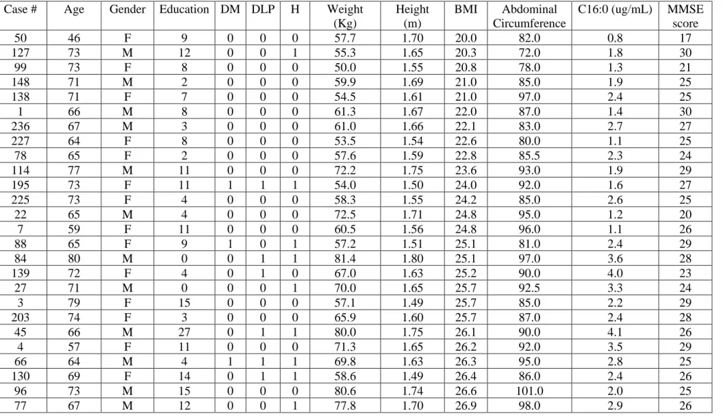
Figure 1. CSF palmitic acid is increased in overweight and obese individuals, correlates with BMI and abdominal circumference, and inversely correlates with MMSE scores in overweight subjects with diabetes, hypertension and dyslipidemia. See also Figure S1 and Table S1.
(A) CSF palmitic acid in normal weight (N = 14), overweight (N = 26) and obese (N= 9)
subjects (classified by BMI). ****p < 0.0001 (one-way ANOVA followed by Holm-Sidak’s
post hoc test).
(B) Correlation between CSF palmitic acid and BMI in normal weight (green), overweight
19
(C) Correlation between CSF palmitic acid and abdominal circumference in normal weight
(green), overweight (blue) and obese (orange) subjects (classified by BMI); linear regression, r2 and p values as indicated.
(D) CSF palmitic acid in overweight subjects according to absence (N = 11) or presence (N
= 15) of diabetes, hypertension or dyslipidemia. p = 0.09 (unpaired t-test).
(E) Inverse correlation between CSF palmitic acid and MMSE score in overweight subjects
with diabetes, hypertension or dyslipidemia (linear regression, r2 and p values as indicated).
(F) Lack of correlation between CSF palmitic acid and MMSE score in overweight subjects
without diabetes, hypertension or dyslipidemia (linear regression, r2 and p values as
indicated).
Figure 2. Palmitate inhibits long term potentiation and induces memory impairment in mice.See also Figure S2 and Figure S3.
(A-C) Long-term potentiation (LTP) was induced by high-frequency stimulation (HFS,
arrow) at the CA3 hippocampal subregion and recording in CA1 in awake freely-moving male 3-5-month-old mice, 10 days after intrahippocampal infusion of vehicle or palmitate (0.3 nmol). N = 6 vehicle-infused/N=8 palmitate-infused mice.
(A) Measurements performed immediately following HFS.
(B) Averaged fEPSP of the time points from 20 minutes to 30 minutes after HFS. Bars
represent means (± SEM). **** p < 0.0001 (unpaired t-test).
(C) Measurements performed on subsequent days following LTP induction. *p < 0.05
(two-way ANOVA followed by Holm-Sidak’s post hoc test).
(D) Novel object recognition (NOR) memory in male 3-5-month-old mice that received an
i.c.v. infusion of vehicle or palmitate (3 nmol) 10 days before testing. Bars represent means (± SEM) exploration times for familiar (F) and novel (N) objects. Symbols represent individual animals. ***p < 0.0005 (unpaired t-test).
(E) Novel object location (NOL) memory in male 3-5-month-old mice that received an i.c.v.
infusion of vehicle or palmitate (3 nmol) 10 days before testing. Bars represent means (± SEM) exploration times for objects in the original location (O) or novel location (N). Symbols represent individual animals. ****p < 0.0001 (unpaired t-test).
(F) Latency to step down in the step-down inhibitory avoidance task of male 3-5-month-old
mice 10 days after i.c.v. infusion of vehicle or palmitate (3 nmol). Bars represent medians with interquartile range and symbols represent individual animals. **p < 0.005 (Mann-Whitney test).
20
(G) Primary latency time in the training phase of the Barnes maze for vehicle- or palmitate
infused 3-5-month-old male mice. Symbols represent means ± standard errors. N = 7 vehicle-infused/6 palmitate-infused mice. **p = 0.005 (two-way ANOVA followed by Sidak’s post
hoc test).
(H) Primary errors in the training phase of the Barnes maze for vehicle- or palmitate infused
3-5-month-old male mice. Symbols represent means ± standard errors. N = 7 vehicle-infused/6 palmitate-infused mice. p = 0.09 (two-way ANOVA followed by Sidak’s post hoc test).
(I) Time in target zone on test day 5 in the Barnes maze for vehicle- or palmitate-infused
3-5-month-old male mice. Bars represent means ± standard errors, and symbols represent individual mice. N = 7 vehicle-infused/6 palmitate-infused mice. **p < 0.005 (unpaired t-test).
Figure 3. Palmitate reduces insulin expression and impairs IRS-1 signaling in hippocampal neurons. See also Figure S4.
(A) Relative insulin expression (mRNA) in cultured hippocampal cells exposed to vehicle or
200 μM palmitate for 24 hours. Bars represent means ± standard errors and symbols represent independent cultures. **p < 0.005 (paired t-test).
(B-I) Neuronal IRS-1pTyr612 (B-C), pSer636 (D-E), pSer307 (E-F) and pSer312 (G-H)
immunoreactivities in cultured hippocampal neurons exposed to vehicle or 200 μM palmitate for 4 hours (10-30 images analyzed per experimental condition per experiment using independent neuronal cultures). Scale bar = 10 μm. Bars represent means ± standard errors and symbols represent independent cultures. *p < 0.05 (unpaired t-test). In representative IRS-1 PTyr612 images, for better viewing, the image color was changed to green using ZEN software.
Figure 4. Palmitate increases GFAP and Iba-1 immunoreactivities in vitro and in vivo.
(A) Representative images of GFAP immunolabeling in hippocampal cultures exposed to
vehicle or 200 μM palmitate for 4 hours (10-15 images analyzed per experimental condition per experiment using independent hippocampal cultures). Scale bar = 10 μm.
(B) GFAP immunoreactivity in hippocampal cultures. Bars represent means ± standard errors
and symbols represent independent cultures. **p = 0.008 (unpaired t-test).
(C, D) Representative images of GFAP immunolabeling in the dentate gyrus of the
hippocampus from 2-4-month-old mice that received i.c.v. infusions of vehicle or palmitate (3 nmol). Animals were perfused 10 days after palmitate infusion. Scale bar = 50 μm; 10 μm (inset). (D) Bars represent means ± standard errors and symbols represent individual animals. * p < 0.05 (unpaired t-test).
21
(E) Representative images of Iba-1 immunolabeling in purified microglial cultures exposed
to vehicle or 200 μM palmitate for 2 hours (10-25 images analyzed per experimental condition per experiment with independent microglial cultures). Scale bar = 10 μm.
(F,G) Iba-1 immunoreactivity (F) and cell area (G) in purified microglial cultures exposed
to vehicle or palmitate. Bars represent means ± standard errors and symbols represent independent cultures. *p < 0.05; **p < 0.005 (unpaired t-test).
(H,I) Representative images of Iba-1 immunolabeling (H) and number of Iba-1 positive cells (I) in the dentate gyrus of the hippocampus from 2-4 month-old mice that received i.c.v.
infusions of vehicle or palmitate (3 nmol). Animals were perfused 10 days after palmitate infusion. Scale bar = 50 μm; 10 μm (inset). (I) Bars represent means ± standard errors and symbols represent individual animals. **p < 0.005 (unpaired t-test).
Figure 5. Microglial activation mediates inhibition of neuronal IRS-1 signaling and memory impairment by palmitate.
(A) Representative images of Iba-1 immunolabeling in purified microglial cultures exposed
to vehicle or 200 μM palmitate for 2 hours in the absence or presence of 1 μM minocycline (10-25 images analyzed per experimental condition per experiment with independent microglial cultures). Scale bar = 10 μm.
(B, C) Iba-1 immunoreactivity (B) and cell area (C) in purified microglial cultures exposed
to vehicle or palmitate, in the absence or presence of minocycline. Bars represent means ± standard errors and symbols represent independent cultures. *p < 0.05 (two-way ANOVA followed by Holm-Sidak’s post hoc test). Vehicle and palmitate conditions were showed in Figure 4 F,G.
(D, E) Neuronal IRS-1pSer636 immunoreactivities in hippocampal cultures exposed for 4
hours to conditioned medium from purified microglial cultures treated with vehicle or 200 μM palmitate (10-30 images analyzed per experimental condition per experiment using independent hippocampal and microglial cultures). Scale bar = 10 μm. Bars represent means ± standard errors and symbols represent experiments with independent hippocampal and microglial cultures. *p < 0.05 (unpaired t-test).
(F, G) Neuronal IRS-1pSer636 immunoreactivities in hippocampal cultures exposed for 4
hours to vehicle or 200 μM palmitate in the absence or presence of 1 μM minocycline (10-30 images analyzed per experimental condition per experiment using independent cultures). Scale bar = 50 μm; 10 μm (inset). Bars represent means ± standard errors and symbols represent experiments from 3 independent cultures. *p < 0.05; **p < 0.005 (two-way ANOVA followed by Holm-Sidak’s post hoc test).
(H) Novel object recognition (NOR) test in male 3-5-month-old mice infused i.c.v. with
vehicle or palmitate (3 nmol) and treated or not with minocycline (50 mg/kg/d, ip, for 3 consecutive days ending on the day of vehicle or palmitate infusion). Testing was performed 10 days after infusions. Bars (means ± SEM) represent time spent exploring the familiar (F)
22
or novel (N) objects, and symbols represent individual mice. *p < 0.05; **p =0.005; ***p < 0.001 (two-way ANOVA followed by Holm-Sidak’s post hoc test).
(I, J) Representative images of Iba-1 immunolabeling (I) and number of Iba-1 positive cells (J) in the dentate gyrus of the hippocampus from 2-4 month-old mice that received i.c.v.
infusions of vehicle or palmitate (3 nmol) and were treated or not with minocycline (50 mg/kg/d, IP, for 3 consecutive days ending on the day of vehicle or palmitate infusion). Animals were perfused 10 days after palmitate infusion. Scale bar = 50 μm; 10 μm (inset). (J) Bars represent means ± standard errors and symbols represent individual animals. **p < 0.005 (two-way ANOVA followed by Dunnett’s post hoc test). Part of the vehicle and palmitate data was shown in Figure 4I.
Figure 6. Increased hippocampal TNF-α expression mediates neuronal IRS-1 signaling impairment in vitro and memory deficits induced by palmitate.
(A) TNF-α released to the culture medium in hippocampal cultures exposed to vehicle or 200
μM palmitate for 4 hours. Symbols represent paired conditions per culture. N= 8 independent cultures. **p = 0.006 (paired t-test).
(B-E) TNF-α mRNA (B, D) and protein levels (C, E) in the hippocampi of 3-5-month-old
mice infused i.c.v. with vehicle or palmitate (3 nmol) 24 hours (B, C) or 10 days (D, E) prior to collection of hippocampi. Bars represent means ± standard errors and symbols represent individual animals. *p < 0.05 (unpaired t-test).
(F, G) Neuronal IRS-1pSer636 immunolabeling in hippocampal cultures exposed for 4 hours
to vehicle or 200 μM palmitate in the absence or presence of 1 μg/ml infliximab (20-30 images analyzed per experimental condition per experiment using independent cultures). Scale bar = 10 μm. Bars represent means ± SEM and symbols represent independent cultures. **p < 0.005; *p < 0.05 (two-way ANOVA followed by Holm-Sidak’s post hoc test).
(H, I) Novel object recognition (NOR) test in male 3-5-month-old mice infused i.c.v. with
vehicle or palmitate (3 nmol) and treated or not with infliximab (100 ng, i.c.v., 30 min previously to the infusion of vehicle or palmitate). Testing was performed 24 hours (H) or 10 days (I) after infusions. Bars (means ± SEM) represent time spent exploring the familiar (F) or novel (N) objects, and symbols represent individual mice. *p < 0.05, **p =0.005 (two-way ANOVA followed by Holm-Sidak’s post hoc test).
(J, K) Representative images of Iba-1 immunolabeling (J) and number of Iba-1 positive cells (K) in the dentate gyrus of the hippocampus from 2-4 month-old mice that received i.c.v.
infusions of vehicle or palmitate (3 nmol) and were treated or not with infliximab (100 ng, i.c.v., 30 min previously to the infusion of vehicle or palmitate). Animals were perfused 10 days after palmitate infusion. Scale bar = 50 μm; 10 μm (inset). (K) Bars represent means ± standard errors and symbols represent individual animals. **p < 0.005 (two-way ANOVA followed by Dunnett’s post hoc test). Part of the vehicle and palmitate data was shown in Figure 4I.
23
Figure 7. Infliximab prevents memory deficits in HFD mice and genetic deletion of TNFR1 prevents memory impairment induced by palmitate.
(A) Novel object recognition (NOR) test in male 6-7-month-old mice fed normal diet (ND)
or high fat diet (HFD) for 12 weeks. Bars (means ± SE) represent time spent exploring the familiar (F) or novel (N) objects, and symbols represent individual animals. *p < 0.05 (unpaired t-test).
(B) Novel object recognition (NOR) test in male 6-7-month-old mice fed high fat diet (HFD)
for 16 weeks and treated or not with infliximab (10 μg, i.c.v., for 3 days before memory task). Bars (means ± SE) represent time spent exploring the familiar (F) or novel (N) objects, and symbols represent individual animals. *p < 0.05 (unpaired t-test).
(C, D) Novel object recognition (NOR) test in 3-5month-old wild type or TNFR1-/- mice
infused i.c.v. with vehicle or palmitate (9 nmol). Testing was performed 24 hours (C) or 8 days (D) after infusions. Bars (means ± SEM) represent time spent exploring the familiar (F) or novel (N) objects, and symbols represent individual animals. *p < 0.05, **p < 0.005, ***p < 0.0005, ****p < 0.0001 (two-way ANOVA followed by Holm-Sidak’s post hoc test).
STAR Methods
CONTACT FOR REAGENT AND RESOURCE SHARING
This study did not generate new unique reagents. Further information and requests for resources and reagents should be directed to and will be fulfilled by the Lead Contact, Fernanda G. De Felice (felice@bioqmed.ufrj.br).
EXPERIMENTAL MODEL AND SUBJECT DETAILS
Human subjects: We studied 49 subjects diagnosed as aMCI recruited from the
Neuropsychology and Dementia clinic, Hospital de Clínicas, University of Campinas invited to participate in the project in order to check their memory (Magalhães et al., 2018; Rabelo et al., 2017; Teixeira et al., 2016, 2018; Weiler et al., 2017). aMCI patients were diagnosed using the core criteria of the National Institute on Aging/Alzheimer's Association for MCI (Albert et al., 2011; Raj et al., 2012) and had a score of 0.5 (with an obligatory memory score of 0.5), estimated on the basis of a semi structured interview. Exclusion criteria for all participants were history of other neurologic/psychiatric diseases or head injury with loss of
24
consciousness, drug or alcohol addiction, a Hachinski ischemic score 15 above 4 and a Fazekas Scale score16 above 1. The Medical Research Ethics Committee of the University of Campinas (UNICAMP) approved our study, and we obtained written informed consent from all participants (or from their responsible guardians if the participants were incapable of consenting) before study initiation. Pre-diagnostic procedures also comprised of laboratory tests including vitamin B12, folate, and thyroid hormones. CSF was obtained from all participants once, by lumbar puncture of the L3/L4 or L4/L5 intervertebral space, using a 25-gauge needle, and collected via a syringe in 12-mL polypropylene tubes (Sarstedt, Nümbrecht, Germany). CSF samples were centrifuged at 700 rpm for 10 minutes at 4ºC and were subsequently aliquoted into 1 ml microtubes (Eppendorf) and stored at -80ºC until the time of analysis.
BMI was calculated by dividing the person´s weight, in kilograms, by their height, in meters squared, or BMI = weight (in kg)/ height2 (in m2). BMI was categorized as: normal weight:
18.5-23.9 kg/m2; overweight: 24.0-27.9 Kg/m2; and obesity: ≥ 28 kg/m2. Abdominal
circumference was measured to the nearest 0.1 cm by a non-elastic flexible tape. We used the method recommended by the World Health Organization (WHO), which consists measuring midway between the lowest rib margin and the iliac crest at the mid-axillary line World Health Organization (WHO Consultation on Obesity, 1999; Geneva, Switzerland)
Abdominal obesity was defined as >102cm cm for men and > 88 cm for women (Garvey et al., 2016).
Patients were divided into groups with or without diabetes, dyslipidemia and hypertension based on the use of medication for these diseases.
Intrahippocampal palmitate administration and placement of recording electrodes: All
experiments were conducted in accordance with the “Principles of Laboratory Animal Care” (NIH publication no. 85–23, revised 1996) and with the Guidelines of the European Union (2003/65/CE) for the use of laboratory animals in chronic experiments. Every effort was made to reduce the number of animals used and to minimize their suffering.
C57BL/6 male mice (3-5 months-old; 25-35g) were obtained from an official supplier (University of Granada Animal House, Granada, Spain) and housed five per cage until
25
experiments began. Mice were kept on a 12 h light/dark cycle with constant ambient temperature (21.5 ± 1.5 °C) and humidity (55 ± 8%). Food and water were available ad libitum. Mice were anesthetized with 4% chloral hydrate and, prior to electrode implantation, received a single intra-CA2 (1.8 mm lateral, 1.5 mm posterior do bregma and 1.1 mm from the brain surface; Franklin and Paxinos, 2004) infusion of palmitate (0.3 nmol) or vehicle in a final volume of 1 µL using a Hamilton syringe, which was kept in place for an additional 30 seconds after drug administration to avoid backflow. Immediately after infusion, bipolar stimulating electrodes were implanted in the CA3 hippocampal subregion of the dorsal hippocampus (2 mm lateral and 1.5 mm posterior to bregma, and 1–1.5 mm from the brain surface; Franklin and Paxinos, 2004) and a bipolar recording electrode was placed in the ipsilateral stratum radiatum underneath the CA1 area (1.2 mm lateral and 2.2 mm posterior to bregma and 1–1.5 mm from the brain surface; Franklin and Paxinos, 2004). Electrodes were made from 50-μm Teflon-coated tungsten wire (Advent Research Materials, UK). The final location of the recording electrodes in the CA1 area was determined after the field potential depth profile evoked by paired (10- to 500-ms interval) pulses presented to the Schaffer collateral. A bare silver wire was fixed to the skull as ground. The four wires were soldered to a four-pin socket (RS Amidata) that was then fixed to the skull using dental cement (Gruart et al., 2006; Madronal et al., 2007). After surgery, animals were housed in individual cages until the end of experiment. Only data from animals with correct electrode placement were analyzed.
Intracerebroventricular infusion of palmitate or oleate in Swiss mice: Male Swiss mice were
obtained from our animal facility (Federal University of Rio de Janeiro) or from Laboratory Animal Breeding Center (Fundação Oswaldo Cruz – CECAL/Fiocruz, Rio de Janeiro) and were 2-5 months-old at the beginning of experiments. Animals were housed in groups of five per cage with free access to food and water, under a 12 h light/dark cycle with controlled room temperature (21 ± 2 °C). The animals weighed approximately 30 – 50 g. Mice were randomly assigned into one of two groups: vehicle or palmitate.
For intracerebroventricular (i.c.v.) infusion of vehicle or palmitate, animals were anesthetized and carefully restrained with 2.5% isoflurane (Cristalia) using a vaporizer system, and a 2.5-mm-long needle was unilaterally inserted 1 mm to the right of the midline
26
point equidistant from each eye and 1 mm posterior to a line drawn through the anterior base of the eyes (Clarke et al., 2015; Figueiredo et al., 2013; Laursen SE, 1986; Lourenco et al., 2013). Three nmol of palmitate or oleate (or an equivalent volume of vehicle) were infused in a final volume of 3 µl. When indicated (see Results and Figure legends), one hour before i.c.v. palmitate infusion mice received 10 ng infliximab (or an equivalent volume of phosphate buffered saline - PBS) in a final volume of 1 µl at the same injection site as the fatty acid. In these animals, 3 nmol palmitate (or an equivalent volume of vehicle) were infused in a final volume of 2 µl so that the final volume injected i.c.v. was 3 µl.
Regarding the LTP experiment, we increased the amount injected palmitate in the behavioral experiments to ensure its distribution to different brain regions following icv infusion. Accurate placement of the needle into the right lateral ventricle was confirmed by macroscopic examination of dissected brains before biochemical analysis. Mice showing any signs of misplaced injections or brain hemorrhage were excluded from further analysis (~ 5% of animals throughout our study). All procedures were performed in the light phase of the daily cycle, followed the “Principles of Laboratory Animal Care” (US National Institutes of Health) and were approved by the Institutional Animal Care and Use Committee of the Federal University of Rio de Janeiro (protocols IBQM045 and IBQM133/15).
Intracerebroventricular infusion of palmitate in TNFR1-/- and C57BL/6 mice: Male 3-4 months-old TNFRp55-/- (TNFR1 KO) mice (Arruda et al., 2011; Pfeffer et al., 1993) were
kindly donated by Dr. Licio A. Velloso from the University of Campinas, São Paulo, Brazil, and male 3-4 months-old C57BL/6 wild type mice from our animal facility (Federal University of Rio de Janeiro) were used as controls. The animals weighed approximately 25-35 g. All animals were housed in groups of five per cage with free access to food and water, under a 12 h light/dark cycle with controlled room temperature (21 ± 2 °C) and were randomly assigned into vehicle or palmitate groups. All procedures were performed in the light phase and followed the “Principles of Laboratory Animal Care” (US National Institutes of Health) and were approved by the Institutional Animal Care and Use Committee of the Federal University of Rio de Janeiro (protocol # IBQM133/15).
TNFR1-/- and controls were randomly assigned into one of two groups: vehicle or palmitate.
27
Nine nmol palmitate (or an equivalent volume of vehicle) were infused in a final volume of 1.5 µl. We increased the amount injected palmitate in the behavioral experiments to ensure its distribution to different brain regions following i.c.v. infusion.
High fat diet (HFD) administration: Male 3-3.5 months-old C57BL/6 wild type from our
animal facility (Federal University of Rio de Janeiro) were randomly divided into two groups fed either standard rodent chow or HFD (35% fat content by weight - PragSoluções®; Razolli
et al., 2015) for 16 weeks. For NOR experiments, HFD mice were treated with infliximab
(10 µg) or PBS for 3 days before memory testing, with the last injection in the day of NOR task. Animals were housed in groups of five per cage with free access to food and water, under a 12 h light/dark cycle with controlled room temperature (21 ± 2 °C). All procedures were performed in the light phase and followed the “Principles of Laboratory Animal Care” (US National Institutes of Health) and were approved by the Institutional Animal Care and Use Committee of the Federal University of Rio de Janeiro (protocol # IBQM133/15).
Mature hippocampal cultures: Primary rat hippocampal cultures were prepared according to
established procedures (De Felice et al., 2009) and adapted from the original protocol described by Brewer (Brewer, 1997). Hippocampi from 18-day-old embryos were dissected and the meninges were removed. Tissue was dissociated in PBS 0.05% trypsin/EDTA (Invitrogen) solution at 37 °C for 5-10 minutes under occasional gentle shaking followed by mechanical dissociation with glass Pasteur pipettes in Dulbecco’s Modified Eagle Medium (DMEM) (Gibco) medium supplemented with 10% horse-serum (Invitrogen) and antibiotics (100 U/ml penicillin/streptomycin) (Invitrogen). Cells in suspension were counted in a Neubauer chamber and were plated on culture plaques or glass coverslips previously coated with 10 μg/ml or 1 mg/ml poly-L-lysine, respectively. The density of culture was 50.000 cells/well (260 cells/mm2) on coverslips for immunocytochemistry experiments and 30.000
cells/well (645 cells/mm2) for the cell viability experiments. Cells were maintained at 37 °C
in a humidified atmosphere containing 5% CO2 for 60 minutes and DMEM was replaced by
Neurobasal medium supplemented with 2% B27 supplement (Invitrogen), 0.5 mM Glutamax (Invitrogen), 100 U/ml penicillin/streptomycin (Invitrogen) and 10 μg/ml amphotericin-B (Invitrogen). Cultures were maintained at 37 °C in a humidified atmosphere containing 5%
28
CO2 for 18-21 days prior to use. Our cultures contain, on average, 50% neurons and 50%
non-neuronal cells. The objective is to have a mixed culture to investigate the interplay between neuronal and non-neuronal cells. Sex of 18-day-old embryos could not be determined at this embryonic age and the embryos were not weighed.
Cultures were exposed at 37 °C to 50-500 µM palmitate for 4, 24 or 48 hours. When present, infliximab (1 μg/ml) or minocycline (1 μM) were added to cultures 30 minutes or 1 hour before application of vehicle or palmitate (200 μM), respectively.
Primary cultures and conditioned media from mouse microglia: Primary microglial cultures
were prepared as described previously (Azevedo et al., 2013; Ledo et al., 2013, 2016). Cortices from neonatal Swiss mice were dissociated, and the resulting cells were plated on poly-L-lysine-coated 75-cm2 flasks. The cells were maintained in DMEM with F12 medium
(1:1 ratio), 10% fetal bovine serum (FBS) and 1% penicillin/streptomycin for 2 weeks at 37 °C in a humidified atmosphere containing 5% CO2. The medium was changed after 1 week
and before cell harvesting. After 14 days, microglia were harvested using an orbital shaker. The cells were counted and plated in 24-well plates for assays. Microglial cells were exposed at 37 °C to 200 µM palmitate, 200 µM oleate or vehicle for 2 hours. E. coli lipopolysaccharide (LPS, 100 ng/ml) was used as a positive control for microglial activation. When present, minocycline (1 μM) were added to cultures 1 hour before application of vehicle or palmitate (200 μM), respectively.
For experiments using conditioned medium, cells were washed 2 times with warm DMEM with F12 medium and changed to Neurobasal medium overnight to obtain conditioned medium. The conditioned medium was then incubated with hippocampal cultures for 4 hours.
METHOD DETAILS
Palmitic acid measurements by GC-MS: For fatty acid profile determination, 170 µL
cerebrospinal fluid were taken in a screw cap glass tube containing 25 µg internal standard (tridecanoic acid - C13:0). Thereafter, 1 mL of 0.5 M NaOH-methanol was added and the
29
sample was boiled at 100 °C for 15 min. After cooling the samples, 2 mL of BF3-methanol was added and the sample was boiled at 100 °C for 20 s. The sample was again cooled to room temperature and 1 mL of isooctane was added. The tubes were shaken and 5 mL of saturated NaCl solution was added. After phase separation, the supernatant was collected and evaporated with the aid of nitrogen gas to concentrate the fatty acid methyl esters (FAME). FAME were resuspended in 50 µL hexane for chromatographic analysis (Shirai et al., 2005). Chromatographic analyses were performed using a gas chromatograph-mass spectrometer (model GCMS-QP2010 Ultra; Shimadzu). A fused silica capillary column Stabilwax (length, 30 m; internal diameter, 0.25 mm; thickness, 0.25 µm; Restek, USA) was used to inject 1 μL of the sample at 250 ºC. High-grade pure helium (He) was used as the carrier gas with a constant flow rate of 1.3 mL/min with a split injection of 2:1. The oven temperature was programmed from 80 to 175 °C at a rate of 5 °C/min, followed by another gradient of 3 °C/min to 230 °C, which was maintained for 10 min. Mass conditions were as follows: ionization voltage, 70 eV; ion source temperature, 200 ºC; full scan mode in the 35–500 mass range with 0.2 s/scan velocity.
Palmitate and oleate preparation: The protocol for preparation of palmitate and oleate
solutions was adapted from the protocol described by Mayer and Belsham (Mayer and Belsham, 2010). Solutions for i.c.v. infusions in mice were prepared from a stock solution of 100 mM sodium palmitate (Sigma) or sodium oleate (Sigma) solubilized in methanol (Tedia). Palmitate or oleate were solubilized in methanol under occasional shaking at 60 ° C (palmitate) or at room temperature (oleate). The stock solution was then diluted in Neurobasal culture medium (Gibco) containing 5% fatty acid free albumin (Sigma) for 1 hour at 37 ° C and shaking. After this period, the solutions were used for icv infusions. For Swiss mice, 2 or 3 μl of the final infused solution contained 3 nmol of fatty acids. For C57BL/6 and TNFR1-/-mice, 1.5 μl of final infused solution contained 9 nmol of palmitate.
Vehicle was Neurobasal medium with 5% fatty acid free albumin plus the corresponding volume of methanol. The methanol in the final solution was a very low volume, with only one residual of the solvent remaining (0,015 - 0,03%).
30
For primary hippocampal cultures, 5 mM palmitate solution in Neurobasal culture medium with 5% fatty acid free albumin was diluted to 50-500 µM in culture medium. The vehicle was Neurobasal with 5% fatty acid free albumin plus the corresponding volume of methanol. The BSA solution was filtered through sterile 0.22 µm filter before palmitate or oleate dilution, and all solutions were performed sterile in a laminar flow hood. Furthermore, the pH of the solutions was measured after preparation, and was in the range of 7.2-7.4.
The amount of palmitate used in our studies in mice (3 or 9 nmol) corresponds to 22 or 66 µg/ml CSF, respectively (assuming uniform distribution in 35 µl of CSF). These concentrations are ~ 3.5-10-fold higher than we found in the CSF of obese subjects, and were chosen to allow investigation of the impact of palmitate in mice within a reasonable experimental timeframe.
Long term potentiation (LTP): Recordings took place 10 days after electrode implantation in
alert behaving mice. Field excitatory post-synaptic potential (fEPSP) baseline values were recorded using 100 µs, square, biphasic paired pulses for 15 min before LTP induction. For construction of I/O curves, stimulus intensities ranged from 0.02mA to 0.4mA and were elicited at a 40ms interstimulus interval. The stimulus intensity was set at ~35% of the intensity necessary for evoking a maximum fEPSP response (i.e., well bellow the threshold for evoking a population spike). For LTP induction, a HFS protocol consisting of five 200 Hz, 100 ms-long trains of pulses at a rate of one per second was used. This protocol was applied six times, at intervals of 1 min. After HFS presentation, recordings of double-pulse stimulation at Schaffer collaterals were conducted for another 20 min to evaluate whether the LTP protocol was effective. During the following three days, mice were submitted to 15 min-long daily recording sessions to assess the persistence of LTP. According to previous studies (Gruart et al., 2006; Madronal et al., 2007), this HFS was enough to evoke a saturating LTP response, lasting 3 days, without the appearance of abnormal spikes in EEG recordings and/or any noticeable epileptic seizure.
All data were stored digitally on a computer through an analog/digital converter (1401 Plus; CED), at a sampling frequency of 11–22 kHz and with an amplitude resolution of 12 bits. Evoked fEPSPs amplitudes were analyzed with the help of commercial software (Spike 2
31
and GraphPad Prism 7) and data are expressed as average amplitude of fEPSP for every 2 min of recording. Statistical analysis for each analysis is described in the corresponding Figure Legends.
Novel object recognition (NOR) and novel object location (NOL) tests: Tests were performed
in a 30 (w) x 30 (d) x 45 (h) cm arena. Before training, each animal was submitted to a 5 min habituation session, in which they were allowed to freely explore the empty arena. Training consisted of a 5 min session during which animals were placed at the center of the arena in the presence of two identical objects and the time spent exploring each object was recorded by a trained researcher. Sniffing and touching the object were considered as exploratory behavior. Approximately one to two hours after training, animals were again placed in the arena for the test session, when one of the two objects used in the training session was replaced by a new one in NOR paradigm or moved to a new location in NOL paradigm, and the time spent exploring familiar and novel objects (or object at the novel location) was measured. The arena and objects were cleaned thoroughly between trials with 40% ethanol to eliminate olfactory cues. Test objects were made of plastic (approximately 3 cm (w) x 3 cm (d) x 4cm (h)) and during behavioral sessions were fixed to the arena floor using tape to prevent displacement caused by exploratory activity of the animals. Preliminary tests showed that none of the objects used in our experiments evoked innate preference by animals, i.e., during the training period, mice explored each object approximately 50% of the time. NOR test in C57BL/6 mice was performed without the habituation phase. In the training phase, the animals explored approximately 45 - 55% of the total time each o object. Results were expressed as percentage of time exploring each object or location (familiar or novel) in relation a total exploration time during the test session, and statistical analysis for each experiment is described in the corresponding Figure Legends.
Step-down inhibitory avoidance test: The test was carried out in an aluminum and acrylic
boxmeasuring 200 mm x 75 mm, a floor consisting of a grid of steel bars with a spacing of 12.5 mm (Insight EP 104R) and an adapted platform of 2 cm of height placed in the center of the grid. In the training phase, each animal was gently placed on the platform. When the animal stepped down from the platform onto the grid with the 4 paws, they received a 0.7
32
mA footshock (stimulus) for 2 seconds. The animal was then withdrawn from the apparatus. After 24h (test session), each animal was placed on the platform again. In this session, no shock was applied when the animal stepped-down on the grid. After stepping down, each animal was withdrawn from the apparatus and placed back in the home cage. The time in platform before stepping down on the grid (latency) was measured during the training and test sessions with a manual chronometer by a trained researcher. Step-down latencies were cut-off at 300 s in the training and test sessions, and animals that did not step down during 300 seconds were excluded from analysis. Results are represented as median and interquartile range of the latency to step down in the test session and the difference was considered statistically if p < 0.05 (Mann-Whitney test).
Barnes maze test: The Barnes maze protocol was adapted from (Sunyer et al., 2014). The
maze consists of a white circular platform (92 cm of diameter) elevated 105 cm above the ground with 20 equally spaced holes (5 cm diameter, 7.5 cm between holes) along the perimeter with a darkened escape box in a constant position under one of the holes at the edge of the platform. Mice are expected to learn the location of the escape hole using extra-maze visual cues including geometric shapes (triangle, rectangle and circle) placed on the walls of the room.
To familiarize mice with the maze and the existence of the escape box, they were subjected to one habituation trial in the first day prior to the initial trial. This habituation consisted of placing the animal in a cylindrical chamber in the middle of the maze and after 10 seconds, the chamber was removed, and the mouse was gently guided to the escape hole, where it remained for 2 minutes.
In the next step, acquisition training was performed with four trials per day for four days. For each trial, mice were placed in the cylindrical chamber in the middle of the maze and, after 10 seconds, the chamber was removed, and mice were allowed to explore the maze for 3 minutes. The trial ended when the animal entered the escape hole or after 3 minutes. If a mouse did not enter the escape hole within 3 minutes, the animal was gently guided to it and kept inside the box for 1 minute. Mice were returned to their home cages until the next trial, with a 15-minute interval between trials. Time (primary latency) and number of errors (primary errors) to reach the escape hole for the first time were measured. Head deflections