Basal sauropodomorphs from the Ischigualasto
Formation
Ricardo N. MartÍnez
a, Cecilia Apaldetti
ab& Diego Abelin
a aInstituto y Museo de Ciencias Naturales , Universidad Nacional de San Juan , 5400 , San
Juan , Argentina
b
Consejo Nacional de Investigaciones Científicas y Técnicas , Buenos Aires , Argentina
Published online: 08 Oct 2013.
To cite this article: Ricardo N. MartÍnez , Cecilia Apaldetti & Diego Abelin (2012) Basal sauropodomorphs from the
Ischigualasto Formation, Journal of Vertebrate Paleontology, 32:sup1, 51-69, DOI: 10.1080/02724634.2013.819361
To link to this article:
http://dx.doi.org/10.1080/02724634.2013.819361
PLEASE SCROLL DOWN FOR ARTICLE
Taylor & Francis makes every effort to ensure the accuracy of all the information (the “Content”) contained
in the publications on our platform. However, Taylor & Francis, our agents, and our licensors make no
representations or warranties whatsoever as to the accuracy, completeness, or suitability for any purpose of the
Content. Any opinions and views expressed in this publication are the opinions and views of the authors, and
are not the views of or endorsed by Taylor & Francis. The accuracy of the Content should not be relied upon and
should be independently verified with primary sources of information. Taylor and Francis shall not be liable for
any losses, actions, claims, proceedings, demands, costs, expenses, damages, and other liabilities whatsoever
or howsoever caused arising directly or indirectly in connection with, in relation to or arising out of the use of
the Content.
This article may be used for research, teaching, and private study purposes. Any substantial or systematic
reproduction, redistribution, reselling, loan, sub-licensing, systematic supply, or distribution in any
form to anyone is expressly forbidden. Terms & Conditions of access and use can be found at http://
www.tandfonline.com/page/terms-and-conditions
Volume 32, Supplement to Number 6: 51–69 ©2013 by the Society of Vertebrate Paleontology
BASAL SAUROPODOMORPHS FROM THE ISCHIGUALASTO FORMATION
RICARDO N. MART´INEZ,*,1CECILIA APALDETTI,1,2and DIEGO ABELIN11Instituto y Museo de Ciencias Naturales, Universidad Nacional de San Juan, 5400 San Juan, Argentina, [email protected];
2Consejo Nacional de Investigaciones Cient´ıficas y T ´ecnicas, Buenos Aires, Argentina, [email protected]
ABSTRACT—Basal sauropodomorphs from the Ischigualasto Formation include Eoraptor lunensis, Panphagia protos, and Chromogisaurus novasi. Few comparisons have been made between these taxa, because Eoraptor was only recently reassessed as a basal sauropodomorph and because Panphagia and Chromogisaurus were described nearly simultaneously. We describe in detail the fully prepared bones of the holotype of Chromogisaurus novasi, examine the evidence for its taxonomic distinction, and analyze the phylogenetic relationships among basal sauropodomorphs. Our results support Chromogisaurus novasi as a valid genus and species and provide weak phylogenetic evidence favoring a series of stem taxa at the base of Sauropodomorpha. The analysis positions Panphagia as the basal-most sauropodomorph, followed by Eoraptor, Pampadromaeus, and a clade that includes Chromogisaurus and Saturnalia.
RESUMEN—Los sauropodomorfos basales de la Formaci ´on Ischigualasto incluyen Eoraptor lunensis, Panphagia protos y Chromogisaurus novasi. Pocas comparaciones se han hecho entre estos taxones, porque Eoraptor fue reevaluado reciente-mente como sauropodomorfo basal y porque Panphagia y Chromogisaurus se describieron de forma casi simult ´anea. De-scribimos en detalle los huesos totalmente preparados del holotipo de Chromogisaurus novasi, examinamos la evidencia de su distinci ´on taxon ´omica, y analizamos la relaci ´on filogen ´etica entre sauropodomorfos basales. Nuestros resultados apoyan a Chromogisaurus novasi como un g ´enero y especie v ´alido y proporcionan d ´ebil soporte filogen ´etico favoreciendo un arreglo parafil ´etico en la base de Sauropodomorpha. El an ´alisis posiciona a Panphagia como el sauropodomorfo m ´as basal, seguido de Eoraptor, Pampadromaeus y un clado que incluye a Chromogisaurus y Saturnalia.
SUPPLEMENTAL DATA—Supplemental materials are available for this article for free at www.tandfonline.com/UJVP
INTRODUCTION
Basal sauropodomorphs are represented in two of the oldest dinosaur-bearing geologic units, namely the Ischigualasto Forma-tion of Argentina (Ezcurra et al., 2008; Ezcurra, 2010; Mart´ınez and Alcober, 2009; Mart´ınez et al., 2011) and the Santa Maria Formation of Brazil (Langer et al., 1999; Cabreira et al., 2011). For many years dinosaurs were thought to comprise a relatively small proportion of the archosaurs in the Ischigualasto fauna, in-cluding only the ornithischian Pisanosaurus (Casamiquela, 1967) and the theropods Herrerasaurus (Reig, 1963; Sereno and Novas, 1992) and Eoraptor (Sereno et al., 1993). Sauropodomorphs had yet to be recorded.
In recent years, however, four new dinosaurs have been de-scribed, including the basal sauropodomorphs Panphagia pro-tos (Mart´ınez and Alcober, 2009) and Chromogisaurus novasi (Ezcurra, 2010), the herrerasaurid theropod Sanjuansaurus gordil-loi (Alcober and Mart´ınez, 2010), and the basal theropod Eo-dromaeus murphi (Mart´ınez et al., 2011). All of these dinosaurs were recovered from the Scaphonyx-Exaeretodon-Herrerasaurus biozone, which is dated to approximately 231 Ma (Rogers et al., 1993; Mart´ınez et al., 2011).
The similar morphology and near-synchronous, independent description of the two aforementioned sauropodomorphs, Pan-phagia (Mart´ınez and Alcober, 2009) and Chromogisaurus (Ezcurra, 2010), leave their validity an open question. Prior to a detailed comparison, we completely prepared the holo-type and only known specimen of Chromogisaurus. The recent reinterpretation of Eoraptor as a basal sauropodomorph closely related to Panphagia (Mart´ınez et al., 2011) underscored the need to effectively compare these three contemporaneous basal sauropodomorphs. Although missing data are a problem for all
*Corresponding author.
taxa considered except Eoraptor and Saturnalia, we analyzed the phylogenetic relationships among known basal sauropodomorphs.
METHODS
Comparative Specimens
Comparisons were made using the holotypic specimens of Chromogisaurus (PVSJ 845), Panphagia (PVSJ 874), and San-juansaurus (PVSJ 605), holotypic and referred specimens of Eo-dromaeus (PVSJ 534, 561, 562), Eoraptor (PVSJ 512, 559), Sat-urnalia (MCP 3844-PV, 3845-PV, 3846-PV), and Guaibasaurus (MCN PV 2355, 2356, UFRGS PV 0725T), and referred speci-mens of Herrerasaurus (PVSJ 373, 380, 407). Other comparisons were based on published figures.
Photography
Some bone surfaces of Chromogisaurus (PVSJ 845) were painted with neutral gray acrylic paint to remove color distractions prior photography.
Anatomical and Taxonomic Terminology
We employ traditional, or ‘Romerian,’ anatomical and direc-tional terms over veterinarian alternatives (Wilson, 2006). ‘An-terior’ and ‘posterior,’ for example, are used as directional terms rather than the veterinarian alternatives ‘rostral’ or ‘cranial’ and ‘caudal.’ We also follow recent recommendations regarding the identification of vertebral laminae (Wilson, 1999).
We used the phylogenetic definitions for basal taxa within Dinosauria proposed by Sereno (2005a, 2005b, 2007). Sauropodomorpha, for example, has a stem-based definition in opposition to Theropoda and does not require the monophyly of Saurischia or Prosauropoda, as do other definitions (e.g., Galton and Upchurch, 2004). In this way, Sauropodomorpha is defined as 51
DINOSAURIA Owen, 1842 SAURISCHIA Seeley, 1887 SAUROPODOMORPHA Huene, 1932
CHROMOGISAURUS Ezcurra, 2010 CHROMOGISAURUS NOVASI Ezcurra, 2010
(Figs. 4–8, 9A–E, 10–13, 14A–E, 15)
Type Specimen—PVSJ 845, partial skeleton comprising one
an-terior and two mid-caudal vertebrae, anan-terior chevron, glenoid re-gion of the left scapulocoracoid, right ilium lacking preacetabu-lar process, acetabupreacetabu-lar portion of the left ilium, shaft of the right femur and proximal and distal ends of the left femur, complete right tibia and proximal end of the left tibia, right fibula lack-ing proximal and distal ends and proximal end of the left fibula, right metatarsal 2, right digit III phalanges, and unidentified bone fragments.
Type Locality—Cancha de Bochas, Hollada de Ischigualasto,
Ischigualasto Provincial Park, San Juan Province, Argentina.
Age and Distribution—The horizon is located at the southern
outcrops of the Ischigualasto Formation, 40 m above the base of the unit in the Cancha de Bochas Member (sensu Currie et al., 2009). The holotypic location belongs to the lower Scaphonyx-Exaeretodon-Herrerasaurus biozone (Mart´ınez et al., 2011). The mid-Carnian age is based on a radioisotopic date near the base of the formation (Rogers et al., 1993) that was recently recalibrated to 231.4 Ma (Mart´ınez et al., 2011).
Revised Diagnosis—Basal sauropodomorph diagnosed by the
following combination of features (asterisk indicates an autapo-morphy): marked posterior projection of the iliac postacetabu-lar process; incipient perforation of the iliac portion of the ac-etabulum; low lateral projection of the iliac supraacetabular crest; strongly concave acetabular surface of the supraacetabular crest; femoral lateral condyle smaller than the medial condyle; medial surface of the proximal end of the fibula with an elongate rugos-ity adjacent to the anterior margin of the shaft∗; metatarsal 2 with strongly dorsoventrally asymmetric distal condyles∗.
DESCRIPTION
We offer alternative identifications for several elements de-scribed by Ezcurra (2010), based on comparisons with other basal saurischians from the Ischigualasto Formation, namely, Eoraptor (Sereno et al., 1993), Panphagia (Mart´ınez and Alcober, 2009), and Eodromaeus (Mart´ınez et al., 2011).
Reidentification of Ulna, Ischium, and Metatarsal
Lower Jaw Fragment—Originally described as the proximal
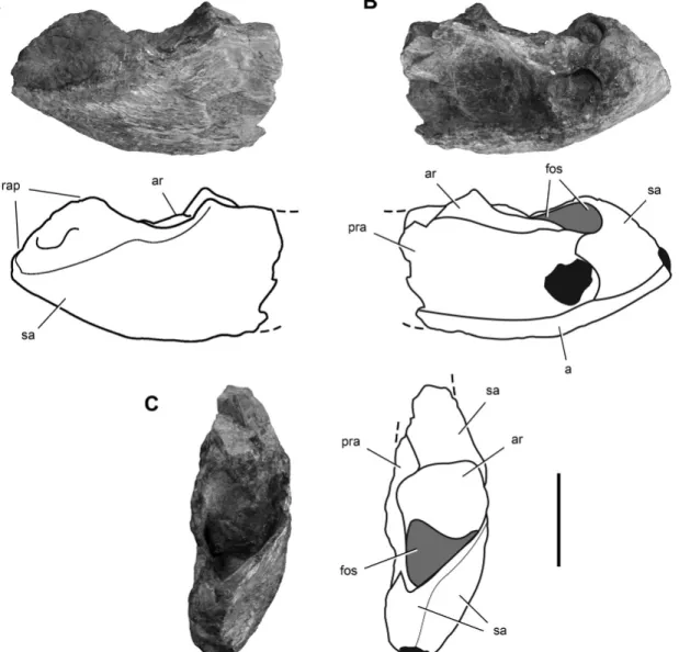
end of the right ulna (Ezcurra, 2010), this fragment is clearly not an ulna because it is formed of four different bones instead of only one (Fig. 1). It is reidentified here as the posterior end of a right lower jaw, possibly of the rhynchosaur Scaphonyx (Fig. 1). Mea-suring 29.5 mm in length, the sutured bones that form the frag-ment preserves the right articular and posterior ends of the angu-lar, suranguangu-lar, and prearticular.
ture of the bone. Ezcurra (2010) interpreted this feature as the strongly striated surface of the olecranon.
The posterior portion of the prearticular is broken and eroded (Fig. 1B). It contacts the angular ventrally, the surangular pos-teriorly, and the articular dorsally. The prearticular forms the medial wall of the adductor fossa and covers most of the medial aspect of the articular, as in Scaphonyx and Hypero-dapedon (Fig. 2B). In basal dinosaurs, in contrast, more of the articular is exposed dorsal to the prearticular (e.g., Panphagia, Herrerasaurus, Lesothosaurus). The angular forms the ventral margin of the posterior end of the lower jaw, extending to the ex-tremity of the retroarticular process (Fig. 1B). The prearticular-surangular suture is not clear. The articular has a subquadrate articular surface as seen in dorsal view and is relatively small (Fig. 1C). The small size of the articular relative to the poste-rior end of the lower jaw is similar to that in Scaphonyx and Hyperodapedon (Fig. 2C) and unlike the proportionately larger articular socket in basal dinosaurs (e.g., Panphagia, Eoraptor, Herrerasaurus). Posterior to the articular, a deep fossa opens dor-sally on the retroarticular process (Fig. 1C), as in Hyperodapedon (Benton, 1983).
The posterior end of the lower jaw is stout, transversely flat-tened, and dorsoventrally high (Fig. 1A, B), resembling Scapho-nyx (Sill, 1970) and Hyperodapedon (Fig. 2A, B), but differ-ing markedly from the slender proportions and low profile of the comparable portion of the lower jaw of the dinosauriform Silesaurus (Dzik, 2003) and basal dinosaurs. This holds for the basal sauropodomorphs Panphagia, Eoraptor, Adeopapposaurus (Mart´ınez, 2009), and Pantydraco (Galton and Kermack, 2011), the basal theropods Eodromaeus, Herrerasaurus (Sereno and No-vas, 1994), Tawa (Nesbitt et al., 2009), and Coelophysis (Raath, 1977), and the basal ornithischians Pisanosaurus (Casamiquela, 1967) and Heterodontosaurus (Crompton and Charig, 1962).
Chevron—Originally described as metatarsal 5 (Ezcurra, 2010),
this bone is reidentified here as an anterior chevron (Fig. 6). The morphology in support of the new interpretation includes the ‘Y’-shaped proximal end with a pair of articular facets, a hemal canal, and the laterally compressed, distally expanding shaft (see De-scription).
Bone Fragments of Unknown Identity and Affinity—One bone
piece originally assigned to the type specimen was identified as a probable section of the ischial shaft (Ezcurra, 2010). The squared section and massive size of the bone (Fig. 3) does not closely resemble the triangular section and slender proportions of the ischium in basal saurischians (e.g., Eoraptor, Saturnalia, Eodro-maeus). Two other bone pieces originally included as part of the holotype have similar size, cross-sectional shape, and surface de-tail, suggesting that they may belong to the same bone (Fig. 3). The bone surface of these three pieces, however, does not preserve the fine, subparallel fractures and adhering hematite that character-ize the other bones of the holotype. The sharp longitudinal ridge (Ezcurra, 2010:fig. 7B) is interpreted here as postmortem crushing, displacing one side with respect to the other. The identification of these pieces and their association with the other bones of the holo-type remain uncertain.
FIGURE 1. Photographs and line drawings of the posterior end of the right lower jaw of a possible rhynchosaur in lateral (A), medial (B), and dorsal (C) views. This specimen was originally included in the holotypic specimen of Chromogisaurus novasi (PVSJ 845) by Ezcurra (2010). Dark tone indicates broken areas; light tone indicates cavities. Abbreviations: a, angular; ar, articular; fos, fossa; pra, prearticular; rap, retroarticular process; sa, surangular. Scale bar equals 1 cm.
Description
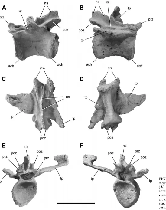
Caudal Vertebrae—Three caudal vertebrae of Chromogisaurus
are preserved. Based on general proportions, size of the trans-verse processes, and location and size of the neural spines, the first is likely to be in the range of caudal vertebrae 4–6 (Fig. 4) and the other two in the range of caudal vertebrae 14–16 (Fig. 5). The posterior-most mid-caudal vertebra (Fig. 5F, G) was not reported in the original description (Ezcurra, 2010). All of the centra are spool-shaped and strongly transversely compressed (Figs. 4D, 5C). In lateral view, the caudal centra are subrectangular with parallel anterior and posterior margins (Figs. 4A, B, 5A).
The mid-caudal centra of Chromogisaurus are slightly shorter (2%) than the anterior caudal centrum. The anterior and posterior faces of the caudal centra are oval, strongly concave, and bordered by a prominent external rim (Figs. 4E, F, 5D, E). The ventral sur-face of the anterior centrum is convex without any groove or keel (Fig. 4D), differing from the mid-caudal vertebrae, which have a
longitudinal groove (Fig. 5C). The articular facets for the chevrons are small (Figs. 4A, B, 5A).
The neural arches of caudal vertebrae of Chromogisaurus are gracile, differing from the more robust neural arches present in Panphagia and Eoraptor. The transverse process of the anterior caudal vertebra is flattened and located in the middle of the cen-trum (Fig. 4C–F). The dorsolaterally directed transverse processes of the caudal vertebrae of Chromogisaurus are subtriangular and taper distally (Figs. 4C, D, F, 5F, G). They are relatively long in Chromogisaurus, constituting 82% of the length of the cen-trum (Ezcurra, 2010). The transverse processes of the distal-most mid-caudal vertebra are flat, strap-shaped, nearly horizontal, and directed posterolaterally (Fig. 5F, G). In the anterior and mid-caudal vertebrae of Chromogisaurus, pre- and postzygapophyses extend slightly beyond the ends of the centrum (Ezcurra, 2010; Figs. 4A, B, 5A), as in Eoraptor, Panphagia, and Eodromaeus. In Herrerasaurus and Sanjuansaurus, in contrast, the postzygapophy-ses terminate flush with the posterior rim of the centrum. In
FIGURE 2. Reconstruction of the right lower jaw of the rhynchosaur Hyperodapedon gordoni in lateral (A), medial (reversed) (B), and dor-sal (C) views (from Benton, 1983). Abbreviations:
a, angular; ar, articular; co, coronoid; d, dentary; pra, prearticular; sa, surangular; sp, splenial; sym,
symphysis. Gray tone indicates the region of the mandible preserved in the posterior end of the lower jaw in Figure 1.
Chromogisaurus, the zygapophyseal articular surfaces are oval and taper distally. Hyposphene-hypantrum articulations are un-known in caudal vertebrae of Chromogisaurus (Ezcurra, 2010). As in other basal dinosaurs, a deep median notch separates the postzygapophyses (Figs. 4F, 5E; contra Ezcurra, 2010). The neural arch of the anterior caudal has a weakly developed interpostzy-gapophyseal lamina (Ezcurra, 2010; Fig. 4A), a feature unknown in other dinosaurs from the Ischigualasto Formation. All the neu-ral spines are damaged, although they seem to be anteroposteri-orly wide, extending along two-thirds of their respective neural arches. The height of the neural spine of the mid-caudal vertebrae appears to be one-third of the height of the neural arch (Fig. 5A, D, E). The tall spine originally reported for mid-caudal vertebrae (Ezcurra, 2010:fig. 4) is an artifact of preservation; a thin fragment of bone was pressed against the neural spine.
Chevron—The single preserved chevron of Chromogisaurus is
from the proximal end of the tail probably in the range of cau-dal vertebrae 4–6 (Fig. 6), based on its large size, lateral shape, size of the articular facets, and distal anteroposterior expansion, and in comparison with the chevrons of Eoraptor. It lacks por-tions of both proximal and distal ends. Its length is approximately 2.5 times the height of the centrum of the anterior caudal vertebra (Fig. 5E). The proximal end is ‘Y’-shaped with two distinct articu-lar facets. The anterior facet faces anterodorsally, and the smaller, posterior facet faces posterodorsally (Fig. 6B–D). The orientation of the anterior facet suggests that the chevron shaft was inclined at approximately 45◦to the longitudinal axis of the tail. Due to trans-verse compression, the anterior opening of the hemal canal has been closed (Fig. 6C). The shaft is laterally compressed, straight, and anteroposteriorly expanded at its distal end.
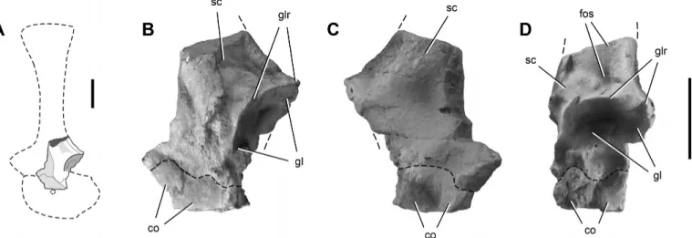
Scapulocoracoid—The left glenoid region is the only preserved
portion of the scapula and coracoid, which are joined along a fused suture (Fig. 7). The scapular glenoid is an oval-shaped concavity with its long axis oriented dorsoventrally (Fig. 7D). The scapular glenoid faces posteroventrally and somewhat laterally, as in Pan-phagia, Eoraptor, and Saturnalia (Langer et al., 2007). A fossa is located dorsal to the posterodorsal rim of the glenoid (Fig. 7B, D), as in Eoraptor, Panphagia, and Eodromaeus. Shallow medial and lateral depressions are present just anterior to the posterodorsal border (Fig. 7B, C). The rim of the glenoid is sharp (Fig. 7B, D). Not enough of the coracoid is preserved to comment on its form.
Ilium—The right ilium is the more complete, lacking only the
preacetabular process; the distal end of the right pubic pedun-cle is weathered laterally (Fig. 8A–D). Only a central piece of the left ilium is preserved (Fig. 8E). The ilium is vertically deep with a relatively long postacetabular process, as in other basal sauropodomorphs (e.g., Eoraptor, Adeopapposaurus). In lateral view, the dorsal margin of the iliac blade is straight and curves ventrally as it approaches the preacetabular process (Fig. 8A, B). In dorsal view, the iliac blade is gently curved with acetab-ular processes diverging laterally (Fig. 8C), as in other basal sauropodomorphs (e.g., Eoraptor, Saturnalia, Adeopapposaurus). A broad, triangular depression covers much of the lateral aspect of the ilium dorsal to the supraacetabular crest (Ezcurra, 2010), as is also present in Saturnalia (Langer, 2003). The posterior projection at the base of the ischial peduncle is poorly developed.
The supraacetabular crest is a well-developed, raised shelf (Fig. 8A, C, D). Although the pubic peduncle is partially damaged, the crest seems to continue to its distal end (Fig. 8A). The supraac-etabular crest has a sinuous curve in lateral view (Fig. 8A). A ventral flange partially closes the acetabulum in Chromogisaurus (Ezcurra, 2010; Fig. 8A, B), but its arched ventral margin suggests that the acetabulum had a central unossified gap.
The pubic peduncle is long, with its distal end facing anteroven-trally (Fig. 8A, C). The distal end of the pubic peduncle has a sub-triangular cross-section with slightly concave medial, dorsolateral, and ventrolateral borders. The broader and shorter ischial pedun-cle has its distal articular surface oriented more ventrally (Ezcurra, 2010; Fig. 8A, B). The distal end of the ischial peduncle is sub-triangular, with flat medial and slightly convex posterior and lat-eral margins (Fig. 8D). The antitrochanter is smooth, poorly devel-oped, and positioned on the anteroventral corner of the peduncle (Ezcurra, 2010).
The long postacetabular process tapers distally to a rounded end in lateral view, which has sustained some weathering. As was noted by Ezcurra (2010), the lateral surface bears a thick and trapezoidal rugose area, which tapers anteriorly along the dorso-lateral border of the iliac blade (Fig. 8A). The well-developed bre-vis shelf does not join the supraacetabular crest (Fig. 8A). The pos-teromedial shelf is moderately developed and similar in transverse width to the brevis shelf in dorsal or ventral view (Fig. 8C, D). In cross-section at midlength, the postacetabular process has an
FIGURE 3. Bone fragments of unknown affinity and identity in dor-sal view. These fragments were originally identified as a possible portion of the ischial shaft and included in the Chromogisaurus novasi holotype (PVSJ 845) by Excurra (2010). Abbreviations: ri, ridge. Scale bar equals 1 cm.
inverted ‘V’-shape. The posteromedial shelf extends anteroven-trally and dissipates as a rounded ridge on the medial surface of the iliac blade dorsal to the supraacetabular crest. Only the base of the preacetabular process is preserved (Fig. 8A, B). The ventral margin is thicker than the dorsal margin, so that the broken cross-section of the process is subtriangular. The ventral margin of the process is everted dorsal to the level of the supraacetabular crest.
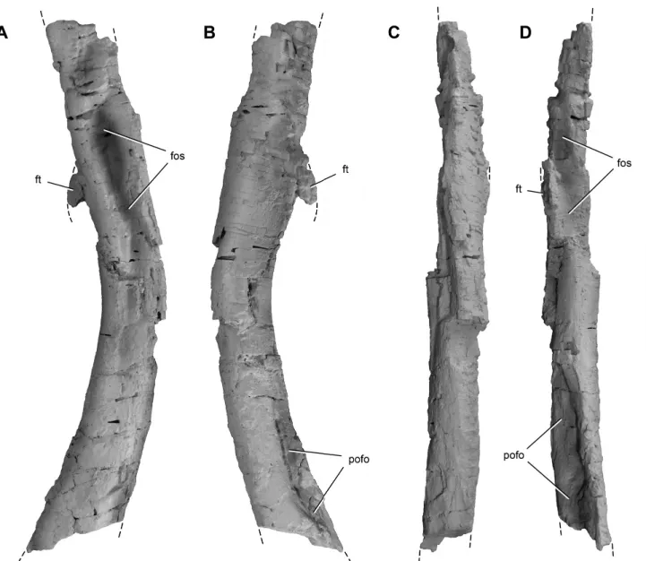
Femur—Although both femora are incomplete (Figs. 9A–E, 10,
11), they overlap to allow an estimate of the form and length (ca. 170 mm) of the bone. The femur is gracile—its minimum circumference-to-length ratio equals 0.28—and slightly shorter than the tibia (Fig. 9F). In lateral view, the shaft of the femur has a sigmoid curve, as in other basal dinosaurs. In anterior view, it is straight with the femoral head rotated anteromedially. The shaft of the right femur of Chromogisaurus is transversely crushed, which may have flattened the medial projection of the head
(com-pare Fig. 9C and F). The proximal surface of the femoral head is kidney-shaped, as in most other basal dinosaurs, but it faces dor-sally and is almost straight in posteromedial view (Fig. 9C, D). The proximal surface bounded by sharp edges, between which lies a groove extending anteromedially from the greater trochanter (Fig. 9E), as in basal dinosaurs such as Eoraptor (PVSJ 855), Sat-urnalia, Herrerasaurus, and Staurikosaurus (Galton, 1977). The greater trochanter is developed as a prominent ridge (Fig. 9A, D, E). The anterior surface of the femoral neck bears a sharp ridge that extends from the anteromedial end of the head and tapers distally (Fig. 9B, C). The trochanteric shelf extends along the lat-eral aspect of the proximal end, dissipating on its posterior aspect dorsal to the fourth trochanter (Ezcurra, 2010; Fig. 9).
A deep, elliptical fossa is present ventral the trochanteric shelf on both femora (Figs. 9A, 10A), which was considered an au-tapomorphy by Ezcurra (2010). Careful observation of the better-preserved left femur reveals compressional fractures surrounding each fossa (Fig. 9A). A similar fossa is present in some specimens of Eodromaeus (PVSJ 534, PVSJ 562), which also shows evidence of postmortem transverse compression (Fig. 9F). We regard this structure as an artifact of crushing and collapse of the internal lu-men of the femoral shaft.
A prominent, crescentic fourth trochanter projects from the proximal half of the femoral shaft (Fig. 9A, B, D, E). The dis-tal portion of the fourth trochanter is damaged on both sides. Whether the trochanter extends distally to end with an asym-metric profile, as in many basal saurischians (e.g., Eoraptor, Sat-urnalia, Herrerasaurus, Staurikosaurus, Eodromaeus), cannot be determined. A marked rugose scar for insertion of the M. caud-ofemoralis longus (Langer, 2003) is present on the medial aspect of the fourth trochanter and extends distally as a shallow striated fossa (Fig. 9B).
The distal end of the left femur, although partially broken, has a nearly subcircular shape as preserved (Fig. 11E). The medial border is flat, and the medial (tibial) condyle is oval and antero-posteriorly longer than the lateral condyle. The posterior surface is marked by a deep popliteal fossa that separates the condyles (Figs. 9F, 11D). At the distal end of the femur, a large rugose at-tachment scar is present facing anteriorly (Fig. 11C).
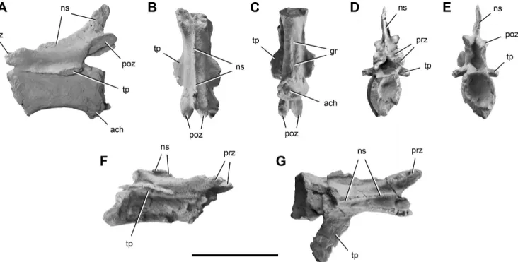
Tibia—The right tibia and proximal end of the left tibia are
pre-served, both of which have been flattened transversely (Fig. 12). The tibia is slender and measures 175 mm long. The tibiofemoral ratio is 1.03, similar to the values of Eoraptor (Sereno et al., 2013) and Eodromaeus (Mart´ınez et al., 2011). The proximal articu-lar surface is subtrianguarticu-lar and deeply concave, which has been exaggerated by transverse compression (Fig. 12E). As noted by Ezcurra (2010), the lateral condyle is located slightly anterior to the medial condyle (Fig. 12E). A median notch separates the prox-imal condyles (Fig. 12D, I). A low and symmetrical cnemial crest forms the anterior margin of the proximal end (Fig. 12A–C, E, H). A very pronounced longitudinal tuberosity is present on the lateral surface posterior to the concavity formed adjacent to the cnemial crest (Ezcurra, 2010; Fig. 12A, G). The shaft of the tibia is straight (Fig. 12A–D). The distal end has been transversely com-pressed postmortem, so that its transverse width is only about 40% of its anteroposterior depth (Fig. 12F). The medial tip of the pos-terolateral process projects distally (Fig. 12A–C). A groove on the lateral aspect of the distal end separates the posterolateral flange from the shaft of the tibia (Fig. 12A). As in other basal dinosaurs, the distal articular surface of the tibia is split between a broad as-tragalar articular surface and a convex ‘L’-shaped surface formed by the posterolateral flange (Fig. 12F).
Fibula—Portions of both fibulae are preserved. On the right
side, both the proximal and distal ends are broken away (Fig. 13A, B). On the left side, the proximal end is well preserved (Fig. 13C–F). The fibula is narrower transversely than the tibia;
FIGURE 4. Proximal caudal vertebra of Chro-mogisaurus novasi (PVSJ 845) in left lateral (A), right lateral (B), dorsal (C), ventral (D), anterior (E), and posterior (F) views.
Abbre-viations: ach, articular surface for a chevron; cr, crest; ns, neural spine; poz,
postzygapoph-ysis; prz, prezygapophpostzygapoph-ysis; tp, transverse pro-cess. Scale bar equals 2 cm.
the anteroposterior width of the midshaft is almost two-thirds that of the tibia. The proximal end is anteroposteriorly expanded to a depth approximately three times its maximum width. The proxi-mal surface is posteriorly concave and slightly anteriorly convex (Fig. 13F). The medial border of the proximal end is straight, al-though the medial surface becomes concave below the border. In lateral view, the anterior border of the proximal end of the fibula is convex, and the posterior border is slightly concave, as in many other basal saurischians. The anterior prominence of the anterior border is well marked (Fig. 13A–D). Two attachment scars are vis-ible on the medial surface (Fig. 13C). The first is an oblique ridge extending anterodistally from the posterior corner of the proximal end of the fibula and is similar to that of Saturnalia (Langer, 2003), Eodromaeus (PVSJ 562), and Herrerasaurus. The second is a more rugose ridge located parallel to and near the anterior border of the fibula, converging distally with the aforementioned ridge
(Fig. 13C). As in many other dinosaurs (e.g., Saturnalia, Eoraptor, Eodromaeus, Herrerasaurus, Sanjuansaurus), a rugosity is present along the anterolateral border of the proximal end (Fig. 13D, E), probably for the insertion of the M. iliofibularis (Bittencourt and Kellner, 2009). The lateral surface of the proximal end is anterior and posteriorly concave and slightly convex at the middle. The shaft of the fibula gradually decreases in anteroposterior depth distally (Fig. 13A, B). The lateral surface of the shaft is convex and has a crest, or anterior trochanter (Fig. 13A), presumably for attachment of the M. iliofibularis (Langer, 2003). An elongate con-cavity extends for most of the length of the medial surface of the shaft due to postmortem collapse of the central lumen of the tibia (Fig. 13B).
Metatarsal 2—Only one metatarsal of Chromogisaurus is
pre-served. The metatarsal is difficult to identify with certainty, given the absence of its proximal end. Ezcurra (2010) regarded the
FIGURE 5. Mid-caudal vertebrae of Chromogisaurus novasi (PVSJ 845). The anterior-most preserved mid-caudal vertebra in left lateral (A), dorsal (B), ventral (C), anterior (D), and posterior (E) views. The posterior-most preserved mid-caudal vertebra in right lateral (F) and dorsal (G) views. Anterior is towards top in B and C and to the right in G. Abbreviations: ach, articular surface for a chevron; gr, groove; ns, neural spine; poz, postzy-gapophysis; prz, prezypostzy-gapophysis; tp, transverse process. Scale bar equals 2 cm.
FIGURE 6. Anterior chevron of Chromo-gisaurus novasi (PVSJ 845) in right lateral (A), anterior (B), posterior (C), and proximal (D) views. Dashed line indicates missing portions of proximal and distal ends. Abbreviations: aace, anterior articular surface for centrum; cr, crest;
hc, hemal canal; pace, posterior articular surface
for centrum; sh, shaft. Scale bar equals 1 cm.
FIGURE 7. Proximal portion of the left scapulocoracoid of Chromogisaurus novasi (PVSJ 845) in left lateral (A, B), medial (C), and posterior (D) views. Dashed lines indicate fused scapulocoracoid suture and the continuation of preserved margins. Abbreviations: co, coracoid; fos, fossa; gl, glenoid;
glr, glenoid rim; sc, scapula. Scale bars equal 1 cm (left scale bar for A only).
FIGURE 8. Ilia of Chromogisaurus novasi (PVSJ 845). Right ilium in lateral (A), medial (B), dorsal (C), and ventral (D) views. Fragmentary left ilium in lateral view (E). Dashed lines indicate the continuation of preserved margins. Abbreviations: acet, acetabulum; ais, articular surface for the ischium;
apu, articular surface for the pubis; aS1r, articular surface for sacral 1 rib; aS2r, articular surface for sacral 2 rib; bfo, brevis fossa; bsh, brevis shelf; fl,
flange; isped, ischial peduncle; poap, postacetabular process; pped, pubic peduncle; ru, rugosity; sac, supraacetabular crest; vfl, ventral flange. Scale bar equals 3 cm.
FIGURE 9. Femora of Chromogisaurus novasi and Eodromaeus murphi. Proximal end of the left femur of Chromogisaurus novasi (PVSJ 845) in lateral (A), medial (B), anterior (C), posterior (D), and proximal (E) views. Proximal end of the left femur of Eodromaeus murphi (PVSJ 562) in lateral view (F). Dashed lines indicate the continuation of preserved margins. Abbreviations: fos, fossa; ft, fourth trochanter; gr, groove; gt, greater trochanter;
hd, head; pmf, postmortem fracturing; ts, trochanteric shelf. Scale bar equals 3 cm.
metatarsal and phalanges as pertaining to left pedal digit II. The size and marked asymmetry between its distal condyles clearly in-dicate that the metatarsal could only pertain to either metatarsal 2 or 4. The distal end is most similar to metatarsal 2 of other basal di-nosaurs (e.g., Eoraptor, Saturnalia, Herrerasaurus). The distal end of metatarsal 4 is typically more asymmetric and divergent, and its lateral condyle is narrower. When identified as metatarsal 2, the asymmetry of the condyles indicates that it is from the right pes and not the left as proposed (Ezcurra, 2010). The greater distal extension of the lateral condyle of metatarsal 2 diverts the pha-langes of pedal digit II medially from a sagittal axis, spreading the weight-supporting digits of the pes (Fig. 14F).
The shaft of metatarsal 2 is parallel-sided and does not change much in diameter along its length. In dorsal view, there is a distinct lateral curvature to the shaft (Fig. 14A), as in Saturnalia, Eoraptor, and Herrerasaurus. In cross-section, the metatarsal is subtriangular at midshaft with medial, anterior, and posterolateral surfaces. A shallow fossa on the dorsal surface of the distal one-third of the metatarsal is probably postmortem deformation
(Fig. 14A). The distal end is transversely broad, with a more distally developed lateral condyle and a larger lateral collateral ligament pit (Fig. 14A, B, D). An asymmetrical, deep, and broad extensor depression is located dorsal to the distal ginglymus. The narrow medial distal condyle protrudes posteriorly and forms a prominent posterolateral edge, which is similar to, although narrower than, that in Sanjuansaurus (Fig. 14E, F). As was noted by Ezcurra (2010), this condition contrasts with the more symmetrical morphology of the distal condyles in other basal dinosaurs (e.g., Eoraptor, Saturnalia, Herrerasaurus).
Pedal Phalanges—The articulated pedal phalanges may
be-long to right pedal digit III, rather than left pedal digit II as proposed by Ezcurra (2010). The most distinctive feature in support of this attribution is the asymmetry of the ungual. The ventromedial border of the ungual of digit III tends to be sharper than the border on the opposite side (e.g., Herrerasaurus, Adeopapposaurus). Only the distal tip of the ungual is lacking (Fig. 15A–D). The first phalanx (phalanx III-2) is longer than the second (phalanx III-3). The lateral, medial, and ventral surfaces
FIGURE 10. Right femoral shaft of Chromogisaurus novasi (PVSJ 845) in lateral (A), medial (B), anterior (C), and posterior (D) views. Dashed lines indicate the continuation of preserved margins. Abbreviations: fos, fossa; ft, fourth trochanter; pofo, popliteal fossa. Scale bar equals 3 cm.
FIGURE 11. Distal end of the left femur of Chromogisaurus novasi (PVSJ 845) in lateral (A), medial (B), anterior (C), posterior (D), and distal (E) views. Abbreviations: fos, fossa; lco, lateral condyle; mco, medial condyle; pofo, popliteal fossa; ru, rugosity. Scale bar equals 3 cm.
FIGURE 12. Tibiae of Chromogisaurus novasi (PVSJ 845). Right tibia in lateral (A), medial (B), anterior (C), posterior (D), proximal (E), and distal (F) views. Proximal end of the left tibia in lateral (G), medial (H), and posterior (I) views. Anterior is towards top in E and F. Dashed lines indicate the continuation of preserved margins. Abbreviations: aas, articular surface for the ascending process of the astragalus; cc, cnemial crest; cr, crest; fos, fossa; gr, groove; lco, lateral condyle; mco, medial condyle; no, notch; plf, posterolateral flange; tu, tuberosity. Scale bar equals 4 cm.
are concave, and the dorsal surface is flat. The proximal ends are transversely slightly wider than the corresponding distal gingly-mus, as in Eoraptor and Saturnalia. Dorsal intercondylar processes are present on both non-ungual phalanges (Fig. 15A, C), and they are better developed than in Eoraptor and Saturnalia (MCP 3844-PV). The medial collateral ligament pits are somewhat more dor-sally oriented than corresponding lateral pits.
The ungual (phalanx III–ungual) is slightly asymmetric, the ven-tromedial edge slightly sharper than its opposite (Fig. 15B). The ventral surface is only slightly arched proximodistally, and there
are marked grooves to each side for attachment of the ungual sheath. The ventral flexor tubercle is low and broad (Fig. 15B).
COMPARISONS AND PHYLOGENETIC POSITION
Comparisons
Chromogisaurus resembles closely the general morphology of basal-most sauropodomorphs from Argentina and Brazil (e.g., Panphagia, Eoraptor, Saturnalia, Guaibasaurus). The vertebral
FIGURE 13. Fibulae of Chromogisaurus no-vasi (PVSJ 845). Right fibula in lateral (A) and medial (B) views. Proximal left fibula in lateral (C), medial (D), anterior (E), and proximal (F) views. Anterior is towards top in F. Dashed lines indicate the continuation of preserved margins.
Abbreviations: afe, articular surface for the
fe-mur; cr, crest; em, eminence; pmf, postmortem fracturing; ri, ridge; ru, rugosity. Scale bar equals 2 cm.
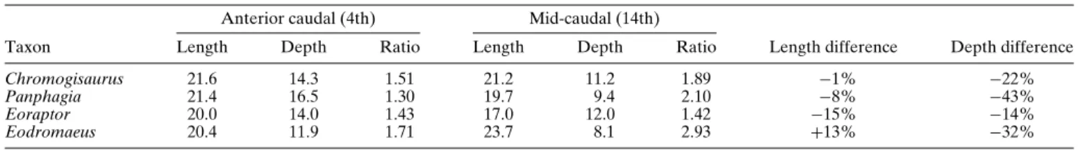
centra of Chromogisaurus are transversely compressed (Figs. 4D, 5C), as in Eoraptor, and less compressed than in Panphagia and Saturnalia (MCP 3846-PV). In Chromogisaurus, centrum length is almost constant in the anterior one-third of the tail. In some other basal sauropodomorphs, caudal centrum length decreases distally in this portion of the tail (Table 1). In the basal theropod Eodro-maeus, in contrast, caudal centrum length increases distally in the proximal caudal vertebrae (Table 1). In these same caudal verte-brae, depth decreases more markedly than in Eoraptor, although less so than in Panphagia and Eodromaeus (Table 1).
Chromogisaurus lacks a groove or keel on the ventral surface of the anterior caudal centra (Fig. 4D), as in Eoraptor, Panpha-gia, Sanjuansaurus, and Herrerasaurus. A groove is present in Eodromaeus. A groove is present in mid-caudal vertebrae in Chromogisaurus (Fig. 5C), as in Panphagia, Eoraptor, and Eodromaeus.
The neural arches of the caudal vertebrae of Chromogisaurus are more gracile than in Panphagia and Eoraptor. The transverse processes of the anterior caudal vertebra of Chromogisaurus are subtriangular and taper distally (Fig. 4C, D). In Eoraptor and Panphagia, in contrast, the comparable processes are leaf-shaped. The relative length of the transverse processes is less in Chro-mogisaurus (82% of centrum length) than in Panphagia (99%) and Eoraptor (111%). In the anterior and mid-caudal vertebrae of Chromogisaurus, pre- and postzygapophyses extend slightly beyond the ends of the centrum (Figs. 4A, B, 5A), as in Eo-raptor, Panphagia, and Eodromaeus. In Herrerasaurus and San-juansaurus, in contrast, the postzygapophyses terminate flush with the posterior rim of the centrum. The neural spines extend along two-thirds of their respective neural arches. In Panphagia, in con-trast, the spine base occupies less than one-half the length of the neural arch.
TABLE 1. Measurements (in mm) and ratios of anterior and mid-caudal vertebrae of several basal dinosaurs from the Ischigualasto Formation. Anterior caudal (4th) Mid-caudal (14th)
Taxon Length Depth Ratio Length Depth Ratio Length difference Depth difference
Chromogisaurus 21.6 14.3 1.51 21.2 11.2 1.89 −1% −22%
Panphagia 21.4 16.5 1.30 19.7 9.4 2.10 −8% −43%
Eoraptor 20.0 14.0 1.43 17.0 12.0 1.42 −15% −14%
Eodromaeus 20.4 11.9 1.71 23.7 8.1 2.93 +13% −32%
Depth measurement was taken at the posterior end of the centrum.
FIGURE 14. Metatarsal 2 of Chromogisaurus novasi and Sanjuansaurus gordilloi. Right metatarsal 2 of Chromogisaurus novasi (PVSJ 845) in dorsal (A), ventral (B), lateral (C), medial (D), and distal (E) views. Left metatarsal 2 (reversed) of Sanjuansaurus gordilloi (PVSJ 605) in distal view (F). Dorsal is towards top in E and F. Dashed lines indicate the continuation of preserved margins. Abbreviations: clp, collateral ligament pit; lco, lateral condyle; mco, medial condyle; pmf, postmortem fracturing. Scale bar equals 1 cm.
The iliac blade is 65% of the maximum depth of the ilium, as in Eoraptor (66%), Panphagia (67%), and Eodromaeus (67%). The depth of the blade is somewhat shallower in Saturnalia (56%) and noticeably deeper in more derived sauropodomorphs, such as Pantydraco and Adeopapposaurus. In lateral view, the straight dorsal margin of the iliac blade is similar to that of Saturna-lia but differs from the gently convex border of Eoraptor and Eodromaeus. The ischial peduncle does not project posteriorly, as in Saturnalia and Guaibasaurus. The peduncle projects more strongly posteriorly in Panphagia, Eoraptor, and more advanced basal sauropodomorphs (e.g., Adeopapposaurus).
The supraacetabular crest of Chromogisaurus extends to the distal end of the pubic peduncle (Fig. 8A), as in Saturnalia, Eoraptor, and Eodromaeus. In Panphagia and other sauropodo-morphs such as Adeopapposaurus, the crest dissipates before the end of the pubic peduncle. The flange of the acetabulum has a straight ventral margin in Saturnalia and Panphagia, which closes
the acetabulum to a greater degree than in Chromogisaurus. The pubic peduncle is directed more ventrally than in Saturnalia, Pan-phagia, and Eoraptor. The subtriangular distal end of the pubic peduncle of Chromogisaurus is similar than that of Eoraptor and Saturnalia (MCP 3846-PV), contrasting with the semicircular, or ‘D’-shaped, cross-section of the peduncle of Panphagia.
There is a thick and trapezoidal rugose area along the dorsolat-eral border of the iliac blade (Fig. 8A), as in Saturnalia (Ezcurra (2010). The development of this muscle scar, which is presumably for attachment of the M. iliotibialis (Langer, 2003), varies in other sauropodomorphs (e.g., Adeopapposaurus), which may be related to maturity or body size.
The femur is slightly shorter than the tibia in Chromogisaurus, as in Eoraptor, Pampadromaeus, and Eodromaeus. In all other sauropodomorphs, the tibia is the shorter than the femur. The femoral shaft of Chromogisaurus is sigmoidal in lateral view, as in all other basal dinosaurs, but in anterior view it is straight,
FIGURE 15. Articulated phalanges 2–4 of right pedal digit III of Chromogisaurus novasi (PVSJ 845) in dorsal (A), ventral (B), lateral (C), medial (D), and proximal (E) views.
Abbre-viations: clp, collateral ligament pit; dip, dorsal
intercondylar process; lco, lateral condyle; mco, medial condyle; un, ungual; vip, ventral inter-condylar process. Scale bar equals 1 cm.
which differs from the sigmoidal shaft in Eoraptor, Saturnalia, and Eodromaeus. The proximal surface of the femoral head faces dorsally and has a straight profile in posteromedial view (Fig. 9C, D), as in Guaibasaurus (Bonaparte et al., 2006:fig. 8; UFRGS PV 0725T). In other basal dinosaurs, in contrast, the proximal surface faces dorsomedially and has a convex profile in posteromedial view (e.g., Saturnalia, Eoraptor, Eodromaeus). The large and rugose attachment scar at the distal end of the femur is similar to that in Guaibasaurus, Eodromaeus, and Herrerasaurus.
The lateral condyle of the tibia is positioned slightly anterior to the medial condyle (Fig. 12E), as in Eoraptor and Panphagia. In Saturnalia, the lateral condyle is located even farther anteri-orly. The symmetrical profile of the cnemial crest in lateral or medial view is similar to that in Eoraptor. In Panphagia, Satur-nalia, and Eodromaeus, the apex of the crest is situated closer to its proximal end. The pronounced tibiofibular crest is similar to that in Saturnalia, Eoraptor, and Eodromaeus, and more marked than in Panphagia. The straight shaft of the tibia (Fig. 12A–D) most closely resembles that in Eodromaeus and differs from the slightly sinuous shaft in Eoraptor (PVSJ 559), Saturnalia (Langer, 2003:fig. 5C), and Panphagia. The posterolateral pro-cess of the tibia projects distally, similar to that in Saturnalia and Eodromaeus.
Taxonomic Status of Chromogisaurus
We were unable to verify several of the characters originally used by Excurra (2010) to differentiate Chromogisaurus novasi from other sauropodomorphs. These include the femoral fossa near the trochanteric shelf, which is here regarded as postmortem deformation, and the absence of a notch between the postzy-gapophyses in caudal vertebrae, which is present in the present material. We were able to confirm only one autapomorphy cited by Ezcurra (2010), the marked dorsoventral asymmetry of the dis-tal condyles of metatarsal 2, although our interpretation of the sid-ing of that bone is opposite to his. A second autapomorphy sup-porting the distinctiveness of Chromogisaurus is the rugose scar near, and parallel to, the anterior border of the fibula (Fig. 13C).
Although there are few autapomorphies in the available material of Chromogisaurus, other features distinguish this basal sauropodomorph from other basal dinosaurs from the Is-chigualasto Formation (Panphagia, Eoraptor, Eodromaeus) and from the basal sauropodomorph Saturnalia from Brazil.
Chromogisaurus novasi differs from Panphagia protos in the following features: the caudal centra are more transversely com-pressed; the transverse processes of the anterior caudal vertebrae are subtriangular and distally tapered; the articular surfaces of caudal zygapophyses are oval; the caudal neural spines are antero-posteriorly broader; the iliac posteromedial shelf equals the bre-vis shelf in transverse width; the proximal articular surface of the tibia is concave; the tibial cnemial crest reaches its most prominent point well below the proximal margin; the lateral side of the cne-mial crest has a rugose crest; the posterolateral flange of the tibia extends distally more prominently; and a more marked groove separates the posterolateral flange from the remainder of the dis-tal end of the tibia.
Chromogisaurus novasi differs from Eoraptor lunensis in the following features: caudal vertebrae with more strongly concave articular faces; iliac blade with a straighter profile in lateral view; less prominent iliac supraacetabular crest; acetabulum with strongly concave dorsal articular surface; femoral head with prox-imal surface facing dorsally and straight in posteromedial view; femoral fibular condyle less developed than the tibial condyle; shaft of the tibia straight in anteroposterior and mediolateral views; medial tip of the posterolateral flange of the tibia distally protruding; and a more marked groove separating the posterolat-eral flange from the remainder of the distal end of the tibia.
Chromogisaurus novasi differs from the basal theropod Eo-dromaeus murphi in a number of features, the most salient of which are shorter mid-caudal vertebrae; caudal vertebrae with more strongly concave articular faces; anterior caudal centra with-out a ventral groove; caudal vertebrae withwith-out any development of hyposphene-hypantrum articulations; robust glenoid region of the scapulocoracoid; iliac blade with a straighter profile in lateral view; less prominent iliac supraacetabular crest; concave surface on the dorsal margin of the acetabulum; straight proximal surface
FIGURE 16. Simplified phylogenetic relationships of basal sauropodomorphs based on a data set of Ezcurra (2010). Strict consensus (A) and reduced consensus trees after the exclusion of Agnosphytis (B). Full black lines indicate Sauropodomorpha in B. Numbers within Sauropodomorpha indicate the decay indices.
of the femoral head facing dorsally in posteromedial view; fibu-lar condyle of the femur less developed than the tibial condyle; the tibial cnemial crest reaches its most prominent point well be-low the proximal margin; the rugose crest on the lateral side of the cnemial crest; the posterolateral flange of the tibia protrudes distally; and a more marked groove separating the posterolateral flange from the remainder of the distal end of the tibia.
Chromogisaurus novasi differs from Saturnalia tupiniquim in the following features: caudal vertebrae with more strongly con-cave articular faces; strongly transversely compressed caudal cen-tra; higher iliac blade of the ilium; pubic peduncle facing more ventrally; slightly concave ventral border of the acetabular wall; poorly developed supraacetabular crest of the ilium; strongly con-cave surface on the dorsal margin of the acetabulum; straight prox-imal surface of the femoral head facing dorsally in posteromedial view; fibular condyle of the femur less developed than the tibial condyle; subtriangular scar on the distal-most region of the an-teromedial surface of the femur; lateral condyle of the tibia much posteriorly located; the tibial cnemial crest reaches its most promi-nent point well below the proximal margin; the rugose crest on the lateral side of the cnemial crest; the posterolateral flange of the tibia protrudes distally; and a more marked groove separating the posterolateral flange from the remainder of the distal end of the tibia.
Phylogenetic Analysis
Reanalysis Based on Ezcurra (2010)—To reexamine
phyloge-netic relationships among basal sauropodomorphs, we modified the data set originally published by Yates (2007) and later mod-ified by Smith and Pol (2007) and Ezcurra (2010). We rescored Chromogisaurus based on the information presented above and added Pampadromaeus as a terminal taxon (Cabreira et al., 2011). The character-state scores for the basal sauropodomorphs Eorap-tor, Panphagia, Chromogisaurus, and Saturnalia were reviewed and rescored (Appendices 1, 2, Supplementary Data).
The data set for reanalysis included 51 taxa and 378 characters. We used TNT 1.1 (Goloboff et al., 2008a, 2008b) in an equally
weighted parsimony analysis that included a heuristic search of 1000 replicates of Wagner trees followed by tree bisection and re-connection (TBR) branch swapping. Following the original anal-ysis of Yates (2007), a total of 36 characters were ordered. The analysis yielded 60 most parsimonious trees (MPTs) of 1192 steps (consistency index= 0.37; retention index = 0.69) (Fig. 16).
The strict consensus tree depicts a polytomy formed by Eo-raptor, Panphagia, Pampadromaeus, the clade Chromogisaurus + Saturnalia, Chindesaurus, Agnosphitys, Guaibasaurus, and Neotheropoda (Fig. 16A). Following Ezcurra (2010), a reduced strict consensus was obtained after exclusion of the poorly known taxon Agnosphitys (Fraser et al., 2002) (Fig. 16B). In the reduced strict consensus, basal sauropodomorphs are depicted in a para-phyletic arrangement with Panphagia as the basal-most member, followed by Eoraptor, Pampadromaeus, and the clade that in-cludes Chromogisaurus+ Saturnalia as the sister clade of more derived sauropodomorphs (Fig. 16B).
The position of Panphagia as basal to other sauropodomorphs is supported by nine unambiguous synapomorphies: pterygoid wing of the quadrate extending for more than 70% of the total quadrate length (71:1); presence of postparietal fenestra between supraoc-cipital and parietals (74:1); supraocsupraoc-cipital wider than high (75:1); coarse serrations of the teeth angled upwards at 45◦(114:1); ab-sence of a postzygodiapophyseal lamina in cervical vertebrae 4–8 (142:1); weakly developed laminae in the neural arches of cervi-cal vertebrae 4–8 (143:1); minimum width of the scapula less than 20% of its length (200:0); posterior end of the fibular condyle of the tibia anterior to the posterior margin of proximal artic-ular surface (304:0); and strongly laterally curved iliac blade in dorsal view (372:1). Sauropodomorpha is well supported by a decay index of 3, although bootstrap confidence is below 50% (Fig. 16B).
The more derived position of Eoraptor is supported by three unambiguous synapomorphies: subtriangular cross-section of the ischial midshaft (274:1); supraacetabular crest of the ilium con-tacting the distal end of pubic peduncle (363:2); and subtriangular distal end of the ischium (377:1). Pampadromaeus and more de-rived sauropodomorphs share four unambiguous synapomorphies:
(362:1); and concave posterolateral corner of the distal end of the tibia (375:1). This clade is particularly weak, with a decay index of 1 and bootstrap frequency below 50% (Fig. 16B).
Comparisons with Recent Analyses—In the analysis of Ezcurra
(2010), Panphagia, Guaibasaurus, and Chromogisaurus + Satur-nalia joined a polytomy within the clade Guaibasauridae (Bona-parte et al., 1999; Ezcurra and Novas, 2009). In the present anal-ysis, Guaibasaurus is recovered as a theropod, the sister taxon to Neotheropoda (Fig. 16B), a very tentative result for this poorly known taxon that mirrors previous analyses (e.g., Smith and Pol, 2007; Yates, 2007; Langer et al., 2011).
Eoraptor was originally viewed as a theropod (Sereno et al., 1993; Novas, 1996; Sereno, 1999; Rauhut, 2003; Yates, 2007; Ezcurra, 2010) or basal saurischian (Langer and Benton, 2006; Brusatte et al., 2010, 2011). Here Eoraptor is retrieved as a basal sauropodomorph (Fig. 16B), as in Mart´ınez et al. (2011) and as discussed in detail in Sereno and Mart´ınez (in review). The sauropodomorph affinity of Eoraptor is generated by alteration of several character states for this taxon (Appendix 2).
Ezcurra (2010) coined the subfamily Saturnaliinae to include Saturnalia plus Chromogisaurus. He based this clade on three synapomorphies: ulna with an extremely enlarged olecranon process; iliac postacetabular process with pointed posteroventral corner and rounded posterodorsal margin; and strong trapezoidal rugosity for the origin of the M. flexor tibialis and iliotibialis. Al-though we show that the ulna of Chromogisaurus is not preserved, our analysis also generates this clade (Fig. 16B), based on only one of the original synapomorphies: strong trapezoidal rugosity for the origin of the M. flexor tibialis and iliotibialis (362:1).
CONCLUSIONS
Most of the small-bodied (<15 kg) primary consumers from the Ischigualasto Formation appear to have been sauro-podomorphs. This trophic niche was shared with at least one silesaurid (Mart´ınez et al., 2013) and possibly the ornithischian Pisanosaurus, which is thus far known only from the upper levels of the formation. The three sauropodomorphs, Panphagia, Eorap-tor, and Chromogisaurus, come from the Scaphonyx-Exaeretodon-Herrerasaurus biozone (Mart´ınez et al., 2011) and thus were likely contemporaries sharing the same ecosystem. The presence in Brazil of the similar-age basal sauropodomorphs Saturnalia and Pampadromaeus suggests that these herbivores had achieved a level of diversity in southern Pangaea during the Carnian. A similar diversity of sauropodomorphs (three) is present in the overlying Los Colorados Formation, which was deposited about 15 million years later in the mid-Norian (Santi Malnis et al., 2011). The main difference between these sauropodomorphs is that in the Los Colorados Formation, they are the most abundant vertebrates and also attained large body size. The fossil record of sauropodomorphs across these two formations suggests that sauropodomorphs have been an important component from the emergence of dinosaurs, but that their size and abundance in-creased markedly in the Norian toward the end of the Triassic.
Alcober, O. A., and R. N. Mart´ınez. 2010. A new herrerasaurid (Di-nosauria, Saurischia) from the Upper Triassic Ischigualasto Forma-tion of northwestern Argentina. ZooKeys 63:55–81.
Benton, M. J. 1983. The Triassic reptile Hyperodapedon from Elgin: func-tional morphology and relationships. Philosophical Transactions of the Royal Society of London, Series B 302:605–717.
Bittencourt, J. S., and A. W. A. Kellner. 2009. The anatomy and phyloge-netic position of the Triassic dinosaur Staurikosaurus pricei Colbert, 1970. Zootaxa 2079:1–56.
Bonaparte, J. F., J. Ferigolo, and A. M. Ribeiro. 1999. A new early Late Triassic saurischian dinosaur from Rio Grande do Sul State, Brazil; pp. 89–109 in Y. Tomida, T. H. Rich, and P. Vickers-Rich (eds.), Pro-ceedings of the Second Gondwanan Dinosaur Symposium, Buenos Aires 26–30 July, 1999. National Science Museum Monographs 15. Bonaparte, J. F., G. Brea, C. L. Schultz, and A. G. Martinelli. 2006. A
new specimen of Guaibasaurus candelariensis (basal Saurischia) from the Late Triassic Caturrita Formation of southern Brazil. Historical Biology 19:73–82.
Brusatte, S. L., M. J. Benton, G. T. Lloyd, M. Ruta, and S. C. Wang. 2011. Macroevolutionary patterns in the evolutionary radiation of ar-chosaurs (Tetrapoda: Diapsida). Earth and Environmental Science Transactions of the Royal Society of Edinburgh 101:367–382. Brusatte, S. L., S. J. Nesbitt, R. B Irmis, R. J. Butler, M. J. Benton, and M.
A. Norell. 2010. The origin and early radiation of dinosaurs. Earth-Science Reviews 101:68–100.
Cabreira, S. F., C. L. Schultz, J. S. Bittencourt, M. B. Soares, D. C. Fortier, L. R. Silva, and M. C. Langer. 2011. New stem-sauropodomorph (Di-nosauria, Saurischia) from the Triassic of Brazil. Naturwissenschaften 938:1035–1040.
Casamiquela, R. M. 1967. Un nuevo dinosaurio ornitisquio Tri ´asico (Pisanosaurus mertii; Ornithopoda) de la Formaci ´on Ischigualasto, Argentina. Ameghiniana 4:47–64.
Crompton, A. W., and A. J. Charig. 1962. A new ornithischian from the Upper Triassic of South Africa. Nature 196:1074–1077.
Currie, B. S., C. E. Colombi, N. J. Tabor, T. C. Shipman, and I. P. Monta ˜nez. 2009. Stratigraphy and architecture of the Upper Trias-sic Ischigualasto Formation, Ischigualasto Provincial Park, San Juan, Argentina. Journal of South American Earth Sciences 27:74–87. Dzik, J. 2003. A beaked herbivorous archosaur with dinosaur affinities
from the early Late Triassic of Poland. Journal of Vertebrate Pale-ontology 23:556–574.
Ezcurra, M. D., A. Lecuona, and R. Irmis. 2008. A new early dinosaur from the Carnian Ischigualasto Formation (NW Argentina) and the origin of dinosaurs; p. 88 in J. O. Calvo, R. J. Valieri, J. D. Porfiri, and D. dos Santos (eds.), Actas de Res ´umenes, III Congreso Latinoamericano de Paleontolog´ıa de Vertebrados, Neuquen 21–24 September 2008. Universidad Nacional del Comahue.
Ezcurra, M. D. 2010. A new early dinosaur (Saurischia: Sauropodomor-pha) from the Late Triassic of Argentina: a reassessment of di-nosaur origin and phylogeny. Journal of Systematic Palaeontology 8:371–425.
Ezcurra, M. D., and F. E. Novas. 2009. Guaibasauridae, a new clade of Tri-assic basal sauropodomorphs. Journal of Vertebrate Paleontology, Program and Abstracts 2009:92A.
Fraser, N. C., K. Padian, G. M. Walkden, and A. L. M. Davis. 2002. Basal dinosauriform remains from Britain and the diagnosis of the Dinosauria. Palaeontology 45:79–95.
Galton, P. M. 1977. On Staurikosaurus pricei, an early saurischian dinosaur from the Triassic of Brazil, with notes on the Herrerasauridae and Poposauridae. Pal ¨aontologische Zeitschrift 51:234–245.
Galton, P. M. 1978. Fabrosauridae, the basal family of ornithischian dinosaurs (Reptilia: Ornithopoda). Pal ¨aontologische Zeitschrift 52:138–159.
Galton, P. M., and D. Kermack. 2011. The anatomy of Pantydraco caducus, a very basal sauropodomorph from the Rhaetian (Upper Triassic) of South Wales, UK. Revue de Pal ´eobiology 29:341–404.
Galton, P. M., and P. Upchurch. 2004. Prosauropoda; pp. 232–258 in D. B. Weishampel, P. Dodson, and H. Osm ´olska (eds.), The Dinosauria, second edition. University of California Press, Berkeley, California. Goloboff, P. A., J. S. Farris, and K. Nixon. 2008a. TNT: Tree
Anal-ysis Using New Technology, version 1.1 (Willi Hennig Society Edition). Available at http://www.zmuc.dk/public/phylogeny/tnt. Ac-cessed November 30, 2008.
Goloboff, P. A., J. S. Farris, and K. Nixon. 2008b. TNT, a free program for phylogenetic analysis. Cladistics 24:774–786.
Huene, F. von. 1932. Die fossile Reptil-Ordnung Saurischia, ihre Entwick-lung und Geschichte. Monographien zur Geologie und Pal ¨aontologie 4:1–361.
Knoll, F. 2002. Nearly complete skull of Lesothosaurus (Dinosauria: Or-nithischia) from the Upper Elliot Formation (Lower Jurassic: Het-tangian) of Lesotho. Journal of Vertebrate Paleontology 22:238–243. Langer, M. C. 2003. The pelvic and hind limb anatomy of the stem-sauropodomorph Saturnalia tupiniquim (Late Triassic, Brazil). Pale-oBios 23:1–30.
Langer, M. C., and M. J. Benton. 2006. Early dinosaurs: a phylogenetic study. Journal of Systematic Palaeontology 4:309–358.
Langer, M. C., and C. L. Schultz. 2000. A new species of the Late Trias-sic rhynchosaur Hyperodapedon from the Santa Mar´ıa Formation of South Brazil. Palaeontology 43:633–652.
Langer, M. C., J. S. Bittencourt, and C. L. Schultz. 2011. A reassessment of the basal dinosaur Guaibasaurus candelariensis, from the Late Tri-assic Caturrita Formation of South Brazil. Earth and Environmental Science Transactions of the Royal Society of Edinburgh 101:1–32. Langer, M. C., M. A. G. Franca, and S. Gabriel. 2007. The pectoral
gir-dle and forelimb anatomy of the stem sauropodomorph Saturnalia tupiniquim (Late Triassic, Brazil). Special Papers in Palaeontology 77:113–137.
Langer, M. C., F. Abdala, M. Richter, and M. J. Benton. 1999. A sauropodomorph dinosaur from the Upper Triassic (Carnian) of southern Brazil. Comptes Rendus de l’Acad ´emie des Sciences, Sci-ences de la Terre et des Plan `etes 329:511–517.
Mart´ınez, R. N. 2009. Adeopapposaurus mognai gen. et sp. nov. (Di-nosauria: Sauropodomorpha) with comments on adaptations of basal Sauropodomorpha. Journal of Vertebrate Paleontology 29:142–164. Mart´ınez, R. N., and O. A. Alcober. 2009. A basal sauropodomorph
(Dinosauria: Saurischia) from the Ischigualasto Formation (Triassic, Carnian) and the early evolution of Sauropodomorpha. PLoS ONE 4:e4397. doi: 4310.1371/journal.pone.0004397.
Mart´ınez, R. N., P. C. Sereno, O. A. Alcober, C. E. Colombi, P. R. Renne, I. P. Monta ˜nez, and B. S. Currie. 2011. A basal dinosaur from the dawn of the dinosaur era in southwestern Pangaea. Science 331:201–210.
Mart´ınez R. N., C. Apaldetti, O. A. Alcober, C. Colombi, P. C. Sereno, E. Fernandez, P. Santi Malnis, G. Correa, and D. Abel´ın. 2013. Ver-tebrate succession in the Ischigualasto Formation; pp. 10–30 in P. C. Sereno (ed.), Basal sauropodomorphs and the vertebrate fossil record of the Ischigualasto Formation (Late Triassic: Carnian–Norian) of Argentina. Society of Vertebrate Paleontology Memoir 12.
Nesbitt, S. J., N. D. Smith, R. B. Irmis, A. H. Turner, A. Downs, and M. A. Norell. 2009. A complete skeleton of a Late Triassic saurischian and the early evolution of dinosaurs. Science 326:1530–1533.
Novas, F. E. 1996. Dinosaur monophyly. Journal of Vertebrate Paleontol-ogy 16:723–741.
Owen, R. 1842. Report on British Fossil Reptiles. Part II. Reports of the British Association for the Advancement of Science 11:60–204. Raath, M. A. 1977. The anatomy of the Triassic theropod Syntarsus
rhode-siensis (Saurischia: Podokesauridae) and a consideration of its bi-ology. Ph.D. dissertation, Rhodes University, Grahamstown, South Africa, 233 pp.
Rauhut, O. W. M. 2003. The interrelationships and evolution of basal theropod dinosaurs. Special Papers in Palaeontology 69:1–213. Reig, O. A. 1963. La presencia de dinosaurios saurisquios en los
“Es-tratos de Ischigualasto” (MesoTri ´asico superior) de las provincias
de la San Juan y La Rioja (Rep ´ublica Argentina). Ameghiniana 3: 3–20.
Rogers, R. R., C. C. Swisher, P. C. Sereno, A. M. Monetta, C. A. Forster, and R. N. Mart´ınez. 1993. The Ischigualasto tetrapod assemblage (Late Triassic, Argentina) and40Ar/39Ar dating of dinosaur origins. Science 260:794–797.
Santi Malnis, P., D. V. Kent, C. E. Colombi, and S. E. Geuna. 2011. Que-brada de la Sal magnetostratigraphic section, Los Colorados Forma-tion, Upper Triassic Ischigualasto Villa Uni ´on Basin, Argentina. Lat-inmag Letters Proceedings 1:1–7.
Seeley, H. G. 1887. On the classification of the fossil animals commonly named Dinosauria. Proceedings of the Royal Society of London 43:165–171.
Sereno, P. C. 1999. The evolution of dinosaurs. Science 284:2137–2147. Sereno, P. C. 2005a. The logical basis for phylogenetic taxonomy.
System-atic Biology 54:595–619.
Sereno, P. C. 2005b. TaxonSearch: a relational database for suprageneric taxa and phylogenetic definitions. PhyloInformatics 8:1–21. Sereno, P. C. 2007. The phylogenetic relationships of early dinosaurs: a
comparative report. Historical Biology 19:145–155.
Sereno, P. C., and F. E. Novas. 1992. The complete skull and skeleton of an early dinosaur. Science 258:1137–1140.
Sereno, P. C., and F. E. Novas. 1994. The skull and neck of the basal thero-pod Herrerasaurus ischigualastensis. Journal of Vertebrate Paleontol-ogy 13:451–476.
Sereno, P. C., C. A. Forster, R. R. Rogers, and A. M. Monetta. 1993. Prim-itive dinosaur skeleton from Argentina and the early evolution of the Dinosauria. Nature 361:64–66.
Sereno, P. C., R. N. Mart´ınez, and O. A. Alcober. 2013. Osteology of Eoraptor lunensis (Dinosauria: Sauropodomorpha); pp. 83–179 in P. C. Sereno (ed.), Basal sauropodomorphs and the verte-brate fossil record of the Ischigualasto Formation (Late Triassic: Carnian–Norian) of Argentina. Society of Vertebrate Paleontology Memoir 12.
Sill, W. D. 1970. Scaphonyx sanjuanensis, nuevo rincosaurio (Reptilia) de la formaci ´on Ischigualasto, Tri ´asico de San Juan, Argentina. Amegh-iniana 7:341–354.
Smith, N. D., and D. Pol. 2007. Anatomy of a basal sauropodomorph dinosaur from the Early Jurassic Hanson Formation of Antarctica. Acta Palaeontologia Polonica 52:657–674.
Wilson, J. A. 1999. A nomenclature for vertebral laminae in sauropods and other saurischian dinosaurs. Journal of Vertebrate Paleontology 19:639–653.
Wilson, J. A. 2006. Anatomical nomenclature of fossil vertebrates: stan-dardized terms or ‘lingua franca.’ Journal of Vertebrate Paleontology 26:511–518.
Yates, A. M. 2007. The first complete skull of the Triassic dinosaur Melanorosaurus Haughton (Sauropodomorpha: Anchisauria); pp. 9–55 in P. M. Barrett and D. J. Batten (eds.), Evolution and Palaeo-biology of Early Sauropodomorph Dinosaurs. Special Papers in Palaeontology 77.
Submitted December 27, 2012; revisions received June 2, 2013; accepted June 21, 2013.
Handling editor: Jeffrey Wilson.
APPENDIX 1. Character-state scores modified from the data matrix of Ezcurra (2010).
Chromogisaurus novasi
Character 215: state (?) instead of (2). Chromogisaurus was originally scored as having greatly enlarged olecranon pro-cess on the proximal ulna, with a separate ossification form-ing a strongly striated portion (2). As we demonstrate, the ulna is unknown in Chromogisaurus, and the element identi-fied by Ezcurra (2010) as an ulna is actually part of a rhyn-chosaur mandible.
Character 374: state (1) instead of (?). The femoral head was scored as unknown in the original description (Ezcurra, 2010), but it can be scored. The head is strongly inturned,