DOI 10.1007/s00281-003-0131-5 © Springer-Verlag 2003
The KRN mouse model of inflammatory arthritis
Diego Kyburz1, Maripat Corr2
1Department of Rheumatology, University Hospital of Zurich, Gloriastrasse 25, 8091 Zurich, Switzerland 2Department of Rheumatology, Allergy and Immunology, and The Sam and Rose Stein Institute
for Research on Aging, University of California, San Diego, La Jolla, CA 92093, USA
Abstract. In 1996 a new murine model of spontaneous arthritis was described by the
group of Benoist and Mathis. Mice transgenic for a T cell receptor recognizing an epitope of bovine RNase and bred onto a NOD background developed severe destructive arthritis, which resembles human rheumatoid arthritis in many respects. The development of disease requires the presence of T and B lymphocytes and is de-pendent on the MHC class II molecule I-Ag7. B cell activation by antigen and an
ad-ditional CD40-CD40 ligand interaction was found to give rise to the production of autoantibodies. Glucose-6-phosphate isomerase was identified as the target of the au-toantibodies; moreover, the transgenic T cells were demonstrated to exhibit a dual specificity for both bovine RNase and glucose-6-phosphate isomerase. Importantly, the arthritis is serum transferable to normal recipients, enabling the examination of the pathogenic mechanisms of joint inflammation and destruction. Recent studies suggest the crucial involvement of the innate immune system in the development of antibody-induced arthritis. Complement components, Fc receptors and neutrophils are indispensable for disease induction. An overview of the existing data is given and the emerging concepts of the pathogenesis of the K/BxN arthritis are discussed with respect to their relevance for human rheumatoid arthritis. Because of the reliable and robust induction of joint inflammation by serum transfer this new disease model has been and will be a valuable means to address the as-yet-unanswered key questions related to the development of arthritis.
The KRN transgenic mouse line, a fortuitously discovered new arthritis model
In a paper in Cell in 1996 the group of Diane Mathis and Christoph Benoist first de-scribed a new spontaneous mouse model of rheumatoid arthritis (RA), the KRN transgenic mouse line [22]. It is a good example of how serendipity is often involved in major scientific advances. The researchers intended to study positive selection of T cells recognizing a peptide of the bovine pancreas ribonuclease in the context of I-Ak[35]. To this end they generated a transgenic mouse line carrying the rearranged
T cell receptor (TCR) genes from the T cell hybridoma R28. Whereas the resulting offspring expressed the transgenic TCR in the thymus as well as in the periphery, the T cell compartments of animals on the selecting H-2k and nonselecting H-2b
back-grounds were very similar, indicating a lack of a significant positive selection. So the KRN mouse line proved unsuitable to answer the questions initally asked by the re-searchers, but Kouskoff et al., “quite fortuitously” as they state, crossed the KRN mice to the NOD strain [22]. Surprisingly, transgene-positive KRNxNOD F1 mice displayed severe joint inflammation of all distal joints. The disease showed a com-plete penetrance with a disease onset around the age of 4–5 weeks. The clinical pic-ture with symmetrical, severe, deforming polyarthritis was much reminiscent of RA (Table 1). The KRN mouse line was therefore proposed as a new model for the hu-man disease rheumatoid arthritis [22]. In this article the pathogenesis of the arthritis in the KRN mouse line, further designated with the newer nomenclature K/BxN, is discussed. Similarities to and differences from RA will be highlighted and the signif-icance of this new disease model for arthritis research will be addressed.
The pathogenesis of arthritis in the K/BxN mouse model
The role of T cells
In the KRN TCR transgenic mice spontaneous arthritis development was only seen in transgene positive F1 offspring of KRN-C57BL/6xNOD (K/BxN) crosses, but not in crosses between KRN-C57BL/6 (KRN-B6) and other inbred strains [22]. The im-portant role of the NOD MHC genes was indicated by experiments using a B6 line congenic for the NOD MHC crossed to KRN transgenic mice, which resulted in ar-thritic offspring. Further studies pinpointed the MHC class II molecule I-Ag7as the
decisive element derived from the NOD background responsible for arthritis devel-opment. BALB/c mice carrying an Aβg7transgene crossed to B6 mice carrying the
TCR transgene resulted in offspring that developed arthritis indistinguishably from K/BxN mice. The crucial dependence of arthritis development on I-Ag7 indicated
that I-Ag7-restricted T cells are involved in the pathogenesis of arthritis [21, 22, 28].
The analysis of the T cell compartment provided evidence for clonal deletion of the R28 TCR transgenic T cells in young K/BxN mice (neonates until 3 weeks of age). However, at 3 weeks of age mature single-positive cells appeared in the thymus and subsequently also in the periphery, albeit at reduced numbers. It was speculated that potentially autoreactive receptors escaped clonal deletion because of incomplete allelic exclusion leading to rearrangement and expression of endogenous TCR-α and Table 1. Similarities and differences to human rheumatoid arthritis
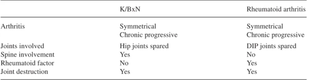
K/BxN Rheumatoid arthritis
Arthritis Symmetrical Symmetrical
Chronic progressive Chronic progressive
Joints involved Hip joints spared DIP joints spared
Spine involvement Yes No
Rheumatoid factor No Yes
-β genes resulting in relatively reduced levels of transgene expression. The TCR
transgene-bearing T cells are functionally impaired with weak responses against p41–61 of bovine RNase. Nevertheless, their expression of surface markers was slightly increased, demonstrating activation in vivo. Kouskoff et al. [22] even suc-ceeded in examining synovial T cells, which showed an enrichment of CD4+T cells,
and among the CD4+population an enrichment for high levels of transgene
expres-sion. Due to the low cell numbers, no functional studies could be undertaken. Further proof of the T cell dependence of the arthritis was demonstrated by injecting the K/BxN mice with a non-depleting anti-CD4 antibody. When administered at the lat-est 5 days before arthritis onset, disease could be blocked completely. Antibody in-jection at later time points did not change the course of the arthritis. This latter find-ing indicated that the arthritis development was dependent on TCR transgene-posi-tive T cells early in the pathogenesis but not at later stages when clinically apparent disease had already developed.
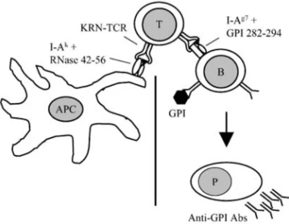
Concerning the T cell specificity, it was initially found that the R28 TCR showed alloreactivity to NOD antigen-presenting cells [22]. This was proposed as the basis of arthritis development. However, later it became clear that T cells in the K/BxN mice were actually autoreactive against glucose-6-phosphate isomerase (GPI) in the context of the self-MHC molecule I-Ag7[2, 29] (Fig. 1). GPI is a ubiquitous enzyme
of the glycolytic pathway and will be discussed in the context of the autoantibody production in K/BxN mice.
The role of B cells
The role of B cells in the pathogenesis of arthritis in the K/BxN mice was initially assessed by introducing the µMT° mutation, which resulted in an absence of mature Fig. 1. Dual specificity of the KRN TCR responsible for an autoimmune response. The KRN T cells (T)
bear a transgenic TCR which has a dual specificity for the bovine RNase peptide 42–56 in the context of I-Ak(shown in the left half of the figure), as well as for the peptide GPI 282–294 in the context of I-Ag7
(right half) [2]. In K/BxN mice B cells (B) displaying I-Ag7 can function as antigen-presenting cells
(APC) and present the autoantigen peptide GPI 282–294 to specific T cells and in turn receive help to dif-ferentiate into plasma cells (P) producing the arthritogenic anti-GPI autoantibodies (GPI glucose-6-phos-phate isomerase)
B cells in the K/BxN mice. K/BxN mice devoid of B cells did not develop arthritis [22]. Further evidence for the mechanism by which B cells are involved in the patho-genesis of arthritis came from arthritis transfer experiments. Lymphocyte-deficient C57BL/6-RAG (B-RAG) mice were injected with splenocytes from K/BxN donors. The recipient mice developed arthritis, however K/BxN splenocytes depleted of either T or B cells were unable to induce arthritis upon transfer into B-RAG mice. Transfer experiments also demonstrated that arthritis is dependent on I-Ag7
expres-sion on B cells. This clearly indicated that B cells were directly involved in the pathogenesis of arthritis via an I-Ag7-restricted recognition mechanism [21]. The fact
that CD40 deficient K/BxN mice were protected from arthritis suggested that B cells were activated, presumably by CD4+T cells via a CD40L-CD40 interaction [21]. To
find out whether a specific B cell product could initiate arthritis, serum from K/BxN mice was transferred into various hosts. Arthritis developed in B cell-deficient as well as normal C57BL/6 hosts, with histological features resembling the spontane-ously occurring disease in K/BxN mice. The main difference was that the serum transfer-induced arthritis was only transient. Joint inflammation starts to subside between 15 and 30 days after serum transfer. Histological analysis 35 days after the serum transfer revealed almost complete disappearance of inflammatory infiltrates and intact cartilage with signs of regeneration. However, arthritis could be main-tained by repeated injection of serum. The IgG fraction of the serum was shown to be responsible for the arthritis induction, indicating the existence of arthritogenic IgG in K/BxN serum [21].
Specificity of arthritogenic immunogobulins
In 1999 Matsumoto et al. [29] described the identification of the arthritogenic IgGs. The glycolytic enzyme GPI was found to be the target of both the disease-inducing T cells as well as the pathogenic IgGs. Analysis of the repertoire of arthritogenic IgGs in the K/BxN model revealed high frequencies of GPI-specific B cell clones in the spleen and other lymphoid organs. For induction of arthritis anti-GPI monoclonal an-tibodies (mAbs) needed to be injected as pools of at least two different mAbs, which recognized different epitopes [27]. Furthermore, arthritis development was depen-dent on the presence of mAbs of the IgG1 isotype in the pools. The requirement of more than one mAb for disease induction was previously reported for the induction of collagen-induced arthritis (CIA), where mAbs of the IgG2 isotype were more effi-cient in inducing arthritis [43]. In general, the more individual mAbs injected the more efficient was the disease induction. This suggested that the formation of large immune complexes enhanced the pathogenicity.
The role of innate immune mechanisms
With the identification of GPI as the autoantigen for CD4 T cells and B cells, a picture emerged in which a break of tolerance leads to the activation of the adaptive immunity resulting in the production of an arthritogenic autoantibody. How this autoantibody pro-duction translates into inflammatory and destructive events in the joints is still unclear. A detailed analysis of the genetic influences on the end-stage effector phase of the ar-thritis in K/BxN mice revealed a prominent role of the C5 locus, coding for the
comple-ment component C5, of the chromosome 2 [14]. The subsequent evaluation of the com-plement system revealed that C5-deficient animals did not develop antibody-induced ar-thritis, and treatment of wild-type mice with anti-C5 mAb could even reverse ongoing disease [15]. Further analysis of the complement pathways demonstrated that the activa-tion of the classical pathway was not needed for arthritis inducactiva-tion, since C4-deficient mice developed arthritis to the same extent as wild-type recipients [15, 42]. In contrast, in the absence of factor B, a member of the alternative pathway, most of the animals did not develop arthritis and in the ones that did it was very weak [15]. How the alternative pathway is activated is still unknown and will be discussed below.
There is a dual requirement for the alternative complement pathway as well as for the presence of FcγR. The finding that injection of serum of arthritic K/BxN mice
in-to FcRγ-deficient mice did not induce arthritis suggests that the pathogenic action of
anti-GPI mAbs depends on FcγR activation [7, 15, 23]. The high-affinity FcγRI as
well as the low-affinity FcγRIII employ the common γ-chain FcγR. Whereas a FcγRI
null mutation was without influence on the arthritis, in the absence of FcγRIII the
in-flammatory response was attenuated compared to wild-type mice, but not completely abolished, indicating the involvement of another receptor depending on the common
γ-chain [7, 15]. Interestingly, FcγRIIIA gene polymorphisms have previously been
associated with susceptibility to RA [34].
FcγR is expressed on mast cells, neutrophils, macrophages and NK cells. Upon
FcγR engagement these cells are activated and secrete tumor necrosis factor (TNF-α)
and interleukin-1 (IL-1), as well as chemokines [38]. FcγR activation (predominantly
through FcγRIII) by GPI-anti-GPI immune complexes could, therefore, lead to the
production of inflammatory cytokines and degradative enzymes by macrophages, neutrophils and mast cells, resulting in arthritis in the K/BxN mice. In this model, mast cell activation and degranulation may heavily contribute to neutrophil recruit-ment and the acute phase of paw swelling, and macrophages may play a role later in the pathogenesis of disease [7, 8, 25]. Thus, the results indicate a prominent role of the pathways of the innate immune system in the pathogenesis of arthritis in the K/BxN model.
Inflammation and joint destruction in K/BxN mice
Although the pathogenesis of RA is still unknown, it is well established that the in-flammatory cytokines TNF-α and IL-1 play an important role in the development of
RA. Clinical studies using anti-TNF-α mAbs, soluble TNF-α-receptor (TNFR) as
well as anti-IL-1β mAbs have clearly shown a beneficial effect in a majority of RA
patients [6, 26, 45]. Early studies in K/BxN mice revealed an increased production of IL-6 and TNF-α in arthritic joints [22]. Given the importance of TNF-α in RA, the
effect of TNF-α blockers were determined in K/BxN mice. Injection of anti-TNF-α
mAbs did not alter the course of arthritis in K/BxN mice [16, 23]. However, the rela-tively mild arthritis following K/BxN serum transfer showed a reduced incidence in TNF-α-deficient recipient mice [16]. This finding suggested a role for TNF-α,
al-though not an absolute requirement, in the arthritis development. Interestingly, envi-ronmental rather than genetic influences governed the susceptibility of the
TNF-α-deficient recipients to arthritis upon serum transfer. The fact that arthritis in
ani-mals deficient for TNFR was similar to controls [16, 23] suggests the involvement of other TNF-α-dependent signaling pathways, using so far non-identified TNFR.
In contrast to the situation in TNFR-deficient mice, IL-1R gene-targeted mice did not display any signs of arthritis after serum transfer [5, 16]. This result is similar to other murine arthritis models [37, 44, 49], establishing a crucial role for this cytokine in arthritis. The dominant role of IL-1 might be explained by the fact that IL-1 seems to function in series with TNF-α. TNF-α can induce IL-1 production by
synovio-cytes and IL-1 blockade is able to inhibit arthritis in TNFα transgenic mice [3, 37].
However, in RA patients IL-1 antagonists are slightly less effective than TNF-α
in-hibitors [11]. In the K/BxN serum transfer model, the administration of Toll-like re-ceptor-4 (TLR-4) agonist was able to circumvent the requirement for signaling through the IL-1R [5], suggesting that targeting IL-1 may be insufficient to prevent signaling through the common pathway that the TLRs and the IL-1R share.
Macrophages are considered the primary source of proinflammatory cytokines in the joints. However, neutrophils, which are abundant in inflamed joints, are also able to secrete TNF-α and IL-1 in addition to degradative enzymes. Administration of
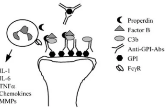
neutrophil-depleting antibodies to recipient mice before or concurrent with K/BxN serum transfer resulted in a complete inhibition of disease [46]. Surprisingly, even established joint inflammation could be reversed with neutrophil-depleting antibod-ies administered as late as 3 days after serum transfer [46]. Thus, neutrophils seem essential for the induction of joint inflammation in the K/BxN serum transfer model. The chemotactic effects of C5a-C5a receptor ligation attracts neutrophils to sites of inflammation [12]. These in turn produce properdin, which binds C3b-IgG complex-es, leading to association with factor B, resulting in activation of the alternative path-way of complement [15, 41] (Fig. 2).
Subsequent inflammatory mechanisms are felt to result in focal bone erosions, which are a hallmark feature of RA. In both the KRN serum transfer model and hu-man RA there is a dissociation between visually detectable joint swelling and the progression of bone erosions [7, 26, 31, 33, 36]. Strong evidence indicates that os-teoclasts are involved in the erosive process. Analysis of the subchondral sites of erosions in RA patients revealed acid phosphatase-positive multinucleated cells [4], and in situ hybridization studies showed mRNA expression of tartrate resistant acid
Fig. 2. Activation of the innate immune system by arthritogenic anti-GPI antibodies. GPI is deposited on
the cartilage surface in the joints and anti-GPI autoantibodies bind to the deposits, forming immune com-plexes. Because of a relative lack of complement regulatory proteins on cartilage surface, complexes with C3b can form. Properdin produced by neutrophils immigrating into the joint then enables binding of factor B resulting in activation of the alternative pathway of complement. Inflammatory cells recruited into the joint are activated by the immune complexes via their FcγR to produce inflammatory cytokines,
phosphatase and cathepsin K at sites of bone erosions, which are typical markers for osteoclasts [9, 13]. Osteoclast differentiation is under the control of TNF-related ac-tivation-induced cytokine (TRANCE), also known as receptor activator of NF-κB
li-gand (RANKL), osteoprotegerin lili-gand or osteoclast differentiation factor [1, 24, 48, 50]. Hence, osteoclasts are absent in TRANCE/RANKL- deficient mice, which dis-play severe osteopetrosis [20]. Transfer of K/BxN serum into TRANCE/RANKL-deficient mice resulted in paw swelling, which was clinically similar to control mice. However, in contrast to the inflammation, bone erosions were dramatically reduced as assessed by micro-CT and histopathological analyses [36]. These results support previous findings in adjuvant arthritis in mice treated with osteoprotegerin [19]. In the light of the critical role of osteoclasts in focal bone erosion, targeting osteoclast differentiation by inhibition of TRANCE/RANKL signaling may represent a promis-ing therapeutic strategy in RA.
Mechanisms responsible for joint specificity
Since the autoantigen GPI is present in all tissues, normally in the cytoplasm but also in soluble form in human serum, the question of the joint specificity arises. Several mechanisms could hypothetically explain the joint specificity of the anti-GPI re-sponse. One simple explanation would be overexpression of GPI in the joints. How-ever, reverse transcription-PCR analysis of GPI mRNA from joints revealed no dif-ferences in GPI transcript levels in joint versus kidney. The sequences of the GPI transcripts in the joints did not differ from the reference sequence in the GenBank, which ruled out that particular GPI isoforms could be responsible for the joint speci-ficity [29, 30]. Moreover, two-dimensional polyacrylamide gel electrophoresis did not reveal evidence for translational and post-translational modifications of GPI in the joints. GPI was expressed homogeneously in all cells including chondrocytes and synoviocytes, as assessed by immunohistochemistry [30]. In addition, GPI was de-tectable extracellularly on the cartilage surface in K/BxN mice and to a lesser extent in normal mice. Double staining with antibodies against IgG confirmed the presence of immune complexes in arthritic K/BxN but not in normal mice [30]. Interestingly, immune complexes were detected also in extra-articular tissues of K/BxN mice, but, in contrast to the joints, the complement component C3 did not colocalize, suggest-ing that extra-articular immune complexes do not fix complement [22, 30]. These immune complexes form locally in the joint, but also circulate in the serum. Injection of preformed GPI immune complexes into naïve mice, however, did not confer ar-thritis, in contrast to injection of anti-GPI antibodies [30]. Consistent with this find-ing, visualization of injected anti-GPI antibodies showed accumulation in the joints within minutes, arguing against the need for a prior immune complex formation in the serum [47].
Based upon these results, Matsumoto et al. [30] put forward the hypothesis that GPI, forming deposits on the cartilage surface in the joints, possibly binding to carti-lage proteoglycans via multiple low affinity interactions, leads to the local formation of immune complexes [30]. The cartilage surface differs from other tissue surfaces in its relative lack of complement-regulatory proteins, such as the cell membrane-bound C3 inactivator decay-accelerating factor and membrane cofactor of proteoly-sis. This could explain the increased susceptibility to the activation of the alternative complement pathway. Taken together the existing evidence indicates that the
proper-ties of the joint cartilage rather than the antigen is responsible for the joint-specific autoimmune manifestations. The findings in the K/BxN mice also demonstrate that, surprisingly, in this autoimmune arthritis the adaptive immune system activates in-nate immune mechanisms rather than the other way round, which is usually seen dur-ing an immune response against invaddur-ing microorganisms.
Relevance for human RA
In spite of great research efforts, the etiology and pathogenesis of RA remain largely in the dark. When rheumatoid factor (RF) was discovered, RA was believed to be caused by autoantibodies. However, RF was found in chronic infections, neoplasms and even in normal individuals. Additionally, the transfer of serum of patients with active RA did not induce disease in the recipients, which argued against a causative role of RF in the pathogenesis of RA [10]. Later it emerged that the expression of the HLA alleles DRB1*0401, DRB1*0404 and DRB1*0101 are associated with severity of RA [32]. Since MHC class II molecules govern positive and negative selection of CD4 T cells in the thymus, a decisive role for these cells as pathogenic effectors was postulated. However, up to the present day no autoantigen with a causal relationship to RA could be identified. Based upon animal models of arthritis type II collagen, heat shock protein 60, human chondrocyte gp39 and others have been proposed as autoantigens. However, only a minority of RA patients have detectable autoantibod-ies against these putative autoantigens. The new arthritis model by Benoist and Mathis now adds GPI to the list of potential autoantigens. Several groups have exam-ined the presence of anti-GPI antibodies in patients with RA and other forms of arthritis. Schaller et al. [39] reported increased anti-GPI titers in 64% of 69 RA patients but not in patients with Lyme arthritis or Sjögren’s syndrome in serum. Cloning of anti-GPI IgGs from RA patients revealed somatic mutations and high re-placement to silence ratios, which indicates an antigen-driven affinity maturation of the autoantibodies. Moreover, they detected significantly increased GPI concentra-tions in RA serum and synovial fluid as compared to Sjögren’s sera and osteoarthritis synovial fluids [39]. These findings contrast those of other groups. Schubert et al. [40] reported only 2 of 61 sera of RA patients as positive for anti-GPI antibodies us-ing recombinant human GPI [40] and similarly, Kassahn et al. [17] studied anti-GPI antibodies in 462 sera of patients with various rheumatic diseases, and detected reac-tivity to GPI in only a few cases of RA as well as in other diseases. Subsequent re-evaluation of their data by Schaller et al. revealed that a large proportion of the pa-tient population they had studied suffered from Felty’s syndrome, which may explain the higher percentage of anti-GPI-positive sera they found. In view of these conflict-ing results, the role of anti-GPI autoantibodies in the pathogenesis of RA is not clear and needs further investigation. Should a clinically identifiable subgroup of RA pa-tients have high titers of anti-GPI antibodies this would argue for an important pathogenetic role of GPI as an autoantigen in RA and implicate new therapeutic op-tions. Anti-GPI Fab fragments administered either systemically or locally into affect-ed joints could be usaffect-ed to block the binding of pathogenic anti-GPI antibodies to GPI deposited in the joint, and thereby prevent FcγR-binding and complement activation.
Moreover, the fact that autoantibodies recognizing ubiquitous antigens cause joint-specific disease revived the interest in B cells as important effector cells in RA, as has been assumed in earlier years following the discovery of RF. Based on the
da-ta with the K/BxN mouse model, B cell-directed therapies could be beneficial in RA. In addition to B cell-depleting therapies, strategies aimed at inhibiting B cell activa-tion seem very promising. Studies in K/BxN mice have shown that blockade of the CD40-CD40L interaction can prevent arthritis development [23]. Transplantation experiments in primates indicate that blockade of B cell costimulation induces a long-term effect without a need for continuous administration of the blocking drug [18]. Treatment of established arthritis would, however, require additional adminis-tration of an anti-inflammatory agent, at least initially, as long as preformed arthrito-genic antibodies are present in the joint. Candidates include agents interfering with the components of the innate immune system providing the link between the activat-ed adaptive immune system and downsteam events of the joint disease, complement components and FcγRIII. In spite of the unclear relevance of the K/BxN model of
arthritis for RA, it will certainly prove useful in the future to test new therapeutic approaches based on the results of studies on the basic pathogenetic mechanisms involved in the arthritis induction.
Conclusions
The new arthritis model K/BxN displays many similarities to human RA (Table 1). The arthritis occurs spontaneously with a penetrance of 100% and is strictly associat-ed with a particular MHC class II allele, the I-Ag7 derived from the NOD
back-ground. Arthritis development is dependent on CD4+T cells and B cells, both
specif-ic for GPI, a ubiquitous enzyme of the glucose metabolism. CD4 T cell-dependent B cell activation by GPI, together with a costimulatory signal via CD40L-CD40 inter-action leads to the production of arthritogenic anti-GPI autoantibodies. How antibod-ies against GPI can induce arthritis is incompletely understood. The available data, however, indicate a crucial involvement of the innate immune system. Arthritis does not develop in FcγR knockout mice, nor in mice deficient for components of the
al-ternative pathway of complement. Similarly, neutrophil- and mast cell-deficient mice show minimal signs of joint inflammation after transfer of arthritogenic antibodies. GPI is deposited on the surface of the joint cartilage, which serves as a ‘molecular sink’ for various proteins. Elegant studies showed binding of anti-GPI antibodies to GPI on the cartilage surface in vivo, forming local immune complexes. In contrast to other organs, e.g. kidney, there is complement activation on the cartilage surface, probably due to a lack of complement regulatory proteins. Activation of the alterna-tive pathway of complement then leads to a local inflammatory reaction. Thus, the joint specificity of the autoantibodies is determined by the properties of the target tis-sue rather than by the expression or distribution of the autoantigen. Whether the situ-ation in the K/BxN mouse model resembles the pathogenic events in RA is unclear. Anti-GPI antibodies have been detected in a proportion of RA patients but also in other forms of arthritides. It is controversial whether there is a predominance in RA, but some evidence suggests that there are significant anti-GPI titers in patients with Felty’s syndrome.
Although the relevance of this model for RA is not known, it is of great value for arthritis research. First, the transfer of arthritogenic antibodies into mutant mice al-lows the investigation of the downstream events of the pathogenesis of RA: the in-flammatory and destructive local processes in the joint. In this respect this model is also a valuable tool to test new therapeutic applications, especially since it can be
as-sumed that these terminal effector functions are similar in the various forms of ar-thritis. Other advantages of the anti-GPI antibody-transfer-induced arthritis are the rapid onset of arthritis within a week after the transfer and the relative strain inde-pendence. The latter allows the use of various knockout and transgenic animal strains as recipients, without the need for backcrossing. Of greater importance is the question of how tolerance to the ubiquitous self-antigen GPI is lost. This break of tolerance and the subsequent activation of innate immune mechanisms represent cru-cial events not only for the development of arthritis, but also for many other organ-specific autoimmune diseases. The careful dissection of the early events in the devel-opment of the spontaneous arthritis in the K/BxN mice might therefore advance our understanding of basic mechanisms of autoimmunity, and hopefully provide new in-sight into the still mysterious pathogenesis of RA.
Note added in proof: Matsumoto et al reported a low prevalence of anti-GPI antibodies in patients with rheumatoid arthritis, thus GPI does not appear to be an autoantigen common to the majority of patients with rheumatoid arthritis (Arthritis Rheum 48: 944).
References
1. Anderson DM, Maraskovsky E, Billingsley WL, et al (1997) A homologue of the TNF receptor and its ligand enhance T-cell growth and dendritic-cell function. Nature 390: 175
2. Basu D, Horvath S, Matsumoto I, et al (2000) Molecular basis for recognition of an arthritic peptide and a foreign epitope on distinct MHC molecules by a single TCR. J Immunol 164: 5788
3. Brennan FM, Chantry D, Jackson A, et al (1989) Inhibitory effect of TNF alpha antibodies on syno-vial cell interleukin-1 production in rheumatoid arthritis. Lancet 2: 244
4. Bromley M, Woolley DE (1984) Chondroclasts and osteoclasts at subchondral sites of erosion in the rheumatoid joint. Arthritis Rheum 27: 968
5. Choe JY, Crain B, Wu SR, et al (2003) Interleukin 1 receptor dependence of serum transferred arthritis can be circumvented by toll-like receptor 4 signaling. J Exp Med 197: 537
6. Cohen S, Hurd E, Cush J, et al (2002) Treatment of rheumatoid arthritis with anakinra, a recombinant human interleukin-1 receptor antagonist, in combination with methotrexate: results of a twenty-four-week, multicenter, randomized, double-blind, placebo-controlled trial. Arthritis Rheum 46: 614 7. Corr M, Crain B (2002) The role of FcgammaR signaling in the K/BxN serum transfer model of
arthritis. J Immunol 169: 6604
8. Cramer T, Yamanishi Y, Clausen BE, et al (2003) HIF-1alpha is essential for myeloid cell-mediated inflammation. Cell 112: 645
9. Gravallese EM, Manning C, Tsay A, et al (2000) Synovial tissue in rheumatoid arthritis is a source of osteoclast differentiation factor. Arthritis Rheum 43: 250
10. Harris J, Vaughn JH (1961) Transfusion studies in rheumatoid arthritis patients. Arthritis Rheum 4: 47 11. Hochberg MC, Tracy JK, Flores RH (2002) The comparative efficacy of anakinra, etanercept,
infliximab and leflunomide when added to methotrexate in patients with active rheumatoid arthritis. Ann Rheum Dis 61 (Suppl I): 170
12. Hopken UE, Lu B, Gerard NP, et al (1996) The C5a chemoattractant receptor mediates mucosal defence to infection. Nature 383: 86
13. Hummel KM, Petrow PK, Franz JK, et al (1998) Cysteine proteinase cathepsin K mRNA is expressed in synovium of patients with rheumatoid arthritis and is detected at sites of synovial bone destruction. J Rheumatol 25: 1887
14. Ji H, Gauguier D, Ohmura K, et al (2001) Genetic influences on the end-stage effector phase of arthri-tis. J Exp Med 194: 321
15. Ji H, Ohmura K, Mahmood U, et al (2002) Arthritis critically dependent on innate immune system players. Immunity 16: 157
16. Ji H, Pettit A, Ohmura K, et al (2002) Critical roles for interleukin 1 and tumor necrosis factor alpha in antibody-induced arthritis. J Exp Med 196: 77
17. Kassahn D, Kolb C, Solomon S, et al (2002) Few human autoimmune sera detect GPI. Nat Immunol 3: 411
18. Kirk AD, Burkly LC, Batty DS, et al (1999) Treatment with humanized monoclonal antibody against CD154 prevents acute renal allograft rejection in nonhuman primates. Nat Med 5: 686
19. Kong YY, Feige U, Sarosi I, et al (1999) Activated T cells regulate bone loss and joint destruction in adjuvant arthritis through osteoprotegerin ligand. Nature 402: 304
20. Kong YY, Yoshida H, Sarosi I, et al (1999) OPGL is a key regulator of osteoclastogenesis, lympho-cyte development and lymph-node organogenesis. Nature 397: 315
21. Korganow AS, Ji H, Mangialaio S, et al (1999) From systemic T cell self-reactivity to organ-specific autoimmune disease via immunoglobulins. Immunity 10: 451
22. Kouskoff V, Korganow AS, Duchatelle V, et al (1996) Organ-specific disease provoked by systemic autoimmunity. Cell 87: 811
23. Kyburz D, Carson DA, Corr M (2000) The role of CD40 ligand and tumor necrosis factor alpha sig-naling in the transgenic K/BxN mouse model of rheumatoid arthritis. Arthritis Rheum 43: 2571 24. Lacey DL, Timms E, Tan HL, et al (1998) Osteoprotegerin ligand is a cytokine that regulates
osteo-clast differentiation and activation. Cell 93: 165
25. Lee DM, Friend DS, Gurish MF, et al (2002) Mast cells: a cellular link between autoantibodies and inflammatory arthritis. Science 297: 1689
26. Lipsky PE, Heijde DM van der, St Clair EW, et al (2000) Infliximab and methotrexate in the treatment of rheumatoid arthritis. Anti-tumor necrosis factor trial in rheumatoid arthritis with concomitant thera-py study group. N Engl J Med 343: 1594
27. Maccioni M, Zeder-Lutz G, Huang H, et al (2002) Arthritogenic monoclonal antibodies from K/BxN mice. J Exp Med 195: 1071
28. Mangialaio S, Ji H, Korganow AS, et al (1999) The arthritogenic T cell receptor and its ligand in a model of spontaneous arthritis. Arthritis Rheum 42: 2517
29. Matsumoto I, Staub A, Benoist C, et al (1999) Arthritis provoked by linked T and B cell recognition of a glycolytic enzyme. Science 286: 1732
30. Matsumoto I, Maccioni M, Lee DM, et al (2002) How antibodies to a ubiquitous cytoplasmic enzyme may provoke joint-specific autoimmune disease. Nat Immunol 3: 360
31. McQueen FM, Stewart N, Crabbe J, et al (1999) Magnetic resonance imaging of the wrist in early rheumatoid arthritis reveals progression of erosions despite clinical improvement. Ann Rheum Dis 58: 156
32. Moxley G, Cohen HJ (2002) Genetic studies, clinical heterogeneity, and disease outcome studies in rheumatoid arthritis. Rheum Dis Clin North Am 28: 39
33. Mulherin D, Fitzgerald O, Bresnihan B (1996) Clinical improvement and radiological deterioration in rheumatoid arthritis: evidence that the pathogenesis of synovial inflammation and articular erosion may differ. Br J Rheumatol 35: 1263
34. Nieto A, Caliz R, Pascual M, et al (2000) Involvement of Fcgamma receptor IIIA genotypes in sus-ceptibility to rheumatoid arthritis. Arthritis Rheum 43: 735
35. Peccoud J, Dellabona P, Allen P, et al (1990) Delineation of antigen contact residues on an MHC class II molecule. EMBO J 9: 4215
36. Pettit AR, Ji H, Stechow D von, et al (2001) TRANCE/RANKL knockout mice are protected from bone erosion in a serum transfer model of arthritis. Am J Pathol 159: 1689
37. Probert L, Plows D, Kontogeorgos G, et al (1995) The type I interleukin-1 receptor acts in series with tumor necrosis factor (TNF) to induce arthritis in TNF-transgenic mice. Eur J Immunol 25: 1794 38. Ravetch JV, Bolland S (2001) IgG Fc receptors. Annu Rev Immunol 19: 275
39. Schaller M, Burton DR, Ditzel HJ (2001) Autoantibodies to GPI in rheumatoid arthritis: linkage be-tween an animal model and human disease. Nat Immunol 2: 746
40. Schubert D, Schmidt M, Zaiss D, et al (2002) Autoantibodies to GPI and creatine kinase in RA. Nat Immunol 3: 411; author reply 412
41. Schwaeble WJ, Reid KB (1999) Does properdin crosslink the cellular and the humoral immune re-sponse? Immunol Today 20: 17
42. Solomon S, Kolb C, Mohanty S, et al (2002) Transmission of antibody-induced arthritis is indepen-dent of complement component 4 (C4) and the complement receptors 1 and 2 (CD21/35). Eur J Immu-nol 32: 644
43. Terato K, Hasty KA, Reife RA, et al (1992) Induction of arthritis with monoclonal antibodies to colla-gen. J Immunol 148: 2103
44. Van den Berg WB, Joosten LA, Helsen M, et al (1994) Amelioration of established murine collagen-induced arthritis with anti-IL-1 treatment. Clin Exp Immunol 95: 237
45. Weinblatt ME, Kremer JM, Bankhurst AD, et al (1999) A trial of etanercept, a recombinant tumor necrosis factor receptor:Fc fusion protein, in patients with rheumatoid arthritis receiving methotrexate. N Engl J Med 340: 253
46. Wipke BT, Allen PM (2001) Essential role of neutrophils in the initiation and progression of a murine model of rheumatoid arthritis. J Immunol 167: 1601
47. Wipke BT, Wang Z, Kim J, et al (2002) Dynamic visualization of a joint-specific autoimmune response through positron emission tomography. Nat Immunol 3: 366
48. Wong BR, Josien R, Lee SY, et al (1997) TRANCE (tumor necrosis factor [TNF]-related activation-induced cytokine), a new TNF family member predominantly expressed in T cells, is a dendritic cell-specific survival factor. J Exp Med 186: 2075
49. Wooley PH, Whalen JD, Chapman DL, et al (1993) The effect of an interleukin-1 receptor antagonist protein on type II collagen-induced arthritis and antigen-induced arthritis in mice. Arthritis Rheum 36: 1305
50. Yasuda H, Shima N, Nakagawa N, et al (1998) Osteoclast differentiation factor is a ligand for osteo-protegerin/osteoclastogenesis-inhibitory factor and is identical to TRANCE/RANKL. Proc Natl Acad Sci USA 95: 3597