0 Springer-Verlag 2004
Encapsulated Zosteric Acid Embedded in Poly[3-
hydroxyalkanoate] Coatings - Protection against
Biofouling
Thomas Geiger' ([XI), Pascal Delavy', Roland Hany', Jurg Schleuniger', Manfred
Zinn'
Swiss Federal Laboratories for Materials Testing and Research (EMPA): 'Laboratory for Functional Polymers, ijberlandstrasse 129, CH-8600 Dubendorf, Switzerland, *Laboratory for Biocompatible Materials, Lerchenfeldstrasse 5, CH-9014 St. Gallen, Switzerland.
e-mail: thomas.geiger@empa,ch; Fax: +41 1 821 62 44
Received: 28 February 20041Revised version: 13 April 20041 Accepted: 4 May 2004
Summary
The natural, non-toxic antifouling compound zosteric acid (ZA, p-coumaric acid sulfate) was encapsulated in polystyrene (PS) microcapsules (30 mg Z N l g PS) with an efficiency of 30 % via an in-liquid drying process. Electron micrographs showed microcapsules with smooth surfaces and a mean diameter of 200 pm. The FIB method was used to cross-section a microcapsule in order to visualize the inner capsule structure and to localize ZA via element analysis. Coatings of a biocompatible polyester, poly[3-hydroxyalkanoate-co-3-hydroxyalkenoate] (PHAE), were prepared on microscopic slides. These coatings contained dispersed ZA (PHAEIZA) or ZA- loaded PS microcapsules (PHAE/PS(ZA)). The release of ZA was monitored via conductivy measurements in water and was 4 pgcm-2d-' for PHAEIZA and 0.9 pgcm-2d-' for PHAE/PS(ZA) coatings. To follow the initial steps of biofilm formation, coated slides were exposed to activated sludge and analyzed for cell adhesion with ESEM. ZA was effective during the burst release time of the PHAE/ZA coating, but no significant differences in biofouling were observed after 48 h. This was attributed to the minimal effective release rate of ZA, which is approximately 10 pgcm-2d-'. Introduction
Marine plants are constantly exposed to harmful attacks by bacteria, spores or fungi building biofilms on the plant surfaces. In order to protect themselves some plants developed repulsion mechanisms, e.g. the seagrass Zostera marina L. (eelgrass). The eelgrass produces and continuously releases the water-soluble (8 % (wiw) in water) antifouling compound zosteric acid (ZA), p-coumaric acid sulfate. This antifoulant does not kill microorganisms but inhibits their adhesion through binding to attachment sites on cell surfaces at non-toxic concentrations [ 1-41.
The aim of our investigation was to mimic this non-toxic protection strategy in combination with a biocompatible surface, namely a coating of poly[3-
hydroxyalkanoate-co-3-hydroxyalkenaote], (PHAE). These polyesters have gained considerable significance in medical and industrial applications, which include
implant materials, scaffolds in tissue engineering, and packaging. ZA was blended with PHAE directly or, to control the release rate, encapsulated in polystyrene and embedded in PHAE, followed by preparation of coatings. Microcapsules have been widely used, e.g. in pharmacy for drug delivery [j]. Several techniques have been developed to produce microparticulate release systems for water-soluble drugs, including the solvent evaporation method [6-121, interfacial reaction techniques [ 13- 161, and suspensioil/dispersion methods [17-191. Here, we report on the successful encapsulation of ZA into polystyrene via the solvent evaporation method and on the characterization of these microcapsules. The preparation of PHAE coatings and release investigations are described. Finally, the adhesion of biological organisms on
unprotected PHAE coatings, PHAE with dispersed ZA, and PHAE with encapsulated ZA are compared.
Experimental
Materials
Bacterial poly[3-hydroxyalkanoate-co-3 -hydroxyalkenaote] (PHAE; Scheme la) with 10 % side-chain double bonds (Mn- 90'000 g/mol, Mw- 210'000 g/mol, calibration with PS-standar4 Tg = -40 "C) was synthesized with Pseudomoizas putida GPol (ATCC 29347) in chemostat cultures under multiple nutrient limited growth conditions [20,2 I]. ZA was obtained from R. C. Zimmermann, Moss Landing Marine
Laboratories, Moss Landmg CA, USA. Polystyrene (Mw- 23O'OOO g/mol, Mn- 140'000 g/mol, Tg = 94 "C) was obtained from Aldnch and poly(viny1 alcohol) (PVA 3-98, Mw- 16'000 g/mol) from Fluka. All other reagents were used as purchased
form Aldrich or Fluka.
Scheme 1 : a) Poly[3-hydroxyalkanoate-co-3-hydroxyalkenaote] (PHAE); m= 0.1; n= 0.9; x= 2 , 4,6; y= 2,4. b) Zosteric acid@-coumaric acid sulfate).
Instruments
The particle size distribution of the microcapsules was measured with a Beckman coulter particle size analyzer LS230. The morphology examination and the elementary analysis of the microcapsules surfaces were carried out with a LEO electron microscopy 1455, equipped with an Oxford Instruments EDS system at 10 kV. Therefore, the samples were coated with a 5
nm
Pt-layer using a BAL-TEC MED 020 Coating System. Cross-sections of the microcapsules were prepared by a dual beam focused ion beam (FIB) milling t e c h q u e with a FEI Strata DB 235 field emission scanning electron microscope (FEG-SEM; &+-ions at 30 kV and 1000 PA). P m - coatings were applied with a Sogolee HP-200 air brush at 2-3 bar. Photographs ofpolished cross-sections of PHAE coatings, which were fixed in epoxy resin, were taken with an Axiovert lOOA (Zeiss) system. The release of ZA was monitored via conductivity by a WTW inoLab pWCond Level3 with a WTW TetraCon 325 probe and a MultiLab pilot program. The biofilm formation was visually analyzed with an environmental electron microscope (Amray Eco-SEM 3200 C) at 150 Ton and 20 "C. Preparation of microcapsules and coatings
Microcapsules were prepared via an "in-liquid-drying process" in a temperature controlled reactor of 250 mL equipped with an impeller stirrer. For the oil in water (OW)-emulsion, 4 g of PS was dissolved in 16 mL methylene chloride. Subsequently, 400 mg ZA was &spersed into this solution with an Ultra Turrax (24'000 rpm, 1 min
under ice cooling). This dispersion was then poured into 160 mL deionized water containing 2 % (w/w) PVA under stirring (2'000 rpm) at 20 "C for 4 h. Solid microcapsules were filtrated, washed with deionized water, and dned under reduced pressure.
Sandblasted microscope slides (area 20 cm2) were used as substrates for all coatings. PHAE coatings containing encapsulated ZA were prepared in three steps. First, a PHAE solution (1 g PHAE dissolved in 10 mL methylene chloride) was sprayed onto microscope slides with an air brush. Second, after short drying, microcapsules (about 150 mg per slide) were evenly spread on these sticky PHAE surfaces with a small brush. Finally, an additional layer of PHAE was applied to cover the microcapsules. For the preparation of PHAE coatings containing dispersed ZA, 100 mg ZA was dispersed in a PHAE solution (1 g PHAE dissolved in 10 mL methylene chloride) with an Ultra Turrax (24'000 rpm, 1 min under ice cooling). This dispersion was sprayed onto microscope slides with an
air
brush. After application, all coatings were dried under reduced pressure at 40 "C for 12 h.Determination of the antifoulant content and its release
The loading of ZA in the microcapsules was determined by weighmg. The loaded microcapsules were dissolved in methylene chloride, ZA was extracted with water, followed by freeze drying and weighmg. The leachmg of ZA from the PHAE coatings was monitored by conductivity measurements. Therefore, the coated microscope slides were stored in Millipore water (80 mL) in a completely closed glass tube. A calibrated conductivity probe recorded the release of ZA at 23 "C.
Determination of cell adhesion
The initial steps of biofilm forination (cell adhesion and attachment) were assessed in a 5 L glass beaker with pretreated, activated sludge. Test coupons were glued with silicone to the inner wall at equal heights in order to guarantee identical test conditions. Activated sludge was settled for 30 min and the supernatant was discarded. The sediment was homogenized with an Ultra T m x at 24'000 rpm for 4 min and resuspended with a filter sterilized salt solution to the original volume. One liter of salt solution contained 85 mg KH2PO4, 217.5 mg K2HP04, 266 mg Na2HP04, 25 mg m C 1 , 22.5 mg MgS04*7H20, 27.5 mg CaC12, and 0.25 mg FeC13*6H20 in demineralized water. The biomass content of the culture was determined by weight measurements and was 4.6
gL-'.
The test coupons were exposed to t h s sludge for 48h. The coupons were sampled with a tweezers, dipped in sterile 0.9 % (w/v> NaC1, and stored in 2.5 % glutaraldehyde at 4
"C
until further analysis by ESEM.Results and discussion
Characterization of the microcapsules
This work demonstrated that the in-liquid-drymg process was successful to encapsulate the hydrophilic sodium salt of ZA in a lipophilic PS matrix. Due to the high viscosity of the PSIZA dispersion, the salt was hindered to completely dissolve in the water phase of the OW-emulsion during the microcapsule formation and hardening. The particle size measurement of the microcapsules revealed a monomodal, relatively narrow size distribution with a mean capsule diameter of 200
pm (Figure 1). The surfaces of the capsules were smooth without failures proven by SEM (Figure 2).
0.1 1 10 loo 1m
Particle Diameter I lun
Figure 1. Particle size distsibution of PS microcapsules, the mean diameter is 200 pm
Figure 2. Electron micrograph of PS
microcapsules.
The
encapsulation efficiency of water-soluble compounds is generally low [5, 61. We determined, after dissolution of the microcapsules in methylene chloride followed by water-extraction of ZA, a loading of 3 % (w/w) ZA in polystyrene capsules. This correspondsto
30 % of the maximum loading, given by the amounts of ZA (400 mg) and PS (4 g) used in the production process.We were interested to know where ZA was localized inside the microcapsule and to visualize the inner microcapsule structure.
Common
methods to open microcapsules are freeze-fracture with following SEM investigations[S,
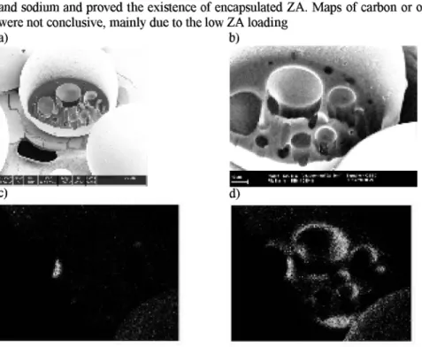
61. We used the FIB method to cross-section a microcapsule. This method has become only recently an important tool for semiconductor device modification, as well as local micro-cross sectioning.An advantage of FIB cross-sectioning is that it can be carried out under permanent SEM control and that the Ga+-ion beam produces smooth cross-sections in the submicrometer range. Figurc 3a shows an electron micrograph of a cross-section of a microcapsule cut by
FIB.
Although the P€IAE/ZA dispersion was treated with ultrasound in order to remove air bubbles, the core of the PS capsule contained still empty caverns. No ZA crystals could be detected inside the microcapsule. The cross- sectioned microcapsule was therefore transferred to a SEM with EDS system for sulfur and sodium elemental analysis. The brighter areas in the electron backscatter images gave first hints of the existence of heavier elements (Figure 3b). Quantitativeelement mapping with the EDS system (Figures 3c,d) clarified the position of sulfur
and sodium and proved the existence of encapsulated ZA. Maps of carbon or oxygen were not conclusive, mainly due to the low ZA loading
Figure 3. a) Electron micrograph of a microcapsule cross-section prepared by FIB milling, b) electron backscatter image, c) sulfur element map of the cross-section region, d) analogous sodium element map.
P M E coatings and release
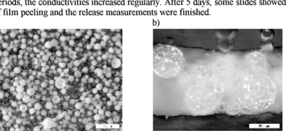
ZA encapsulated microcapsules were embedded in a PHAE coating (named PHAEDJS(ZA)). Figure 4a shows this coating applied to
an
objective slide (area 20 cm2, approximately 4.5 mg ZA per slide), and Figure 4b its cross-section, respectively. The film thickness was about 450 pm. In contrast, the film thickness ofthe PHAE coating with dispersed ZA (named PHAEIZA) was only about 50 pm (approximately 10 mg ZA per slide). Two slides of PHAEPS(ZA) and one slide of PHAE/ZA were separately immersed in Millipore water (80 mL), and the release of
ZA was monitored by changes in the water conductivity. In keeping with the biofilm formation experiments (see below), Figure 5 displays the time-course of the conductivity of both PHAE coatings for the initial 48 h. After
a
burst release, observable by a short increase of the conductivity during the fast few hours, encapsulated ZA was released atan
almost constant rate. After 48 h the release (per slide) of dispersed ZA was nearly four timesthan
the release of encapsulated ZA. Due to the high glass transition temperature and the hydrophilicity of PS, the leaching ofZA out of the coating was hindered. On the other hand, the pure
PHAE
coating, whichhas a low glass transition temperature, had
a
smaller barrier effect against water. Overall, 0.9 pgcm-’d*’ encapsulated ZA and 4 pgcm-2d-’ dispersed ZA were released. This corresponds to approximately 0.8 % and 1.6YO
of ZA applied. For longerJ 1.6- 1.4- -L . E 1.2-
z
.
.
1.0-g
0.81 " 0.67periods, the conductivities increased regularly. After 5 days, some slides showed signs of film peeling and the release measurements were finished.
Figure 4. a) Photograph of a PHAE/PS(ZA) coating with embedded microcapsules, b) cross- section of this coating with a film thickness of about 450 pm.
2.2, I
2.01
1.8::l
0.0 ***** -pH- +PHwzA **** , 0 12 24 36 48 t l hFigure 5. Release of ZA &om the PHAE coatings with embedded microcapsules (PHAE/PS(ZA)) and with dispersed ZA (PHAEIZA) monitored by changes in the water conductivity.
Bio91m formation
To examine the mode of action, biofilm formation experiments on protected and unprotected PHAE coatings were carried out. Therefore, PHAE, PKAE/PS(ZA) and PHAE/ZA coatings were exposed to diluted activated sludge in an aerated
tank
reactor over 48 h, a period suffcient to follow the attachment of microorganisms and theinitial
stages of biofilm formation [22]. Electron micrographs of o r i w coatings and after 48 h are shown in Figure 6. Before exposure, the PHAE and PHAE/PS(ZA) coatings (Figures 6a and 6e) showed both smooth and undamaged surfaces. Smallpores and elevations were visible for the PHAE/ZA coating in Figure 6c. They can be attributed to ZA crystals located near the surface; these crystals dissolved during the first three hours in contact with water and small craters were formed (Figure 6d). In contrast, the unprotected and microcapsules containing coating surfaces remained unchanged (Figures 6b, 6f). The extent of cell adhesion on the three coatings after 48 h is comparable and the unprotected PHAE surface (Figure 6b) showed not sigdicant more bacterial settlement
than
the coatings containing ZA (Figure 6d, Sf). Thiscan
beexplained with the low ZA release rates determined for these coatings. The minimal effective release rate (MERR) of ZA is approximately 10
-
50 pgcm-’d-’ [3], considerably higherthan
the 0.9 pgcm-’d-’ found for the PHAE/PS(ZA) coating. Therefore, a substantial reduction in bacterial settlement should not be expected for this coating. On the other hana the release rate for the PHAE/ZA coating was 4 pgcm- d- , only slightly lowerthan
the MEER This agrees with the observation that during the first 2 h no cell attachment was observed on this coating. During this time of the burst release (Figure 5), the leaching rate was considerable higher and the antifouling2 1
properties of ZA became evident.
a) b’l
Figure 6. ESEM micrographs of original coatings and coatings after 48 h of exposure to activated sludge; a) and b) unprotected PHAE coating, c) and d) PHAE coating with dispersed 24, e ) and f ) PHAE coating with encapsulated ZA; in b), d) and f ) biological organisms are observable as small parhcles covering the coating surfaces.
Conclusion
Conventional strategies to remove a biofilm or protect a surface use highly toxic compounds that are aimed at killing the microorganisms. However, these antifoulants
are often also toxic to non-target organisms, can be accumulated through food chain amplification and cause environmental damage. This work demonstrates first results towards a more environmentally safe way to control biofouling. The flux of encapsulated ZA was lower
than
the minimal effective release rate. However, for ZAdispersed in PHAE, the antifouling effect was apparent during the first 2 hours of the burst release. Therefore, the encapsulation process and the loading with ZA have to be adjusted. If the size of the capsules is reduced, the increase of the capsule surface would presumably lead to a corresponding increase in the release rate. With the capsule being small enough (nanocapsules), it could even come close to the rate of the burst release of the ZA dispersion. Also, the PHAE hydrophilic-hydrophobic balance and the improvement of the coating adhesion are further steps to be optimized. Therefore, parts of the PHAE side-chain double bonds will be transformed into hydrophilic carboxyl or hydroxyl groups 1231, and the remaining double bonds in the coatings cross-linked through irradiation.
Acknowledgements.
The authors thank P. Gasser for the FIB cross-sectioning, M. Trottmann for his help with the elemental analysis, and E. Pletscher for producing PHAE.
References
1. Stanley MS, Callow ME, Perry R, Alberte RS, Smith R; Callow JA (2002) Phytopathology 92: 378
2. Callow ME, Callow JA (1998) Biofouling 13: 87
3. Shin
H W ,
Smith CM, Haslbeck EG (2001) Journal of Environmental Biology 22: 243 4. Todd JS, Zimmerman RC, Crews P, Alberte RS (1 993) Phytochemistry 34: 4015. Benita S (1996) Microencapsulation: methods and industnal application. Marcel Dekker, New York
6. Zydowicz N, Nzimba-Ganyanad E (2002) Polymer Bulletin 47: 457
7. Mandal TK, Bostanian LA, Graves RA, Chapman SR, Womack I (2002) Drug Development and Industrial Pharmacy 28: 339
8. Mandal TK, Bostanian LA, Graves RA, Chapman SR, Idodo TU (2001) European Journal of Pharmaceutics and Biopharmaceutics 52: 91
9. Schlicher E, Postma NS, Zuidema J, Talsma
9
He& WE (1 997) International Journal of Pharmaceutics 153: 23510. Tabata Y, Langer R (1 993) Pharmaceutical Research 10: 391
1 1. Iwata M, Nakamura Y, McGinity JW (1999) Journal of Mcroencapsulation 16: 49 12. De Rosa G, Quaglia F, Bochot A, Ungaro F, Fattal E (2003) Biomacromolecules 4: 529 13. Montasser I, Fessi H, Briancon S, Lieto J (2003) Polymer Bulletin 50: 169
14. Hirech
K,
Payan S, Carnelle G, Brujes L, Legrand J (2003) Powder Technology 130: 324 15. Yeom CK, Kim YH, Lee JM (2002) Journal of Applied Polymer Science 84: 1025 16. Yeom CK, Oh SB, Rlum JW, LeeJ
M
(2000) Journal of Applied Polymer Science 78: 1645 17. Shukla PG, Kalidhass B, Shah A, Palaskar DV (2002) Journal of Microencapsulation 19: 18. Kriwet B, Walter E, Kissel T (1 998) Journal of Controlled Release 56: 14919. Okubo M, Minami I-r, Jing Y (2003) Journal of Applied Polymer Science 89: 706 20. Duma R, Zinn M, Witholt B, Egli T (2001) Biotechnology and Bioengineering 72: 278 21. Egli T, Zinn M (2003) Biotechnology Advances 22: 35
22. Zinn M, Zinunermann RC, White DC (2000) In: Evans LV (ed) Biofilms: Recent 23. Hany R, Bohlen Ch, Geiger T, Hartmann R, Kawada J, Schmid M,
Zinn
M, Marchessault293
Advances in Their Study and Control. Harwood Academic Publishers p 361 RH (2004) Macromolecules 37: 385