HAL Id: hal-03180714
https://hal.archives-ouvertes.fr/hal-03180714
Submitted on 25 Mar 2021
HAL is a multi-disciplinary open access
archive for the deposit and dissemination of
sci-entific research documents, whether they are
pub-lished or not. The documents may come from
teaching and research institutions in France or
abroad, or from public or private research centers.
L’archive ouverte pluridisciplinaire HAL, est
destinée au dépôt et à la diffusion de documents
scientifiques de niveau recherche, publiés ou non,
émanant des établissements d’enseignement et de
recherche français ou étrangers, des laboratoires
publics ou privés.
Isolated from New World Sand Flies in Ecuador, Is the
First Representative of a Novel Clade in the Genus
Flavivirus
Cigdem Alkan, Sonia Zapata, Laurence Bichaud, Grégory Moureau, Philippe
Lemey, Andrew Firth, Tamara Gritsun, Ernest Gould, Xavier de Lamballerie,
Jérôme Depaquit, et al.
To cite this version:
Cigdem Alkan, Sonia Zapata, Laurence Bichaud, Grégory Moureau, Philippe Lemey, et al.. Ecuador
Paraiso Escondido Virus, a New Flavivirus Isolated from New World Sand Flies in Ecuador, Is the
First Representative of a Novel Clade in the Genus Flavivirus. Journal of Virology, American Society
for Microbiology, 2015, 89 (23), pp.11773-11785. �10.1128/JVI.01543-15�. �hal-03180714�
World Sand Flies in Ecuador, Is the First Representative of a Novel
Clade in the Genus Flavivirus
Cigdem Alkan,a,bSonia Zapata,cLaurence Bichaud,a,bGrégory Moureau,a,bPhilippe Lemey,d Andrew E. Firth,eTamara S. Gritsun,f
Ernest A. Gould,a,bXavier de Lamballerie,a,bJérôme Depaquit,gRémi N. Charrela,b
Aix Marseille Université, IRD French Institute of Research for Development, EHESP French School of Public Health, EPV UMR_D 190 Emergence des Pathologies Virales, Marseille, Francea; IHU Mediterranée Infection, APHM Public Hospitals of Marseille, Marseille, Franceb; Instituto de Microbiología, Universidad San Francisco de Quito,
Quito, Ecuadorc; Department of Microbiology and Immunology, Rega Institute, KU Leuven, Leuven, Belgiumd; Division of Virology, Department of Pathology, University of
Cambridge, Cambridge, United Kingdome; School of Biological Sciences, University of Reading, Whiteknights, Reading, United Kingdomf; Université de Reims
Champagne-Ardenne, ANSES, EA 4688 USC VECPAR Transmission Vectorielle et Épidémiosurveillance de Maladies Parasitaires, SFR Capsanté, Reims, Franceg
ABSTRACT
A new flavivirus, Ecuador Paraiso Escondido virus (EPEV), named after the village where it was discovered, was isolated from sand flies (Psathyromyia abonnenci, formerly Lutzomyia abonnenci) that are unique to the New World. This represents the first sand fly-borne flavivirus identified in the New World. EPEV exhibited a typical flavivirus genome organization. Nevertheless, the maximum pairwise amino acid sequence identity with currently recognized flaviviruses was 52.8%. Phylogenetic analysis of the complete coding sequence showed that EPEV represents a distinct clade which diverged from a lineage that was ancestral to the nonvectored flaviviruses Entebbe bat virus, Yokose virus, and Sokoluk virus and also the Aedes-associated mosquito-borne flavi-viruses, which include yellow fever virus, Sepik virus, Saboya virus, and others. EPEV replicated in C6/36 mosquito cells, yielding high infectious titers, but failed to reproduce either in vertebrate cell lines (Vero, BHK, SW13, and XTC cells) or in suckling mouse brains. This surprising result, which appears to eliminate an association with vertebrate hosts in the life cycle of EPEV, is discussed in the context of the evolutionary origins of EPEV in the New World.
IMPORTANCE
The flaviviruses are rarely (if ever) vectored by sand fly species, at least in the Old World. We have identified the first representa-tive of a sand fly-associated flavivirus, Ecuador Paraiso Escondido virus (EPEV), in the New World. EPEV constitutes a novel clade according to current knowledge of the flaviviruses. Phylogenetic analysis of the virus genome showed that EPEV roots the
Aedes-associated mosquito-borne flaviviruses, including yellow fever virus. In light of this new discovery, the New World origin
of EPEV is discussed together with that of the other flaviviruses.
T
he genus Flavivirus currently includes 86 viruses, of which 73 are grouped into 53 species (1). More than 40 of these flavivi-ruses are known to be pathogenic for humans and other verte-brates, in which they cause a variety of clinical diseases from mild febrile illness to severe encephalitis and/or hemorrhagic fever. Fla-viviruses are small enveloped viruses with positive-sense single-stranded RNA genomes consisting of an open reading frame (ORF) flanked by 5= and 3= noncoding regions (NCR). The poly-protein encodes three structural poly-proteins (the capsid [C], mem-brane [M], and envelope [E] proteins) and seven nonstructural (NS) proteins (NS1, NS2a, NS2b, NS3, NS4a, NS4b, and NS5) (2,3). Flaviviruses have extensive geographic distributions and di-verse arthropod vectors, and many of them infect vertebrate hosts (4). Among the arthropod-borne flaviviruses there is a correlation between phylogenetic relationships and virus-vector-host interac-tions (5–8). On the basis of virus neutralization studies and, sep-arately, the association of arthropod vectors with vertebrates, 4 major groups of flaviviruses are recognized: the tick-borne flavi-viruses (TBFVs), the mosquito-borne flaviflavi-viruses (MBFVs), no-known-vector flaviviruses (NKVs), and no-known-vertebrate-host flaviviruses (5,6,9,10).The mosquito- and tick-borne borne flaviviruses contain im-portant animal and human pathogens, including yellow fever vi-rus (YFV), dengue vivi-rus (DENV), West Nile vivi-rus (WNV), St.
Louis encephalitis virus (SLEV), Japanese encephalitis virus (JEV), and tick-borne encephalitis virus (TBEV), which, in total, annually cause millions of human infections worldwide. Subse-quently, on the basis of phylogenetic analysis of a relatively limited number of viral envelope gene sequences, the mosquito-borne flaviviruses were subdivided into the Aedes- or Culex-associated
Received 15 June 2015 Accepted 4 September 2015 Accepted manuscript posted online 9 September 2015
Citation Alkan C, Zapata S, Bichaud L, Moureau G, Lemey P, Firth AE, Gritsun TS, Gould EA, de Lamballerie X, Depaquit J, Charrel RN. 2015. Ecuador Paraiso Escondido virus, a new flavivirus isolated from New World sand flies in Ecuador, is the first representative of a novel clade in the genus Flavivirus. J Virol
89:11773–11785.doi:10.1128/JVI.01543-15. Editor: A. García-Sastre
Address correspondence to Cigdem Alkan, cgdmalkan@gmail.com, or Rémi N. Charrel, remi.charrel@univ-amu.fr.
Supplemental material for this article may be found athttp://dx.doi.org/10.1128 /JVI.01543-15.
Copyright © 2015 Alkan et al. This is an open-access article distributed under the terms of theCreative Commons Attribution-Noncommercial-ShareAlike 3.0 Unported license, which permits unrestricted noncommercial use, distribution, and reproduction in any medium, provided the original author and source are credited.
viruses. Some of the Aedes-transmitted viruses induce hemor-rhagic fevers in humans and primates, whereas many of the Culex-transmitted viruses are associated with encephalitic infection in avian species (5). In addition to NKVs, Sokoluk virus (SOKV), Entebbe bat virus (ENTV), and Yokose virus (YOKV), which share ancestral roots with the Aedes-associated MBFVs, were iso-lated from vertebrates but not from arthropods. They form a clade normally referred to as the “Entebbe bat virus group,” which is closely related to the YFV and Edge Hill virus (EHV) clades. It was previously suggested that these viruses may have lost their associ-ation with arthropods during their evolution and divergence (11). In addition to these three groups of flaviviruses which infect vertebrates, additional groups have been isolated only from mos-quitoes or sand flies and under experimental conditions appear to infect and replicate only in insect cell lines. Accordingly, they have tentatively been defined as insect-specific flaviviruses, until appro-priate taxonomic criteria are devised and approved. The cell-fus-ing-agent virus (CFAV) was the first of many insect-specific flaviviruses (classical insect-specific flaviviruses [clSFs]) to be identified. CFAV was first isolated from an Aedes aegypti cell line in 1975 (12), and its genomic sequence was characterized in 1992 (13). CFAV and a subsequently identified heterogeneous group of related clSFs form a distinct lineage in flavivirus phylogenies. These viruses have subsequently been isolated from a wide range of mosquito species in many countries throughout the world (14– 22). An additional separate group of flaviviruses that do not ap-pear to infect vertebrate cells currently consists of nine viruses: Lammi virus (LAMV) (23), Ilomantsi virus (ILOV) (24), Marisma mosquito virus (MMV) (19), Donggang virus (DONV) (unpub-lished data; GenBank accession numberNC_016997), Chaoy-ang virus (CHAOV) (25, 26), Nounane virus (NOUV) (27), Barkedji virus (BJV) (28), Nhumirim virus (NHUV) (29), and Nanay virus (NANV) (30). These nine viruses form a distinct clade that sits within the MBFV group of viruses. Moreover, flavi-virus-like genomic sequences integrated within the genomes of
Aedes mosquitoes (21,31) have also been identified. Finally, three viruses with very highly divergent genetic lineages, viz., Tamana bat virus (TABV), Ngoye virus (NGOV), and Mogiana tick virus (MGTV), are also considered to be flaviviruses because they share similar genome organizations with the recognized flaviviruses (1,
32).
The only representative of a sand fly-borne flavivirus, Saboya virus (SABV), sits in the group of mosquito-borne flaviviruses primarily associated with Aedes mosquitoes in Africa (5,33). Fla-vivirus RNA has also been discovered in phlebotomine sand flies from Algeria (34) and Portugal (unpublished data; GenBank ac-cession number HM563684). However, these sequences align closely with those of the clSFs.
We report here on the detection, isolation, complete genome sequence, and phylogenetic assignment of a novel sand fly-borne flavivirus in Psathyromyia abonnenci (Pa. abonnenci) sand flies. We propose the name Ecuador Paraiso Escondido virus (EPEV), based on the village where the sand flies were collected. We also discuss the possible significance of the discovery of a New World (NW) sand fly-associated virus that shares a common ancestral lineage with nonvectored Old World (OW) flaviviruses.
MATERIALS AND METHODS
Trapping of sand flies. Sand flies were trapped during March 2011 in the
locality of Paraiso Escondido (00°85=03⬙N, 79°17=49⬙W), Pichincha
Prov-ince, Ecuador. CDC miniature light traps were placed from dusk to dawn. Individual sand flies were identified morphologically using a microscope (35,36). The identified sand flies were pooled on the basis of species and sex with up to 50 individuals per pool and placed in 1.5-ml tubes for storage at⫺80°C.
Detection of virus. Pools of sand flies were ground in 600l of Eagle
minimal essential medium (supplemented with 7% fetal bovine serum, 1% penicillin-streptomycin, and 1%L-glutamine [200 mM]) in the
pres-ence of 3-mm tungsten beads using an MM300 mixer mill (Qiagen, Courtaboeuf, France) as described previously (37). A 200-l aliquot was used for viral nucleic acid (NA) extraction with a BioRobot EZ1-XL Ad-vanced virus extraction minikit (Qiagen) and eluted in a 90-l volume. Five microliters of this solution was used for SYBR green reverse tran-scription (RT)-PCR and seminested PCR assays with the primers and by the protocol described previously (34,38). Bands of the expected size were purified (Amicon Ultra centrifugal filters; Millipore) and directly se-quenced.
Isolation of virus. A 100-l volume of the PCR-positive sand fly pool
was inoculated onto C6/36 cell monolayers in 25-cm2tissue culture flasks after being mixed with 900l of L15 medium enriched with 1% penicillin-streptomycin, 1%L-glutamine (200 mM), 5% kanamycin, 3%
amphoter-icin B (Fungizone), and 5% tryptose phosphate broth solution. After in-cubation at room temperature for 1 h, 5 ml fresh medium containing 5% fetal bovine serum (FBS) was added. The flasks were incubated at 28°C and examined daily for the presence of a cytopathic effect (CPE).
In vitro culture of EPEV. Cell lines of different vertebrate species,
including human (SW13), hamster (BHK), monkey (Vero), and amphib-ian (XTC), were inoculated with the supernatant medium of EPEV-in-fected C6/36 cells obtained at passage 6. Two flasks were inoculated for each cell line and incubated at either 32°C or 37°C. The flasks were exam-ined daily for the presence of a CPE. A 100-l volume of the PCR-positive sand fly homogenate was also inoculated onto Vero cells. In the absence of a CPE, the cells were harvested after 7 days, and nucleic acids were puri-fied. Regardless of the absence of a CPE, 5 serial passages were performed, and each was tested by real-time RT-PCR (38) for the presence of EPEV RNA.
Mouse brain inoculation. A total of 15l of undiluted
EPEV-con-taining supernatant medium (passage 4) or 15l of EPEV-containing supernatant medium (passage 4) diluted 1:10 with minimal essential me-dium was injected intracerebrally into 2-day-old newborn OF1 mice. The baby mice were observed for 14 days and then euthanized. Nucleic acids were purified from the brain tissues and used for the detection of EPEV RNA by a specific real-time RT-PCR assay (38). Additional mice were injected with supernatant medium containing a pool of infected brain tissue from the previously infected mice. They were observed for 14 days and then euthanized, and nucleic acids were purified from the brain tis-sues and used for detection of EPEV RNA by a specific real-time RT-PCR assay (38). Veterinary Services of the Ministry of Agriculture has approved animal experimentation under the number A1301309.
Complete genome sequencing. The EPEV strain (passage 6 in C6/36
cells) and the original homogenate of the EPEV-positive sand fly pool were used independently for complete genome characterization through next-generation sequencing (NGS). Briefly, 140l of each sample was incubated at 37°C for 7 h in 30 U of Benzonase endonuclease (catalog number 70664-3; Novagen) to eliminate cellular DNA and RNA and pre-serve encapsidated viral particles. The encapsidated viral particles were then processed for RNA extraction using a BioRobot EZ1-XL Advanced viral RNA minikit (Qiagen) without an RNA carrier. Random amplifica-tion was performed using a tagged random primer for RT and using tag-specific and random primers for PCR amplification (Applied Biosys-tems). The PCR products were purified (Amicon Ultra centrifugal filters; Millipore), and quantification was done using a Qubit fluorometer. Two hundred nanograms of the sample was processed for sequencing using an Ion PGM sequencer (Life Technologies SAS, Saint Aubin, France) (39).
Analyses of the sequencing data were performed using the CLC Genomics Workbench program (v6.5; Qiagen).
Ultracentrifugation and genome circularization. The clarified
su-pernatant medium of EPEV-infected C6/36 cells was incubated at 4°C overnight in a polyethylene glycol 6000 (Sigma-Aldrich) solution, to con-centrate the virions. The concon-centrate was centrifuged at 3,000 rpm for 30 min, and the pelleted sediments were resuspended and ultracentrifuged at 30,000 rpm for 3 h using a Fiberlite F14-6⫻ 250y fixed-angle rotor with a Thermo Scientific Heraeus Multifuge X3R ultracentrifuge. The phase 4 fraction of the centrifuge tube was collected to provide the sample that was then used for viral RNA extraction. A 400-l volume was used for viral RNA extraction as mentioned above and treated with 10 U/l tobacco acid pyrophosphatase (Epicentre); circularization was performed with 5 U/l T4 RNA ligase (Ambion) at 10°C overnight. Specific primers were designed to perform RT-PCR and to amplify the extremities using an Access RT-PCR one-step kit (Promega). The positive samples were gel purified using a QIAquick gel extraction kit (Qiagen), and both strands were sequenced.
Phylogenetic analysis. Flavivirus sequences available in the GenBank
database were collected to obtain a data set including a representative of at least one sequence for each species of the genus Flavivirus available as an amino acid sequence of the complete ORF. Alignments of the amino acid sequences of the complete ORF were generated using both the Clustal W2 (40,41) and MUSCLE (42) programs, available at the EMBL server (http: //www.ebi.ac.uk/Tools/), and refined manually for comparison.
The effect of mass removal of regions of ambiguous alignment by use of the GBlocks algorithm (43) was also investigated. Phylogenetic trees were reconstructed using Markov chain Monte Carlo (MCMC) analysis implemented in the MrBayes software program (v3.1.2) (44). The analysis was performed using the WAG substitution model with a gamma-distrib-uted rate variation among sites and default priors. Five independent Markov chains were run for 10 million generations, with the first 10% of samples being discarded as burn-in. Stationarity was confirmed on the basis of effective sample sizes of⬎400 using the Tracer program (v1.4.1) (45). A maximum clade credibility tree was summarized using the Tree-Annotator program, which annotates all nodes with posterior probability support values.
Genetic distances. The amino acid distances among the
representa-tives of the flaviviruses were calculated for the complete ORF and NS5 protein by use of the p-distance method in MEGA software (v5) (46). Nucleotide distances were also calculated for the 1-kb region in the NS5 protein reported by Kuno et al. (4).
Comparison of cleavage sites, glycosylation sites, cysteine residues, and conserved motifs of EPEV and known flaviviruses. Putative cleavage
sites from amino acid sequence alignments that included EPEV and other flaviviruses were deduced and compared according to the proteolytic pro-cessing cascade pattern previously described for the flavivirus ORFs (47). Predicted glycosylation and conserved cysteine residue sites were deter-mined for EPEV and compared with those of known flaviviruses. Con-served enzymatic patterns of EPEV were also analyzed and compared with those of known flaviviruses.
Search for ribosomalⴚ1 frameshifting. Possible ribosomal
frame-shifting sites on the EPEV genome were investigated using the methods described previously (48–50).
Nucleotide sequence accession numbers. The complete genomic
se-quence of EPEV has been submitted to the GenBank database and may be found under accession numberKJ152564.
RESULTS
Trapping of sand flies and detection of the virus. One thousand
three hundred sand flies (1,150 females and 150 males) were col-lected in Paraiso Escondido village, and the following species were identified morphologically: Nyssomyia trapidoi, Psychodopygus
panamensis, Psathyromyia aragoi, Pa. abonnenci, Lutzomyia hartmanni, Trichophoromyia reburra, and a Trichophoromyia sp.
Twenty-six pools were tested by PCR for the presence of flavivirus RNA. One pool of 30 female Pa. abonnenci sand flies contained flavivirus RNA when the pan-flavivirus RT-PCR (38) was used, and on the basis of quantitative real-time PCR, the pool contained ⬎1012genome copies. Importantly, this EPEV-positive pool
con-sisted exclusively of females that were neither gravid nor en-gorged, thus precluding the possibility that a blood meal from a vertebrate host might be the source of EPEV in the tested sample. On the basis of the fact that all the other sand fly pools were negative and the positive pool contained nonengorged sand flies, it seems reasonable to assume that a single sand fly might have been positive for EPEV RNA. Thus, the evidence strongly sup-ports the concept that EPEV must have infected and reproduced efficiently in the positive sand fly. However, we cannot rule out the possibility that EPEV might have been detected in mosquitoes if they had been collected from the same area where the sand flies were trapped.
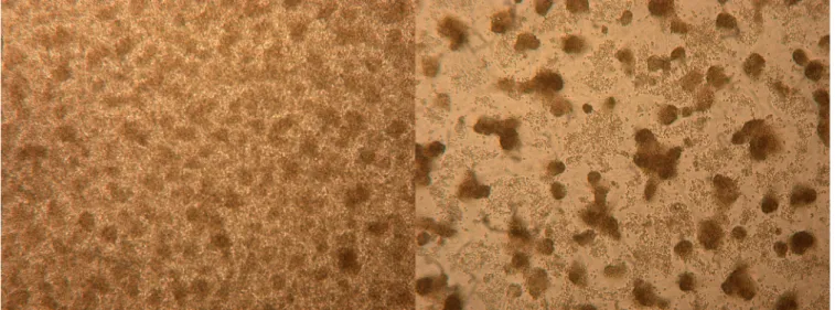
Virus isolation, mouse brain inoculation, and investigation of host range in vitro. C6/36 cells infected with the sand fly
ho-mogenate from the PCR-positive pool showed a cytopathic effect within 3 days and 1 day at the 4th passage and 6th passage, respec-tively (Fig. 1). Amplification of the virus using the pan-flavivirus RT-PCR assay (38) for the first three passages was also confirmed, with threshold cycle values being 18.32, 16.21, and 19.01, respec-tively. In order to test whether or not EPEV could infect human and/or other vertebrate cells in culture, Vero, BHK, SW13, and XTC cell cultures were inoculated with passage 6 EPEV and incu-bated at 32°C and 37°C. None showed signs of a CPE during 5 serial passages. Moreover, no CPE was detected following five se-rial passages of Vero cells inoculated with the original EPEV-pos-itive sand fly homogenate. Viral RNA detection tests using the pan-flavivirus RT-PCR assay also produced negative results, indi-cating that EPEV did not replicate in any of the vertebrate cell lines tested. Finally, newborn mice inoculated with the original EPEV-positive sand fly homogenate showed no signs of disease, and EPEV RNA could not be detected in the brains of these newborn mice. Thus, under the conditions employed in these experiments, EPEV did not infect or replicate in vertebrate cells or newborn mice.
EPEV genome strategy. (i) Complete genome sequencing and cyclization. Initially, an almost complete sequence of C6/36
cell-derived virus was obtained by NGS. Unfortunately, the sand fly homogenate-derived virus gave very poor sequence readouts after NGS. Reads with minimum lengths of 30 nucleotides and with a minimum quality of 99% per base were trimmed using the CLC Genomic Workbench program (v6.5) and mapped to refer-ence sequrefer-ences previously obtained by Sanger sequencing. Pa-rameters were set to ensure that each accepted read mapped to the reference sequence for at least 50% of its length and had a mini-mum of 80% identity to the reference sequence.
Thus, the reported sequence was derived from NGS of the C6/36 cell-derived virus and Sanger sequencing of the gaps. Miss-ing regions, includMiss-ing segments of the capsid protein, the Pr pro-tein, and the 5= and 3= noncoding regions, were successfully am-plified after cyclization of the genome and sequencing of the concatenated 3= and 5= termini. This resulted in determination of the complete genomic sequence of EPEV (GenBank accession numberKJ152564), which was 10,761 nucleotides (nt) in length and which had an ORF of 10,323 nt (positions 120 to 10,442). The single complete ORF was predicted to encode a polyprotein of
3,441 amino acids (aa). The lengths and positions of the genes or genomic regions are shown inTable 1.
(ii) Terminal dinucleotides. At each terminus of the genome,
2 nucleotides which are conserved among members of the entire
Flavivirus genus (51–53), i.e., 5=-AG and CT-3=, were detected. Mutagenesis studies have previously shown that CT-3= is probably a recognition site for the replication complex to initiate RNA syn-thesis (54,55).
(iii) 5= NCR and 3= NCR. The 5= and 3= NCRs are thought to
function as recognition sites for virus translation, replication, and possibly, assembly (56). The lengths of the EPEV 5= and 3= NCRs were 119 and 316 nt, respectively. On the basis of comparative alignment of the nucleotide sequences of all flaviviruses, the EPEV 316-nt 3= NCR is significantly shorter than that of any of the pre-viously studied flaviviruses and lacks a number of conserved se-quences found in the mosquito-borne flaviviruses. The only con-served sequence detected, concon-served sequence 1 (CS1), was 5=-C ATATTGACACCAGGGAAAAGAC-3=, which is located near the
3= terminus of the 3= NCR. This is conserved in all mosquito-borne flaviviruses. CS1 is complementary to a conserved sequence within the N-terminal coding region of the capsid protein. This complementarity results in a long-range RNA interaction that promotes cyclization of the flavivirus genome, forming a panhan-dle-like RNA structure required for viral RNA synthesis (57). In the EPEV genome, the first 12 nucleotides of CS1 are complemen-tary to a region in the capsid protein. Conserved sequence 2 (CS2) is also found in all mosquito-borne flaviviruses. The nucleotides in a short stretch prior to CS1 in EPEV showed very poor homol-ogy with the nucleotides at the equivalent positions of other flavi-viruses, and the degenerate reverse primer 3=UTR-MOS (5=-GGT CTCCWMTAACCTCTAG-3=), which anneals to CS2 (58), was unable to anneal to this related region in the EPEV 3= untranslated region.
The highly conserved pentanucleotide motif 5=-CACAG-3= at the terminus of the 3= NCR was identified. This pentanucleotide is conserved among almost all mosquito- and tick-borne flaviviruses (the exception is 5=-CACCG-3= in Murray Valley encephalitis vi-rus [MVEV]), and it has been reported to be an element essential for viral RNA synthesis, whereas its functional role is not known (59–61). It is more variable in the NKV group.
Comparison of the EPEV polyproteins with the polyproteins of all available flaviviruses. The predicted pentapeptide cleavage
sites and conserved domains of EPEV were compared with the corresponding sites of all available flavivirus polyprotein se-quences generated using amino acid sequence alignments.
(i) Cleavage sites. The predicted cleavage sites for EPEV are
shown inTable 2. A comparison of these cleavage sites with those of other flaviviruses is shown in Table S1 in the supplemental material. The similarities detected are consistent with the phylo-genetic position of EPEV, which is explained below in “Phyloge-netic analysis” and “Ge“Phyloge-netic distances.”
(a) Structural genes. The mature virion capsid protein (VirC)
is cleaved from the nascent capsid protein (AnchC) after a dibasic amino acid sequence at positions 101 and 102 and before a C-ter-minal hydrophobic domain (CTHD) by the viral serine protease (VSP) in the cytoplasm of the host cell. For EPEV the proposed residues Arg-Arg were also identified in other flaviviruses.
How-FIG 1 C6/36 cell cultures at the 6th passage after 1 day. (Left) Noninfected cells; (right) cells infected with EPEV.
TABLE 1 Genome organization of EPEV
Protein type Gene(s) or region Gene size (no. of nucleotides) Genome position Protein size (no. of amino acids) Structural 5= NCR 119 1–119 VirC 306 120–425 102 AnchC 51 426–476 17 prM 333 477–809 111 M 234 810–1043 78 E 1,476 1044–2519 492 Nonstructural NS1 1,059 2520–3578 353 NS2A 690 3579–4268 230 NS2B 393 4269–4661 131 NS3 1,872 4662–6533 624 NS4A 378 6534–6911 126 2K 69 6912–6980 23 NS4B 750 6981–7730 250 NS5 2,712 7731–10442 904 3= NCR 316 10446–10761
ever, Asn at position⫺1 has not been reported for VirC/AnchC but it was reported for other cleavage sites.
The CTHD protein is cleaved from the prM polyprotein after Cys (at position 119 for EPEV) by a host signalase (HS) in the lumen of the endoplasmic reticulum (ER). Cys at position⫺1 was not reported for flaviviruses, but it is consistent with the (⫺3, ⫺1) rule of von Heijne (62).
The prM protein is the glycosylated precursor of the M protein. Pr is cleaved from the M protein by the host cell furin or a similar enzyme (63,64) in the lumen of the ER. A specific pattern of Arg-X-(Arg/Lys)-Arg was reported for this cleavage site (65). For EPEV, we propose the signature His-Arg-(Arg/Arg)-Ser after the amino acid at position 230 or Ser-Pro-Arg/Ser-Ile after the amino acid at position 234.
The M protein is cleaved from the envelope protein after Gly at position 308 by an HS in the lumen of the ER, which is consistent with the rule of von Heijne (62).
The envelope protein is cleaved from the nonstructural NS1 protein after Ala at position 799 by a signalase (HS) in the lumen of the ER, which is consistent with the rule of von Heijne (62).
(b) Nonstructural genes. The NS1/NS2A cleavage site occurs
after Ala at position 1153, which is consistent with the rule of von Heijne (62).
The NS2A/NS2B cleavage site occurs after Arg at position 1384, which is cleaved by the viral serine protease, but Thr at position ⫺2 is not consistent with the dibasic requirement at positions ⫺1 and⫺2. Experimental evidence is required to determine whether the proposed cleavage site is functional or not.
The NS2B/NS3 cleavage site occurs after two Arg residues at positions 1514 and 1515, which are cleaved by the viral serine protease.
The NS3/NS4A cleavage site occurs after a double Arg at posi-tions 2138 and 2139, which are cleaved by the viral serine protease. The NS4A/2K cleavage site occurs after Gln and Arg at posi-tions 2264 and 2265, respectively, which are cleaved by the viral serine protease.
The 2K/NS4B cleavage site occurs after Ala at position 2288, which is cleaved by a host signalase. This is consistent with the rule of von Heijne (62).
The NS4B/NS5 cleavage site occurs after a double Arg at posi-tions 2537 and 2538, which are cleaved by the viral serine protease.
(ii) Glycosylation sites and conserved cysteine residues. The
Pr gene region contains 1 possible N-linked glycosylation site and 6 Cys residues which are conserved among other flaviviruses
ex-cept the clSFs, the E-gene region contains 2 possible N-linked glycosylation sites and 12 Cys residues which are conserved in all other flaviviruses except the clSFs, and the NS1 protein contains 2 possible N-linked glycosylation sites and 12 Cys residues which are highly conserved. Cysteine residues are required for stabilizing disulfide bridges in all mosquito-borne viruses (3).
(iii) Fusion peptide. A sequence homologous to the fusion
peptide, a 14-aa motif thought to be involved in virus fusion with cellular membranes (66), is present at positions 406 to 419 in the NS1 protein. It conforms with the Asp-Arg-Gly-Trp-X-X-(Gly/ His)-Cys-X-X-Phe-Gly-Lys-Gly motif observed for all mosquito-borne flaviviruses (67,68).
(iv) Conserved enzymatic patterns. The N-terminal sequence
of the flavivirus NS3 protein contains conserved regions (boxes 1 to 4) that have significant similarity to serine proteases and belong to the trypsin superfamily (69,70). This protease activity was re-ported to be required for the polyprotein processing of flavivi-ruses. On the other hand, the C-terminal sequence contains con-served regions (motifs I, IA, and II to VI) which are similar to RNA helicases of the DEAD family (71). These motifs are shown inFig. 2for EPEV. Compared with the sequences of other flaviviruses, these patterns are highly conserved in the EPEV genome. The EPEV NS5 protein contains the conserved sequences (motifs A to D) associated with RNA-dependent RNA polymerase activ-ity (72) and also the Gly-Asp-Asp sequence of motif C, which is the active site of the polymerase. In the N-terminal domain, two conserved motifs, viz., motifs 1 and 2, are similar to meth-yltransferases (Fig. 2).
Comparative alignment of the amino acid sequences of EPEV and all available flaviviruses. With the ultimate objective
of generating a phylogenetic tree, the nucleotide and amino acid sequences of the entire genome of EPEV were aligned with the corresponding sequences of all available flaviviruses. Despite the fact that EPEV was genetically significantly different from the other flaviviruses, the results of the alignment clearly identified EPEV to be a flavivirus. Nevertheless, the EPEV genome was distinct from the genomes of all other recognized flaviviruses in that the capsid protein contained a unique sequence of 17 amino acids from po-sitions 141 to 157. Use of the BLAST search engine revealed no equivalent sequence in any known virus or cellular sequences. However, fragments from this sequence of up to 10 amino acids matched fragments of sequences found in a variety of bacterial enzymes.
We then prepared a truncated flavivirus alignment containing the amino acid sequences of only the capsid and prM, and we attempted to align manually a 26-aa fragment that contained the 17-aa fragment of the unique EPEV sequence. This truncated alignment was manually adjusted as described previously when the untranslated regions of the flaviviruses were analyzed (73–76). Using this method, residual amino acids within the 26-aa frag-ment aligned with amino acids in the upstream region of the prM protein, particularly with those of YFV, Sepik virus (SEPV), and Wesselsbron virus (WSLV), i.e., the most closely related viruses (Fig. 3).
Phylogenetic analysis. The topology of the phylogenetic tree
based on the genomic sequence is in agreement with that in pre-viously published analyses (11,23,33,58,77,78) and clearly dem-onstrates the segregation of the major clusters consisting of the mosquito-borne, tick-borne, and no-known-vector flaviviruses with 100% support. The MBFVs showed a divergence of the two
TABLE 2 Putative polyprotein cleavage sites of EPEV polyprotein
Cleavage site Protease Amino acid sequence
VirC/AnchC VSP KKKRR/NGGTA AnchC/prM HS MMIVC/YVNAR Pr/M Furin HRHRR/RSPRS or RRSPR/SIAMP M/E HS GPAYG/THCLT E/NS1 HS TGVGA/DVGCS NS1/NS2A ? SLVSA/GNGMV NS2A/NS2B VSP WRKTR/SWPIS NS2B/NS3 VSP RSKRR/AGILW NS3/NS4A VSP AEGRR/NWTGL NS4A/2K VSP EPGCQR/SAQDN 2K/NS4B HS AGVAA/NELGW NS4B/NS5 VSP KSFRR/GKAQA
major clades, i.e., the viruses primarily associated with either
Aedes or Culex mosquitoes (as delineated inFig. 3). Interestingly, the most recent common ancestor for EPEV was shared with the nonvectored ENTV clade and the larger MBFV clade that includes SEPV, YFV, and several other viruses. It is also important to note that, apart from EPEV, in these three clades that share an ancestor, only YFV is found in the NW, where it is believed to have been introduced during the slave trade. Nevertheless, EPEV diverged from these other lineages and was clearly distinct from these other flavivirus species, the closest of which was WSLV, with which it had a genome sequence homology of 52.8%.
In view of the discovery of a unique sequence fragment in the EPEV capsid region and the knowledge that some flavivirus genes possess regions with ambiguous, highly variable sequences, it was decided to conduct two phylogenetic analyses. In the first, a tree
was constructed in which all ambiguous/highly variable sequence regions, including the unique EPEV capsid sequence, were tained in the alignment. In the second tree, these ambiguous re-gions were removed using GBlocks trimming. Despite this cau-tious approach, the overall topology, branching patterns, and support for the trees were closely similar (data not shown). More-over, the relative position of EPEV remained the same in both trees with 100% support. Independent phylogenetic analyses based on the complete ORF and, separately, the E-gene sequence also resulted in a similar position of EPEV, in which it appeared outside the ENTV and YFV clusters (data not shown). However, in trees based on the NS3 and NS5 gene sequences, EPEV was positioned between the ENTV and YFV clusters (data not shown).
Genetic distances. The pairwise distances of the amino acid
sequences between EPEV and other flaviviruses are shown in
Ta-FIG 2 Conserved motifs in the polyprotein of EPEV. The numbers 1 to 4 indicate the conserved enzymatic motifs in the proteins encoded by the NS3 and NS5
ble S2 in the supplemental material. More than 15 years ago, in a pioneer study, it was proposed that virus species and clades within the genus Flavivirus should be based on nucleotide distances using a 1-kb region of the NS5 gene (4), and cutoff values were set at
69% and 84% for discriminating clades and species, respectively. Using these values, EPEV, which is at best 68% similar to the 13 most closely related viruses (SOKV, ENTV, YOKV, SEPV, WSLV, YFV, EHV, Bouboui virus [BOUV], Banzi virus [BANV],
Uganda S virus, Jugra virus [JUGV], Potiskum virus [POTV], and SABV), would represent a new species. The maximum 68.0% pairwise nucleotide sequence identity of EPEV with these viruses was⬍69.0%, which was the cutoff value that grouped viruses in the same clade, and⬍84%, which was the cutoff to consider two viruses to be the same species. Moreover, the maximum pairwise amino acid sequence identities of the complete ORF and NS5 gene (52.8% and 65.4%, respectively) between EPEV and the YFV-re-lated clade were lower than the minimum pairwise amino acid identities of the complete ORF and the NS5 gene (61% and 69.4%, respectively) between the clades defined at that time (4). In a sep-arate analysis, the cutoff value based on complete ORF sequences was recorded to be ⬍55% amino acid sequence identity for a number of arthropod-borne flaviviruses (77), which is also higher than the maximum identity of 52.8% with EPEV.
A search for ribosomalⴚ1 frameshift sites. Many RNA virus
genomes contain specific sequences that direct a proportion of ribosomes into an alternative reading frame. Where functionally utilized, this is known as programmed ribosomal frameshifting (PRF). The most common type of PRF involves⫺1 tandem slip-page of the P- and A-site tRNAs on a slippery heptanucleotide sequence with the consensus motif X_XXY_YYZ, where X_XX represents any three identical nucleotides (with some exceptions, such as U_CC and G_GA); Y_YY represents A_AA or U_UU; Z represents A, C, or U; and underscores separate zero-frame codons. For efficient⫺1 PRF to occur, an extra stimulatory ele-ment is required, and this normally takes the form of a down-stream RNA stem-loop or pseudoknot structure separated from the heptanucleotide shift site by a spacer sequence of 5 to 9 nt (79). Ribosomal⫺1 frameshifting in the gene expression of JEV se-rogroup viruses (33,48,80) and the clSFs (i.e., CFAV, Culex fla-vivirus [CxFV], and relatives [49]) has been described previously. On the basis of comparative genomic analysis,⫺1 PRF has also been predicted to be utilized by viruses in the CHAOV-LAMV-DONV-ILOV and WSLV SEPV flavivirus clades (33,49). These frameshift sites occur within the region of the genome encoding NS2A-NS2B, with the site being conserved within a clade but not obviously conserved between clades, suggesting that⫺1 PRF in the flaviviruses may have evolved independently on several occa-sions. Analysis of EPEV for potential⫺1 PRF sites was inconclu-sive. On the one hand, the genome contains several slippery hep-tanucleotide motifs, a few of which have potential downstream RNA structures. However, predictions based on a single sequence are inadequate, as such motifs have a high probability of occurring in random sequences. None of the potential⫺1 PRF sites identi-fied aligned with the previously predicted⫺1 PRF site in the re-lated WSLV SEPV clade. Moreover, the high degree of divergence
of EPEV from all other sequenced flavivirus species precluded robust comparative genomic analyses to identify (or tentatively rule out) the occurrence of purifying selection (i.e., evidence for functionality) at other sites with a potential⫺1 PRF.
DISCUSSION
During the past 10 years, thousands of sand flies (Phlebotomus and
Sergentomyia spp.) have been collected in Europe, Africa, and the
Middle East and analyzed for the presence of flavivirus RNA. We also systematically inoculated sand fly extracts onto Vero cells. None of these analyses produced evidence of infection by EPEV or any other flavivirus, suggesting that the well-known flaviviruses are rarely (if at all) vectored by sand fly species, at least in these regions of the OW. In the current study, we analyzed pools of sand flies collected in Ecuador for the presence of flavivirus-related RNA. One pool of Pa. abonnenci sand flies yielded a PCR product, the sequence of which suggested a genetic relationship with vi-ruses in the genus Flavivirus. This PCR-positive pool of sand flies was also inoculated onto C6/36 cells, and a cytopathic virus was subsequently isolated. We have named that virus Ecuador Paraiso Escondido virus (EPEV). However, attempts to isolate the virus using Vero cells failed, implying that EPEV is another example of an insect-specific flavivirus. Thus, EPEV appears to be unique, in that it was isolated in Ecuador, i.e., the NW, from Pa. abonnenci sand flies. Detailed comparative analysis of the entire viral RNA sequence of EPEV revealed a genomic organization very similar to that of viruses in the genus Flavivirus (2,3). However, alignment of the corresponding regions of EPEV and other flaviviruses re-vealed in EPEV a unique insert of 51 nucleotides (17 amino acids) between amino acid positions 141 and 157 (as determined from the 5=-terminal starting codon), i.e., near the boundary between the viral capsid and prM genes. The lack of hydrophobic amino acids in this region indicates that this unique sequence is more representative of the newly formed N-terminal segment of the prM protein than the hydrophobic transmembrane (TM) domain of immature C protein. The TM domain is proteolytically cleaved to generate mature C and prM proteins following coordinated signalase- and NS2B/3pro
-mediated cleavage (81,82). By manually adjusting the alignment as described previously (73–76), residual amino acids within the region unique to EPEV aligned with amino acids in the upstream region of the prM protein, particularly but not exclusively with those of YFV, SEPV, and WSLV. Importantly, no similar nucleotide sequences were identified among other virus and cellular sequences when the BLAST search engine was used. This unique EPEV sequence therefore appears to contain ancestral elements of the flaviviral prM sequence which have presumably
FIG 3 Bayesian phylogeny of the ORFs of the members of the genus Flavivirus. Only posterior probabilities are included. The tree is rooted at the midpoint.
Strain names and GenBank accession numbers are given after the names of the viruses, which are abbreviated as follows: KUNV, Kunjin virus; KOUV, Koutango virus; YAOV, Yaounde virus; USUV, Usutu virus; ALFV, Alfuy virus; CPCV, Cacipacore virus; ITV, Israel turkey meningoencephalitis virus; BAGV, Bagaza virus; NTAV, Ntaya virus; TMUV, Tembusu virus; STWV, Sitiawan virus; DEDSV, duck egg drop syndrome virus; ROCV, Rocio virus; ILHV, Ilheus virus; IGUV, Iguape virus; AROAV, Aroa virus; BSQV, Bussuquara virus; NJLV, Naranjal virus; KOKV, Kokobera virus; STRV, Stratford virus; NMV, New Mapoon virus; DENV_1 to DENV_4, dengue virus serotypes 1 to 4, respectively; ZIKV, Zika virus; SPOV, Spondweni virus; KEDV, Kedougou virus; DGV, Donggang virus; UGSV, Uganda S virus; LIV, Louping ill virus; SSEV, Spanish sheep encephalitis virus; TSEV, Turkish sheep encephalitis virus; GGEV, Greek goat encephalitis virus; OHFV, Omsk hemorrhagic fever virus; LGTV, Langat virus; AHFV, Alkhurma virus; KFDV, Kyasanur Forest disease virus; RFV, Royal Farm virus; KSIV, Karshi virus; GGYV, Gadgets Gully virus; KADV, Kadam virus; MEAV, Meaban virus; SREV, Saumarez Reef virus; TYUV, Tyuleniy virus; PPBV, Phnom Penh bat virus; BCV, Batu Cave virus; MMLV, Montana myotis leukoencephalitis virus; RBV, Rio Bravo virus; JUTV, Jutiapa virus; MODV, Modoc virus; MosFV, mosquito flavivirus virus; QBV, Quang Ninh virus; Cv_theileri, Culex theileri flavivirus; NAKV, Nakiwogo virus; PCV, Palm Creek virus; KRV, Kamiti River virus; HANKV, Hanko virus. The definitions of the other abbreviations for viruses are provided in the text.
survived during the insertion and deletion process of RNA evolu-tion, as described previously (73–76).
The unique single polyprotein with conserved cleavage sites, the 5=-to-3= orientation of structural and nonstructural proteins, and the highly conserved amino acid sequence domains in the virus genome-encoded enzymes justify the inclusion of EPEV in the genus Flavivirus. The cysteine residues, 6 in the prM, 12 in the envelope, and 12 in the NS1, proteins which play an important role in protein folding through disulfide bridges, are all conserved among other flaviviruses. There are also N-glycosylation motifs which were speculated to be very important for viral replication, virulence, and maturation of viral particles (83–86) in the prM, envelope, and NS1 proteins, some of which are conserved in EPEV. EPEV has the shortest 3= NCR, with 316 nt, among the flaviviruses. It contains only conserved sequence 1, which is found in all MBFVs, whereas conserved sequence 2 was highly variable. This conserved sequence 2 was reported to have a minor effect on viral RNA synthesis but seems to decrease viral pathogenicity (87– 90).
The presence of a given virus in an arthropod does not confirm that the corresponding vector species plays a role in the natural cycle of the virus. However, in the case of the EPEV-positive sand fly pool, the Pa. abonnenci females were neither gravid nor en-gorged, and an extremely high viral load was detected. This is compelling evidence that EPEV replicates in this sand fly species. Thus, Pa. abonnenci and possibly other NW sand flies may play a direct role in the natural infectious life cycle of EPEV.
EPEV is not the first flavivirus isolated from sand flies, al-though few viruses have been found in phlebotomine flies until now. Saboya virus was isolated from sand flies in Senegal (91), although it was originally isolated from the gerbil species Tatem
itempi in Saboya Village in Senegal (92) and was later recovered from rodents of the Mastomys, Aivicantis nilaticus, and Mus
mus-culus species (93,94) and chiropters in the Republic of Guinea (95). It was also isolated from Anopheles mosquitoes and ticks (77). In addition, two flavivirus sequences were detected in
Phle-botomus perniciosus sand flies in Algeria. These formed a
mono-phyletic group more closely related to Culex-associated insect-only flaviviruses (34). Flavivirus RNA has also been discovered in phlebotomine sand flies from Portugal (unpublished data; GenBank accession numberHM563684).
The phylogenetic analysis showed that EPEV diverges from the common ancestral lineage of Aedes-associated mosquito-borne flaviviruses that include YFV and also the NKV-like flaviviruses (YOKV, ENTV, SOKV). EPEV is more distantly related to the other group of Aedes-borne flaviviruses that include dengue vi-ruses and the Culex-associated group that includes WNV, SLEV, and Zika virus. Interestingly, EPEV is also only distantly related to the clSFs (CFAV, Kamiti River virus, Hanko virus, and CxFV) and dual-host-affiliated ISFs (dISFs) (NOUV, LAMV, CHAOV, Donggang virus (DGV) and ILOV) and the NKVs that include bat- and rodent-associated flaviviruses, such as Modoc virus and Montana myotis leukoencephalitis virus. EPEV appeared alone as a lineage which was separated from a major phylogenetic clade that included the YFV, EHV, and ENTV groups. The phylogenetic analysis showed that EPEV appears at the root of these groups with 100% bootstrap support. However, the phylogenetic position among the mosquito-borne viruses of EPEV conflicts with its lack of an ability to infect and replicate in vertebrate cell cultures or newborn mice. The insect-specific flaviviruses NOUV and LAMV
also appear to infect and replicate only in insect cell lines (23,27), although they are phylogenetically grouped with mosquito-borne flaviviruses.
These data therefore support the identification of the first rep-resentative of a novel group of NW sand fly-borne flaviviruses. When the origins of EPEV and the other flaviviruses are consid-ered, most of the earliest phylogenetic lineages of the mosquito-borne viruses, tick-mosquito-borne viruses, and NKVs are found in Europe, Asia, Australia, and, in particular, Africa. On the basis of the close relationships of the NW and OW MBFVs, the current opinion is that these viruses were introduced to the NW at various times before, during, or after the period of slave trading (33). The only representative of the tick-borne flaviviruses in the NW is Powas-san virus (POWV) and its relatively recent descendant variant, deer tick virus (DTV). On the basis of historical, ecological, an-thropological, and molecular epidemiological evidence, it was proposed that POWV was introduced into the NW between 15,000 and 11,000 years ago, possibly during the tick and mam-malian migrations, when the Bering land bridge connected Asia and North America (78,96). Among the NKVs, the OW virus Apoi virus (APOIV), which roots all other NKVs, is the only ro-dent-associated virus related to the NKV group, whereas the bat-associated NKVs are found in both the OW and the NW. This supports the idea of a single dispersion into the NW, most likely through bats (11,33). It has recently been suggested that the in-troduction of viruses into the NW occurred several times during two evolutionary time periods (33). It seems highly probable that the most recent introductions occurred from the period of in-creasing slave and commercial trading across the Atlantic Ocean from the 16th to the 19th centuries. The most convincing evidence of such dispersions includes the mosquito-borne viruses YFV, DENV, and, in 1999, WNV. For each of these viruses there are historical records and robust phylogenetic data (97–102). In the case of the potentially earlier introductions to the NW, which include Aroa virus, Ilheus virus, SLEV, Cacipacore virus, and the NKVs, their dispersion appears to have occurred thousands of years before slave and commercial trading (33). Whether or not these introductions to the NW occurred during the period when the Bering land bridge connected Asia and North America cannot be determined. However, it seems unlikely, since there are no recognized OW sand flies in the NW. On the other hand, it is recognized that estimation of the time of occurrence of such as-sumed introductions is highly dependent on the choice of dates used for calibration (33,78). As analytical methods improve and more viruses are identified and characterized, it should become possible to provide more robust estimates for these virus disper-sion patterns. The genus Flavivirus comprises both vectored and nonvectored viruses, raising the possibility that arthropod-medi-ated transmission is either a derived trait or a secondary loss. The former traditional hypothesis is supported extensively (4,5,10,
77, 103,104), but the alternative hypothesis should not be dis-carded at this stage of our comprehension. The insect-specific flavivirus group consisting of NOUV, BJV, DONV, ILOV, CHAOV, LAMV, and the newly discovered NANV from Peru clusters phylogenetically with the MBFVs, but none of these in-sect-specific viruses have been found to replicate in mammalian cells (23, 24, 104). In contrast, the other nonvectored viruses, ENTV and YOKV, replicate in mosquito cells. This reflects the need for more in-depth field investigations to identify the possible arthropod vectors of these viruses. For example, the isolation of
SOKV, a close relative of YOKV, from Argasidae ticks, birds, and bats and serological evidence of human infections by YOKV were recently reported (105).
Our phylogenetic analysis places EPEV at the root of the YOKV, SOKV, and ENTV clade, which itself roots the Aedes-as-sociated mosquito-borne flaviviruses (EHV, BOUV, Uganda S vi-rus, BANV, JUGV, POTV, SABV, WSLV, SEPV, and YFV). In contrast to EPEV, all of these viruses have been found only in the OW. This finding requires a cautious interpretation before em-barking on a definitive hypothesis. As previously elaborated (106), sampling issues should be taken into consideration in generating these apparently conflicting results. The situation with EPEV is further complicated when one considers that it was isolated from a pool of NW sand flies. However, it replicates in mosquito cells but does not appear to replicate in mammalian cells, and it is ancestral to the ENTV and YFV clades. Since EPEV roots clades of OW viruses, is closely related to YFV, and has also been shown to replicate to high titers in mosquito cells, we can speculate that the vectors for the transmission cycle of EPEV in the OW may include mosquitoes. In this case, the introduction of EPEV might then be attributable to the slave trade, in common with several other fla-viviruses (33). This argument is further supported by the compel-ling evidence that the alphavirus chikungunya virus (CHIKV) caused many disease outbreaks in humans about 200 years ago in the Americas (107). It therefore seems highly likely that CHIKV was also introduced into the NW during the period of slave trad-ing across the Atlantic Ocean. In this case, however, the intro-duced CHIKV does not appear to have become permanently es-tablished in NW reservoir hosts. The alternative possibility, that EPEV was introduced into the NW via infected sand flies from the OW, seems unlikely, since OW sand fly species have never been found in the NW. Clearly, mosquito trapping and screening are required in the region where the EPEV-positive sand fly pool was collected to discover whether or not EPEV circulates among the indigenous mosquito species. If this proves to be the case, it would support the hypothesis of an early divergence of the vector-borne viruses during the evolution of the flaviviruses. Moreover, the insect-specific flaviviruses (lSFs) also replicate only in mosquito cells, and the number of viruses in this group has been rapidly increasing. Most flaviviruses are associated with mosquitoes and vertebrates, but the ISF group is associated only with mosquitoes, appearing at the root of the MBFV, NKV, and TBFV branches. The highly divergent TABV, isolated in 1973 from an insectivo-rous bat in Trinidad (108), is now included as a tentative flavivirus (1). Recently, MGTV was isolated from ticks in Brazil (32). It appears to be highly divergent but associated with the genus
Fla-vivirus on the basis of a partial NS3 and NS5 analysis. TABV and
MGTV are highly divergent from each other, but they were both isolated in the NW. Nevertheless, they are related, albeit distantly, to the other flaviviruses. Thus, together with EPEV, TABV, and MGTV in the NW, viruses in the genus Flavivirus appear to have evolved following their introduction into the NW from the OW. However, the wide variety of different flaviviruses and their attri-butes are potentially biased by the small number of representatives of different lineages that have currently been identified (24).
At this stage, we cannot rule out the possibility that EPEV may be vectored by mosquitoes. However, the results in this study sug-gest that EPEV represents a new flavivirus species and could be the first recognized representative of a novel clade including other yet-to-be-discovered sand fly-borne flaviviruses. There is support
for this proposal: (i) EPEV has been isolated from Pa. abonnenci, a species of NW sand flies that is known to be present in Bolivia, Brazil, Colombia, French Guiana, Panama, Peru, Suriname, Ven-ezuela, and Ecuador (CIPA: Computer-Aided Identification of Phlebotomine Sand flies of the Americas,http://cipa.snv.jussieu.fr
[109]), (ii) Pa. abonnenci is not listed among the species of medical interest (110), and (iii) the absence of viral replication in various vertebrate cells and in the brains of newborn mice suggests that EPEV could be an insect-only flavivirus rather than an arbovirus. Further studies are required to understand if there are other sand fly species infected with EPEV in the region where the Pa.
abonnenci samples were collected. Alternatively, if mosquitoes are
the natural vectors, it would seem sensible to look first in the region of Ecuador where EPEV was discovered.
REFERENCES
1. Pletnev A, Gould E, Heinz FX, Meyers G, Thiel HJ, Bukh J, Stiasny K,
Collett MS, Becher P, Simmonds P, Rice CM, Monath TP. 2011.
Flaviviridae, p 1003–1020. In King AMQ, Adams MJ, Carstens EB, Lefkowitz EJ (ed), Virus taxonomy. Elsevier, Oxford, United Kingdom. 2. Rice CM, Lenches EM, Eddy SR, Shin SJ, Sheets RL, Strauss JH. 1985. Nucleotide sequence of yellow fever virus: implications for flavivirus gene expression and evolution. Science 229:726 –733.http://dx.doi.org /10.1126/science.4023707.
3. Chambers TJ, Hahn CS, Galler R, Rice CM. 1990. Flavivirus genome organization, expression, and replication. Annu Rev Microbiol 44:649 – 688.http://dx.doi.org/10.1146/annurev.mi.44.100190.003245. 4. Kuno G, Chang GJ, Tsuchiya KR, Karabatsos N, Cropp CB. 1998.
Phylogeny of the genus Flavivirus. J Virol 72:73– 83.
5. Gaunt MW, Sall AA, Lamballerie X, Falconar AK, Dzhivanian TI,
Gould EA. 2001. Phylogenetic relationships of flaviviruses correlate with
their epidemiology, disease association and biogeography. J Gen Virol
82:1867–1876.http://dx.doi.org/10.1099/0022-1317-82-8-1867. 6. Gould EA, Solomon T. 2008. Pathogenic flaviviruses. Lancet 371:500 –
509.http://dx.doi.org/10.1016/S0140-6736(08)60238-X.
7. Grard G, Moureau G, Charrel RN, Lemasson JJ, Gonzalez JP, Gallian
P, Gritsun TS, Holmes EC, Gould EA, de Lamballerie X. 2007. Genetic
characterization of tick-borne flaviviruses: new insights into evolution, pathogenetic determinants and taxonomy. Virology 361:80 –92.http: //dx.doi.org/10.1016/j.virol.2006.09.015.
8. Zanotto PM, Gao GF, Gritsun T, Marin MS, Jiang WR, Venugopal K,
Reid HW, Gould EA. 1995. An arbovirus cline across the northern
hemisphere. Virology 210:152–159.http://dx.doi.org/10.1006/viro.1995 .1326.
9. Porterfield JS. 1980. Antigenic characteristics and classification of Toga-viridae, p 13– 46. In Schlesinger RW (ed), The togaviruses. Academic Press, New York, NY.
10. Gould EA, de Lamballerie X, Zanotto PM, Holmes EC. 2003. Origins, evolution, and vector/host coadaptations within the genus Flavivirus. Adv Virus Res 59:277–314.http://dx.doi.org/10.1016/S0065-3527(03)59008-X. 11. Cook S, Holmes EC. 2006. A multigene analysis of the phylogenetic
relationships among the flaviviruses (family: Flaviviridae) and the evo-lution of vector transmission. Arch Virol 151:309 –325.http://dx.doi.org /10.1007/s00705-005-0626-6.
12. Stollar V, Thomas VL. 1975. An agent in the Aedes aegypti cell line (Peleg) which causes fusion of Aedes albopictus cells. Virology 64:367– 377.http://dx.doi.org/10.1016/0042-6822(75)90113-0.
13. Cammisa-Parks H, Cisar LA, Kane A, Stollar V. 1992. The complete nucleotide sequence of cell fusing agent (CFA): homology between the nonstructural proteins encoded by CFA and the nonstructural proteins encoded by arthropod-borne flaviviruses. Virology 189:511–524.http: //dx.doi.org/10.1016/0042-6822(92)90575-A.
14. Cook S, Moureau G, Kitchen A, Gould EA, de Lambellerie X, Holmes
EC, Harbach RE. 2012. Molecular evolution of the insect-specific
flavi-viruses. J Gen Virol 93(Pt 2):223–234.http://dx.doi.org/10.1099/vir.0 .036525-0.
15. Huanyu W, Haiyan W, Shihong F, Guifang L, Hong L, Xiaoyan G,
Lizhi S, Rayner S, Aiqiang X, Guodong L. 2012. Isolation and
collected in mainland China. Virol J 9:73.http://dx.doi.org/10.1186 /1743-422X-9-73.
16. Zuo S, Zhao Q, Guo X, Zhou H, Cao W, Zhang J. 2014. Detection of Quang Binh virus from mosquitoes in China. Virus Res 180:31–38.http: //dx.doi.org/10.1016/j.virusres.2013.12.005.
17. Parreira R, Cook S, Lopes A, de Matos AP, de Almeida AP, Piedade
J, Esteves A. 2012. Genetic characterization of an insect-specific
flavivirus isolated from Culex theileri mosquitoes collected in southern Portugal. Virus Res 167:152–161. http://dx.doi.org/10.1016/j.virusres .2012.04.010.
18. Hobson-Peters J, Yam AW, Lu JW, Setoh YX, May FJ, Kurucz N,
Walsh S, Prow NA, Davis SS, Weir R, Melville L, Hunt N, Webb RI, Blitvich BJ, Whelan P, Hall RA. 2013. A new insect-specific flavivirus
from northern Australia suppresses replication of West Nile virus and Murray Valley encephalitis virus in co-infected mosquito cells. PLoS One
8:e56534.http://dx.doi.org/10.1371/journal.pone.0056534.
19. Vázquez A, Sánchez-Seco MP, Palacios G, Molero F, Reyes N, Ruiz S,
Aranda C, Marqués E, Escosa R, Moreno J, Figuerola J, Tenorio A.
2012. Novel flaviviruses detected in different species of mosquitoes in Spain. Vector Borne Zoonotic Dis 12:223–229.http://dx.doi.org/10.1089 /vbz.2011.0687.
20. Grisenti M, Vázquez A, Herrero L, Cuevas L, Perez-Pastrana E,
Ar-noldi D, Rosá R, Capelli G, Tenorio A, Sánchez-Seco MP, Rizzoli A.
2015. Wide detection of Aedes flavivirus in north-eastern Italy—a Euro-pean hotspot of emerging mosquito-borne diseases. J Gen Virol 96(Pt 2):420 – 430.http://dx.doi.org/10.1099/vir.0.069625-0.
21. Rizzo F, Cerutti F, Ballardini M, Mosca A, Vitale N, Radaelli MC,
Desiato R, Prearo M, Pautasso A, Casalone C, Acutis P, Peletto S, Mandola ML. 2014. Molecular characterization of flaviviruses from
field-collected mosquitoes in northwestern Italy, 2011-2012. Parasit Vec-tors 7:395.http://dx.doi.org/10.1186/1756-3305-7-395.
22. Goenaga S, Fabbri CM, García JB, Rondán JC, Gardenal N, Calderón
GE, Enria DA, Levis SM. 2014. New strains of Culex flavivirus isolated
in Argentina. J Med Entomol 51:900 –906.http://dx.doi.org/10.1603 /ME13172.
23. Huhtamo E, Putkuri N, Kurkela S, Manni T, Vaheri A, Vapalahti O,
Uzcátegui NY. 2009. Characterization of a novel flavivirus from
mos-quitoes in northern Europe that is related to mosquito-borne flaviviruses of the tropics. J Virol 83:9532–9540.http://dx.doi.org/10.1128/JVI .00529-09.
24. Huhtamo E, Cook S, Moureau G, Uzcátegui NY, Sironen T, Kuivanen
S, Putkuri N, Kurkela S, Harbach RE, Firth AE, Vapalahti O, Gould EA, de Lamballerie X. 2014. Novel flaviviruses from mosquitoes:
mos-quito-specific evolutionary lineages within the phylogenetic group of mosquito-borne flaviviruses. Virology 464-465:320 –329.http://dx.doi .org/10.1016/j.virol.2014.07.015.
25. Wang Z, An S, Wang Y, Han Y, Guo Y. 2009. A new virus of flavivirus: Chaoyang virus isolated in Liaoning Province. Chin J Pub Health 25: 769 –772. (In Chinese.).
26. Lee JS, Grubaugh ND, Kondig JP, Turell MJ, Kim HC, Klein TA,
O’Guinn ML. 2013. Isolation and genomic characterization of
Chaoy-ang virus strain ROK144 from Aedes vexans nipponii from the Republic of Korea. Virology 435:220 –224.http://dx.doi.org/10.1016/j.virol.2012 .10.020.
27. Junglen S, Kopp A, Kurth A, Pauli G, Ellerbrok H, Leendertz FH. 2009. A new flavivirus and a new vector: characterization of a novel flavivirus isolated from Uranotaenia mosquitoes from a tropical rainfor-est. J Virol 83:4462– 4468.http://dx.doi.org/10.1128/JVI.00014-09. 28. Kolodziejek J, Pachler K, Bin H, Mendelson E, Shulman L, Orshan L,
Nowotny N. 2013. Barkedji virus, a novel mosquito-borne flavivirus
identified in Culex perexiguus mosquitoes, Israel, 2011. J Gen Virol 94(Pt 11):2449 –2457.http://dx.doi.org/10.1099/vir.0.056200-0.
29. Pauvolid-Corrêa A, Solberg O, Couto-Lima D, Kenney J, Serra-Freire
N, Brault A, Nogueira R, Langevin S, Komar N. 2015. Nhumirim virus,
a novel flavivirus isolated from mosquitoes from the Pantanal, Brazil. Arch Virol 160:21–27.http://dx.doi.org/10.1007/s00705-014-2219-8. 30. Evangelista J, Cruz C, Guevara C, Astete H, Carey C, Kochel TJ,
Morrison AC, Williams M, Halsey ES, Forshey BM. 2013.
Character-ization of a novel flavivirus isolated from Culex (Melanoconion) ocossa mosquitoes from Iquitos, Peru. J Gen Virol 94(Pt 6):1266 –1272.http: //dx.doi.org/10.1099/vir.0.050575-0.
31. Crochu S, Cook S, Attoui H, Charrel RN, De Chesse R, Belhouchet M,
Lemasson JJ, de Micco P, de Lamballerie X. 2004. Sequences of
flavi-virus-related RNA viruses persist in DNA form integrated in the genome of Aedes spp. mosquitoes. J Gen Virol 85:1971–1980.http://dx.doi.org /10.1099/vir.0.79850-0.
32. Maruyama SR, Castro-Jorge LA, Ribeiro JM, Gardinassi LG, Garcia
GR, Brandão LG, Rodrigues AR, Okada MI, Abrão EP, Ferreira BR, da Fonseca BA, de Miranda-Santos IK. 2014. Characterisation of divergent
flavivirus NS3 and NS5 protein sequences detected in Rhipicephalus mi-croplus ticks from Brazil. Mem Inst Oswaldo Cruz 109:38 –50.http://dx .doi.org/10.1590/0074-0276130166.
33. Moureau G, Cook S, Lemey P, Nougairede A, Forrester NL,
Khasnati-nov M, Charrel RN, Firth AE, Gould EA, de Lamballerie X. 2015. New
insights into flavivirus evolution, taxonomy and biogeographic history, extended by analysis of canonical and alternative coding sequences. PLoS One 10:e0117849.http://dx.doi.org/10.1371/journal.pone.0117849. 34. Moureau G, Ninove L, Izri A, Cook S, De Lamballerie X, Charrel RN.
2010. Flavivirus RNA in phlebotomine sand flies. Vector Borne Zoonotic Dis 10:195–197.http://dx.doi.org/10.1089/vbz.2008.0216.
35. Galati EAB. 2011. Phlebotominae (Diptera, Psychodidae) classificação, morfologia, terminologia e identificação de adultos. Apostila Disciplina HEP 5752 Bioecologia e Identificação de Phlebotominae, vol I. Departa-mento de Epidemiologia, Faculdade de Saúde Pública, Universidade de São, São Paulo, SP, Brazil.
36. Young DG, Duncan MA. 1994. Guide to the identification and geo-graphic distribution of Lutzomyia sand flies in Mexico, the West Indies, Central and South America (Diptera: Psychodidae). Mem Ann Entomol Institut 54:1– 88.
37. Charrel RN, Moureau G, Temmam S, Izri A, Marty P, Parola P, da
Rosa AT, Tesh RB, de Lamballerie X. 2009. Massilia virus, a novel
phlebovirus (Bunyaviridae) isolated from sand flies in the Mediterra-nean. Vector Borne Zoonotic Dis 9:519 –530.http://dx.doi.org/10.1089 /vbz.2008.0131.
38. Moureau G, Temmam S, Gonzalez JP, Charrel RN, Grard G, De
Lamballerie X. 2007. A real-time RT-PCR method for the universal
detection and identification of flaviviruses. Vector Borne Zoonotic Dis
7:467– 477.http://dx.doi.org/10.1089/vbz.2007.0206.
39. Rothberg JM, Hinz W, Rearick TM, Schultz J, Mileski W, Davey M,
Leamon JH, Johnson K, Milgrew MJ, Edwards M, Hoon J, Simons JF, Marran D, Myers JW, Davidson JF, Branting A, Nobile JR, Puc BP, Light D, Clark TA, Huber M, Branciforte JT, Stoner IB, Cawley SE, Lyons M, Fu Y, Homer N, Sedova M, Miao X, Reed B, Sabina J, Feierstein E, Schorn M, Alanjary M, Dimalanta E, Dressman D, Kasinskas R, Sokolsky T, Fidanza JA, Namsaraev E, McKernan KJ, Williams A, Roth GT, Bustillo J. 2011. An integrated semiconductor
device enabling non-optical genome sequencing. Nature 475:348 –352.
http://dx.doi.org/10.1038/nature10242.
40. Larkin MA, Blackshields G, Brown NP, Chenna R, McGettigan PA,
McWilliam H, Valentin F, Wallace IM, Wilm A, Lopez R, Thompson JD, Gibson TJ, Higgins DG. 2007. Clustal W and Clustal X version 2.0.
Bioinformatics 23:2947–2948.http://dx.doi.org/10.1093/bioinformatics /btm404.
41. Thompson JD, Higgins DG, Gibson TJ. 1994. CLUSTAL W: improving the sensitivity of progressive multiple sequence alignment through se-quence weighting, position-specific gap penalties and weight matrix choice. Nucleic Acids Res 22:4673– 4680.http://dx.doi.org/10.1093/nar /22.22.4673.
42. Edgar RC. 2004. MUSCLE: multiple sequence alignment with high ac-curacy and high throughput. Nucleic Acids Res 32:1792–1797.http://dx .doi.org/10.1093/nar/gkh340.
43. Talavera G, Castresana J. 2007. Improvement of phylogenies after re-moving divergent and ambiguously aligned blocks from protein se-quence alignments. Syst Biol 56:564 –577.http://dx.doi.org/10.1080 /10635150701472164.
44. Huelsenbeck JP, Ronquist F. 2001. MRBAYES: Bayesian inference of phylogenetic trees. Bioinformatics 17:754 –755.http://dx.doi.org/10 .1093/bioinformatics/17.8.754.
45. Drummond AJ, Rambaut A. 2007. BEAST: Bayesian evolutionary anal-ysis by sampling trees. BMC Evol Biol 7:214.http://dx.doi.org/10.1186 /1471-2148-7-214.
46. Tamura K, Peterson D, Peterson N, Stecher G, Nei M, Kumar S. 2011. MEGA5: molecular evolutionary genetics analysis using maximum like-lihood, evolutionary distance, and maximum parsimony methods. Mol Biol Evol 28:2731–2739.http://dx.doi.org/10.1093/molbev/msr121.