TRANSACTIONS OFTHE ROYAL SOCIETY OFTROPICAL MEDICINE AND HYGIENE (1997) 91,361-363 361
PCR amplification
of DNA from malaria parasites
on fixed and stained thick and
thin blood films
Dominic Edoh’, Sylvia SteigeG, Blaise Genton and Hans-Peter Beck2 ‘Zfakara Centre, Zfakara, Tanzania; 2Swiss Tropical Institute, Base& Switzerland; 3Papua New Guinea Institute of Medical Research, Madang, Papua New Guinea
Abstract
Under some circumstances, polymerase chain reaction (PCR) amplification of deoxyribonucleic acid (DNA) from Plasmodium may become necessary from infections for which only blood slides are available. Established methods used for DNA preparation do not work in that case. We have developed a reliable and controlled method for DNA preparation from malaria parasites on fixed and stained blood films. 162 slides from 2 different locations, some stored for at least one year, have been analysed by PCR amplifica- tion of the polymorphic loci for MSAl and MSA2. In 92% of microscopically positive slides, a PCR product could be detected using material derived from thick blood films. When thin blood films with scanty parasitaemia were used, a PCR product could be obtained with only 7 1% of samples. In all unsuc- cessful cases, DNA preparation was the limiting factor, which was controlled for by amplification of a control human template.
Keywords: malaria, Plasmodium falciparum, polymerase chain reaction, blood films
Introduction
During the past few years, the polymerase chain reac- tion (PCR) has become a major diagnostic and research technique. It is valuable for the detection of parasites present at low concentrations in blood or serum sam- ples. For several parasitic species, in particular for Plas- modium species, analysis of amplified products encoding polymorphic proteins allowed the discrimination not only of species, but also of subspecies or strains (BATIK- ER et al., 1994; FELGER et al., 1994: CONTAMIN et al.. 1995; I’..& et al., 1995). Genetic traits, such as drug re: sistance, can easily be detected by PCR (PETERSON et al., 1990), provided the genetic locus is known. PCR amplification has been used with I? falciparum to study parasite diversity (F%LGER et al., 1994; CONTAMIN et al., 1995), recombination rates (BABIKER et al., 1994), multiplicity of infections (BECK et al., in press) and the development and frequency of drug resistance. But very often sample material is limited, or material is not avail- able for preparation of deoxyribonucleic acid (DNA). Historical samples, collected many years ago, are avail- able only as fixed and stained microscope slides. In or- der to be able to use such samples for retrospective analyses, we attempted to utilize material isolated from fixed and stained malaria blood films. Here we present a reliable technique for PCR amplification using fixed and stained thick and thin blood films, previously used for microscopical diagnosis of I? falciparum, as a source of DNA for amplification.
Material and Methods Samples
Blood slides were obtained from different locations; 112 were derived from the Wosera area of Papua New Guinea, an area highly endemic for I? falciparum (for details see GENTON et al., 1995). Venous blood was col- lected and thick and thin blood films were made on sep- arate slides, fixed and stained with 4% Giemsa’s stain and examined at 1000x magnification. Four hundred microscope fields of a thick film were examined before the slide was declared negative. Most slides showed scanty parasitaemia; the mean density of positive slides was 1870 parasites/& of blood (range 40-4000 para- sites). All thin films were used for PCR analysis, irre- spective of the parasitaemia determined by microscopy of the thick films.
Address for correspondence: Hans-Peter Beck, Swiss Tropical Institute, Socinsuasse 57, CH 4002 Base], Switzerland; phone +41 61 284 8116, fax +41 61 271 8654, e-mail
clu.unibas.ch beckhp@uba-
Further blood slides were collected from children in the Kilombero valley of Tanzania, an area holoendemic for I? falciparum (for details see SMITH et al., 1993). All slides were Giemsa-stained and microscopically exam- ined. Forty parasitaemic slides were selected from chil- dren presenting with more than 5000 parasites/& of blood and a temperature >37.5”C (mean parasite den- sity 111000 parasites/& range 2000420000 para- sites). Blood from 10 children was also collected in tubes containing ethylenediaminetetraacetic acid for comparison. Another set of 10 slides collected one year previously and stored at room temperature was selected on the same criteria (mean density 94000 parasites/@, range 8000-250000 parasites).
Additionally, thin films were prepared from a culture of I? falciparum (strain Kl) at decreasing parasite con- centrations from 10 000 to 10 parasites/&, and treated similarly.
PCR analysis
Two techniques were tried for DNA preparation from fixed and stained slides. Thick blood films derived from Tanzanian children were washed with ether and air- dried. Subsequently, the slides were washed with 3 changes of 100 mu Na2HP04 and the whole thick film (equivalent to approximately 4 & of blood; SHIJTE, 1986) was scraped with a sterile scalpel blade into 10 ~.IL of 5 mM Na,HPO,. After 2 washes in the same buffer, the remaining material was resuspended in 20 & of wa- ter and boiled for 10 min. Five & of the supematant were used for the PCR. All slides were amplified in a nested PCR reaction for the gene for merozoite surface antigen (MSA) 1 as previously described (FOLEY et al., 1992). Amplified products were visualized on 1% agar- ose gels.
All thin blood films derived from Papua New Guinea and slides of cultured material were cleaned with chlo- roform and air-dried. Fifty l.iL of 10 mu Tris buffer (pH 7.2) were spotted on to one-quarter of the dried blood on the thin film and the material (equivalent to approx- imately 0.35 & of blood; SHUTE, 1986) was wiped off within the buffer drop using sterile Whatman 3MM pa- per (25 mm2). To avoid cross-contamination between slides, forceps were briefly soaked in 2.5 N HCl and flamed with ethanol. The &er paper was transferred to a 1.5 mL CUD and treated with OIAamu Tissue-KitTM (catalogue no. 29304, Qiagen, J&den,-Germany) ac- cording to the supplier’s instructions for dried blood samples but, before addition of isopropanol, the filter paper was removed from the cup. PCR amplification for
362 DOMINICEDOH ETAL.
l? falciparum MSA2 was performed with one-third of the isolated DNA (10 pL) as described previously (FELGER et al., 1993) and PCR products were visual- ized on 10% polyacrylamide gels. All negative PCR re- actions were repeated with DNA from a second quarter of the thin blood film. In order to test the aualitv of the DNA, all the remaining negative samples -were- ampli- fied for the human promoter for tumour necrosis factor (TNF) a using previously described primers (MCGUIRE
et al., 1994). Ten /.L of DNA were amplified in 100 pL reactions containing 20 mu Tris (pH 8.4), 50 mu KCl, 3 mM MgC12, 0.25% Tween 20 @‘, 10 pi of each nucle- otide, 0.1 ~JM of each primer, and 1.5 units of Taq polymerase (Life Technologies). The amplification pro- gramme used was that previously described (MCGUIRE et aZ., 1994). Ten JL of amplified product were analysed on a 15% agarose gel.
Results
Thick blood films from Tanzanian children could be amplified by the nested PCR for MSAl, although 3 of 40 microsconicallv nositive slides (8%) failed to oroduce an amplificaiion i&duct. Storage‘ for’over one iear did not influence the quality of DNA for PCR detection, and all 10 stored blood films yielded PCR products of equal quality.
busing the same technique as that used for the thick films from Tanzania. onlv a few of the samnles from Pa- pua New Guinea, oi whfch only thin films were availa- ble, produced an amplification signal. Therefore, an improved DNA purification system had to be used and affinity purification yielded the most sensitive and relia- ble results. Microscopical examination of 112 slides re- vealed 68 with l? falciparum, 48 of which were identified by PCR (70%). Of the remaining 20 slides, 5 could not be amplified, either for l? falciparum MSA2 or for hu- man TNF promoter as a control. Similarly, with 10 slides the TNF-promoter locus could be amplified only very weakly, indicating that only a limited amount of DNA had been recovered, and with 5 slides the TNF promoter region could be adequately amplified, but not the MSA2 locus. In addition. 4 slides on which oarasites had not been detected by &icroscopy, gave a-positive PCR result for MSA2.
All the thin films derived from cultured material could be amplified for MSA2 by the nested PCR, to a minimum density of about 10 parasites/& of culture suspension.
Discussion
The ability to use Plasmodium DNA Tom fixed and stained microscope slides for the PCR is very valuable, and analysis of genetic markers directly from slides will be useful in certain applications. This technique can be used with samples from places where no facility exists for blood collection and storage, such as remote health stations, in basic epidemiological field studies, or by travellers to diagnose their illness retrospectively. Fur- thermore, in many places microscope slides have been kept for many years and it will be possible to use this val- uable historical material to investigate, for example, the spread of drug resistance of l? falciparum or other genet- ic traits, when suitable markers become available. Only
a few studies have been conducted on such historical material, all with only limited success, and in most cases the techniques were rather cumbersome and unreliable (KIMURA et al., 1994). The simple washing and boiling method which we used for DNA nreoaration with the Tanzanian slides appeared to be adequate for high par- asitaemias, but it failed with low-grade infections such as those found on the slides Tom Panua New Guinea. in particular when only thin films were’ available. Also, ;s- ing only one-quarter of a thin blood film, as we did with the samples from Papua New Guinea, decreases the amount of blood tested to one-sixth of that on a whole thick blood film, as used with the Tanzanian material. It
appears unlikely that differences in the method of clean- ing the slides (ether versus chloroform) or amplification of different loci CMSAl versus MSA2’, were resoonsible for the differenck between the 2 sets of slides. The sen- sitivity of the PCR has been reported to be similar when amplifying either locus (FOLEY et al., 1992). Hence, for thin films, affinity purification using simple DNA prep- aration kits seems to be the technique of choice when large numbers of samples have to be processed. This technique is easy and involves only a few handling steps, thus reducing the probability of cross-contamination. All infections with more than 3 parasites/200 micro- scope fields from Papua New Guinea were readily de- tected with this technique. The PCR amplification of a control template is a crucial necessity to test the quality of DNA used for amplification. A human marker gene is always present in approximately similar amounts in whole blood slides, and can be amplified. In 10 cases, detection of the control locus was only just above the de- tection limit, producing only a weak band, and no l? fal-
ciparum PCR product could be obtained. In these cases,
l? falciparum DNA may well have been scanty and be- low the detection limit of the PCR. This may explain the
negative results obtained with microscopically positive slides, all of which had only one or 2 parasites per 200 fields of the thick film. Similarly, the 5 slides from which the TNF promoter locus could easily be amplified, but not the MSA2 locus, all had scanty parasitaemia with one or 2 parasites per 200 fields. All but one also had mixed infections with P. falciparum and l? vivax or I?
malariae reported by microscopy, therefore an error in identification during slide examination cannot be ex- cluded. On the other hand, the PCR was able to detect 4 infections which were overlooked by microscopy. It is well established that the PCR, under optimal conditions and with sufficient volume, is better than microscopy at detecting low parasitaemias (FELGER et al, 1995). In conclusion, DNA isolated from fixed and stained blood films used for malaria diagnosis can be used for PCR analvsis. Certainlv. this will not be the startine material of choice for large scale studies or accurate assessment of parasite prevalence, when blood samples can be col- lected conventionally, but it may enable researchers to study and analyse amplified DNA for several purposes from blood films collected before the develooment of the PCR.
Acknowledgements
We are grateful to the field teams and microscopists at the Papua New Guinea Institute of Medical Research and the Ifakara Centre, Tanzania. We also thank Dr H. Matille, Hoff- manr-LaRoche, for providing cultured material of Z? falci- parum (Kl). This work was partly funded through Schweizerischen Nationalfond, project no. 3200-045616.95/l.
References
Babiker, H. A., Ranford-Cartwright, L. C., Currie, D., Charl- wood, J. D., Billingsley, P., Teuscher, T. & Walliker, D.
(1994). Random mating in a natural population of the ma- laria parasite Plasmodium falciparum. Parasitology, 109, 413-421.
Beck, H. I?, Felger, I., Huber, W., Steiger, S., Smith,T., Weiss, N., Alonso, I? L. &Tanner, M. (in press). Analysis of multi- ple Plasmodium falciparum infections in Tanzanian children during the phase III trial of the malaria vaccine SPf66. Jour-
nal of Infectious Diseases.
Contamin, H., Fandeur, T., Bonnefoy, S., Skouri, E, Ntoumi, F. & Mercereau-Puijalon, 0. (1995). PCR typing of field iso- lates of Plasmodium falciparum. Journal of Clinical Microbiol- ogy, 33,944-95 1.
Felger, I., Tavul, L. & Beck, H. l? (1993). PZasmodium falci- parum: a rapid technique for genotyping the merozoite sur- face protein 2. Experimental Parasitology, 77, 372-377. Felger, I.,Tavul, L., Kabintik, S., Marshall,V., Genton, B., Al-
pers, M. & Beck, H. P. (1994). Plasmodium falciparum: ex- tensive polymorphism in merozoite surface antigen 2 alleles in an area with endemic malaria in Papua New Guinea. Ex- perimental Parasitology, 79, 106-l 16.
WITHPCRDNAFROMSTAINEDBLOODFILMS 363
Beck, H. I? (1995).The use of the polymerase chain reaction for more sensitive detection of Plasmodium falciparum. Papua New Guinea MedicalJournal, 38, 52-56.
Foley, M., Ranford-Cartwrigbt, L. C. & Babiker, H. A. (1992). Rapid and simple method for isolating malaria DNA from fingerprick samples of blood. Molecular and Biochemical
Parasitology, 53,241-244.
Genton, B., Al-Yaman, F., Beck, H. I’., Hii, J., Mellor, S., Rare, L., Ginny, M., Smitb,T. & Alpers, M. I? (1995). The epide- miology of malaria in the Wosera area, East Sepik Province, Pauua New Guinea. in orenaration for vaccine trials. II. Mortality and morbidity. &&s of Tropical Medicine and Par-
asitology, 89, 377-390.
Kimura, M., Kaneko, O., Inoue, A., Ishii, A. & Tanabe, K. (1995). Amplification by polymerase chain reaction of plus-
modium falciparum DNA from Giemsa-stained thin blood smears. Molecular and Biochemical Parasitology, 70, 193-197. McGuire, W., Hill, A. V S., Allsopp, C. E. M., Greenwood, B. M. & Kwiatkowski, D. (1994). Variation in the TNF-o pro- moter region associated with susceptibility to cerebral malar- ia. Nature, 371, 508-511.
Paul. R. E.. Packer. M. I.. Walmslev. M.. Lagog, M., Ranford Cartwright, L. C., Pa&; R. & Day;K. P. I (1995). Mating pat- terns in-malaria narasite nonulations of Pauua New Guinea.
Science, 269, 1769-l 7 1 1 .- *
Peterson, D. S., Milhous, W. K. & Wellems, T E. (1990). Mo- lecular basis of differential resistance to cycloguanil and py- rimethamine in PZasmodium falciparum malaria. Proceedings of the National Academy of Sciences of the USA, 87,
3018-3022.
Shute, G. (1986). The microscopic diagnosis of malaria. In:
Malaria, Wernsdorfer, W. & McGregor, I. (editors). Edin- burgh: Churchill Livingstone, pp. 781-814.
Smith, T. A., Charlwood, J. D., Kihonda, J., Mwankuye, S., Billingsley, l?, Meuwissen, J., Lyimo, E., Takken, W., Teuscher, T. & Tanner, M. (1993). Absence of seasonal var- iation in malaria parasitaemia in an area of extremely high, but seasonal transmission. Acta Tropica, 54, 55-72.
Received 30 September 196; revised 28 November 1996; ac- cepted for publication 28 November 1996
TRANSACTIONS OFTHE ROYALSOCIETYOFTROPICALMEDICINEANDHYGIENE(1997)91,363-365
An alternative
to serum for cultivation
of Plasmodium
falciparum
in vitro
Susan L. Cranmerl*, Cathleen Magowan*, Joy Liang*, Ross L. Coppel’ and Brian M. Cooke’
‘Department of Microbiology, Monash University, Clayton 3168, Victoria, Australia; 2Laye Sciences Division, Law- rence Berkeley Laboratory, Berkeley, California 94720, USA
Keywords: malaria, Plasmodium falciparum, cultivation
in vitro, serum substitute
It is generally accepted that optimum growth of the malaria parasite Plasmodium falciparum in vitro is de- pendent on the presence of human serum in the culture medium. Numerous disadvantages associated with hu- man serum, including variation in its growth-promoting effectiveness between different batches (JENSEN, 1979; DIVO & JENSEN, 1982; ZOLG et al., 1982), which nec- cessitates batch testing, limited availability, relatively high cost, requirement for frozen storage, necessity for red cell/serum ABO group compatibility, and the obvi- ous biohazard considerations, continue to provide an impetus toward the development of serum substitutes. Several alternative supplements, some quite complex, have been described; however, in general, these have been less effective at supporting parasite growth than se- rum-supplemented media, their effectiveness has been strain specific, or the parasites have required several weeks of culture to adapt to the new medium (IFEDIBA & VANDERBERG, 1980; SAX & RECKMANN, 1980; WILLET 8z CANFIELD, 1984;RAMos etal., 1986; OF- ULLA et al., 1993; ASAHI & KANUAWA, 1994). Thus,
although there is a great need for a suitable replacement, human serum remains the most widely used supplement in malaria culture media.
*Present address: Monash University Deparmrent of Medi- cine, Box Hill Hospital, Box Hill 3 128, Victoria, Australia. Address for correspondence: Dr B. M. Cooke, Department of Microbiology, Monash University, Clayton 3168, Victoria, Australia; phone +61 3 9905 4827, fax +61 3 9905 4811, e- mail: [email protected]
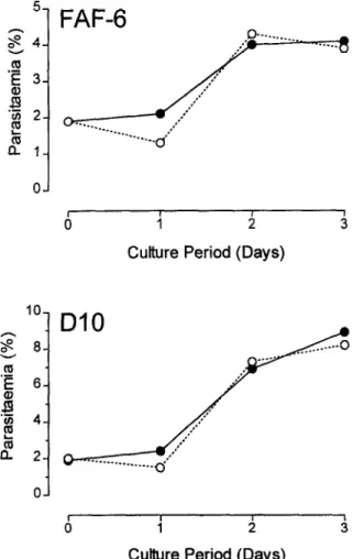
During the past 3 years, we have been successfully culturing l? falciparum without human serum using RPM1 1640 medium supplemented with a commercial- ly available lipid-rich bovine serum albumin called Al- bumax II@ (Gibco BRL, Grand Island, New York, USA). To our surprise, a literature search revealed no report comparing growth of l? falciparum in media sup- plemented with Albumax II@ with growth in serum-sup- plemented media. One recent report (GEROLD et al.,
51 FAF-6
0-J
I I , ,
0 1 2 3
Culture Period (Days)
i Culture Period (Days)
Fig. 1 .Two separate experiments comparing growth of 2 differ- ent lines of Rfalciparum (FAF-6 and DIO) in BPMI-S (0) or IU’MI-A (*) medium for 3 consecutive days.