Supporting Information
Amide Neighbouring-Group Effects in Peptides:
Phenylalanine as Relay Amino Acid in Long-Distance
Electron Transfer
Joses G. Nathanael,
[a]Luke F. Gamon,
[a]Meike Cordes,
[b]Paul R. Rablen,
[c]Thomas Bally,
[d]Katharina M. Fromm,
[d]Bernd Giese,*
[d]and Uta Wille*
[a]Supporting Information
Contents
1. Supplementary figures and tables ... S2
2. Peptide synthesis ... S7
3. Spectroscopic details for synthesised compounds ...S11
4. Spectra of substrates used in the laser flash photolysis study ...S20
4.1.
1H and
13C NMR spectra ... S20
4.2. HPLC spectra ... S37
5. Laser flash photolysis studies ...S46
6. Reaction of NO
3●with Ac-Phe-Phe-OMe (28) ...S49
7. Gaussian archive entries for computational data ...S50
8. References ...S58
1.
Supplementary figures and tables
Figure S1. UV/Vis spectrum (black) 40 ns after the laser flash of a nonapeptide with
2,4,6-trimethoxybenzene as functional group at the central amino acid. Subtraction by red line (electron
acceptor) leads to blue and green lines (oxidized electron donor and relay amino acid, respectively).
Data taken from: M. Cordes, A. Köttgen, C. Jasper, O. Jacques, H. Boudebous, B. Giese, Angew. Chem.
2008, 120, 3511-3513; Angew. Chem. Int. Ed. 2008, 47, 3461-3663.
Figure S2. UV/Vis spectrum (black) 40 ns after the laser flash of 1b. Subtraction by red line (electron
acceptor) leads to blue and green lines (oxidized electron donor and relay amino acid, respectively).
Data taken from: B. Giese, M. Wang, J. Gao, M. Stoltz, M. Gruber, J. Org. Chem. 2009, 74, 3621-3625.
O OMe OR OMe OMe MeO
Figure S3. UV/Vis spectrum (black) 40 ns after the laser flash of 1c (Met as central amino acid).
Subtraction by red line (electron acceptor) leads to blue (oxidized electron donor) and pink lines. The
pink absorption corresponds well to the absorption of a thioether radical cation, which is stabilized by
a neighbouring pyrrolidine amide (ref. [13] in the paper).
Figure S4. UV/Vis spectrum 40 ns after the laser flash of 1d (Phe as central amino acid). Subtraction
by red line (electron acceptor) leads to blue (oxidized electron donor) and pink lines. The pink
absorption is blue-shifted compared to the toluene radical cation (ref. [17] in the paper) and could
indicate the Phe radical cation 4d, which is stabilized by a neighbouring amide.
-0.10
-0.08
-0.06
-0.04
-0.02
0.00
0.02
0.04
0.06
0.08
0.10
350
400
450
500
550
600
650
700
Ab
sor
banc
e
Wavelength (nm)
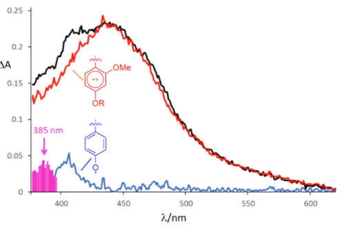
Figure S5a. UV/Vis spectra 180 ns after the laser flash of a 0.3 mM CAN solution in the absence (red
line) and presence of 10 mM Ac-Phe-OMe (7) (blue line). Black line: blue line and red line difference.
Spectra taken at 60 ns, 240 ns and 1400 ns after the laser flash showed a similiar behaviour. It was
shown in a separate experiment that the decay of the absorption at 500 nm has the same kinetic
behaviour as that of the NO
3•absorption at 630 nm. Because of the strong CAN depletion possible
transient formation below 480 nm cannot be detected.
CAN
depletion
Laser flash
@ 355 nm
NO
3●
+ Ac-Phe-OMe (7)
NO
3●
difference
Figure S5b. UV/Vis spectra 180 ns after the laser flash of a 0.3 mM CAN solution in the absence (red
line) and presence of 10 mM Ac-Phe-NHMe (6) (blue line). Black line: blue line and red line difference.
Spectra taken at 60 ns, 240 ns and 1400 ns showed a similiar behaviour. The decay of the absorption
at 500 nm shows the same kinetics as that of the NO
3•absorption at 630 nm. Because of the strong
CAN depletion possible transient formation below 480 nm cannot be detected.
-0.10
-0.08
-0.06
-0.04
-0.02
0.00
0.02
0.04
0.06
0.08
0.10
350
400
450
500
550
600
650
700
Ab
sor
banc
e
Wavelength (nm)
CAN
depletion
Laser flash
@ 355 nm
NO
3 ●+ Ac-Phe-NHMe (6)
NO
3●difference
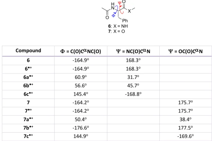
Table S1. Calculated peptide backbone dihedral angles for structures shown in Schemes 2 and 4
(M062X/6-31G*).
Compound
F
= C(O)C
a
NC(O)
Y
= NC(O)C
a
N
Y
= OC(O)C
a
N
6
-164.9
o168.3
o6
●+-164.9
o168.3
o6a
●+60.9
o31.7
o6b
●+56.6
o45.7
o6c
●+145.4
o-168.8
o7
-164.2
o175.7
o7
●+-164.2
o175.7
o7a
●+50.4
o38.4
o7b
●+-176.6
o177.5
o7c
●+144.9
o-169.6
oO H N Ph X 6: X = NH 7: X = O Ψ Φ O
2.
Peptide synthesis
The N-acetyl amino acid methyl esters were prepared by N-acetylation of the amino acids,
followed by methylation of the C-termini. Dipeptides were obtained by coupling the
N-protected and C-N-protected amino acids.
Tripeptides were synthesised sequentially either from (i) the terminus (starting with the
C-protected methyl ester hydrochloride salt) via N-Boc-C-protected intermediates (Procedure A,
scheme S1), or (ii) the N-terminus (starting with the N-acetyl amino acid) via N-acetyl
dipeptides (Procedure B, Scheme S2). Compounds for which no spectroscopic details are
provided, were obtained commercially (Sigma Aldrich, AK Scientific).
Scheme S1. Sequential synthesis of tripeptides from the C-terminus (Procedure A).
Scheme S2. Sequential synthesis of tripeptides from the N-terminus (Procedure B).
1
H and
13C spectra were recorded on either an Agilent MR 400 MHz NMR spectrometer or an
Agilent DD2 500 MHz NMR spectrometer, in either deuterated dimethylsulfoxide (DMSO-d
6),
deuterated acetonitrile (Acetonitrile-d
3) or deuterated methanol (Methanol-d
4). Chemical
shifts are reported in ppm (δ) using respective residual solvents as reference (DMSO: δ = 2.50
ppm for
1H NMR, δ = 39.52 ppm for
13C NMR; Acetonitrile: δ = 1.94 ppm for
1H NMR, δ =
118.26 ppm for
13C NMR; and Methanol: δ = 3.31 ppm for
1H NMR, δ = 49.00 ppm for
13C
NMR).
High Resolution Mass Spectrometry (HRMS) was conducted by ionising the samples using ESI
into a Thermo Scientific Exactive Plus Orbitrap mass spectrometer.
The crude products were purified by silica column chromatography with approximately 30 g
of dry silica per 1 g of crude product mixture. The eluting solvent consisted of a mixture of
petroleum ether and ethyl acetate or dichloromethane and methanol. Purity was assessed by
analytical reversed-phase HPLC on an Alltech Hypersil BDS-C18 5 μm 150 x 4.6 mm (Gradient:
100% water buffered with 0.1% TFA to 100% acetonitrile buffered with 0.1% TFA over 25
minutes, 4%/min, flow rate: 1 mL/min).
2.1. General procedure for the N-acetylation of the amino acids
Amino acid (53.4 mmol) was suspended in 5% aq. NaHCO
3(150 mL) and cooled to 0
oC. Acetic
anhydride (6.1 mL, 64.5 mmol, 1.2 eq.) was added dropwise over a period of 1 hour. The
mixture was stirred at room temperature for 2-4 hours and the reaction was monitored by
TLC (9:1 ethanol/1 M acetic acid, ninhydrin stain) until consumption of the starting material
was observed. The mixture was then acidified to pH 2-3 with 6 M HCl and cooled overnight.
The resulting precipitate was filtered off, washed with cold water (2 x 10 mL) and dried to
give the N-acetylated amino acid as a white solid.
2.2. General procedure for the esterification of the amino acids
Amino acid (78.8 mmol) was suspended in methanol (250 mL) and cooled at 0
oC. Thionyl
chloride (10 mL, 138 mmol, 1.7 eq.) was added dropwise. The mixture was stirred overnight
at room temperature and the reaction was monitored by TLC (9:1 ethanol/1 M acetic acid,
ninhydrin stain) until consumption of the starting material was observed. The solvent was
removed under reduced pressure to give the amino acid as the methyl ester hydrochloride
salt.
2.3. General procedure for the esterification of the N-acetylated amino acids
Amino acid (12.0 mmol) was suspended in methanol (11 mL) and cooled to 0
oC. Thionyl
chloride (1.0 mL, 13.8 mmol, 1.2 eq.) was added dropwise. The mixture was stirred for 4 hours
and the reaction was monitored by TLC (ethyl acetate, PMA or Hanessian’s stain) until
consumption of starting material was observed. The solvent was removed under reduced
pressure and water (20 mL) was added. The crude product was extracted with
dichloromethane (3 x 15 mL). The combined organic layer was dried over Na
2SO
4and the
solvent was removed under reduced pressure to give the N- and C- protected amino acids as
white crystals.
2.4. General procedure for the peptide coupling
The N-protected amino acid (10.0 mmol), amino acid methyl ester salt (10.1 mmol) and
(2-(1H-benzotriazol-1-yl)-1,1,3,3-tetramethyluronium hexafluorophosphate (HBTU) (3.75 g, 9.9
mmol) were suspended in anhydrous DMF (15 mL) and cooled to 0
oC. Triethylamine (4.2 mL,
30.0 mmol) was added dropwise and the mixture was stirred overnight at room temperature.
The reaction was monitored by TLC (ethyl acetate, PMA or ninhydrin stain) until consumption
of starting material was observed. The mixture was then partitioned between 1 M HCl (100
mL) and ethyl acetate (100 mL). The aqueous phase was extracted with ethyl acetate (2 x 100
mL), and the combined extracts were washed with 5% aq. NaHCO
3(100 mL) and brine (100
mL). The organic layer was dried over MgSO
4and the solvent was removed under reduced
pressure to give a sticky oil or a white solid. The residue was purified by silica column
chromatography or by recrystallisation from ethyl acetate.
2.5. General procedure for the N-Boc deprotection
The N-Boc protected peptide methyl ester (10.0 mmol) was dissolved in dichloromethane (8
mL) and cooled to 0
oC. Trifluoroacetic acid (8 mL, 104.5 mmol) was added dropwise. The
mixture was stirred overnight and the reaction was monitored by TLC (ethyl acetate,
ninhydrin stain) until consumption of the starting material was observed. The solvent was
removed under reduced pressure, followed by azeotroping with toluene to remove residual
trifluoroacetic acid, to give a white or yellow solid. The crude material was obtained as the
trifluoroacetate salt and used without further purification.
2.6. General procedure for the hydrolysis of C-terminal methyl esters
The N-acetyl protected peptide methyl ester (1.82 mmol) was dissolved in tetrahydrofuran
(28 mL) and cooled to 0
oC. A solution of lithium hydroxide (0.60 g, 25.2 mmol) in water (28
mL) was cooled to 0
oC and then added to the first solution. After stirring overnight, a solution
of 3 M HCl (10 mL) was added, followed by an addition of brine (20 mL). The mixture was then
extracted with ethyl acetate (4 x 20 mL), and the combined organic extracts were washed
with brine (50 mL). The organic layer was dried over MgSO
4and the solvent was removed
under reduced pressure to give the product as a white solid.
2.7. Preparation of N-acetyl-
L
-phenylalanine amide (Ac-Phe-NH
2(14))
L
-Phenylalanine methyl ester hydrochloride (2.40 g, 11.1 mmol) was dissolved in 28% aqueous
ammonia solution (25 mL). After stirring overnight, the mixture was concentrated under
reduced pressure to give a white-off solid. The solid was dissolved in acetonitrile (5 mL) and
acetic anhydride (1 mL, 10.6 mmol) was added dropwise. The mixture was stirred for 1 day at
room temperature and filtered off to give the product as a white solid.
2.8. Preparation of N-acetyl-
L
-phenylalanine methyl amide (Ac-Phe-NHMe (6))
N-Methyl morpholine (1.2 mL, 10.9 mmol) was added to a solution of N-acetyl-
L-phenylalanine (2.10 g, 10.1 mmol) in dimethylformamide (7 mL) and tetrahydrofuran (7 mL)
at -10
oC. Trimethylacetyl (pivaloyl) chloride (1.25 mL, 10.2 mmol) was added dropwise. After
10 minutes, a solution of methylamine hydrochloride (3.36 g, 49.8 mmol) and triethylamine
(7.0 mL, 50.2 mmol) in water (7 mL) was added. The resulting mixture was stirred for 1.5 hours
and the solvent was removed under reduced pressure. The residue was dissolved in
chloroform (30 mL) and washed with water (2 x 30 mL). The organic layer was dried over
Na
2SO
4and the solvent was removed under reduced pressure to give a white solid. The crude
product was purified by silica column chromatography to give the pure product.
2.9. Preparation of N-acetyl-
L
-phenylalanine t-butyl amide (Ac-Phe-NHtBu (13))
N-acetyl-
L-phenylalanine (3.50 g, 16.9 mmol) and p-nitrophenol (2.37 g, 17.1 mmol) were
dissolved in dichloromethane (20 mL). N,N’-Dicyclohexylcarbodiimide/DCC (3.48 g, 16.9
mmol) was added in portions. The resulting mixture was heated at 40
oC for 8 hours and
filtered through a bed of celite. The filter cake was washed with dichloromethane and the
filtrate was concentrated under reduced pressure. The yellow solid was dissolved in
dichloromethane (150 mL) and t-butylamine (2.0 mL, 18.8 mmol) was added dropwise. The
resulting mixture was heated at 40
oC for 16 hours and filtered through a bed of celite. The
filtrate was concentrated under reduced pressure and the crude material was recrystallised
from hot ethanol to give a colourless crystal.
3.
Spectroscopic details for the synthesised compounds
3.1. N-Acetyl-
L
-phenylalanine methyl ester (Ac-Phe-OMe (7))
(a) N-Acetyl-L-phenylalanine:
1H NMR (400 MHz, DMSO-d
6) δ 12.65 (s, 1H), 8.18 (d, J = 8.1 Hz,
1H), 7.31 – 7.25 (m, 2H), 7.24 – 7.17 (m, 3H), 4.40 (ddd, J = 9.6, 8.1, 4.9 Hz, 1H), 3.03 (dd, J =
13.8, 4.9 Hz, 1H), 2.83 (dd, J = 13.8, 9.6 Hz, 1H), 1.77 ppm (s, 3H).
13C NMR (101 MHz,
DMSO-d
6) δ 173.15, 169.19, 137.71, 129.03, 128.16, 126.38, 53.48, 36.77, 22.33 ppm. HRMS (ESI)
m/z calcd. for [C
11H
14NO
3]
+: 208.0978 [M+H]
+, found 208.0975, HRMS (ESI) m/z calcd. for
[C
22H
27N
2O
6]
+: 415.1869 [2M+H]
+, found 415.1893.
(b) N-Acetyl-L-phenylalanine methyl ester:
1H NMR (400 MHz, DMSO-d
6) δ 8.33 (d, J = 7.8 Hz,
1H), 7.33 – 7.23 (m, 2H), 7.26 – 7.16 (m, 3H), 4.44 (ddd, J = 9.1, 7.7, 5.6 Hz, 1H), 3.59 (s, 3H),
3.00 (dd, J = 13.8, 5.6 Hz, 1H), 2.87 (dd, J = 13.7, 9.3 Hz, 1H), 1.79 ppm (s, 3H).
13C NMR (101
MHz, DMSO-d
6) δ 172.19, 169.29, 137.25, 128.99, 128.23, 126.52, 53.60, 51.79, 36.72, 22.23
ppm. HRMS (ESI) m/z calcd. for [C
12H
16NO
3]
+: 222.1130 [M+H]
+, found 222.1137, HRMS (ESI)
m/z calcd. for [C
12H
15NO
3Na]
+: 375.2495 [M+Na]
+, found 375.2517.
3.2. N-acetyl-
L
-phenylalanine amide (Ac-Phe-NH
2(14))
(a) L-Phenylalanine methyl ester hydrochloride:
1H NMR (400 MHz, DMSO-d
6) δ 8.74 (s, 3H),
7.38 – 7.28 (m, 2H), 7.32 – 7.20 (m, 3H), 4.23 (dd, J = 7.4, 5.9 Hz, 1H), 3.65 (s, 3H), 3.21 (dd, J
= 14.0, 5.7 Hz, 1H), 3.10 ppm (dd, J = 14.0, 7.5 Hz, 1H).
13C NMR (101 MHz, DMSO-d
6) δ 169.34,
134.69, 129.38, 128.57, 127.24, 53.22, 52.53, 35.83 ppm. HRMS (ESI) m/z calcd. for
[C
10H
14NO
2]
+: 180.1019 [M-Cl]
+, found 180.1010.
(b) N-Acetyl-L-phenylalanine amide:
1H NMR (400 MHz, DMSO-d
6) δ 8.01 (d, J = 8.5 Hz, 1H),
7.42 (s, 1H), 7.29 – 7.21 (m, 4H), 7.22 – 7.13 (m, 1H), 7.01 (s, 1H), 4.41 (td, J = 9.2, 4.7 Hz, 1H),
2.98 (dd, J = 13.7, 4.7 Hz, 1H), 2.72 (dd, J = 13.7, 9.7 Hz, 1H), 1.75 ppm (s, 3H).
13C NMR (101
MHz, DMSO-d
6) δ 173.24, 168.97, 138.22, 129.07, 127.99, 126.15, 53.78, 37.65, 22.50 ppm.
HRMS (ESI) m/z calcd. for [C
11H
15N
2O
2]
+: 207.1134 [M+H]
+, found 207.1130, HRMS (ESI) m/z
calcd. for [C
11H
14N
2O
4Na]
+: 229.0953 [M+Na]
+, found 229.0949.
3.3. N-Acetyl-
L
-phenylalanine methyl amide (Ac-Phe-NHMe (6))
(a) N-Acetyl-L-phenylalanine: see section 2.1(a)
(b) N-Acetyl-L-phenylalanyl methyl amide:
1H NMR (400 MHz, DMSO-d
6) δ 8.10 (d, J = 8.5 Hz,
1H), 7.88 (q, J = 4.6 Hz, 1H), 7.30 – 7.21 (m, 2H), 7.24 – 7.13, (m, 3H), 4.40 (ddd, J = 9.5, 8.4,
5.0 Hz, 1H), 2.95 (dd, J = 13.7, 5.0 Hz, 1H), 2.72 (dd, J = 13.7, 9.6 Hz, 1H), 2.56 (d, J = 4.5 Hz,
3H), 1.75 ppm (s, 3H).
13C NMR (101 MHz, DMSO-d
6) δ 171.58, 169.00, 138.16, 129.03, 128.02,
126.19, 54.07, 37.78, 25.52, 22.50 ppm. HRMS (ESI) m/z calcd. for [C
12H
17N
2O
2]
+: 221.1290
[M+H]
+, found 221.1283, HRMS (ESI) m/z calcd. for [C
24H
33N
4O
4]
+: 441.2502 [2M+H]
+, found
441.2495.
3.4. N-Acetyl-
L
-phenylalanine t-butyl amide (Ac-Phe-NHtBu (13))
(a) N-Acetyl-L-phenylalanine: see section 2.1(a)
(b) N-Acetyl-L-phenylalanyl t-butyl amide:
1H NMR (400 MHz, DMSO-d
6) δ 7.96 (d, J = 8.4 Hz,
1H), 7.49 (s, 1H), 7.29 – 7.18 (m, 4H), 7.22 – 7.13, (m, 1H), 4.46 (td, J = 8.7, 6.1 Hz, 1H), 2.85
(dd, J = 13.5, 5.7 Hz, 1H), 2.72 (dd, J = 13.5, 8.9 Hz, 1H), 1.75 (s, 3H), 1.19 ppm (s, 9H).
13C NMR
(101 MHz, DMSO-d
6) δ 170.47, 168.77, 137.90, 129.25, 127.86, 126.10, 54.07, 50.02, 38.33,
28.38, 22.48 ppm. HRMS (ESI) m/z calcd. for [C
15H
23N
2O
2]
+: 263.1760 [M+H]
+, found 263.1752,
HRMS (ESI) m/z calcd. for [C
15H
22N
2O
2K]
+: 301.1318 [M+K]
+, found 301.1310.
3.5. N-Acetyl-
L
-leucyl-
L
-phenylalanine methyl ester (Ac-Leu-Phe-OMe (15))
(a) L-Phenylalanine methyl ester hydrochloride: see section 2.2(a)
(b) N-Acetyl-L-leucine:
1H NMR (400 MHz, DMSO-d
6) δ 12.46 (s, 1H), 8.07 (d, J = 7.9 Hz, 1H),
4.19 (ddd, J = 9.1, 8.1, 6.0 Hz, 1H), 1.83 (s, 3H), 1.69 – 1.54 (m, 1H), 1.48 ppm (ddd, J = 8.5,
5.5, 3.0 Hz, 2H), 0.89 (d, J = 6.5 Hz, 3H), 0.84 (d, J = 6.5 Hz, 3H).
13C NMR (101 MHz, DMSO-d
6)
δ 174.26, 169.24, 50.18, 40.00, 24.32, 22.84, 22.33, 21.30 ppm. HRMS (ESI) m/z calcd. for
[C
8H
16NO
3]
+: 174.1130 [M+H]
+, found 174.1133, HRMS (ESI) m/z calcd. for [C
16H
31N
2O
6]
+:
347.2182 [2M+H]
+, found 347.2182.
(c) N-Acetyl-leucyl-L-phenylalanine methyl ester:
1H NMR (400 MHz, DMSO-d
6) δ 8.30 (d, J =
7.4 Hz, 1H), 7.90 (d, J = 8.4 Hz, 1H), 7.29 – 7.24 (m, 2H), 7.24 – 7.18 (m, 3H), 4.48 – 4.40 (m,
1H), 4.32 (td, J = 8.7, 6.0 Hz, 1H), 3.56 (s, 3H), 3.02 (dd, J = 13.9, 5.9 Hz, 1H), 2.94 (dd, J = 13.9,
8.6 Hz, 1H), 1.80 (s, 3H), 1.61 – 1.49 (m, 1H), 1.44 – 1.28 (m, 2H), 0.87 (d, J = 6.6 Hz, 3H), 0.82
ppm (d, J = 6.5 Hz, 3H).
13C NMR (101 MHz, DMSO-d
6) δ 172.30, 171.79, 168.90, 137.12,
129.04, 128.19, 126.50, 53.45, 51.75, 50.51, 40.85, 36.43, 24.09, 22.93, 22.44, 21.71 ppm.
HRMS (ESI) m/z calcd. for [C
18H
27N
2O
4]
+: 335.1965 [M+H]
+, found 335.1976, HRMS (ESI) m/z
calcd. for [C
18H
26N
2O
4Na]
+: 357.1785 [M+Na]
+, found 357.1790.
3.6. N-Acetyl-
L
-phenylalanyl-
L
-leucine methyl ester (Ac-Phe-Leu-OMe (16))
(a) L-Leucine methyl ester hydrochloride:
1H NMR (400 MHz, DMSO-d
6) δ 8.73 (s, 3H), 3.90
(t, J = 7.0, 1H), 3.73 (s, 3H), 1.81 – 1.72 (m, 1H), 1.72 – 1.58 (m, 2H), 0.88 ppm (d, J = 6.4 Hz,
6H).
13C NMR (101 MHz, DMSO-d
6) δ 170.25, 52.69, 50.47, 39.11, 23.71, 22.16, 21.97 ppm.
HRMS (ESI) m/z calcd. for [C
7H
16NO
2]
+: 146.1181 [M-Cl]
+, found 146.1182.
(b) N-Acetyl-L-phenylalanine: see section 2.1(a)
(c) N-Acetyl-phenylalanyl-L-leucine methyl ester:
1H NMR (400 MHz, DMSO-d
6) δ 8.37 (d, J =
7.7 Hz, 1H), 8.06 (d, J = 8.4 Hz, 1H), 7.26 (d, J = 4.4 Hz, 4H), 7.19 (p, J = 4.5 Hz, 1H), 4.54 (ddd,
J = 10.1, 8.5, 4.3 Hz, 1H), 4.29 (ddd, J = 9.6, 7.7, 5.3 Hz, 1H), 3.61 (s, 3H), 2.99 (dd, J = 13.9, 4.3
Hz, 1H), 2.71 (dd, J = 13.9, 10.0 Hz, 1H), 1.74 (s, 3H), 1.68 – 1.57 (m, 1H), 1.60 – 1.44 (m, 2H),
0.90 (d, J = 6.4 Hz, 3H), 0.84 ppm (d, J = 6.3 Hz, 3H).
13C NMR (101 MHz, DMSO-d
6) δ 172.78,
171.68, 169.00, 137.95, 129.12, 127.97, 126.19, 53.55, 51.83, 50.28, 39.68, 37.56, 24.18,
22.74, 22.42, 21.31 ppm. HRMS (ESI) m/z calcd. for [C
18H
27N
2O
4]
+: 335.1965 [M+H]
+, found
335.1975, HRMS (ESI) m/z calcd. for [C
18H
26N
2O
4Na]
+: 357.1785 [M+Na]
+, found 357.1784.
3.7. N-Acetyl-
L
-valyl-
L
-phenylalanine methyl ester (Ac-Val-Phe-OMe (17))
(a) L-Phenylalanine methyl ester hydrochloride: see section 2.2(a)
(b) N-Acetyl-valyl-L-phenylalanine methyl ester:
1H NMR (400 MHz, DMSO-d
6) δ 8.39 (d, J =
7.3 Hz, 1H), 7.79 (d, J = 9.0 Hz, 1H), 7.31 – 7.22 (m, 2H), 7.25 – 7.15 (m, 3H), 4.45 (dt, J = 8.5,
6.5 Hz, 1H), 4.18 (dd, J = 9.1, 7.0 Hz, 1H), 3.55 (s, 3H), 3.01 (dd, J = 13.9, 6.1 Hz, 1H), 2.93 (dd,
J = 13.9, 8.8 Hz, 1H), 1.91 (hept, J = 7.0 Hz, 1H), 1.83 (s, 3H), 0.82 (d, J = 7.0 Hz, 3H), 0.79 ppm
(d, J = 7.0 Hz, 3H).
13C NMR (101 MHz, DMSO-d
6) δ 171.79, 171.32, 169.04, 137.12, 129.02,
128.19, 126.52, 57.18, 53.53, 51.68, 36.50, 30.56, 22.44, 19.06, 18.08 ppm. HRMS (ESI) m/z
calcd. for [C
17H
25N
2O
4]
+: 321.1814 [M+H]
+, found 321.1814, HRMS (ESI) m/z calcd. for
[C
17H
24N
2O
4Na]
+: 343.1633 [M+Na]
+, found 343.1638.
3.8. N-Acetyl-
L
-phenylalanyl-
L
-valine methyl ester (Ac-Phe-Val-OMe (18))
(a) L-Valine methyl ester hydrochloride:
1H NMR (400 MHz, DMSO-d
6) δ 8.66 (s, 3H), 3.83 (d,
J = 4.7, 1H), 3.74 (s, 3H), 2.19 (heptd, J = 6.9, 4.5 Hz, 1H), 0.98 (d, J = 6.9 Hz, 3H), 0.93 ppm (d,
J = 6.9 Hz, 3H).
13C NMR (101 MHz, DMSO-d
6) δ 169.20, 57.23, 52.23, 29.31, 18.44, 17.56 ppm.
HRMS (ESI) m/z calcd. for [C
6H
14NO
2]
+: 132.1019 [M-Cl]
+, found 132.1021.
(b) N-Acetyl-L-phenylalanine: see section 2.1(a)
(c) N-Acetyl-phenylalanyl-L-valine methyl ester:
1H NMR (400 MHz, DMSO-d
6) δ 8.24 (d, J =
8.1 Hz, 1H), 8.07 (d, J = 8.3 Hz, 1H), 7.28 – 7.24 (m, 4H), 7.24 – 7.13 (m, 1H), 4.62 (ddd, J =
10.0, 8.4, 4.4 Hz, 1H), 4.17 (dd, J = 8.1, 6.3 Hz, 1H), 3.62 (s, 3H), 2.96 (dd, J = 13.9, 4.4 Hz, 1H),
2.71 (dd, J = 13.9, 10.1 Hz, 1H), 2.04 (h, J = 6.8 Hz, 1H), 1.74 (s, 3H), 0.90 (d, J = 6.9 Hz, 3H),
0.87 ppm (d, J = 6.8 Hz, 3H).
13C NMR (101 MHz, DMSO-d
6) δ 171.87, 171.81, 169.08, 137.91,
129.15, 127.95, 126.18, 57.42, 53.50, 51.68, 37.47, 29.88, 22.40, 18.92, 18.25 ppm. HRMS
(ESI) m/z calcd. for [C
17H
25N
2O
4]
+: 321.1814 [M+H]
+, found 321.1809, HRMS (ESI) m/z calcd.
for [C
17H
25N
2O
4Na]
+: 343.1633 [M+Na]
+, found 343.1628.
3.9. N-Acetyl-
L
-leucyl-
L
-leucyl-
L
-phenylalanine methyl ester (Ac-Leu-Leu-Phe-OMe
(19))
This tripeptide was synthesised from the C-terminus according to Procedure A, Scheme S1.
(a) L-Phenylalanine methyl ester hydrochloride: see section 2.5(a)
(b) N-Boc-L-leucyl-L-phenylalanine methyl ester:
1H NMR (400 MHz, DMSO-d
6) δ 8.12 (d, J =
7.7 Hz, 1H), 7.30 – 7.22 (m, 2H), 7.22 – 7.18 (m, 3H), 6.80 (d, J = 8.5 Hz, 1H), 4.48 (td, J = 8.2,
5.7 Hz, 1H), 3.96 (td, J = 9.0, 5.7 Hz, 1H), 3.57 (s, 3H), 3.02 (dd, J = 13.8, 5.8 Hz, 1H), 2.94 (dd,
J = 13.9, 8.7 Hz, 1H), 1.61 – 1.44 (m, 1H), 1.37 (s, 9H), 1.31 (d, J = 7.5 Hz, 2H), 0.85 (d, J = 6.6
137.04, 129.07, 128.17, 126.48, 77.97, 53.25, 52.62, 51.79, 40.78, 36.61, 28.17, 24.12, 22.84,
21.62 ppm. HRMS (ESI) m/z calcd. for [C
21H
33N
2O
5]
+: 393.2384 [M+H]
+, found 393.2383, HRMS
(ESI) m/z calcd. for [C
21H
32N
2O
5Na]
+: 415.2204 [M+Na]
+, found 415.2203.
(c)
L-Leucyl-L-phenylalanine methyl ester trifluoroacetate: 1H NMR (400 MHz, DMSO-d
6) δ
8.96 (d, J = 7.3 Hz, 1H), 8.14 (s, 3H), 7.35 – 7.26 (m, 2H), 7.28 – 7.19 (m, 3H), 4.61 – 4.49 (m,
1H), 3.77 (dd, J = 8.5, 5.3 Hz, 1H), 3.60 (s, 3H), 3.07 (dd, J = 14.0, 5.9 Hz, 1H), 2.99 (dd, J = 14.0,
8.6 Hz, 1H), 1.73 – 1.58 (m, 1H), 1.53 (dd, J = 7.7, 5.9 Hz, 2H), 0.90 (d, J = 5.1 Hz, 3H), 0.88 ppm
(d, J = 4.7 Hz, 3H).
13C NMR (101 MHz, DMSO-d
6) δ 171.30, 169.35, 136.83, 129.02, 128.37,
126.72, 53.88, 52.00, 50.58, 40.22, 36.32, 23.36, 22.76, 21.71 ppm. HRMS (ESI) m/z calcd. for
[C
16H
25N
2O
3]
+: 293.1865 [M-CF
3CO
2]
+, found 293.1860.
(d) N-Acetyl-L-leucine: see section 2.5(b)
(e) N-Acetyl-L-leucyl-L-leucyl-L-phenylalanine methyl ester:
1H NMR (400 MHz, DMSO-d
6) δ
8.21 (d, J = 7.4 Hz, 1H), 7.95 (d, J = 8.1 Hz, 1H), 7.83 (d, J = 8.4 Hz, 1H), 7.27 (dd, J = 8.0, 6.1 Hz,
2H), 7.20 (td, J = 6.6, 1.7 Hz, 3H), 4.46 (td, J = 8.2, 6.1 Hz, 1H), 4.28 (dq, J = 15.0, 7.8 Hz, 2H),
3.56 (s, 3H), 3.01 (dd, J = 13.9, 6.0 Hz, 1H), 2.94 (dd, J = 14.0, 8.5 Hz, 1H), 1.82 (s, 3H), 1.55
(dh, J = 13.5, 6.8 Hz, 2H), 1.38 (q, J = 7.1 Hz, 4H), 0.86 (d, J = 6.6 Hz, 6H), 0.82 ppm (dd, J = 6.6,
3.1 Hz, 6H).
13C NMR (101 MHz, DMSO-d
6) δ 171.96, 171.85, 171.69, 169.12, 137.02, 128.97,
128.20, 126.50, 53.38, 51.75, 50.91, 50.63, 40.86, 40.71, 36.49, 24.15, 24.03, 23.04, 22.93,
22.48, 21.72, 21.59 ppm. HRMS (ESI) m/z calcd. for [C
24H
38N
3O
5]
+: 448.2812 [M+H]
+, found
448.2809, HRMS (ESI) m/z calcd. for [C
24H
37N
3O
5K]
+: 486.2370 [M+K]
+, found 486.2366.
3.10. N-Acetyl-
L
-leucyl-
L
-phenylalanyl-
L
-leucine methyl ester (Ac-Leu-Phe-Leu-OMe
(20))
This tripeptide was synthesised from the N-terminus according to Procedure B, Scheme S2.
(a) N-Acetyl-L-leucyl-L-phenylalanine methyl ester: see section 2.5
(b) N-Acetyl-L-leucyl-L-phenylalanine:
1H NMR (400 MHz, DMSO-d
6) δ 12.66 (s, 1H), 8.08 (d, J
= 7.8 Hz, 1H), 7.90 (d, J = 8.5 Hz, 1H), 7.30 – 7.21 (m, 2H), 7.24 – 7.15 (m, 3H), 4.40 (td, J = 8.2,
5.3 Hz, 1H), 4.31 (td, J = 8.8, 5.9 Hz, 1H), 3.04 (dd, J = 13.9, 5.2 Hz, 1H), 2.91 (dd, J = 13.9, 8.8
Hz, 1H), 1.80 (s, 3H), 1.55 (dp, J = 13.4, 6.7 Hz, 1H), 1.43 – 1.30 (m, 2H), 0.86 (d, J = 6.6 Hz, 3H),
0.82 ppm (d, J = 6.6 Hz, 3H).
13C NMR (101 MHz, DMSO-d
6) δ 172.73, 172.13, 168.93, 137.49,
129.12, 128.12, 126.38, 53.27, 50.61, 40.82, 36.51, 24.09, 23.02, 22.46, 21.66 ppm. HRMS
(ESI) m/z calcd. for [C
17H
25N
2O
4]
+: 321.1814 [M+H]
+, found 321.1811, HRMS (ESI) m/z calcd.
for [C
17H
24N
2O
4Na]
+: 343.1634 [M+Na]
+, found 343.1630.
(c) L-Leucine methyl ester hydrochloride: see section 2.6(a)
(d) N-Acetyl-L-leucyl-L-phenylalanyl-L-leucine methyl ester:
1H NMR (400 MHz, DMSO-d
6) δ
8.18 (d, J = 7.6 Hz, 1H), 7.93 (dd, J = 8.3, 5.0 Hz, 2H), 7.29 – 7.11 (m, 5H), 4.51 (td, J = 8.9, 4.9
Hz, 1H), 4.29 (td, J = 9.1, 5.2 Hz, 1H), 4.19 (q, J = 7.6 Hz, 1H), 3.60 (s, 3H), 3.03 (dd, J = 14.1,
4.8 Hz, 1H), 2.80 (dd, J = 13.9, 9.3 Hz, 1H), 1.80 (s, 3H), 1.67 – 1.52 (m, 2H), 1.54 – 1.42 (m,
2H), 1.30 (t, J = 7.3 Hz, 2H), 0.89 (d, J = 6.3 Hz, 3H), 0.83 (d, J = 6.4 Hz, 6H), 0.79 ppm (d, J = 6.5
Hz, 3H).
13C NMR (101 MHz, DMSO-d
6) δ 172.65, 171.87, 171.01, 169.21, 137.67, 129.15,
127.95, 126.17, 53.31, 51.82, 51.09, 50.23, 40.65, 39.66, 37.05, 24.05, 24.04, 22.90, 22.81,
22.44, 21.67, 21.22 ppm. HRMS (ESI) m/z calcd. for [C
24H
38N
3O
5]
+: 448.2812 [M+H]
+, found
448.2809, HRMS (ESI) m/z calcd. for [C
24H
37N
3O
5Na]
+: 470.2631 [M+Na]
+, found 470.2628.
3.11. N-Acetyl-
L
-phenylalanyl-
L
-leucyl-
L
-leucine methyl ester (Ac-Phe-Leu-Leu-OMe
(21))
This tripeptide was synthesised from the N-terminus according to Procedure B, Scheme S2.
(a) N-Acetyl-L-phenylalanyl-L-leucine methyl ester: see section 2.6
(b) N-Acetyl-L-phenylalanyl-L-leucine:
1H NMR (400 MHz, DMSO-d
6) δ 12.55 (s, 1H), 8.23 (d, J
= 7.9 Hz, 1H), 8.04 (d, J = 8.5 Hz, 1H), 7.29 – 7.20 (m, 4H), 7.22 – 7.14 (m, 1H), 4.54 (ddd, J =
10.2, 8.6, 4.0 Hz, 1H), 4.22 (ddd, J = 9.2, 7.8, 5.6 Hz, 1H), 3.00 (dd, J = 14.0, 4.0 Hz, 1H), 2.70
(dd, J = 13.8, 10.2 Hz, 1H), 1.73 (s, 3H), 1.70 – 1.56 (m, 1H), 1.56 – 1.49 (m, 2H), 0.90 (d, J = 6.5
Hz, 3H), 0.85 ppm (d, J = 6.4 Hz, 3H).
13C NMR (101 MHz, Acetonitrile-d
3) δ 174.80, 172.43,
171.81, 137.84, 129.83, 128.85, 127.14, 54.92, 51.44, 40.55, 37.88, 25.02, 22.74, 22.33, 21.29
ppm. HRMS (ESI) m/z calcd. for [C
17H
25N
2O
4]
+: 321.1814 [M+H]
+, found 321.1810, HRMS (ESI)
m/z calcd. for [C
17H
24N
2O
4Na]
+: 343.1634 [M+Na]
+, found 343.1630.
(c) L-Leucine methyl ester hydrochloride: see section 2.6(a)
(d) N-Acetyl-L-phenylalanyl-L-leucyl-L-leucine methyl ester:
1H NMR (400 MHz, DMSO-d
6) δ
8.20 (d, J = 7.7 Hz, 1H), 8.04 (d, J = 8.0 Hz, 2H), 7.24 (d, J = 4.3 Hz, 4H), 7.20 – 7.15 (m, 1H),
4.51 (td, J = 9.8, 4.2 Hz, 1H), 4.39 – 4.23 (m, 2H), 3.60 (s, 3H), 2.97 (dd, J = 13.9, 4.1 Hz, 1H),
2.70 (dd, J = 13.9, 9.9 Hz, 1H), 1.74 (s, 3H), 1.68 – 1.52 (m, 3H), 1.54 – 1.41 (m, 3H), 0.90 (dd,
J = 6.3, 1.6 Hz, 6H), 0.85 ppm (dd, J = 8.5, 6.4 Hz, 6H).
13C NMR (101 MHz, Acetonitrile-d
3) δ
173.89, 173.01, 172.04, 171.47, 138.37, 130.22, 129.29, 127.57, 55.87, 52.58, 52.35, 51.69,
41.48, 40.95, 38.07, 25.43, 25.29, 23.34, 23.17, 22.96, 21.82, 21.67 ppm. HRMS (ESI) m/z
calcd. for [C
24H
38N
3O
5]
+: 448.2812 [M+H]
+, found 448.2809, HRMS (ESI) m/z calcd. for
[C
24H
37N
3O
5Na]
+: 470.2631 [M+Na]
+, found 470.2629.
3.12. N-Acetyl-
L
-valyl-
L
-valyl-
L
-phenylalanine methyl ester (Ac-Val-Val-Phe-OMe
(22))
This tripeptide was synthesised from the C-terminus according to Procedure A, Scheme S1.
(a) L-Phenylalanine methyl ester hydrochloride: see section 2.2(a)
(b) N-Boc-L-valyl-L-phenylalanine methyl ester:
1H NMR (400 MHz, DMSO-d
6) δ 8.25 (d, J =
7.6 Hz, 1H), 7.30 – 7.22 (m, 2H), 7.25 – 7.15 (m, 3H), 6.56 (d, J = 9.1 Hz, 1H), 4.49 (ddd, J = 9.4,
7.7, 5.8 Hz, 1H), 3.78 (dd, J = 9.0, 7.3 Hz, 1H), 3.57 (s, 3H), 3.02 (dd, J = 13.9, 5.7 Hz, 1H), 2.92
(dd, J = 13.9, 8.9 Hz, 1H), 1.93 – 1.77 (m, 1H), 1.37 (s, 9H), 0.77 (d, J = 6.6 Hz, 3H), 0.76 ppm
(d, J = 6.7 Hz, 3H).
13C NMR (101 MHz, DMSO-d
6) δ 171.83, 171.44, 155.24, 137.07, 129.02,
128.19, 126.51, 77.99, 59.50, 53.37, 51.74, 36.65, 30.50, 28.17, 19.02, 18.11 ppm. HRMS (ESI)
m/z calcd. for [C
20H
31N
2O
5]
+: 379.2233 [M+H]
+, found 379.2222, HRMS (ESI) m/z calcd. for
[C
20H
30N
2O
5Na]
+: 401.2052 [M+Na]
+, found 401.2052.
(c)
L-Valyl-L-phenylalanine methyl ester trifluoroacetate: 1H NMR (400 MHz, DMSO-d
6) δ
8.90 (d, J = 7.1 Hz, 1H), 8.10 (s, 3H), 7.35 – 7.26 (m, 2H), 7.28 – 7.19 (m, 3H), 4.55 (dt, J = 7.4,
6.0 Hz, 1H), 3.65 (d, J = 5.1 Hz, 1H), 3.59 (s, 3H), 3.07 (dd, J = 14.0, 5.9 Hz, 1H), 2.98 (dd, J =
14.0, 8.5 Hz, 1H), 2.18 – 2.04 (m, 1H), 0.95 (d, J = 6.9 Hz, 3H) 0.91 ppm (d, J = 6.9 Hz, 3H).
13C
NMR (101 MHz, DMSO-d
6) δ 171.34, 168.22, 136.77, 129.03, 128.36, 126.73, 57.01, 53.90,
51.94, 36.38, 29.87, 18.25, 17.10 ppm. HRMS (ESI) m/z calcd. for [C
15H
23N
2O
3]
+: 279.1709
[M-CF
3CO
2]
+, found 279.1719.
(d) N-Acetyl-L-valyl-L-valyl-L-phenylalanine methyl ester:
1H NMR (500 MHz, DMSO-d
6) δ
8.34 (d, J = 7.4 Hz, 1H), 7.87 (d, J = 8.8 Hz, 1H), 7.66 (d, J = 9.0 Hz, 1H), 7.25 (t, J = 7.4 Hz, 2H),
7.20 (d, J = 7.2 Hz, 3H), 4.49 (ddd, J = 8.7, 7.2, 5.7 Hz, 1H), 4.16 (dd, J = 8.9, 6.9 Hz, 2H), 3.55
(s, 3H), 3.02 (dd, J = 14.0, 5.8 Hz, 1H), 2.92 (dd, J = 14.0, 8.9 Hz, 1H), 1.90 (dt, J = 13.8, 6.9 Hz,
2H), 1.85 (s, 3H), 0.80 (d, J = 7.7 Hz, 6H), 0.77 ppm (d, J = 6.9 Hz, 6H).
13C NMR (101 MHz,
DMSO-d
6) δ 171.70, 170.91, 169.15, 137.01, 128.86, 128.19, 126.49, 57.72, 57.21, 53.29,
51.70, 36.49, 30.75, 30.18, 22.46, 19.20, 19.00, 18.21, 18.07 ppm. HRMS (ESI) m/z calcd. for
[C
22H
34N
3O
5]
+: 420.2499 [M+H]
+, found 420.2496, HRMS (ESI) m/z calcd. for [C
22H
33N
3O
5Na]
+:
442.2318 [M+Na]
+, found 442.2314.
3.13. N-Acetyl-
L
-valyl-
L
-phenylalanyl-
L
-valine methyl ester (Ac-Val-Phe-Val-OMe
(23))
This tripeptide was synthesised from the C-terminus according to Procedure A, Scheme S1.
(a) L-Valine methyl ester hydrochloride: see section 2.8(a)
(b) N-Boc-L-phenylalanyl-L-valine methyl ester:
1H NMR (400 MHz, DMSO-d
6) δ 8.08 (d, J =
8.3 Hz, 1H), 7.30 – 7.24 (m, 4H), 7.23 – 7.15 (m, 1H), 6.93 (d, J = 8.6 Hz, 1H), 4.25 (ddd, J =
23.0, 9.3, 5.2 Hz, 2H), 3.63 (s, 3H), 2.94 (dd, J = 13.9, 4.3 Hz, 1H), 2.73 (dd, J = 13.8, 10.4 Hz,
1H), 2.05 (h, J = 6.7 Hz, 1H), 1.30 (s, 9H), 0.89 ppm (dd, J = 9.0, 6.8 Hz, 6H).
13C NMR (101 MHz,
DMSO-d
6) δ 172.09, 171.86, 155.21, 138.06, 129.19, 127.95, 126.14, 78.01, 57.23, 55.44,
51.71, 37.21, 30.11, 28.10, 18.88, 18.12 ppm. HRMS (ESI) m/z calcd. for [C
20H
31N
2O
5]
+:
379.2233 [M+H]
+, found 379.2228, HRMS (ESI) m/z calcd. for [C
20H
30N
2O
5Na]
+: 401.2052
[M+Na]
+, found 401.2048.
(c) L-Phenylalanyl-L-valine methyl ester trifluoroacetate:
1H NMR (400 MHz, DMSO-d
6) δ 8.73
(d, J = 8.1 Hz, 1H), 8.25 (s, 3H), 7.32 (dd, J = 7.8, 6.1 Hz, 2H), 7.32 – 7.21 (m, 3H), 4.21 (dd, J =
8.1, 6.3 Hz, 1H), 4.16 (t, J = 6.8 Hz, 1H), 3.63 (s, 3H), 3.08 (dd, J = 14.0, 6.1 Hz, 1H), 2.97 (dd, J
= 14.0, 7.4 Hz, 1H), 2.11 – 1.97 (m, 1H), 0.90 ppm (t, J = 7.0 Hz, 6H).
13C NMR (101 MHz,
DMSO-d
6) δ 171.18, 168.32, 134.73, 129.51, 128.47, 127.13, 57.64, 53.09, 51.87, 36.96, 30.06, 18.84,
18.18 ppm. HRMS (ESI) m/z calcd. for [C
15H
23N
2O
3]
+: 279.1709 [M-CF
3CO
2]
+, found 279.1703,
HRMS (ESI) m/z calcd. for [C
30H
45N
4O
6]
+: 557.3339 [2M-C
4HO
4F
6]
+, found 557.3332.
(d) N-Acetyl-L-valyl-L-phenylalanyl-L-valine methyl ester:
1H NMR (400 MHz, DMSO-d
6) δ
8.13 (d, J = 7.7 Hz, 1H), 8.09 (d, J = 8.1 Hz, 1H), 7.85 (d, J = 8.7 Hz, 1H), 7.29 – 7.18 (m, 4H),
7.22 – 7.13 (m, 1H), 4.60 (ddd, J = 9.7, 8.1, 4.7 Hz, 1H), 4.16 (dd, J = 8.1, 6.4 Hz, 1H), 4.09 (dd,
J = 8.9, 7.0 Hz, 1H), 3.60 (s, 3H), 2.99 (dd, J = 14.0, 4.7 Hz, 1H), 2.80 (dd, J = 13.9, 9.7 Hz, 1H),
2.04 (hept, J = 6.9 Hz, 1H), 1.94 – 1.84 (m, 1H), 1.83 (s, 3H), 0.88 (d, J = 7.0 Hz, 3H), 0.86 (d, J
= 7.0 Hz, 3H), 0.74 (d, J = 2.7 Hz, 3H), 0.72 ppm (d, J = 2.7 Hz, 3H).
13C NMR (101 MHz,
DMSO-d
6) δ 171.65, 171.35, 170.93, 169.14, 137.69, 129.16, 127.94, 126.18, 57.75, 57.43, 53.59,
51.65, 37.31, 30.34, 29.91, 22.47, 19.12, 18.88, 18.18, 18.09 ppm. HRMS (ESI) m/z calcd. for
[C
22H
34N
3O
5]
+: 420.2499 [M+H]
+, found 420.2496, HRMS (ESI) m/z calcd. for [C
22H
33N
3O
5Na]
+:
442.2318 [M+Na]
+, found 442.2315.
3.14. N-Acetyl-
L
-phenylalanyl-
L
-valyl-
L
-valine methyl ester (Ac-Phe-Val-Val-OMe
(24))
This tripeptide was synthesised from the C-terminus according to Procedure A, Scheme S1.
(a) L-Valine methyl ester hydrochloride: see section 2.8(a)
(b) N-Boc-L-valyl-L-valine methyl ester:
1H NMR (400 MHz, DMSO-d
6) δ 7.97 (d, J = 7.9 Hz,
1H), 6.69 (d, J = 9.1 Hz, 1H), 4.18 (dd, J = 8.0, 6.2 Hz, 1H), 3.86 (dd, J = 9.2, 7.0 Hz, 1H), 3.61 (s,
3H), 2.04 (hept, J = 6.8 Hz, 1H), 1.91 (hept, J = 6.8 Hz, 1H), 1.38 (s, 9H), 0.91 – 0.80 ppm (m,
12H).
13C NMR (101 MHz, DMSO-d
6) δ 171.83, 171.78, 155.36, 77.96, 59.49, 57.24, 51.58,
30.32, 29.86, 28.15, 19.12, 18.86, 18.21, 18.17 ppm. HRMS (ESI) m/z calcd. for [C
16H
31N
2O
5]
+:
331.2233 [M+H]
+, found 331.2227, HRMS (ESI) m/z calcd. for [C
16H
30N
2O
5Na]
+: 353.2052
[M+Na]
+, found 353.2046.
(c) L-Valyl-L-valine methyl ester trifluoroacetate:
1H NMR (400 MHz, DMSO-d
6) δ 8.61 (d, J =
7.4 Hz, 1H), 8.14 (d, J = 4.4 Hz, 3H), 4.20 (dd, J = 7.4, 5.9 Hz, 1H), 3.73 (t, J = 4.8 Hz, 1H), 3.64
(s, 3H), 2.17 – 2.00 (m, 2H), 1.00 – 0.88 ppm (m, 12H).
13C NMR (101 MHz, DMSO-d
6) δ 171.36,
168.35, 57.77, 56.99, 51.77, 29.94, 29.67, 18.84, 18.20, 18.15, 17.45 ppm. HRMS (ESI) m/z
calcd. for [C
11H
23N
2O
3]
+: 231.1709 [M-CF
3CO
2]
+, found 231.1706, HRMS (ESI) m/z calcd. for
[C
22H
45N
4O
6]
+: 461.3339 [2M-C
4HO
4F
6]
+, found 461.3338.
(d) N-Acetyl-L-phenylalanine: see section 2.1(a)
(e) N-Acetyl-L-phenylalanyl-L-valyl-L-valine methyl ester:
1H NMR (400 MHz, DMSO-d
6) δ 8.18
(d, J = 7.7 Hz, 1H), 8.13 (d, J = 8.4 Hz, 1H), 7.95 (d, J = 8.9 Hz, 1H), 7.27 – 7.19 (m, 4H), 7.21 –
7.12 (m, 1H), 4.56 (ddd, J = 10.1, 8.5, 4.2 Hz, 1H), 4.28 (dd, J = 8.9, 6.9 Hz, 1H), 4.14 (dd, J =
7.5, 6.4 Hz, 1H), 3.60 (s, 3H), 2.98 (dd, J = 13.9, 4.2 Hz, 1H), 2.72 (dd, J = 14.0, 9.9 Hz, 1H), 2.11
– 1.99 (m, 1H), 2.03 – 1.91 (m, 1H), 1.74 (s, 3H), 0.89 (dd, J = 9.5, 5.5 Hz, 6H), 0.85 ppm (dd, J
= 7.3, 5.6 Hz, 6H).
13C NMR (101 MHz, DMSO-d
6) δ 171.76, 171.30, 171.26, 169.15, 138.07,
129.17, 127.96, 126.16, 57.54, 57.33, 53.88, 51.57, 37.32, 30.79, 29.66, 22.43, 19.08, 18.91,
18.30, 18.13 ppm. HRMS (ESI) m/z calcd. for [C
22H
34N
3O
5]
+: 420.2499 [M+H]
+, found 420.2494,
HRMS (ESI) m/z calcd. for [C
22H
33N
3O
5Na]
+: 442.2318 [M+Na]
+, found 442.2313.
3.15. N-Acetyl-
L
-phenylalanyl-
L
-leucyl-
L
-phenylalanine methyl ester
(Ac-Phe-Leu-Phe-OMe (25))
This tripeptide was synthesised from the C-terminus according to Procedure A, Scheme S1.
(a) L-Leucyl-L-phenylalanine methyl ester trifluoroacetate: see section 2.9(a) – (c)
(b) N-Acetyl-L-phenylalanine: see section 2.1(a)
(c) N-Acetyl-L-phenylalanyl-L-leucyl-L-phenylalanine methyl ester:
1H NMR (400 MHz,
DMSO-d
6) δ 8.29 (d, J = 7.4 Hz, 1H), 8.04 (d, J = 8.4 Hz, 1H), 7.99 (d, J = 8.3 Hz, 1H), 7.31 – 7.13 (m,
10H), 4.54 – 4.42 (m, 2H), 4.43 (td, J = 8.3, 6.4 Hz, 1H), 3.56 (s, 3H), 3.03 (dd, J = 13.9, 6.0 Hz,
1H), 3.00 – 2.88 (m, 2H), 2.67 (dd, J = 13.9, 10.2 Hz, 1H), 1.73 (s, 3H), 1.61 – 1.48 (m, 1H), 1.47
– 1.34 (m, 2H), 0.88 (d, J = 6.6 Hz, 3H), 0.83 ppm (d, J = 6.5 Hz, 3H).
13C NMR (101 MHz,
DMSO-d
6) δ 171.94, 171.72, 171.11, 169.10, 138.04, 137.05, 129.09, 129.00, 128.22, 127.95, 126.50,
126.14, 53.74, 53.43, 51.77, 50.75, 40.99, 37.42, 36.48, 24.03, 22.94, 22.43, 21.78 ppm. HRMS
(ESI) m/z calcd. for [C
27H
36N
3O
5]
+: 482.2655 [M+H]
+, found 482.2656, HRMS (ESI) m/z calcd.
for [C
27H
35N
3O
5Na]
+: 504.2474 [M+Na]
+, found 504.2469.
3.16. N-Acetyl-
L
-phenylalanyl-
L
-phenylalanyl-
L
-leucine methyl ester
(Ac-Phe-Phe-Leu-OMe (26))
This tripeptide was synthesised from the N-terminus according to Procedure B, Scheme S2.
(a) L-Phenylalanine methyl ester hydrochloride: see section 2.5(a)
(b) N-Acetyl-L-phenylalanine: see section 2.1(a)
(c) N-Acetyl-L-phenylalanyl-L-phenylalanine methyl ester:
1H NMR (400 MHz, DMSO-d
6) δ
8.44 (d, J = 7.5 Hz, 1H), 8.03 (d, J = 8.5 Hz, 1H), 7.30 – 7.16 (m, 10H), 4.57 – 4.51 (m, 1H), 4.51
– 4.45 (m, 1H), 3.58 (s, 3H), 3.04 (dd, J = 13.8, 5.9 Hz, 1H), 2.95 (dd, J = 13.0, 5.3 Hz, 1H), 2.94
(dd, J = 14.5, 7.7 Hz, 1H), 2.67 (dd, J = 13.9, 10.0 Hz, 1H), 1.71 ppm (s, 3H).
13C NMR (101 MHz,
DMSO-d
6) δ 171.71, 171.54, 168.92, 137.89, 137.02, 129.10, 129.06, 128.24, 127.96, 126.55,
126.18, 53.57, 53.45, 51.82, 37.49, 36.55, 22.39 ppm. HRMS (ESI) m/z calcd. for [C
21H
25N
2O
4]
+:
369.1814 [M+H]
+, found 369.1804, HRMS (ESI) m/z calcd. for [C
21H
24N
2O
4Na]
+: 391.1634
[M+Na]
+, found 391.1604.
(d) N-Acetyl-L-phenylalanyl-L-phenylalanine:
1H NMR (400 MHz, DMSO-d
6) δ 12.77 (s, 1H),
8.29 (d, J = 7.8 Hz, 1H), 8.10 (d, J = 8.6 Hz, 1H), 7.32 – 7.16 (m, 9H), 7.20 – 7.12 (m, 1H), 4.51
(ddd, J = 10.1, 8.5, 4.2 Hz, 1H), 4.44 (td, J = 8.4, 5.1 Hz, 1H), 3.08 (dd, J = 13.9, 5.1 Hz, 1H), 2.97
(dd, J = 10.0, 4.0 Hz, 1H), 2.92 (d, J = 14.4 Hz, 1H), 2.67 (dd, J = 13.9, 10.2 Hz, 1H), 1.71 ppm (s,
3H).
13C NMR (101 MHz, DMSO-d
6) δ 172.68, 171.46, 168.95, 138.03, 137.47, 129.17, 129.14,
128.16, 127.94, 126.43, 126.14, 53.66, 53.48, 37.44, 36.58, 22.42 ppm. HRMS (ESI) m/z calcd.
for [C
20H
23N
2O
4]
+: 355.1658 [M+H]
+, found 355.1654, HRMS (ESI) m/z calcd. for
[C
20H
22N
2O
4Na]
+: 377.1477 [M+Na]
+, found 377.1474.
(e) L-Leucine methyl ester hydrochloride: see section 2.6(a)
(f) N-Acetyl-L-phenylalanyl-L-phenylalanyl-L-leucine methyl ester:
1H NMR (400 MHz,
DMSO-d
6) δ 8.30 (d, J = 7.7 Hz, 1H), 8.09 (d, J = 8.2 Hz, 1H), 8.00 (d, J = 8.3 Hz, 1H), 7.31 – 7.23 (m,
4H), 7.25 – 7.11 (m, 6H), 4.54 (td, J = 8.8, 4.7 Hz, 1H), 4.45 (ddd, J = 9.9, 8.2, 4.2 Hz, 1H), 4.31
(ddd, J = 9.7, 7.7, 5.1 Hz, 1H), 3.61 (s, 3H), 3.04 (dd, J = 14.0, 4.7 Hz, 1H), 2.91 (dd, J = 13.9, 4.2
Hz, 1H), 2.81 (dd, J = 13.9, 9.2 Hz, 1H), 2.64 (dd, J = 13.9, 9.9 Hz, 1H), 1.71 (s, 3H), 1.69 – 1.42
(m, 3H), 0.90 (d, J = 6.4 Hz, 3H), 0.85 ppm (d, J = 6.3 Hz, 3H).
13C NMR (101 MHz,
Acetonitrile-d
3) δ 173.53, 172.16, 171.87, 171.68, 137.27, 137.12, 129.63, 129.39, 128.63, 128.61, 126.95,
126.93, 55.09, 54.21, 52.36, 51.15, 39.97, 37.41, 37.24, 24.58, 22.38, 21.96, 21.02 ppm. HRMS
(ESI) m/z calcd. for [C
27H
36N
3O
5]
+: 482.2655 [M+H]
+, found 482.2655, HRMS (ESI) m/z calcd.
for [C
27H
35N
3O
5Na]
+: 504.2474 [M+Na]
+, found 504.2472.
3.17. N-Acetyl-
L
-phenylalanyl-
L
-phenylalanyl-
L
-valine methyl ester
(Ac-Phe-Phe-Val-OMe (27))
This tripeptide was synthesised from the C-terminus according to Procedure A, Scheme S1.
(a) L-Phenylalanyl-L-valine methyl ester trifluoroacetate: see section 2.13(a) – (c)
(b) N-Acetyl-L-phenylalanine: see section 2.1(a)
(c) N-Acetyl-L-phenylalanyl-L-phenylalanyl-L-valine methyl ester:
1H NMR (400 MHz,
DMSO-d
6) δ 8.20 (d, J = 8.1 Hz, 1H), 8.10 (d, J = 8.1 Hz, 1H), 8.00 (d, J = 8.4 Hz, 1H), 7.28 – 7.24 (m,
4H), 7.25 – 7.11 (m, 6H), 4.62 (td, J = 8.8, 4.8 Hz, 1H), 4.46 (td, J = 9.2, 4.1 Hz, 1H), 4.19 (dd, J
= 8.3, 6.2 Hz, 1H), 3.63 (s, 3H), 3.02 (dd, J = 14.0, 4.7 Hz, 1H), 2.92 (dd, J = 13.9, 4.2 Hz, 1H),
2.82 (dd, J = 14.0, 9.2 Hz, 1H), 2.64 (dd, J = 13.9, 9.9 Hz, 1H), 2.04 (h, J = 6.7 Hz, 1H), 1.70 (s,
3H), 0.89 ppm (t, J = 7.4 Hz, 6H).
13C NMR (101 MHz, Methanol-d
4) δ 173.29, 173.28, 173.16,
173.04, 138.43, 138.10, 130.45, 130.18, 129.39, 129.38, 127.73, 127.69, 59.28, 55.89, 55.63,
52.49, 38.89, 38.62, 31.91, 22.36, 19.44, 18.62 ppm. HRMS (ESI) m/z calcd. for [C
26H
34N
3O
5]
+:
468.2499 [M+H]
+, found 482.2655, HRMS (ESI) m/z calcd. for [C
26H
33N
3O
5Na]
+: 490.2318
[M+Na]
+, found 490.2305.
4.
Spectra of substrates used in the laser flash photolysis study
4.1.
1H NMR and
13C NMR spectra
4.1.9. N-Acetyl-
L
-leucyl-
L
-leucyl-
L
-phenylalanine methyl ester (Ac-Leu-Leu-Phe-OMe
4.1.10. N-Acetyl-
L
-leucyl-
L
-phenylalanyl-
L
-leucine methyl ester
4.1.11. N-Acetyl-
L
-phenylalanyl-
L
-leucyl-
L
-leucine methyl ester
4.1.12. N-Acetyl-
L
-valyl-
L
-valyl-
L
-phenylalanine methyl ester (Ac-Val-Val-Phe-OMe
4.1.13. N-Acetyl-
L
-valyl-
L
-phenylalanyl-
L
-valine methyl ester (Ac-Val-Phe-Val-OMe
4.1.14. N-Acetyl-
L
-phenylalanyl-
L
-valyl-
L
-valine methyl ester (Ac-Phe-Val-Val-OMe
4.1.15. N-Acetyl-
L
-phenylalanyl-
L
-leucyl-
L
-phenylalanine methyl ester
4.1.16. N-Acetyl-
L
-phenylalanyl-
L
-phenylalanyl-
L
-leucine methyl ester
4.1.17. N-Acetyl-
L
-phenylalanyl-
L
-phenylalanyl-
L
-valine methyl ester
4.2. HPLC spectra
4.2.1. N-Acetyl-
L
-phenylalanine methyl ester (Ac-Phe-OMe (7))
4.2.2. N-Acetyl-
L
-phenylalanine amide (Ac-Phe-NH
2(14))
m in 0 5 1 0 1 5 2 0 2 5 m A U 0 1 0 0 2 0 0 3 0 0 4 0 0 5 0 0 D A D 1 B , S ig = 2 2 0 ,8 R e f= 6 0 0 ,1 0 0 (2 0 1 5 \0 9 1 5 \1 5 0 9 1 7 \J N _ 0 -2 F 1 _ A 1 _ 1 _ H P L C 2 .D ) min 0 5 10 15 20 25 mAU 0 50 100 150 200 250 300 350
4.2.3. N-Acetyl-
L
-phenylalanine methyl amide (Ac-Phe-NHMe (6))
4.2.4. N-Acetyl-
L
-phenylalanine t-butyl amide (Ac-Phe-NHtBu (13))
min 0 5 10 15 20 25 mAU 0 50 100 150 200 250 300 350
DAD1 B, Sig=220,8 Ref=600,100 (2016\1016\161020\JN_0-2F(N)1_A1_1_F12-19_HPLC1.D)
min 0 5 10 15 20 25 mAU 0 50 100 150 200 250 300
4.2.5. N-Acetyl-
L
-leucyl-
L
-phenylalanine methyl ester (Ac-Leu-Phe-OMe (15))
4.2.6. N-Acetyl-
L
-phenylalanyl-
L
-leucine methyl ester (Ac-Phe-Leu-OMe (16))
min 0 5 10 15 20 25 mAU 0 50 100 150 200 250 300
DAD1 B, Sig=220,8 Ref=600,100 (160621\JN_0-2LF1_A1_2_F5-15_HPLC2.D)
min 0 5 10 15 20 25 mAU -50 0 50 100 150 200 250 300 350
4.2.7. N-Acetyl-
L
-valyl-
L
-phenylalanine methyl ester (Ac-Val-Phe-OMe (17))
4.2.8. N-Acetyl-
L
-phenylalanyl-
L
-valine methyl ester (Ac-Phe-Val-OMe (18))
m in 0 5 1 0 1 5 2 0 2 5 m A U 0 5 0 1 0 0 1 5 0 2 0 0 D A D 1 B , S ig = 2 2 0 ,8 R e f= 6 0 0 ,1 0 0 (1 6 0 5 1 7 \J N _ 0 -2 V F 1 _ A 1 _ 2 _ F 1 1 -2 2 _ H P L C 1 .D ) min 0 5 10 15 20 25 mAU 0 100 200 300 400 500
4.2.9. N-Acetyl-
L
-leucyl-
L
-leucyl-
L
-phenylalanine methyl ester (Ac-Leu-Leu-Phe-OMe
(19))
4.2.10. N-Acetyl-
L
-leucyl-
L
-phenylalanyl-
L
-leucine methyl ester
(Ac-Leu-Phe-Leu-OMe (20))
min 0 5 10 15 20 25 mAU 0 50 100 150 200 250 300
DAD1 B, Sig=220,8 Ref=600,100 (2016\1116\161111\JN_0-2LLF1_A1_1A_HPLC1.D)
min 0 5 10 15 20 25 mAU 0 50 100 150 200 250 300 350
4.2.11. N-Acetyl-
L
-phenylalanyl-
L
-leucyl-
L
-leucine methyl ester
(Ac-Phe-Leu-Leu-OMe (21))
4.2.12. N-Acetyl-
L
-valyl-
L
-valyl-
L
-phenylalanine methyl ester (Ac-Val-Val-Phe-OMe
(22))
min 0 5 10 15 20 25 mAU 0 50 100 150 200 250 300 350
DAD1 B, Sig=220,8 Ref=600,100 (170503\JN_0-2FLL1_A1_1_HPLC1.D)
min 0 5 10 15 20 25 mAU 0 50 100 150 200 250 300 350
4.2.13. N-Acetyl-
L
-valyl-
L
-phenylalanyl-
L
-valine methyl ester (Ac-Val-Phe-Val-OMe
(23))
4.2.14. N-Acetyl-
L
-phenylalanyl-
L
-valyl-
L
-valine methyl ester (Ac-Phe-Val-Val-OMe
(24))
min 0 5 10 15 20 25 mAU 0 50 100 150 200 250 300
DAD1 B, Sig=220,8 Ref=600,100 (2016\1116\161108\JN_0-2VFV1_A1_1A_HPLC2.D)
min 0 5 10 15 20 25 mAU 0 50 100 150 200 250 300 350
4.2.15. N-Acetyl-
L
-phenylalanyl-
L
-leucyl-
L
-phenylalanine methyl ester
(Ac-Phe-Leu-Phe-OMe (25))
4.2.16. N-Acetyl-
L
-phenylalanyl-
L
-phenylalanyl-
L
-leucine methyl ester
(Ac-Phe-Phe-Leu-OMe (26))
min 0 5 10 15 20 25 mAU 0 50 100 150 200 250 300DAD1 B, Sig=220,8 Ref=600,100 (2016\1116\161115\JN_0-2FLF1_A1_1D_HPLC1.D)
min 0 5 10 15 20 25 mAU 0 50 100 150 200 250 300 350


![Figure S7. Plot of pseudo-first order rate coefficient (k obs ) versus [phenylalanine] with different C-terminal protecting groups (see Table 1)](https://thumb-eu.123doks.com/thumbv2/123doknet/14878855.643491/47.892.130.738.618.980/figure-pseudo-coefficient-versus-phenylalanine-different-terminal-protecting.webp)
![Figure S8. Plot of pseudo-first order rate coefficient (k obs ) versus [dipeptides] containing a Phe residue (see Table 2)](https://thumb-eu.123doks.com/thumbv2/123doknet/14878855.643491/48.892.118.746.115.476/figure-pseudo-coefficient-versus-dipeptides-containing-residue-table.webp)
![Figure S10. Plot of pseudo-first order rate coefficient (k obs ) versus [tripeptides] containing Phe and Val residues (see Table 2)](https://thumb-eu.123doks.com/thumbv2/123doknet/14878855.643491/49.892.95.749.146.506/figure-pseudo-coefficient-versus-tripeptides-containing-residues-table.webp)