EDITORIAL
Hybrid CCTA/IVUS: breaking the traditional
boundaries of coronary imaging
Oliver Gaemperli
1,2*
and Philipp A. Kaufmann
2,31
Interventional Cardiology;2
Cardiac Imaging, Cardiovascular Center, University Hospital Zurich, Ramistrasse 100, 8091 Zurich, Switzerland; and3
Zurich Center for Integrative Human Physiology, University of Zurich, Switzerland
Online publish-ahead-of-print 26 January 2012
This editorial refers to ‘Automated quantification of coron-ary plaque with computed tomography: comparison with intravascular ultrasound using a dedicated registration al-gorithm for fusion-based quantification’†, by M.J. Boogers et al., on page 1007
The modern paradigm of unstable plaques has led to the abandon-ment of the central role of luminal narrowing as a risk factor of plaque rupture and embraced an integrated concept of plaque vul-nerability including a variety of microanatomical features such as plaque size, lipid content of the core, fibrous cap thickness, and positive plaque remodelling.1,2 A large number of studies have documented a high diagnostic accuracy of coronary computed tomography angiography (CCTA) to detect coronary stenoses (a finding with strong clinical implications). However, unlike inva-sive angiography, CCTA can provide non-invainva-sive visualization of the vessel wall, including information regarding plaque size, extent, composition, and arterial remodelling. Several studies have demonstrated that beyond coronary stenoses, the presence of non-obstructive plaques is associated with an increased risk for cardiovascular events.3 Confirmation of these findings emerged from the recent multinational, multiethnic CONFIRM registry, including .23 000 patients where the presence of non-obstructive coronary disease was associated with a 50 – 70% increase in total mortality.4
CCTA has an excellent accuracy to detect coronary plaques compared with the gold standard intravascular ultrasound (IVUS), with an area under the curve for the receiver operating characteristics analysis of 0.94, a sensitivity of 90%, and a specificity of 92%.5 The sensitivity is highest for the right coronary artery (RCA), and lowest for the circumflex artery (LCX). Several studies have assessed the value of CCTA for the quantification of the extent of coronary atheroma. Leber and colleagues showed that plaque volumes assessed with 64-slice CCTA corre-lated well with IVUS (r ¼ 0.83).6 Unfortunately, depiction of
further microanatomical details such as the presence of a thin fibrous cap is beyond the spatial resolution of current CCTA devices, although some thin-cap fibroatheromas may produce a ring-like enhancement on CCTA.7
Several plaque characteristics on CCTA are associated with acute ischaemic events. A few small retrospective studies have consistently shown that culprit lesions of acute coronary syndromes (ACS) had larger vessel areas, more positive remodel-ling, and a higher proportion of non-calcified and mixed plaque components.5Hoffmann et al. demonstrated a significantly larger plaque area (17.5 vs. 13.5 mm2) and a higher remodelling index (1.4 vs. 1.2) in culprit lesions of ACS patients, compared with patients with stable angina.8One retrospective study in 71 patients showed that culprit lesions in patients with ACS had more positive remodelling (87% vs. 12%), more low-density [,30 Hounsfield units (HU)] plaque components (79% vs. 9%), and a higher preva-lence of ‘spotty’ calcifications (63% vs. 21%).9A prospective valid-ation study confirmed the former two features as significant predictors of ACS in more than .1000 patients.10 Thus, the CCTA-based evaluation of plaque size and composition appears an attractive new avenue to determine individual risks of cardiovas-cular events.
Boogers and co-workers have validated the use of an automated quantitative software (QCT) developed at their institution for the assessment of coronary plaques and stenoses with CCTA.11The standard of reference is IVUS in 51 patients, and co-registration of CT and IVUS images is performed using a dedicated fusion soft-ware. This ‘hybrid’ approach allows superposition of IVUS runs on multiplanar straight CCTA reconstructions using anatomical land-marks such as side branches or calcified spots to ascertain appro-priate co-registration along the long axis and the circumference of the vessel. By this means, it facilitates obtaining and comparing measurements from corresponding lesions interrogated by QCT and IVUS. The authors find a reasonably good correlation between both techniques for minimal luminal area (MLA), lumen
*Corresponding author. Tel:+41 44 255 10 52, Fax: +41 22 255 44 01, Email:Oliver.gaemperli@usz.ch †doi:10.1093/eurheartj/ehr465.
The opinions expressed in this article are not necessarily those of the Editors of the European Heart Journal or of the European Society of Cardiology. Published on behalf of the European Society of Cardiology. All rights reserved.&The Author 2012. For permissions please email: journals.permissions@oup.com
European Heart Journal (2012) 33, 941–943 doi:10.1093/eurheartj/ehs006
area stenosis, plaque burden, and vascular remodelling index, with correlation coefficients ranging from 0.56 (for remodelling index) to 0.79 (for lumen area stenosis). They report a significant under-estimation of MLA with a significant overunder-estimation of luminal area stenosis by QCT. Similarly, mean plaque burden was significantly overestimated by QCT. Importantly, the authors also highlight that low image quality and coronary calcifications may interfere with accurate QCT.
With this report, Boogers and co-workers add important data to the existing albeit small body of published literature comparing CCTA measurements of coronary plaque extent and vascular re-modelling with an invasive gold standard (IVUS), and their results deserve closer attention.5Studies of this kind can only be success-fully performed in an environment where experienced operators collaborate in the fields of non-invasive and invasive imaging, and the investigators of this study should be commended for their high level of skills. As outlined above, the reported indices repre-sent important measurements of plaque size and extent which have proven to correlate with cardiovascular events on follow-up and therefore in the future could be used to assess non-invasively individual ‘plaque risks’ and tailor personalized treatment strat-egies. Additionally, repeat imaging studies would offer the potential to assess the effect of treatment strategies on individual measures of plaque risk. Yet, significant barriers stand between these early promising reports and their wide clinical application. Previous studies have shown moderate correlation coefficients for luminal area stenosis12 and plaque volumes6 between CCTA and IVUS. Interobserver variability for determining plaque volumes with CCTA ranges from 17% to 37% depending on image quality, vessel size, and presence or absence of calcifications.6,13,14Given the lower spatial resolution of CCTA compared with intravascular
imaging modalities, this raises questions about the reproducibility of CCTA measurements of plaque size and extent. Naturally, some of these inaccuracies could be partly explained by the fact that the exact site of measurement along a diseased coronary artery did not correspond between different observers due to the lack of a standardized co-registration algorithm. With the use of dedicated automated quantification software with fusion of cor-responding CCTA and IVUS segments, Boogers and colleagues eliminate such confounding factors. Nonetheless, the reported variability (as assessed by Bland – Altman limits of agreement) between QCT and IVUS is 53% for luminal area stenosis and 43% for mean plaque burden, demonstrating that indeed when compared with IVUS, QCT offers limited accuracy and robustness to determine plaque size and extent. Whether this is enough for adopting CCTA quantitative plaque measurements as a surrogate marker for efficacy of therapeutic treatment strategies remains questionable.
Interestingly, Boogers and co-workers reported a systematic underestimation of MLA and a systematic overestimation of luminal area stenosis with QCT compared with IVUS. Other reports indicated good agreement between CTA and IVUS for plaque area and volume measurements, albeit with a certain level of statistical heterogeneity.5On average, CCTA slightly overesti-mates plaque luminal area and plaque volume and underestioveresti-mates luminal area stenosis (i.e. in contrast to the present results by Boogers); however, conflicting results can be found among differ-ent study groups.5,12This is probably related to the presence or absence of coronary calcifications which, owing to the limited spatial resolution of CCTA, may result in partial volume effects. Accordingly, calcified lesions appear larger due to significant partial volume effects (blooming artefacts) from the very dense
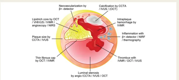
Figure 1 Conceptual graphics illustrating how invasive and non-invasive imaging modalities could combine to provide an integrated assess-ment of microanatomical and biological features of an unstable coronary plaque. CCTA, CT coronary angiography; IVMR, intravascular magnetic resonance; IVUS, intravascular ultrasound; NIRF, near-infrared fluoroscopy; NIRS, near-infrared spectroscopy; OCT, optical coherence tomography.
Editorial
942
calcific deposits, and therefore plaque volume is overestimated and MLA underestimated. Conversely, in non-calcified lesions there is a partial volume effect from the contrast-filled coronary lumen which results in overestimation of luminal area and under-estimation of plaque volume. Boogers’ study population had high calcium scores (mean 595) which explains the lower MLA and higher luminal area stenosis. Moreover, image quality strongly affects interobserver variability, which is lowest for plaque volumes in the proximal segments of the left anterior descending artery (LAD) (17%) and highest for the RCA (32%).14 Plaque volume and variability have a significant inverse relationship, and reproducibility is better in non-calcified than in calcified lesions.13 However, the exact relationship between estimates of local calcifications and measurements of plaque size and extent on CCTA remain uncertain and should be subject to further sys-tematic investigations.
These open questions should encourage further research into the value CCTA for the non-invasive evaluation of coronary lesions. Technical developments in the field of CCTA march at an incredible pace. The spatial and temporal resolution of CCTA is ever improving in fourth-generation devices. The use of dual energy may allow spectral CT, which could allow the identification of types of tissue based on their characteristic energy-dependent photon attenuation, and may thereby allow identification and selective extraction of different qualitative plaque components such as calcifications, or fibrous and lipid-rich components. The ‘hybrid’ approach proposed by Boogers and colleagues provides an excellent platform to validate these new advances against inva-sive gold standards such as virtual histology – IVUS or optical co-herence tomography. An unresolved issue is whether CCTA can distinguish between predominantly lipid-rich and fibrous plaques based on the CT density measured in HU. Several comparisons between CCTA and IVUS have delivered conflicting results, par-ticularly with a large overlap in HU between predominantly lipid-rich and fibrous plaques. Presumably the lack of a standar-dized co-registration algorithm between CCTA and IVUS would explain some of this overlap. Additionally, a hybrid approach would allow combining other intravascular techniques such as near-infrared spectroscopy, intravascular magnetic resonance, thermography, or intravascular positron detection, with CCTA closing a bridge between invasive and non-invasive coronary imaging (Figure 1). By breaking the boundaries of traditional imaging and combining information from different domains, we could potentially improve our understanding of the vulnerability of individual coronary plaques and in the future offer our patients a tailored and personalized approach to reducing individual risk. Conflict of interest: none declared.
References
1. Falk E, Shah P, Fuster V. Coronary plaque disruption. Circulation 1995;92:657 – 671. 2. Naghavi M, Libby P, Falk E, Casscells SW, Litovsky S, Rumberger J, Badimon JJ, Stefanadis C, Moreno P, Pasterkamp G, Fayad Z, Stone PH, Waxman S,
Raggi P, Madjid M, Zarrabi A, Burke A, Yuan C, Fitzgerald PJ, Siscovick DS, de Korte CL, Aikawa M, Juhani Airaksinen KE, Assmann G, Becker CR, Chesebro JH, Farb A, Galis ZS, Jackson C, Jang IK, Koenig W, Lodder RA, March K, Demirovic J, Navab M, Priori SG, Rekhter MD, Bahr R, Grundy SM, Mehran R, Colombo A, Boerwinkle E, Ballantyne C, Insull W Jr., Schwartz RS, Vogel R, Serruys PW, Hansson GK, Faxon DP, Kaul S, Drexler H, Greenland P, Muller JE, Virmani R, Ridker PM, Zipes DP, Shah PK, Willerson JT. From vulner-able plaque to vulnervulner-able patient: a call for new definitions and risk assessment strategies: part I. Circulation 2003;108:1664 – 1672.
3. Abdulla J, Asferg C, Kofoed KF. Prognostic value of absence or presence of cor-onary artery disease determined by 64-slice computed tomography corcor-onary angiography a systematic review and meta-analysis. Int J Cardiovasc Imaging 2011; 27:413 – 420.
4. Min JK, Dunning A, Lin FY, Achenbach S, Al-Mallah M, Budoff MJ, Cademartiri F, Callister TQ, Chang HJ, Cheng V, Chinnaiyan K, Chow BJ, Delago A, Hadamitzky M, Hausleiter J, Kaufmann P, Maffei E, Raff G, Shaw LJ, Villines T, Berman DS. Age- and sex-related differences in all-cause mortality risk based on coronary computed tomography angiography findings results from the Inter-national Multicenter CONFIRM (Coronary CT Angiography Evaluation for Clin-ical Outcomes: an International Multicenter Registry) of 23,854 patients without known coronary artery disease. J Am Coll Cardiol 2011;58:849 – 860. 5. Voros S, Rinehart S, Qian Z, Joshi P, Vazquez G, Fischer C, Belur P, Hulten E,
Villines TC. Coronary atherosclerosis imaging by coronary CT angiography: current status, correlation with intravascular interrogation and meta-analysis. JACC Cardiovasc Imaging 2011;4:537 – 548.
6. Leber AW, Becker A, Knez A, von Ziegler F, Sirol M, Nikolaou K, Ohnesorge B, Fayad ZA, Becker CR, Reiser M, Steinbeck G, Boekstegers P. Accuracy of 64-slice computed tomography to classify and quantify plaque volumes in the proximal coronary system: a comparative study using intravascular ultrasound. J Am Coll Cardiol 2006;47:672 – 677.
7. Kashiwagi M, Tanaka A, Kitabata H, Tsujioka H, Kataiwa H, Komukai K, Tanimoto T, Takemoto K, Takarada S, Kubo T, Hirata K, Nakamura N, Mizukoshi M, Imanishi T, Akasaka T. Feasibility of noninvasive assessment of thin-cap fibroatheroma by multidetector computed tomography. JACC Cardiovasc Imaging 2009;2:1412 – 1419.
8. Hoffmann U, Moselewski F, Nieman K, Jang IK, Ferencik M, Rahman AM, Cury RC, Abbara S, Joneidi-Jafari H, Achenbach S, Brady TJ. Noninvasive assessment of plaque morphology and composition in culprit and stable lesions in acute coron-ary syndrome and stable lesions in stable angina by multidetector computed tom-ography. J Am Coll Cardiol 2006;47:1655 – 1662.
9. Motoyama S, Kondo T, Sarai M, Sugiura A, Harigaya H, Sato T, Inoue K, Okumura M, Ishii J, Anno H, Virmani R, Ozaki Y, Hishida H, Narula J. Multislice computed tomographic characteristics of coronary lesions in acute coronary syn-dromes. J Am Coll Cardiol 2007;50:319 – 326.
10. Motoyama S, Sarai M, Harigaya H, Anno H, Inoue K, Hara T, Naruse H, Ishii J, Hishida H, Wong ND, Virmani R, Kondo T, Ozaki Y, Narula J. Computed tomo-graphic angiography characteristics of atherosclerotic plaques subsequently resulting in acute coronary syndrome. J Am Coll Cardiol 2009;54:49 – 57. 11. Boogers MJ, Broersen A, van Velzen JE, de Graaf FR, El-Naggar HM, Kitslaar PH,
Dijkstra J, Delgado V, Boersma E, de Roos A, Schuijf JD, Schalij MJ, Reiber JHC, Bax JJ, Jukema JW. Automated quantification of coronary plaque with computed tomography: comparison with intravascular ultrasound using a dedicated registration algorithm for fusion-based quantification. Eur Heart J 2012;33: 1007 – 1016.
12. Leber AW, Knez A, von Ziegler F, Becker A, Nikolaou K, Paul S, Wintersperger B, Reiser M, Becker CR, Steinbeck G, Boekstegers P. Quantification of obstructive and nonobstructive coronary lesions by 64-slice computed tomography: a com-parative study with quantitative coronary angiography and intravascular ultra-sound. J Am Coll Cardiol 2005;46:147 – 154.
13. Klass O, Kleinhans S, Walker MJ, Olszewski M, Feuerlein S, Juchems M, Hoffmann MH. Coronary plaque imaging with 256-slice multidetector computed tomography: interobserver variability of volumetric lesion parameters with semi-automatic plaque analysis software. Int J Cardiovasc Imaging 2010;26:711 – 720. 14. Pflederer T, Schmid M, Ropers D, Ropers U, Komatsu S, Daniel WG,
Achenbach S. Interobserver variability of 64-slice computed tomography for the quantification of non-calcified coronary atherosclerotic plaque. Rofo 2007; 179:953 – 957.