Case report
Delayed heart perforation after blunt trauma
Thierry Roth
a, Beat Kipfer
b, Jukka Takala
c, Ralph A. Schmid
a,*
a
Division of General Thoracic Surgery, University Hospital, 3010 Berne, Switzerland
bDivision of Cardiovascular Surgery, University Hospital, Berne, Switzerland cDivision of Intensive Care Medicine, University Hospital, Berne, Switzerland
Received 30 April 2001; received in revised form 21 September 2001; accepted 14 October 2001
Abstract
A 33-year-old patient was hospitalized after a blunt chest trauma with a left flail chest. Six hours after admission to the intensive care unit the patient suddenly developed hypotension and tachycardia. His left chest tube drained 1.5 l of blood within minutes. Immediate resuscita-tion and emergency sternotomy with left anterolateral extension was performed for pericardial tamponade secondary to left ventricular perforation due to a sharp rib fragment. Outcome was favourable and the patient was operated on for his flail chest by internal stabilization the next day. q 2002 Elsevier Science B.V. All rights reserved.
Keywords: Heart perforation; Ventricle laceration; Lung perforation; Blunt chest trauma; Flail chest
1. Introduction
Traumatic injuries, following cardiovascular disease and cancer, are the third most common cause of death in occi-dental countries. In the United States approximately 90 000 accidental deaths and 9 million disabling injuries are committed annually, and 75% of the casualties from blunt trauma are due to chest injuries [1].
2. Case report
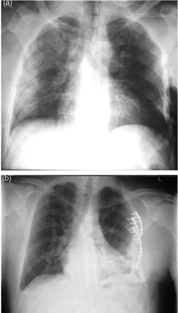
A 33-year-old man was referred from a regional hospital to our centre after a blunt chest trauma. At work, he was crushed between a tractor and a tree. He complained of left thoracic pain without dyspnea. The chest X-ray disclosed multiple rib fractures from the fourth to the eighth rib on the left side, with subcutaneous emphysema (Fig. 1a). The patient’s previous medical history was uneventful. On admission, 3 h after the accident, he was conscious and stable with arterial blood pressure of 150/90 mmHg and heart rate of 109/min, saturation was 94% with 6 l/min oxygen. Clinically a paradoxal motion of the left chest wall could be observed. Blood analysis showed a hyperleu-cocytosis of 20.8 g/l, creatine kinase elevation of 861 IU/l
but troponin , 0:6 mg/l. A left lateral chest tube was
imme-diately inserted which drained 150 ml blood. A computed
tomography (CT) scan of the thorax and abdomen was performed confirming a left haemothorax and the rib frac-tures which were moderately displaced. In addition, a diffuse contralateral lung contusion was present (Fig. 2). An antibiotic prophylaxis with amoxicillin/clavulanic acid was initiated. The operative indication for flail chest stabi-lization was considered and planned for the next day. A peridural analgesia (with bupivacaine 1 mg/ml, fentanyl 2 mg/ml and adrenaline 2 mg/ml) was initiated and the patient was transferred to the intensive care unit (ICU) with mini-mal chest pain. Six hours after admission, he turned in his bed and immediately complained of transfixiant chest pain, the arterial blood pressure fell abruptly to non-measurable levels, and the chest tube drained 1500 ml blood within minutes. Immediate resuscitation with volume perfusion, vasoactive drugs (adrenaline) i.v., intubation and heart massage was instituted while the patient was transferred to the adjacent operative room. An emergency sternotomy with left anterolateral extension (hemi-clamshell) was performed. After drainage of a pericardial blood tamponade, systemic blood pressure rose immediately. A left ventricular perforation of 15 mm length close to the apex was quickly discovered and controlled by finger pressure, then closed with non-absorbable monofilament suture reinforced by Teflon felt. The pericardium was partially closed. A lung perforation of the lingula with air leak was resected using an endo-GIA 45-mm stapler. Two sharp, distal, rib fragments were removed, including the one responsible for the heart perforation. A pericardial tube and two thoracic tubes were
European Journal of Cardio-thoracic Surgery 21 (2002) 121–123
1010-7940/02/$ - see front matter q 2002 Elsevier Science B.V. All rights reserved. PII: S 1 0 1 0 - 7 9 4 0 ( 0 1 ) 0 1 0 5 4 - 5
www.elsevier.com/locate/ejcts
* Corresponding author. Tel.: 141-31-632-2330; fax: 141-31-632-2327. E-mail address: [email protected] (R.A. Schmid).
inserted. After completion of surgery the patient was haemodynamically stable, after receiving 11 blood concen-trates, 6 fresh frozen plasmas and 4000 ml colloid.
On the next day the patient showed no neurological defi-cit and was operated on as planned for his left flail chest stabilization, with reconstruction plates and 4.0-mm cancel-lous screws. The patient did not develop any post-operative respiratory failure and was immediately extubated. The post-operative chest X-ray disclosed no relevant findings (Fig. 1b), and post-operative echocardiography demon-strated good left ventricle function (ejection fraction 70%) with slight pericardial effusion. After removal of chest drains, the patient was discharged from hospital on the 12th post-operative day.
3. Discussion
Rib fractures occur frequently after any chest trauma and are also often disclosed after some abdominal or pelvic major trauma. Surgical treatment is usually not required, merely good analgesia to avoid any occurrence of lung atelectasis or pneumonia. Flail chest with paradoxal respira-tion or thoracic deformity may be considered for an opera-tive rib stabilization.
Heart perforation occurs very exceptionally after blunt chest trauma. In most cases it results from penetrating inju-ries such as stab wound (69%), gun shot (26%) or others (5%), but less than 20% of the injured patients reach the hospital alive [1]. Most frequently the right ventricle is penetrated (46%), followed by the left ventricle (30%) and the right atrium (11%). Heart perforation by rib fragments occur in general immediately after the injury, are fatal in most cases, and are hence not documented. Secondary heart perforation should not be misdiagnosed with secondary cardiac rupture (right atrium 41%, right ventricle 31%, left atrium 25% and left ventricle 9%) after heart contusion, which is also a relatively uncommon complication (0.3%), with a consecutive mortality rate as high as 80% [2]. In the literature, heart perforation due to a fractured rib after blunt chest trauma has been reported in only four patients [3–5]. All of these were caused by motor vehicle accidents. Only one patient was haemodynamically stable prior to surgery. These four heart injuries happened with a delay of 3 h to 15 days after the accident. It seems, however, that secondary
perforation with haemodynamically unstable patients
occurs within the first 24 h (3, 4 and in our case, 6 h). In three cases the left ventricle, and in one case the right ventri-cle, was injured.
A CT scan of the chest is usually performed in the case of flail chest to demonstrate the chest deformity and the poten-tial underlying visceral lesions [3], but secondary
perfora-T. Roth et al. / European Journal of Cardio-thoracic Surgery 21 (2002) 121–123 122
Fig. 1. Chest X-ray on admission (a) and after operative internal fixation with removal of the chest tubes (b).
Fig. 2. CT of the thorax on admission with the rib fractures and the chest-wall deformity.
tion cannot be definitively precluded by this examination. Only two of the four patients reported in the literature survived after secondary heart lesion [4,5]. This mechanism of heart perforation after blunt trauma should be known, because emergency surgery is life-saving in this condition. This case demonstrates the potentially fatal complica-tions which may occur in patients after major chest trauma, particularly with flail chest, even if they have been initially haemodynamically stable. This should be considered in post-traumatic care of patients after blunt chest trauma. Therefore we suggest an observation on ICU-setting for 24 h in a centre with cardiothoracic surgery abilities. Early internal rib stabilization of left flail chest might prevent the occurrence of secondary heart perforation.
References
[1] Calhoon JH, Grover FL, Trinkle JK. Chest trauma, approach and management. Clin Chest Med 1992;13:55–67.
[2] Brathwaite CE, Rodriguez A, Turney SZ, Dunham CM, Cowley R. Blunt traumatic cardiac rupture: a 5-year experience. Ann Surg 1990;212:701–704.
[3] Galvin IF, Costa R, Murton M. Fractured rib with penetrating cardio-pulmonary injury. Ann Thorac Surg 1993;56(3):558–559.
[4] Glock Y, Roux D, Dalous P, Puel P. Left ventricular perforation by rib fracture after closed chest injury. A propos of 2 cases. Ann Chir 1986;40(2):98–101.
[5] Bourguignon N, Godier S, Genevois A, Kaeffer N, Dureuil B. Heart injury following closed thoracic injury. Ann Fr Anesth Reanim 1996;15(7):1088–1089.