ARTICLES
Role of
␣1 Acid Glycoprotein in the In Vivo Resistance
of Human BCR-ABL
+Leukemic Cells to the Abl
Inhibitor STI571
Carlo Gambacorti-Passerini, Rossella Barni, Philipp le Coutre, Massimo Zucchetti,
Gonc¸alo Cabrita, Loredana Cleris, Francesca Rossi, Elisabetta Gianazza, Josef
Brueggen, Robert Cozens, Pietro Pioltelli, Enrico Pogliani, Gianmarco Corneo,
Franca Formelli, Maurizio D’Incalci
Background: Chronic myeloid leukemia is caused by a chro-mosomal translocation that results in an oncogenic fusion protein, Bcr-Abl. Bcr-Abl is a tyrosine kinase whose activity is inhibited by the antineoplastic drug STI571. This drug can cure mice given an injection of human leukemic cells, but treatment ultimately fails in animals that have large tumors when treatment is initiated. We created a mouse model to explore the mechanism of resistance in vivo. Methods: Nude mice were injected with KU812 Bcr-Abl+ human leukemic
cells. After 1 day (no evident tumors), 8 days, or 15 days (tumors >1 g), mice were treated with STI571 (160 mg/kg every 8 hours). Cells recovered from relapsing animals were used for in vitro experiments. Statistical tests were two-sided. Results: Tumors regressed initially in all STI571-treated mice, but all mice treated 15 days after injection of tumor cells eventually relapsed. Relapsed animals did not respond to further STI571 treatment, and their Bcr-Abl kinase ac-tivity in vivo was not inhibited by STI571, despite high plasma concentrations of the drug. However, tumor cells from resistant animals were sensitive to STI571 in vitro, sug-gesting that a molecule in the plasma of relapsed animals may inactivate the drug. The plasma protein␣1 acid glyco-protein (AGP) bound STI571 at physiologic concentrations in vitro and blocked the ability of STI571 to inhibit Bcr-Abl kinase activity in a dose-dependent manner. Plasma AGP concentrations were strongly associated with tumor load. Erythromycin competed with STI571 for AGP binding. When animals bearing large tumors were treated with STI571 alone or with a combination of STI571 and erythro-mycin, greater tumor reductions and better long-term tu-mor-free survival (10 of 12 versus one of 13 at day 180; P<.001) were observed after the combination treatment. Conclusion: AGP in the plasma of relapsed animals binds to STI571, preventing this compound from inhibiting the Bcr/Abl tyrosine kinase. Molecules such as erythromycin that compete with STI571 for binding to AGP may enhance the therapeutic potential of this drug. [J Natl Cancer Inst 2000;92:1641–50]
BCR-ABL, a human oncogenic fusion gene, encodes the hy-brid Bcr-Abl protein. Through its enhanced, constitutive tyro-sine kinase activity, BCR-ABL causes chronic myeloid leu-kemia (CML) and some cases of acute lymphoblastic leuleu-kemia and acute myeloid leukemia (1–3). Inhibition of Bcr-Abl
ki-nase activity is an innovative and rational strategy for treating CML and other neoplasias caused by this oncogenic fusion pro-tein (4).
STI571 (formerly known as CGP57148B) is an active and relatively specific inhibitor of Bcr-Abl kinase activity (5) that blocks cell proliferation and induces apoptotic cell death in BCR-ABL+cells in vitro (6), inhibits the growth of clonogenic bone marrow cells from CML patients (4,7), and can eradicate leukemic cell growth in vivo (8). The in vivo activity of STI571 depends on the stable and continuous inhibition of Bcr-Abl; thus, multiple daily administrations are required in the murine model studied (8). Currently, STI571 is being tested in initial clinical trials of patients with CML and other BCR-ABL-associated diseases.
In our earlier studies (8), treatment with STI571 failed in some animals when it was started 1 week after leukemic cells were injected and a measurable mass was present. Limited in-formation, however, is available on the mechanism of STI571 resistance. Two cell lines have been selected in vitro for resis-tance to STI571 (9). The mechanism of resisresis-tance involves am-plification of the BCR-ABL gene and protein overexpression in one line (10) but is unknown in the other line. However, the biologic relevance of these in vitro-selected sublines remains to be established because the selection conditions in vitro do not necessarily reproduce the in vivo situation.
To our knowledge, no in vivo experimental model of resis-tance to STI571 has been developed and characterized. In this article, we describe the molecular characterization of a murine model for in vivo resistance to STI571.
Affiliations of authors: C. Gambacorti-Passerini, Department of Experimental
Oncology, Istituto Nazionale Tumori, Milan, Italy, and Section of Hematology, University of Milano Bicocca, S. Gerardo Hospital, Monza, Italy; R. Barni, P. le Coutre, G. Cabrita, L. Cleris, F. Rossi, F. Formelli, Department of Experimental Oncology, Istituto Nazionale Tumori, Milan; M. Zucchetti, M. D’Incalci, Mario Negri Institute for Pharmacological Research, Milan; E. Gianazza, Department of Pharmacological Sciences, University of Milano; J. Brueggen, R. Cozens, In Vivo Pharmacology Unit, Oncology Research, Novartis Pharma AG, Basel, Switzerland; P. Pioltelli, E. Pogliani, G. Corneo, Section of Hematology, Uni-versity of Milano Bicocca, S. Gerardo Hospital.
Correspondence to: Carlo Gambacorti-Passerini, M.D., Department of
Ex-perimental Oncology, Istituto Nazionale Tumori, Via Venezian 1, 20133 Milan, Italy (e-mail: Gambacorti@istitutotumori.mi.it).
See “Notes” following “References.”
M
ATERIALS ANDM
ETHODS STI571STI571 was provided by Novartis Pharma AG, Basel, Switzerland. It is a derivative of 2-phenylaminopyrimidine, with a molecular weight of 590. For in
vitro experiments, stock solutions of STI571 were prepared at 1 and 10 mM in
distilled water, filtered, and stored at −20 °C. Preparations used for animal experiments were made daily at 16 mg/mL in water or 5% methylcellulose (Methocel MC high viscosity; Sigma–Aldrich Chemie, Steinheim, Germany), and the solution was kept at 4 °C.
Erythromycin
For in vitro experiments, we used an erythromycin base (Sigma Chemical Co., St. Louis, MO). Erythromycin was dissolved in ethanol and then diluted 1 : 1000 in distilled water. For in vivo experiments, we dissolved erythromycin estolate (provided by DSM Antiinfectives, Capua, Italy) at 35 mg/mL in a solution containing 5% methylcellulose and 16 mg/mL STI571. The estolate formulation of erythromycin was chosen because it has good bioavailability when given orally to mice (11).
In Vivo Administration of STI571
Seven- to 9-week-old female CD1 nu/nu mice purchased from Charles River Breeding Laboratories (Calco, Italy) were kept under standard laboratory con-ditions according to the guidelines of the National Cancer Institute, Milan, Italy. This study was approved by the institutional ethics committee for laboratory animals used in experimental research of the National Cancer Institute. Approxi-mately 50 × 106BCR-ABL+KU812 cells were injected subcutaneously in the
left flank of each animal. STI571 (at 160 mg/kg every 8 hours for 11–21 con-secutive days) was administered orally through a syringe connected to a soft plastic tube introduced into the esophagus (gavage). In all experiments evaluat-ing the antitumor activity of STI571 in vivo, 12–15 animals were used per group. Tumor weight (TW) and total weight were monitored every 3 or 4 days. TW was calculated by the formula TW (mg)⳱ (d2× D/2), where d and D are the shortest
and longest diameters of the tumor, respectively, measured in millimeters. Treat-ment was started 1, 8, or 15 days after leukemia cells were injected; the three groups of animals were designated as groups I, II, and III, respectively.
Cell Lines
The BCR-ABL+ human leukemia cell line KU812 was used (12). It was
obtained from a CML patient in blast crisis and was maintained in RPMI-1640 medium (BioWhittaker, Walkersville, MD) supplemented with 5% fetal calf serum (BioWhittaker).
Determination of the In Vitro Cell Proliferation Activity ([3H]Thymidine Uptake Assay)
Six to eight replicate cultures (200L), each containing 104KU812 cells,
were incubated with 0–10M STI571 in 96-well microtiter plates (Corning Costar Corp., Cambridge, MA) for 54 hours at 37 °C. After this period, 20L of RPMI-1640 medium and 10% fetal calf serum containing [3H]thymidine at a
dose of 1Ci per well (Du Pont NEN, Boston, MA) were added to each well. After an additional 18 hours, cells were harvested and transferred to a filter (Spot-on Filtermat; Pharmacia Biotech Europe, Brussels, Belgium). [3
H]Thymi-dine uptake was measured in a 1205 betaPlate liquid scintillation counter (Wal-lac Inc., Turku, Finland). The IC50 inhibitory concentration (defined as the
concentration of a compound producing a 50% decrease in proliferation com-pared with untreated controls) was calculated. In some experiments, human␣1 acid glycoprotein (AGP) (Sigma–Aldrich Chemie) or human albumin (Centeon Pharma, Marburg, Germany) was added to the wells.
Western Blot Analysis
heated at 95 °C for 10 minutes, and stored at −80 °C. Proteins in the cell lysates and tumor extracts were separated by sodium dodecyl sulfate–polyacrylamide gel electrophoresis on 7.5% gels and transferred to protran nitrocellulose trans-fere membrane (Schelecher & Schuell, Dassel, Germany). Endogenous Bcr-Abl, tyrosine-phosphorylated Bcr-Abl, and actin were detected with the correspond-ing mouse monoclonal antibodies or rabbit antiserum and visualized by en-hanced chemiluminescence detection (Amersham Corp., Little Chalfont, U.K.) by use of horseradish peroxidase-linked goat anti-mouse or anti-rabbit immu-noglobulin G as the secondary antibody (Amersham Corp.). Anti-abl monoclonal antibody (clone Ab-3) was purchased from Calbiochem Corp. (La Jolla, CA). Antiphosphotyrosine monoclonal antibody (clone 4G10) was purchased from Upstate Biotechnology (Lake Placid, NY). Rabbit anti-actin polyclonal antibody was purchased from Sigma Chemical Co.
Densitometric analysis of western blots was performed with an Eagle Eye II photodensitometer (Stratagene Cloning Systems, La Jolla, CA), and the inten-sities of tyrosine-phosphorylated BCR-ABL bands were normalized against the BCR-ABL expression levels.
AGP Determination
Levels of the inducible plasma protein AGP in serum were evaluated by immunodiffusion or by isoelectric focusing. For radial immunodiffusion to mea-sure AGP in mouse serum, we used the single radial immunodiffusion plate test and antiserum specific for murine or human AGP (Cardiotech Services, Inc., Louisville, KY). Five microliters of serum was placed in a well and incubated for 24 hours at 37 °C. The amount of AGP was determined from the diameter of the precipitin ring by use of AGP standards of 1000, 250, and 125g/mL that were provided with the test kit. Determinations were performed in duplicate.
For isoelectric focusing, an immobilized pH gradient (13) from pH 2.5 to pH 5 was cast between an acid solution (i.e., 3 mM Immobiline™ of pK 1 [negative logarithm of the dissociation constant] 1, 11.83 mM Immobiline of pK 3.6, 0.76 mM Immobiline of pK 9.3, and 12.9 mM Tris) and a basic solution (9.28 mM Immobiline of pK 3.6, 9.50 mM Immobiline of pK 4.6, and 16.13 mM Immo-biline of pK 9.3) (Pharmacia, Uppsala, Sweden). After polymerization, the gel was washed three times with 1% glycerol, dried, and rehydrated in a solution of 8 M urea and 0.5% carrier ampholytes from pH 2.5 to pH 5 (Pharmacia). Subsequently, 7.5L of serum, diluted to 25 L with 2% 2-mercaptoethanol, was loaded near the cathode, and the sample was subjected to electrophoresis overnight at 55 V/cm and then to isoelectric focusing for 90 minutes at 165 V/cm. The gel was then stained with Coomassie blue.
Estimation of Binding Parameters
Plasma or purified human AGP (Sigma Chemical Co.) or albumin (diluted in PBS) was incubated at room temperature for 30 minutes with various concen-trations of STI571, and the concentration of STI571 was determined by high-pressure liquid chromatography. This procedure has a lower limit of detection of 0.1M STI571. Free STI571 was determined by ultrafiltration with Centriplus filters (30-kd cutoff, Amicon; Millipore Corp., Bedford, MA). From these data, modified Scatchard plots were constructed, and the association constant of STI571 with each plasma protein was calculated as reported previously (14).
Statistical Analysis
Statistical analysis was performed with two-sided Fisher’s exact test in Fig. 8, B, and Student’s t test in all of the other experiments by use of the GraphPad software analysis program (Prism, San Diego, CA). For survival analysis, data were compared with the log-rank test. P values of less than .05 were considered to be statistically significant and were derived from two-sided statistical tests. All data are presented as the mean (95% confidence interval [CI]). CIs are displayed when they exceed 5% of the respective mean.
given an injection of tumor cells but treated with water only) was detected between days 5 and 12 (data not shown). Tumor re-gression was initially observed in all groups. However, although all animals in group I were cured (by 80 days or longer), 33% of the mice in group II relapsed between days 24 and 34 (range, 33%–40%; n ⳱ 3 experiments) (Fig. 1, A). No cure was ob-served in group III (data not shown). Relapses usually developed 1–3 weeks after treatment was discontinued. When data from groups I, II, and III were compared (Fig. 1, B), a clear relation-ship was observed between the amount of tumor present at the beginning of treatment and the outcome of the therapy. To de-termine whether the period of treatment with STI571 was long enough in groups II and III, we treated two groups of mice in group II with STI571 for either 11 or 18 days. However, we observed the same percentage (40%) of relapses in both groups (data not shown), indicating that a longer period of treatment did not substantially improve the rate of cure. These results indicate that, in this model, treatment with STI571 can cure animals bearing 8-day-old tumors only if the neoplasia is eradicated in the first 11 days of treatment; if this does not happen, cure cannot be achieved, even with longer exposure to STI571.
Relapsed Tumors With In Vivo STI571 Resistance but In Vitro STI571 Sensitivity
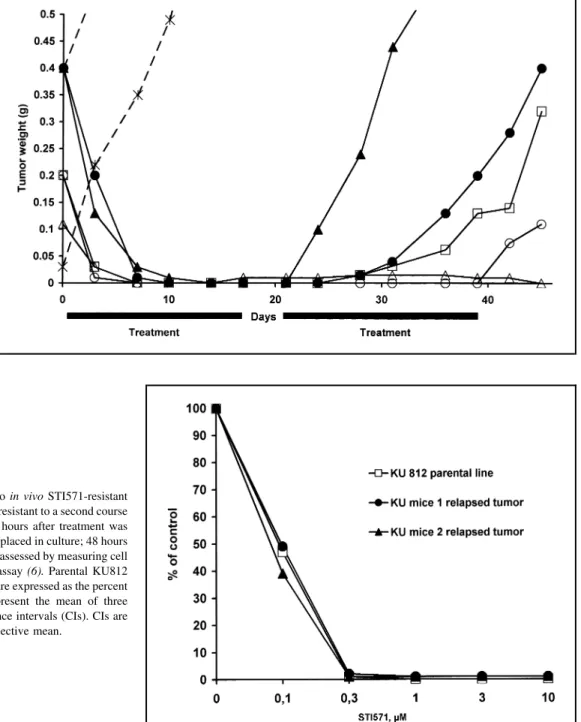
Animals with recurrent tumors were treated a second time with STI571 on the same 11-day schedule. Treatment was started as soon as tumors were measurable. Recurrent tumors responded poorly to the treatment and eventually started to grow at a rate resembling that of tumors in untreated animals (Fig. 2). Only one of five tumors (from the animal with the smallest initial tumor) failed to regrow during the second round of treat-ment. To determine the intrinsic in vitro sensitivity of the recur-rent tumor cells to STI571, we excised tumors from STI571-resistant animals and dissociated, cultured, and tested the cells for their sensitivity to STI571 within 48 hours, as described previously (6) (Fig. 3).
Tumor cells from STI571-resistant animals were found to still be sensitive to STI571. The STI571 IC50values of these cells
and the parental KU812 cell line were similar. These results prompted us to determine whether the Bcr-Abl kinase activity in the tumors of STI571-resistant mice was still inhibited by STI571 by performing molecular pharmacokinetics experiments
Fig. 1. Initial tumor load and the ability of
STI571 to cure tumor-bearing animals. Panel A: Nude mice were given an injection of 50 × 106
BCR-ABL+ KU812 leukemic cells on day 1.
Treatment with STI571 (160 mg/kg orally every 8 hours for 11 days) was started on day 2 (solid
squares⳱ no measurable tumor present) or on
day 9 (solid diamonds⳱ mean tumor weight ⳱ 276 mg; 95% confidence interval [CI]⳱ 179– 373). The percentage of animals with tumor-free survival is presented as a function of time after tumor cell injection. Numbers in parentheses indicate the number of animals at risk. The P value is from a two-sided test. Panel B: Nude mice given an injection of KU812 cells were treated with STI571 (same schedule as in panel
A) on day 2 (group I), on day 9 (group II: mean
tumor weight⳱ 253 mg; 95% CI ⳱ 131–375), or on day 16 (group III; mean tumor weight⳱ 1054 mg; 95% CI⳱ 796–1312). The percentage of animals cured is presented for each group. The results represent the mean of three consecu-tive experiments with 95% CIs. CIs are dis-played only when they exceed 5% of the respec-tive mean.
(8). In these experiments, we investigated the degree and dura-tion of Bcr-Abl inhibidura-tion in tumor-bearing animals that either were sensitive or were resistant to STI571. Mice in both groups were treated acutely with STI571 (160 mg/kg orally) and killed 2, 4, or 6 hours later. The levels of Bcr-Abl kinase activity (measured as autophosphorylation) are shown in Fig. 4. Al-though tumor extracts from STI571-sensitive mice showed the previously reported inhibition (8) of Bcr-Abl
phosphoryla-STI571 compensated by increasing their metabolism of phosphoryla-STI571, thereby decreasing the concentration of STI571 in plasma to a level that would not inhibit Bcr-Abl activity. Tumor-bearing mice (three or four animals per group, one that was sensitive to STI571 and one that was resistant to STI571) were killed at 30 minutes, 2 hours, and 5 hours after an acute treatment with STI571 (160 mg/kg). Total concentrations of STI571 in the plasma and the tumor were determined by reverse-phase high-M
Fig. 2. Appearance of STI571-resistant
tumors in animals that had an initial positive response to STI571. Weights of tumors in five animals are shown. Tumors first responded to an STI571 treatment of 160 mg/kg orally every 8 hours for 18 days. If the tumor re-curred, animals were treated with STI571 as soon as the tumor was mea-surable. Weights of the tumor at vari-ous times after the initiation of treat-ment are shown. Dashed lines show the growth of two untreated tumors.
Different symbols indicate individual
animals.
Fig. 3. In vitro sensitivity to STI571 of two in vivo STI571-resistant
tumors. Tumors from two animals that were resistant to a second course of treatment with STI571 were excised 72 hours after treatment was discontinued. Tumor cells were isolated and placed in culture; 48 hours later, their in vitro sensitivity to STI571 was assessed by measuring cell proliferation with a [3H]thymidine uptake assay (6). Parental KU812
cells were included for comparison. Values are expressed as the percent of untreated control cells. The results represent the mean of three consecutive experiments with 95% confidence intervals (CIs). CIs are displayed when they exceed 5% of the respective mean.
⳱ 1.4–15.4]; P ⳱ .36) and 5 hours (2.1 M [95% CI ⳱ 1.3– 2.9] versus 5.7 M [95% CI ⳱ 4.5–6.9]; P<.001), an even greater decrease was observed in STI571 levels in STI571-sensitive animals as compared with STI571-resistant animals. There was a tendency toward lower STI571 concentrations in tumors of STI571-resistant animals (data not shown). These pre-liminary results did not support our initial hypothesis that
ani-mals treated with STI571 compensate by increasing the metabo-lism of this compound. These data were instead compatible with the presence of a factor in the blood of STI571-resistant mice that binds to and thus decreases the biologic activity of STI571. AGP Binding of STI571
Two plasma proteins that can bind drugs are albumin and AGP (14), and AGP preferentially binds basic molecules; STI571, in fact, contains several residues that can confirm a basic potential to this. BCR-ABL+ KU812 cells were used to
assess the ability of AGP to alter the STI571-mediated inhibition of cell proliferation. Because murine AGP was not available in quantities sufficient for this experiment (15), human AGP was used. AGP inhibited the effect of STI571 (measured as IC50) in
a concentration-dependent manner (Fig. 5, A). The IC50 value
for STI571 increased from 0.05 M in the absence of AGP to greater than 3.0 M in the presence of AGP at 2 mg/mL. In a separate experiment, the IC50value for STI571 in the presence
of AGP at 2 mg/mL was calculated to be 4.5M, an increase of 90-fold (data not shown). By contrast, albumin did not substan-tially increase the IC50for STI571, even at 35 mg/mL (Fig. 5, B).
The association constants for the binding of STI571 to AGP and to albumin were then calculated. We incubated 5M AGP with various STI571 concentrations and determined the amount of unbound drug (i.e., the free fraction) by ultrafiltration. Scatchard analyses were done for AGP and albumin to deter-Fig. 4. In vivo STI571-mediated inhibition of BCR-ABL autophosphorylation.
Tumor-bearing mice were acutely treated with STI571 (160 mg/kg orally) and killed 2, 4, or 6 hours later. Tumor extracts were made and used for western blot analysis with antiphosphotyrosine (ptyr) to detect phosphorylated Bcr-Abl or anti-Abelson (abl) to detect total Bcr-Abl. Ctrl⳱ tumor extract from animals not pretreated with STI571; Rel⳱ tumor extract from animals pretreated with STI571 that relapsed and were resistant to further STI571 treatment; and n.t.⳱ not treated. Similar data were obtained from three other animals.
Fig. 5. Effect of␣1 acid glycoprotein (AGP) (panel A) and albumin (panel B) on the in vitro sensitivity of
KU812 cells to STI571. KU812 cells were cultured with AGP (0–2 mg/mL), with albumin (0–35 mg/mL), or with neither, and the sensitivity to STI571 was as-sessed after 48 hours as described previously (6) by [3H] thymidine uptake assay. Data from one
represen-tative experiment are shown. The experiment was per-formed three times with similar results. The results represent one representative experiment with 95% confidence intervals (CIs). CIs are displayed when they exceed 5% of the respective mean.
mine the association constants of STI571 to this protein. The association constant for AGP was 4.9 × 10⁶ L/mol, which is approximately 21 times higher than the association constant cal-culated for albumin (2.3 × 10⁵ L/mol). These results indicate that, although albumin and AGP can bind STI571, the latter binds STI571 with a much higher affinity and thus lowers the concentration of active STI571 more effectively.
We confirmed these results in a [3H]thymidine uptake assay
with KU812 cells with the use of sera containing different amounts of AGP. KU812 cells were incubated with serum con-taining either 330 (serum A) or 1150 (serum B)g of AGP per mL that was added to the culture medium at a final concentration of 25% (vol/vol) for 72 hours. The calculated IC50 values for STI571 were 0.1M without added serum, 0.4 M with serum A, and greater than 2M with serum B. Although these results do not exclude other serum factors, the IC50value for STI571
appears to increase in an AGP concentration-dependent manner. Thus, AGP can bind STI571, and this binding blocks the ability of STI571 to inhibit the proliferative activity of BCR-ABL+cells and, consequently, has important biologic consequences. Relationship of AGP Serum Levels, Tumor Load, and STI571 Treatment
Because AGP potently interfered with the ability of STI571 to inhibit the proliferation of BCR-ABL+cells in vitro, we mea-sured the levels of AGP in the plasma of nude mice 8 and 15 days after the injection of KU812 cells. We used these data to determine whether there was an association between the in vivo sensitivity of KU812 cells to STI571 and the level of AGP in the corresponding mouse plasma. As measured by immunodiffu-sion, basal levels of AGP in control mice that had not received an injection of tumor cells were very low (96g/mL [95% CI ⳱ 75–117]; Fig. 6, A). In tumor-bearing mice, the AGP concen-tration increased proportionally with the tumor load. Mice with
a tumor load of 200–300 mg (8 days after tumor cell injection) showed a fourfold increase in AGP (383 g/mL; 95% CI ⳱ 252–514), and mice with a tumor load of 0.8–1 g (15–20 days after tumor cell injection) had a mean AGP level of 1580g/mL (95% CI ⳱ 1346–1814). Animals with measurable tumors showed a progressive decrease in plasma AGP levels as the tumors were cured; these AGP levels returned to normal in cured animals 4–8 weeks after the start of the treatment. The amount of AGP observed in the plasma of normal mice corresponded to an in vitro AGP-bound STI571 fraction of 42% at an STI571 concentration of 7M, and the amount observed in the plasma of mice bearing large tumors corresponded to an AGP-bound STI571 fraction of more than 99% (data not shown). After treat-ment of control mice with STI571 at 160 mg/kg orally for 11 days, treated mice had a lower, but statistically significant, in-crease in the amount of plasma AGP (213g/mL; 95% CI ⳱ 170–256) compared with non-STI571-treated mice. Increased AGP levels in tumor-bearing mice were also detected by iso-electrofocusing (Fig. 6, B). Thus, tumor load (and, to a lesser extent, STI571 pretreatment) induced the synthesis of AGP, a plasma protein that, in turn, could bind and effectively inactivate STI571.
Erythromycin and Its Competition With STI571 for AGP Binding
Several drugs, including erythromycin (15), can bind to AGP. If the binding of STI571 to AGP effectively inhibits the ability of STI571 to block cell division and inhibit Bcr-Abl kinase activity, then a molecule that binds to the same site on AGP could compete with STI571 and thus increase the concentration of free STI571. To test this possibility, we added 5–30 M erythromycin to KU812 cultures containing STI571 and/or AGP and assessed cell proliferation (Fig. 7, A). In the presence of AGP, erythromycin restored the sensitivity of KU812 cells to
STI571 but did not modify the IC50of STI571 in the absence of
AGP. Thus, a direct interaction between erythromycin and STI571 was excluded. To determine whether erythromycin af-fected the STI571-mediated inhibition of Bcr-Abl kinase activity (Fig. 7, B), we cultured KU812 cells with STI571, AGP, and/or erythromycin at 37 °C for 60 minutes; cells were then lysed, and Bcr-Abl kinase activity in the lysates was measured. STI571 inhibited the autophosphorylation of Bcr-Abl, AGP decreased the ability of STI571 to inhibit this activity, and erythromycin restored it. Thus, results from two assays demonstrate that eryth-romycin and STI571 compete for binding to AGP, and thus the presence of erythromycin increases the amount of free STI571. In Vivo Effects of Erythromycin Treatment and STI571 Pretreatment
To confirm that erythromycin could reverse the observed in vivo resistance to STI571, we injected mice with KU812 cells and started STI571 treatment 11 days later, when the tumor load was approximately 400 mg. (At this stage, we expected few or no cures from a standard STI571 treatment.) The treatment schedule that we used was STI571 at a dose of 160 mg/kg given orally every 8 hours, either alone or in combination with eryth-romycin estolate (350 mg/kg every 8 hours) for 21 days. This formulation produced peak concentrations of more than 20M erythromycin (11). The combined treatment produced a statisti-cally significantly higher tumor reduction on day 6 and then from day 16 onward (Fig. 8, A). Tumors regressed progressively in mice receiving the combined treatment, whereas some tumors started to regrow during the last days of treatment in the group given STI571 alone.
The effect of erythromycin was even more apparent when the cure rates in the two groups were compared (Fig. 8, B). Of the 15 mice receiving STI571 alone, the tumor initially disappeared from five, but it reappeared in four of these animals between
days 25 and 40, so that only one of 13 animals was cured at day 180 (last day of follow-up) (two tumor-bearing animals in this group were accidentally killed at some point during treatment). In the group receiving the combined treatment of erythromycin and STI571, 14 of 15 mice became tumor free and a tumor had recurred in only one mouse by day 30 (three tumor-free animals were accidentally killed during the treatment procedure). There-fore, 10 of 12 animals were cured by the combined treatment at day 180, a value that is statistically significantly different (P<.001) from the value (one of 13 animals) obtained in the group receiving STI571 only. Control groups receiving erythro-mycin alone did not show any evidence of tumor regression (data not shown).
The effects of STI571 pretreatment on tumor growth by in-jected leukemic cells were also evaluated. Mice that had been pretreated with STI571 for 11 days or had not been pretreated received an injection of 50 × 106KU812 cells and were treated with STI571 after 1 day (160 mg/kg every 8 hours for 11 days), a treatment regimen that is expected to cure all of the animals. None of the 14 controls (nonpretreated mice) developed tumors, whereas seven of the 14 pretreated mice had detectable tumors (P ⳱ .006). These results indicate that the elevated levels of AGP induced by a previous STI571 treatment can also produce a statistically significant biologic effect.
Thus, these results support the hypothesis that the binding of STI571 to AGP inhibits its therapeutic activity and provide a partial experimental confirmation for the use of erythromycin or similar compounds to bypass in vivo resistance mediated by AGP.
D
ISCUSSIONSTI571 is a new type of antineoplastic drug that is specifi-cally tailored to an oncogenic protein causally linked to several Fig. 7.␣1 Acid glycoprotein (AGP) and erythromycin effects on the ability of
STI571 to inhibit KU812 cell proliferation and BCR/ABL autophosphorylation.
Panel A: KU812 cells were cultured with STI571 (䊏 ⳱ 1 M), AGP (䉱 ⳱ 1
mg/mL), or both STI571 (1M) and AGP (1 mg/mL) (⽧) in the presence of increasing concentrations of erythromycin (0–30M). Cells were then pulse labeled with [3H]thymidine and harvested. Data are the percent of untreated
control cells. All data are presented as means, with 95% confidence intervals (CIs). CIs are displayed when they exceed 5% of the respective mean. Panel B: Approximately 3 × 10⁶KU812 cells were incubated per well at 37 °C with an
eryrthromycin base (100 M), STI571 (3 M), and/or AGP (2 mg/mL), as indicated. After 1 hour, cells were washed twice with ice-cold phosphate-buffered saline and subsequently lysed in 500L of 1× Laemmli’s buffer (8). Cell lysates corresponding to 90 000 cells were analyzed by sodium dodecyl sulfate–polyacrylamide electrophoresis on 7.5% gels. Total endogenous Bcr-Abl (anti-Abl) and tyrosine-phosphorylated Bcr-Abl (anti-PTyr) were detected with the mouse monoclonal antibody indicated. The results of one representative experiment are presented. The experiment was performed two times with similar results.
human cancers. The antileukemic activity of STI571 is well documented in vitro and in vivo (4–8). STI571 is not absolutely specific; in fact, it inhibits the normal Abl protein and the re-ceptor tyrosine kinases c-kit and platelet-derived growth factor-. However, the toxic profile of STI571 seems benign, with few normal cells being affected and very limited side effects ob-served so far in treated patients (6,16).
Because STI571 is an inhibitor of Bcr-Abl, the development of resistance is an important issue. Resistance can develop as a
selected in vitro, and this type of resistance has not yet been observed in vivo. In this article, we report the induction and characterization of resistance to STI571 in vivo. The mechanism underlying this resistance was traced to the induction of AGP, a plasma protein that binds STI571 tightly and inhibits its ability to interact with Bcr-Abl kinase. Consistent with the fact that resistance did not originate intracellularly in the tumor cell, in vitro assays did not demonstrate any intrinsic resistance to STI571, although differences in sensitivity to low STI571 con-M) cannot be ruled out. We showed that
Fig. 8. Co-administration of erythromycin and
STI571 to tumor-bearing mice. Animals bearing a tumor 11 days after tumor cell injection were randomly assigned to one of two groups. Fifteen mice were treated with STI571 and erythromy-cin (average tumor weight⳱ 385 mg; 95% con-fidence interval [CI]⳱ 332–438), and 15 addi-tional animals received STI571 only (mean tumor weight⳱ 390 mg; 95% CI ⳱ 276–504). The treatment schedule that we used was STI571 at a dose of 160 mg/kg given orally every 8 hours, either alone or in combination with eryth-romycin estolate (350 mg/kg every 8 hours) for 21 days. Control animals received erythromycin in 5% methylcellulose (five mice) or in 5% methylcellulose alone (six mice). Panel A: Mean tumor weights measured during treatment (days 0–21) from mice treated with STI571 alone or with erythromycin and STI571 are shown. Error bars represent 95% CIs. Panel B: Percent of tumor-bearing mice is shown as a function of time after the start of treatment. Mice were treated for 21 days with STI571 (160 mg/ kg) and erythromycin (350 mg/kg) administered every 8 hours. Error bars represent 95% CIs.
Numbers in parentheses indicate the number of
20M, erythromycin substantially increased the therapeutic ac-tivity of STI571 in vivo. It is important to note that erythromycin had no effect in vitro in the absence of AGP, which excludes the possibility that AGP directly affected the sensitivity of KU812 cells to STI571. Although erythromycin can interfere with the metabolism of several drugs, in our in vivo model, erythromycin augmented the antitumor activity of STI571 only when levels of AGP were also increased. In fact, when mice were given an injection of COLIA/PDGFB transfectants, which are sensitive to STI571 treatment (22) but do not induce increased AGP levels in mice (Gambacorti C: unpublished results), the therapeutic ef-fects of a combined treatment with erythromycin and STI571 and a treatment with STI571 alone (Greco MA: personal com-munication) were similar.
The increased AGP levels associated with advanced tumors provide an explanation for the limited efficacy of STI571 in our model. The increased AGP level apparently induced by STI571 pretreatment substantially reduced the therapeutic activity of STI571. These data strongly suggest that AGP, when present in increased concentrations, bound most of the STI571 adminis-tered, blocked its diffusion from the blood to tissues and cells, and thus blocked access to its biologic targets. It is also worth noting that another molecule, UCN-01, with some structural similarity to STI571, was reported to bind strongly to AGP and thus reduce its biologic availability (15).
Because an initial tumor reduction was noted even in animals bearing large tumors, some sort of selection is probably taking place in the relapsed tumor. This selection, however, is possible by the exposure of leukemic cells to STI571 at marginally active concentrations (because most of the STI571 is bound to AGP), a condition that was, in fact, exploited for selecting resistance in vitro (10).
AGP is an acute-phase protein that is synthesized in the liver and has an average molecular weight of 40 000 (14). AGP binds neutral and basic drugs in a one-to-one molar ratio. Increased levels of AGP have been described in a variety of pathologic conditions, such as chronic inflammation, myocardial infarction, and advanced cancer (14). The data presented herein are derived from an animal model, and their clinical relevance remains to be demonstrated. However, it has been observed in an ongoing clinical trial that the majority of CML patients in blast crisis and patients with relapsed Philadelphia chromosome-positive acute lymphoblastic leukemia show only temporary responses to STI571, which are soon followed by the development of resis-tance, while the therapeutic effects seem more durable in CML patients treated in chronic phase (16,23).
Substantial differences exist, however, between mice and hu-mans. Mice have very low baseline AGP levels (<100 mg/mL), whereas humans have higher and wider baseline AGP levels (400–800 mg/mL). Thus, under normal conditions, the fraction of STI571 bound to AGP should be considerably higher in hu-mans than in mice. For example, at STI571 concentrations found in patients (1–10M) and at murine AGP levels (<2.5 M), the AGP-bound fraction of STI571 can be between 40% and 60%, whereas at the same STI571 concentrations and normal human AGP levels (10–20M), the AGP-bound fraction of STI571 is less than 10%. Thus, a mouse with normal AGP values retains a substantial fraction of free STI571, but a leukemia patient, who has higher levels of AGP, does not. Furthermore, exposure of CML cells to marginally active STI571 concentrations could facilitate the emergence of resistant leukemic cells, especially in
patients in blast crisis when additional genetic alterations induce a higher degree of heterogeneity.
Therefore, the bioavailability of STI571 can be substantially modified by the presence of AGP. The four to eight times higher levels of AGP in humans than in mice could increase the half-life of STI571, thus permitting STI571 to reach and maintain more constant concentrations in humans than in mice. However, the high AGP levels, including those associated with pathologic conditions, could alter the tissue distribution of STI571 and the cellular uptake of the drug, which would interfere with the ac-cess of STI571 to its intracellular target protein, Bcr-Abl. The concentration of free STI571 would be expected to increase dramatically as dosages approached the stoichiometric equiva-lence of STI571 to AGP. Drugs that compete with STI571 for AGP binding could increase the concentration of free STI571 without the need to increase the total dose of STI571, a strategy that we have used in our model. Extreme caution must be ex-ercised, however, in transferring these data to clinical studies. The results presented in this article were obtained in a murine model that is characterized by baseline low AGP levels and that used high doses of STI571 and erythromycin. Clearly, appropri-ate pilot clinical studies will be needed to validappropri-ate these results in humans. Our data also indicate the need to assess free STI571 levels in addition to total plasma concentrations in designing dosage and treatment schedules, to ensure an optimal tissue dis-tribution of the drug, ideally combined with the concurrent evaluation of surrogate markers of activity (e.g., the phosphor-ylation status of Bcr-Abl) within the tumor.
In conclusion, we have identified the molecular nature of in vivo resistance to STI571 in this model and have devised a strategy to circumvent it. Our results indicate that in vivo resis-tance to STI571 is caused by AGP, a plasma protein that is not found in the target leukemic cells, but do not exclude the pos-sibility that resistance to STI571 can develop by other mecha-nisms (10). The decreased expression of BCL-XLin CML cells
treated with STI571 seems to be important in mediating the apoptotic effect of STI571 (24). However, BCL-XLlevels did
not change in BCR-ABL+cells selected for resistance to STI571 (25). Further studies in vivo and in vitro will be needed to elu-cidate the molecular mechanisms involved in determining sen-sitivity or resistance to STI571 and to increase the therapeutic potential of this promising and innovative drug.
R
EFERENCES(1) Heisterkamp N, Stam K, Groffen J, de Klein A, Grosveld G. Structural
organization of the bcr gene and its role in the Ph’ translocation. Nature 1985;315:758–61.
(2) Barila D, Superti-Furga G. An intramolecular SH3-domain interaction
regulates c-Abl activity. Nat Genet 1998;18:280–2.
(3) Goga A, McLaughlin J, Afar DE, Saffran DC, Witte ON. Alternative
signals to RAS for hematopoietic transformation by the BCR-ABL onco-gene. Cell 1995;82:981–8.
(4) Druker BJ, Tamura S, Buchdunger E, Ohno S, Segal GM, Fanning S, et al.
Effects of a selective inhibitor of the Abl tyrosine kinase on the growth of Bcr-Abl positive cells. Nat Med 1996;2:561–6.
(5) Buchdunger E, Zimmermann J, Mett H, Meyer T, Muller M, Druker BJ, et
al. Inhibition of the Abl protein–tyrosine kinase in vitro and in vivo by a 2-phenylaminopyrimidine derivative. Cancer Res 1996;56:100–4.
(6) Gambacorti-Passerini C, le Coutre P, Mologni L, Fanelli M, Bertazzoli C,
Marchesi E, et al. Inhibition of the ABL kinase activity blocks the prolif-eration of BCR/ABL+ leukemic cells and induces apoptosis. Blood Cells Mol Dis 1997;23:380–94.
(7) Deininger MW, Goldman JM, Lydon N, Melo JV. The tyrosine kinase
inhibitor CGP57148B selectively inhibits the growth of BCR-ABL-positive cells. Blood 1997;90:3691–8.
(8) le Coutre P, Mologni L, Cleris L, Marchesi E, Buchdunger E, Giardini R,
et al. In vivo eradication of human BCR/ABL-positive leukemia cells with an ABL kinase inhibitor. J Natl Cancer Inst 1999;91:163–8.
(9) le Coutre P, Marchesi E, Bertazzoli C, Pogliani E, Gambacorti-Passerini C.
Generation of BCR-ABL positive leukemic cell lines resistant to a selective ABL kinase inhibitor [absract]. Blood 1997;90:496a.
(10) le Coutre P, Tassi E, Varella-Garcia M, Barni R, Mologni L, Cabrita G, et
al. Induction of resistance to the Abelson inhibitor STI571 in human leu-kemic cells through gene amplification. Blood 2000;95:1758–66.
(11) Vainio PJ, Viluksela M, Mannisto PT. Absorption of various erythromycin
esters and salts in mice after intragastric intubation. Methods Find Exp Clin Pharmacol 1992;14:367–72.
(12) Kishi K. A new leukemia cell line with Philadelphia chromosome
charac-terized as basophil precursors. Leuk Res 1985;9:381–90.
(13) Gianazza E, Celentano F, Magenes S, Ettori G, Righetti PG. Formulations
for immobilized pH gradients including pH extremes. Electrophoresis 1989;10:806–8.
(14) Kremer JM, Wilting J, Janssen LH. Drug binding to human alpha-1-acid
glycoprotein in health and disease. Pharmacol Rev 1988;40:1–47.
(15) Fuse E. Tanii H, Kurata N, Kobayashi H, Shimada Y, Tamura T, et al.
Unpredicted clinical pharmacology of UCN-01 caused by specific finding to human alpha1-acid glycoprotein. Cancer 1998;3248–53.
(16) Druker BJ, Talpaz M, Resta D, Peng B, Buchdunger E, Ford J, et al.
Clinical efficacy and safety of an ABL specific tyrosine kinase inhibitor as targeted therapy for chronic myelogenous leukemia. 41st Meeting of the American Society of Hematology. New Orleans (LA). Blood 1999. p 368a.
(17) Goker E, Waltham M, Kheradpour A, Trippett T, Mazumdar M, Elisseyeff
Y, et al. Amplification of the dihydrofolate reductase gene is a mechanism of acquired resistance to methotrexate in patients with acute lymphoblastic leukemia and is correlated with p53 gene mutations. Blood 1995;86: 677–84.
(18) Shao W, Benedetti L, Lamph WW, Nervi C, Miller WH Jr. A
retinoid-resistant acute promyelocytic leukemia subclone expresses a dominant negative PML-RAR alpha mutation. Blood 1997;89:4282–9.
(19) Muindi JR, Frankel SR, Miller WH Jr, Jekubowski A, Scheinberg DA,
Young CW, et al. Continuous treatment with all-trans retinoic acid causes a progressive reduction in plasma drug concentrations: implications for relapse and retinoid “resistance” in patients with acute promyelocytic leu-kemia [published erratum appears in Blood 1992;80:855]. Blood 1992;79: 299–303.
(20) Dermime S, Grignani F, Clerici M, Nervi C, Sozzi G, Talamo GP, et al.
Occurrence of resistance to retinoic acid in the acute promyelocytic
leu-kemia cell line NB4 is associated with altered expression of the pml/RAR alpha protein. Blood 1993;82:1573–7.
(21) Fanelli M, Minucci S, Gelmetti V, Nervi C, Gambacorti-Passerini C,
Pelicci PG. Constitutive degradation of PML/RARalpha through the pro-teasome pathway mediates retinoic acid resistance. Blood 1999; 93: 1477–81.
(22) Shimizu A, O’Brien KP, Sjoblom T, Pietras K, Buchdunger E, Collins VP,
et al. The dermatofibrosarcoma protuberans-associated collagen type Ial-pha1/platelet-derived growth factor (PDGF) B-chain fusion gene generates a transforming protein that is processed to functional PDGF-BB. Cancer Res 1999;59:3719–23.
(23) Druker BJ, Kantarjian H, Sawyers LC, Resta D, Fernandes Rees S, Ford J,
et al. Activity of an ABL specific tyrosine kinase inhibitor in patients with BCR-ABL positive acute leukemias, including chronic myelogenous leu-kemia in blast crisis. 41st Meeting of the American Society of Hematology. New Orleans (LA). Blood 1999. p 697a.
(24) Horita M, Andreu EJ, Benito A, Arbona C, Sanz C, Benet I, et al. Blockade
of the Bcr-Abl kinase activity induces apoptosis of chronic myelogenous leukemia cells by suppressing signal transducer and activator of transcrip-tion 5-dependent expression of Bcl-xL. J Exp Med 2000;191:977–84.
(25) Weisberg E, Griffin JD. Mechanism of resistance to the ABL tyrosine
kinase inhibitor STI571 in BCR/ABL-transformed hematopoietic cell lines. Blood 2000;95:3498–505.
N
OTESEditor’s note: C. Gambacorti-Passerini and E. Pogliani are co-principal
in-vestigators of three clinical studies on STI571 sponsored by Novartis Pharma AG (Basel, Switzerland). P. le Coutre is the principal investigator on a clinical trial of STI571 sponsored by Novartis Pharma AG. J. Brugeggen and R. Cozens are employed by and own stock in Novartis Pharma AG; the research reported was performed as a part of their employment. E. Pioltelli is involved in three clinical trials on STI571, sponsored by Novartis Pharm AG, which are presently being conducted at their institution. G. Corneo is the principal investigator on three clinical trials on STI571, sponsored by Novartis Pharma AG, that are being conducted at his institution.
C. Gambacorti-Passerini and R. Barni contributed equally to this article. Supported in part by the Italian Association for Cancer Research (420.198.662), the Italian Research Council (95.00842,9600225.CT04), the Is-tituto Superiore di Sanita` (881A/10), and the Jose Carreras International Foun-dation.
We thank Dott. Luca Mantovani and Giuseppe Palladino (DSM-Anti-infectives SPA, Capua, Italy) for the supply of erythromycin estolate and Dr. Roberta Rostagno for editorial assistance.
Manuscript received February 28, 2000; revised August 11, 2000; accepted August 15, 2000.