Unilateral and bilateral corticotomies for correction of
maxillary transverse discrepancies
C. F. Mossaz\ F. K. Byloff\ and M. Richter"
'Department of Orthodontics, School of Dentistry, and "Department of Surgery, Maxillofacial Surgery Unit, University Hospital of Geneva, Switzerland
SUMMARY Surgically-assisted rapid maxillary expansion in adults has been proved effective in overcoming the strong resistance of the maxillary complex after growth is completed, particularly after the second decade of life.
The aim of this study was to describe the dental and the skeletal expansion and relapse, as well as the amount of tipping of the two maxillary bones and first permanent molars, during a rapid maxillary expansion procedure combined with unilateral and bilateral corticotomies.
The sample consisted of four adult patients, two presenting with bilateral and two with unilateral cross-bite. Records were taken before and after rapid maxillary expansion, at the end of retention and at least 12 months post-retention.
In the cases of bilateral cross-bite the same amount of skeletal expansion was observed on both sides. The angular changes measured at the upper first molars indicated important tipping on both sides, which tended to relapse moderately during the retention and post-retention period. Following unilateral surgery, the operated side showed more than twice the amount of skeletal expansion than the non-operated side. The angular changes presented twice as much tipping and relapse on the operated side.
The results of this study demonstrate that unilateral cross-bites in adults can be corrected with unilateral corticotomy and rapid maxillary expansion using the contralateral non-operated side as anchorage.
Stability appeared satisfactory in all cases.
Introduction
Rapid maxillary expansion is the method com-monly used to correct maxillary transverse dis-crepancies, especially during the growth period. Haas (1970) considered the 'growth spurt' as the ideal time for this procedure, as alveolar remodelling occurs with minimal tooth tipping. In older patients the risk of failure as well as the tendency towards relapse increases (Wertz, 1970). Timms and Vero (1981) reported the absence of mid-palatal suture opening in a 15-year-old girl. Using cadavers, Persson and Thi-lander (1977) found that synostosis in the mid-palatal suture begins between 15 and 19 years of age, and increases in the third decade. For those reasons, Timms and Vero (1981) recom-mended normal rapid maxillary expansion until the age of 25 years, and a palatal osteotomy when the suture had not opened after 1 week of daily activation of the expansion device.
Lines (1975) advocated both lateral cortico-tomy as well as surgical opening of the mid-palatal suture to eliminate resistance before the activation of a maxillary expansion appliance. Using selected maxillary osteotomies as an adjunct to rapid maxillary expansion in monkeys, Kennedy and Bell (1976) found that the most effective osteotomy to reduce the resistance to lateral movement of the two hemi-maxillae was a corticotomy through the zygomatic buttress, nasomaxillary and pterygomaxillary areas. Glassmann (1984), Alpern and Yurosko (1987), and Lehmann et al. (1984) confirmed these find-ings in human adults. The expansion was success-fully performed with a Hyraxmm (Dentaurum
AG, Pforzheim, Germany) appliance, following a lateral osteotomy from the piriform rim to the pterygoid plate without palatal surgery. Their study did not consider the amount of skeletal versus dental expansion, and the corresponding relapse following a retention period.
111 The aim of this investigation is to describe
the dental and skeletal expansion as well as the amount of dento-alveolar tipping in adults pro-duced by rapid maxillary expansion following lateral maxillary osteotomies. In two patients presenting a unilateral cross-bite, a corticotomy was performed unilaterally, and the changes on each side were analysed separately and com-pared to the bilateral osteotomy subjects.
Subjects and methods
The sample consisted of four adult patients, two males and two females, aged 21 to 35 years, two presenting a bilateral and two with unilat-eral cross-bite without mandibular dis-placement.
A Hyraxmin appliance was soldered in the
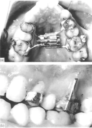
laboratory to bands previously fitted to the upper first premolars and upper first molars, and then cemented prior to the surgery (Fig. la).
Figure I (a) Patient J.M.: occlusal view of the Hyraxmio
appliance cemented on the patient pre-operatively. (b) Patient J.M.: lateral view showing head gear tubes soldered vertically on the molar bands and pins inserted before taking the P.A. head-films to measure angular changes on the radiographs.
On the buccal side of the permanent molars, headgear tubes were soldered vertically (Fig. lb). The surgical intervention was carried out under general anaesthesia. An incision was made at the depth of the vestibule from the first molar area to the distal aspect of the lateral incisor. The mucoperiosteum was then elevated and the maxillary bone exposed from the piri-form aperture anteriorly to the pterygomaxillary fissure posteriorly via a subperiosteal tunnelling technique. Subsequently, an osteotomy was made horizontally well above the apices of the teeth from the piriform aperture to the pterygo-maxillary fissure (Fig. 2). A curved osteome was then used to separate the pterygoid plate from the maxilla. Anteriorly, the maxilla was separ-ated by malleting a thin osteotome between the central incisors at a level below the anterior nasal spine. In bilateral cross-bite cases, this surgical procedure was carried out on both sides.-The surgical site was then closed with continuous arid interrupted sutures of polyam-ide sheathed multifil (Supramid 3-0).
The previously cemented appliance was activ-ated four quarter-turns (1 mm) at the time of surgery.
The patients were later instructed to activate the appliance one quarter-turn a day, until the necessary amount of expansion was achieved. The patients were seen once a week during the expansion procedure. After completion of expansion, the appliances were left in place for 12 weeks, and then removed and replaced by a conventional maxillary retainer for another 3-month period. In the unilateral cross-bite cases, the palatal acrylic lining the premolars and molars of the contralateral side was ground
Figure 2 Lateral view of a dry skull illustrating the
off to allow the teeth to tip back into their original positions (Fig. 3). Following the reten-tion phase, all patients were treated with fixed applicances over a period of 12-18 months. No transpalatal arch was used.
Results
Maxillary impressions were taken prior to treat-ment (Tl), at the end of expansion (T2), at the end of retention (T3), and at least 12 months after the end of retention (T4). For each set of models, a dot was marked with a 3H pencil on the tip of the canines and in the centre of the enamel ridge between the distobuccal and the mesiopalatal cusps of the first permanent molars. The intercanine and intermolar dis-tances were recorded twice to the nearest 0.1 mm with a boley gauge and averaged.
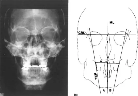
Postero-anterior head-films and occlusal radiographs were taken prior to surgery (Tl), immediately after the end of expansion (T2), at the end of retention (T3), and at least 12 months post-retention (T4) (Fig. 4a). For each head-film the patients were positioned in a cephalostat with the horizontal and the vertical nasal rest position recorded in order to re-orientate the patient as closely as possible to the original position. On the head-films a midline reference point was chosen. A cranial base reference line was drawn by connecting each orbit at the intersection of the greater wing of the sphenoid bone (orbital oblique line) with the inner cortex of the orbit at a landmark described as 'latero orbitale' (LO). The midpoint between the right
Figure 3 Patient I.D.: on the unilateral cross-bite cases, the acrylic palatal to the molars and premolars of the non-operated side was trimmed off to allow more dental relapse on that side.
and left LO landmarks was chosen as the upper reference point for the midline. The lower refer-ence point was defined as a point in the middle of the narrowest inferior part of the nasal septum.
The skeletal expansion for each side was measured on a perpendicular line to the midline 5 mm above the upper end of the pin inserted into the head gear tubes. The distance was measured from the midline to the intersection of the perpendicular line with the outline of the zygomatico-maxillary ridge. Changes in angular position of the upper right and left molars were measured as the angles formed by the extension drawn from each pin inserted in the head gear tubes and the midline. All measurements were recorded three times and averaged (Fig. 4b). General findings
The pre-operatively inserted Hyraxmin appli-ance, as well as the upper retention plate, were well tolerated by all patients. Following the surgical procedure and the immediate activation of the expansion screw, a small diastema appeared between the upper central incisors which increased significantly during the active period of expansion (Fig. 5). Three months after the end of expansion, the Hyraxmln appliance removal was a simple and painless procedure. Occlusal contacts did not appear to interfere with the expansion.
During the retention period the patients were seen once a month.
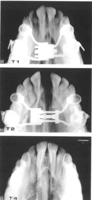
On the occlusal radiograph taken at the end of the expansion period, an opening of the mid-palatal suture was observed in all four patients. The sutural widening was wedge shaped, with the base of the triangle located anteriorly. After 3 months of retention, radiotranslucency was no longer visible along the suture and 12 months post-retention a regenerated suture could be observed (Fig. 6a,b,c).
Individual findings
Patient J.M. (age 27 years, bilateral corticotomy). Measurements made on casts showed an increase of the intermolar distance of 9.8 mm followed by a decrease of 0.2 mm during the retention period and 0.5 mm during the post-retention period. The inter-canine width gained 10.3 mm during the expansion and lost 0.3 mm during the retention and 2.7 mm during the post-retention period (Fig. 7).
SURGICAL MAXILLARY EXPANSION 113
Figure 4 (a) Postero-anterior (P.A.) head-film from patient J.M. (T,). (b) Reference points, lines and angles measured on
the P.A. radiographs.
I 1 '
Fipire5 Patient I.B.: midline diastema produced by the
maxillary expansion.
The skeletal expansion measured on the PA head-films was identical on both sides at 1.9 mm. It remained stable on the right side during the retention and the post-retention period. On the left side, following a slight relapse during the retention period (0.5 mm), it remained stable during the observation period (Fig. 8a).
The expansion produced a 6-degree angular
change measured on the right molar and 5.5 degrees on the left side.
During the retention period it tipped 3 degrees more on the right side and returned 1 degree on the left (Fig. 9a). No post-retention measure-ment was available as the buccal tubes and the upper first molar bands were removed at the end of retention.
Patient I.B. (age 23 years, bilateral corticotomy). The expansion measured on the
models was 10.0 mm at the molars and 6.8 mm at the canines. It relapsed 0.6 mm during the retention period and 3.7 mm during the post-retention period at the molar level. At the canines, the distance decreased 0.8 mm during the retention and 4.4 mm during the post-reten-tion period (Fig. 7). On the right side, the skeletal expansion was 0.8 mm and on the left side 1.2 mm. This side remained stable during the retention period, while the right side decreased only 0.1 mm. A relapse of 0.1 mm on the right and 0.6 mm on the left side were recorded during the post-retention interval (Fig. 8a).
Figure 6 Patient I.B.: (a) occlusal radiograph taken
preop-eratively (T,); (b) occlusal radiograph at the end of expan-sion (T2); (c) occlusal radiograph 12 months after the end
of retention (T4).
degrees on the right side and lost 2.5 degrees during the retention period. On the opposite side it tipped 13 degrees and relapsed 3.5 degrees during retention (Fig. 9a).
Patient F.F. (age 28 years, unilateral
corticotomy). The intermolar width increased 9.5 mm and the intercanine width 4.9 mm with the expansion. During retention, 3.6 mm of
Expansion (ti-t2) (1,-1 J
12 10 8 8 4 2 0 2 4 8 6
traarmofer I I ^ H IrttrcanJnt
Figure 7 Overall expansion and relapse measured between
the upper canines and between the first molars on the casts.
( 1 0 0 )V( 4 0 )
(a)
w««k*
(b) _V_ ..It"..
Figure 8 (a) Skeletal expansion and relapse in the patients
with bilateral corticotomy. (b) Skeletal expansion and relapse in the patients with unilateral corticotomy.
intermolar distance was lost with no change in the canine area. Following the retention period, 2.5 mm of relapse was recorded at the intermolar and 0.1 mm at the intercanine dis-tance (Fig. 7). The skeletal expansion was min-imal on the non-operated side (NOS) (0.3 mm) compared to the operated side (OS) (1.3 mm). It relapsed totally during the retention period on the NOS and partially on the OS (0.4 mm) (Fig. 8b). The lateral rotation of the OS was more pronounced at 11 degrees measured at the molar buccal tube than the NOS (6 degrees).
115
(40) (10) <tO) (1«) 0 10 10 30 40
Non-op*r*t»d aid* Operated ttd*
(30) (10) (10) 0 10 10 30
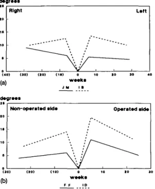
Figure 9 (a) Angular changes in the patients with bilateral corticotomy. (b) Angular changes in the patients with unilateral corticotomy.
This buccal tipping recovered 6 degrees on the OS and 2 degrees on the NOS during the retention phase (Fig. 9b).
Patient I.D. (age 35 years, unilateral corticotomy). A very similar response to the
previous case was noted in this older patient. The expansion recorded between the molars was 7.7 and 2.7 mm between the canines. It decreased 1.5 mm between the molars and 0.5 mm between the canines during retention, and 1.7 mm between the molars during the post-retention time. It regained 0.3 mm during that time between the canines (Fig. 7). The skeletal expansion was again minimal with I.Omm on the OS and 0.4 mm on the NOS. The relapse was even smaller during retention ( - 0 . 1 mm on the OS and - 0.4 mm on the NOS) as well as during the post-retention period ( - 0 . 1 mm on the OS and none on the NOS; Fig. 8b).
The operated hemi-maxilla rotated 19.5 degrees while the non-operated side rotated 14 degrees. It came back lingually 8.5 degrees on the OS and 5.5 degrees on the NOS during the retention period (Fig. 9b).
Discussion
Maxillary expansion, surgically assisted with lateral corticotomies only, appeared to be a simple and effective procedure to achieve cross-bite correction in adult patients. Although the surgery has been advocated under local anaes-thesia with presedation (Glassmann 1984, Bell and Epker 1976), our opinion remains that excessive bleeding from the nasal and . sinus mucosa, and particularly from the spheno-palat-ine, descending-palatine or even internal maxil-lary arteries, could present a surgical risk. Therefore, general anaesthesia is' preferred so that, should a large vessel be damaged during bone sectioning, the maxilla could be immedi-ately downfractured to allow access to the bleed-ing site. Post-operatively we did not observe any sinus infection or devitalization of teeth.
During expansion, a significant midline dias-tema appeared which was more pronounced in the bilateral cases. It seemed very important that the patients be informed of the unaesthetic aspect of the procedure prior to treatment. The diastema was closed during the fixed appliance phase of treatment and resulted in a decrease in the inter-canine distance (particularly in the bilateral corticotomy cases), which should not be considered as relapse.
The skeletal expansion was measured on the PA radiographs at a level above the apices of the teeth and relatively close to the osteotomy site. As previously observed by Davis and Kron-man, (1969) minimal expansion was found (0.3-1.9 mm) and, although twice as much expansion was observed on the operated side in the unilat-eral cases, no significant conclusion can be drawn. More skeletal expansion was clearly detected at the palatal level on the occlusal radiographs, which indicated that the two max-illary bones separate from each other more in a rotational than a translational movement. This finding is in agreement with Hicks (1978) who used maxillary implants to demonstrate skeletal tipping of the two maxillary parts in growing patients.
Haas (1970) and Wertz (1970) stated that during rapid maxillary expansion in children and adolescents, the maxillary halves tip buc-cally as do the teeth, with a centre of rotation located approximately at the fronto-maxillary suture. In our experiment, this centre of rotation was probably situated more inferiorly. Follow-ing the expansion, we observed 5-20 degrees of
angular change per side, which was twice the amount of tipping measured between both upper first molars in normal RME on young patients in different studies (Hicks, 1978; Moss-az-Jackson and Mossaz, 1989). Proffit and White (1991) mentioned that there was an important transverse relapse tendency after sur-gical cross-bite corrections with Le Fort I pro-cedures involving two or three pieces.
In the present study stability appeared satis-factory. The cross-bite correction remained stable 12 months post-retention in all patients. This could be related to the better transverse control following the surgically assisted maxil-lary expansion without palatal surgery, as a maxillary plate could be immediately inserted following the Hyraxmin removal, which also served as a retainer for the first 3 months after the end of expansion. The period of fixed appli-ance therapy which immediately followed the retention period could also be a factor in achiev-ing acceptable stability.
More data will be gathered in the future to confirm these hypotheses.
The indication for unilateral corticotomy and maxillary expansion remains rare and is often related to early extraction of permanent teeth without orthodontic therapy or reconstruction. Nevertheless, this study demonstrates that uni-lateral expansion is possible using the contralat-eral side as an anchorage. Dental tipping occurred on the same side, which later on relapsed during the retention and post-retention period. Better control of this movement can be achieved by selective grinding of the acrylic on the maxillary retainer.
Address for correspondence
Dr Claude Mossaz University of Geneva School of Dentistry Department of Orthodontics 19, rue Barth.-Menn CH-1205 Geneva Switzerland References
Alpern M C, Yurosko J J 1987 Rapid palatal expansion in adults with and without surgery. Angle Orthodontist 57: 245-263
Bell W H, Epker B N 1976 Surgical orthodontic expansion of the maxilla. American Journal of Orthodontics 79: 517-528
Davis W M , Kronman J H 1969 Anatomical changes induced by splitting of the midpalatal suture. Angle Orthodontist 39: 126-132
Glassmann A S 1984 Conservative surgical orthodontic adult rapid palatal expansion: Sixteen cases. American Journal of Orthodontics 86: 207-213
Haas A J 1970 Just the beginning of dentofacial orthopedics. American Journal of Orthodontics 57: 219-255 Hicks E P 1978 Slow maxillary expansion: a clinical study
of the skeletal vs dental response to low magnitude force. American Journal of Orthodontics 73: 121-141 Kennedy J W, Bell W H 1976 Osteotomy as an adjunct to
rapid maxillary expansion. American Journal of Ortho-dontics 70: 123-137
Lehmann J A, Haas A J, Haas D G 1984 Surgical ortho-dontic correction of transverse maxillary deficiency: A simplified approach. Journal of Plastic and Recon-structive Surgery 73: 62-68
Lines P A 1975 Adult rapid maxillary expansion with corticotomy. Angle Orthodontist 67: 44-56
Mossaz-Joelson K, Mossaz C F 1989 Slow maxillary expan-sion: a comparison between banded and bonded appli-ances. European Journal of Orthodontics II: 67-76 Persson M, "Philander B 1977 Palatal suture closure in man
from 15-35 years of age. American Journal of Orthodont-ics 72: 42-52
Proffit W R, White R P, Jr 1991 Surgical orthodontic treat-ment. Mosby Year Book. C V Mosby, New York Timms D J, Vero D 1981 The relationship of rapid maxillary
expansion to surgery with special reference to midpalatal synostosis. British Journal of Oral Surgery 19: 180-196 Wertz R A 1970 Skeletal and dental changes accompanying
rapid midpalatal suture opening. American Journal of Orthodontics 58: 41-66