HAL Id: hal-03226402
https://hal-amu.archives-ouvertes.fr/hal-03226402
Submitted on 14 May 2021
HAL is a multi-disciplinary open access
archive for the deposit and dissemination of sci-entific research documents, whether they are pub-lished or not. The documents may come from teaching and research institutions in France or abroad, or from public or private research centers.
L’archive ouverte pluridisciplinaire HAL, est destinée au dépôt et à la diffusion de documents scientifiques de niveau recherche, publiés ou non, émanant des établissements d’enseignement et de recherche français ou étrangers, des laboratoires publics ou privés.
Endothelial-Specific Deletion of CD146 Protects Against
Experimental Glomerulonephritis in Mice
Ahmed Abed, Aurélie Leroyer, Panagiotis Kavvadas, Florence Authier,
Richard Bachelier, Alexandrine Foucault-Bertaud, Nathalie Bardin, Clemens
Cohen, Maja Lindenmeyer, Ahmad Joshkon, et al.
To cite this version:
Ahmed Abed, Aurélie Leroyer, Panagiotis Kavvadas, Florence Authier, Richard Bachelier, et al.. Endothelial-Specific Deletion of CD146 Protects Against Experimental Glomerulonephritis in Mice. Hypertension, American Heart Association, 2021, 77 (4), pp.1260-1272. �10.1161/HYPERTENSION-AHA.119.14176�. �hal-03226402�
For Hypertension Peer Review. Do not distribute.
Destroy after use.
Disclaimer: The manuscript and its contents are confidential,
intended for journal review purposes only, and not to be further
disclosed.
URL: http://hype-submit.aha-journals.org
Title: Endothelial-specific deletion of CD146 protects against
experimental glomerulonephritis in mice
Manuscript number: HYPE201914176
Author(s): Christos Chadjichristos, INSERM UMR S1155
Ahmed Abed, Inserm U702
Aurelie Leroyer, Aix-Marseille Université
Panagiotis Kavvadas, Inserm U702
Florence Authier, INSERM UMR S702
Richard Bachelier, C2VN INSERM UMR 1263
Alexandrine Foucault-Bertaud, INSERM UMRS 1076
Nathalie Bardin, Aix-Marseille Université
Clemens Cohen, Krankenhaus Harlaching
For Hypertension Peer Review. Do not distribute.
Destroy after use.
Policlinic IV, University of Munich, Munich, Germany
Magali Genest, INSERM UMR-S1155
Noémie Jourde-Chiche, INSERM INRA, C2VN, Marseille
Stephane Burtey, Aix Marseille Université
Marcel Blot-Chabaud, INSERM INRA, C2VN, Marseille
Francoise Dignat-George, Aix-Marseille Univ
For Hypertension Peer Review. Do not distribute.
Destroy after use.
1
Endothelial-specific deletion of CD146 protects against experimental
glomerulonephritis in mice
Ahmed Abed1,2*, Aurélie S. Leroyer3*, Panagiotis Kavvadas1*, Florence Authier1, Richard Bachelier3, Alexandrine Foucault-Bertaud3, Nathalie Bardin3, Clemens D. Cohen4, Maja T. Lindenmeyer4, Magali Genest1, Noémie Jourde-Chiche3,5, Stéphane Burtey3,5, Marcel Blot-Chabaud3, Françoise Dignat-George3¶ and Christos E. Chadjichristos1,2
1
INSERM UMR-S1155, Tenon Hospital, 4 rue de la Chine 75020, Paris, France.
2
Sorbonne Université, Paris, France.
3
Aix-Marseille Univ, INSERM INRA, C2VN, Marseille, France. 4
Nephrological Center, Medical Clinic and Policlinic IV, University of Munich, Munich, Germany.
5
Aix-Marseille Univ, Department of Nephrology, AP-HM Hôpital de la Conception, Marseille, France.
Running title: Targeting endothelial CD146 protects against GN *,
Equal contribution Address correspondence to:
Dr Christos E. Chadjichristos, INSERM UMR S1155, Batiment Recherche, Tenon Hospital, 4 rue de la Chine, 75020 Paris, France. Email: christos.chadjichristos@inserm.fr
For Hypertension Peer Review. Do not distribute.
Destroy after use.
2
Abstract
CD146 is an endothelial junctional adhesive molecule which expression is increased in human glomerular diseases. However, the pathological significance of this overexpression remains unknown. By using a mouse model of glomerulonephritis, we showed that CD146 expression was highly induced within damaged glomeruli and was associated with renal inflammation and fibrosis. Interestingly, two weeks after glomerulonephritis induction, CD146 knock-out mice showed preserved renal function as proteinuria and blood urea nitrogen levels were significantly lower compared to wild type littermates. Furthermore, renal structure was considerably conserved, since crescents formation, tubular dilation, monocyte and lymphocyte infiltration and interstitial renal fibrosis were highly reduced. Colocalization with markers for different types of glomerular cells showed that CD146 expression was mainly increased within the injured endothelium of the glomerular tuft. Consequently, we generated a new transgenic strain in which CD146 was specifically deleted in the vascular endothelium. Similarly to CD146 knock-out, these mice showed preservation of renal structure and function after the induction of glomerulonephritis compared to wild type animals. These data show that endothelial CD146 plays a major role in glomerulonephritis and may represent a novel therapeutic target to reduce glomerular damage and the progression of renal disease.
Keywords: CD146, experimental GN, inflammation, renal fibrosis
For Hypertension Peer Review. Do not distribute.
Destroy after use.
3
Glomerular diseases can lead to renal fibrosis and chronic kidney disease (CKD), either progressively or rapidly. Rapidly progressive or crescentic glomerulonephritis (GN) are a group of rare kidney diseases that may rapidly progress to end stage renal failure as a consequence of pronounced inflammation with glomerular damage and development of crescents1. Other kidney compartments are also affected, including tubulo-interstitium which shows an inflammation leading to renal interstitial fibrosis. The functional outcome of such structural lesions is a rapid deterioration of renal function2. Even though several studies, mostly on preclinical models of experimental GN, provided pathophysiological advances, both on the inflammatory process and in the consequent fibrosis, molecular insights in the regulation and the progression of the disease are still limited and current treatments remain only partially effective.
CD146 is an adhesion molecule detected in all endothelial cells of the vascular tree, regardless of the vessel caliber or anatomical region3,4. It mainly localizes at the intercellular junctions and controls inter-endothelial cell cohesion and paracellular permeability5-7 but also monocyte transmigration8 and angiogenesis9-12. CD146 exists as different isoforms: a short isoform with a
putative PDZ binding motif13-16, which may mediate its anchoring to the cytoskeleton, a long isoform with a putative endocytosis motif17, and a soluble form (sCD146) as the result of metalloprotease-dependent shedding of membrane CD14618,19. sCD146 is detectable in the human serum and its level is modulated in different pathologies such as inflammatory bowel diseases20, abnormal pregnancies21 or chronic renal failure18. Interestingly, we have reported that both membrane expression of CD146 and sCD146 were elevated in patients suffering from different types of renal diseases. Moreover, in human biopsies, high expression of CD146 was associated with renal damage and inflammation22. Furthermore, increased levels of sCD146 in sera of patients were consistent with changes of vascular permeability during chronic renal failure and suggested that CD146 function may be altered in these pathological settings18.
For Hypertension Peer Review. Do not distribute.
Destroy after use.
4
However, the pathological significance of CD146 overexpression in renal diseases remains unknown.
In this study, to decipher the role of CD146 in renal disease, we used a well-established model of experimental crescentic GN in mice. We demonstrated that the expression of this junctional molecule is highly increased within injured glomerular endothelium leading to severe structural tissue damage thus contributing to the progression of the disease. More importantly, CD146 knock-out (KO) mice or mice in which CD146 was specifically deleted in the vascular endothelium (CD146-EC-del), showed preservation of both renal functional and structural parameters during experimental GN.
Methods
Methods
Mice strains
CD146-floxed and CD146-KO mice were generated as previously described and backcrossed for more than 10 generations into the C57BL/6J background8. CD146-floxed mice were first crossed with the B6.Cg-Gt(ROSA)26Sortm6(CAG-ZsGreen1)Hze/J mice (Jackson laboratories) which express the fluorescent protein ZsGreen1 as a reporter for CRE-recombinase activity. Then, mice with endothelial cell-specific deletion of the CD146 gene were generated by further crossing CD146flox-ZsGreen animals with the Cdh5 (PAC)-CreERT2 mouse strain, a gift from Dr Ralf Adams23. The deletion of the CD146 gene was induced by I.P. injections of tamoxifen diluted in corn oil (10 mg/ml solution, 1 mg tamoxifen/injection) and administered on 3 consecutive days. Primers for genotyping CD146 alleles were 5’-TCACTTGACAGTGTGATGGT-3' (forward primer used to detect CD146 WT, floxed and KO alleles), 5’-CCTTAGAAAGCAGGGATTCA-3' (reverse primer used to detect
For Hypertension Peer Review. Do not distribute.
Destroy after use.
5
CD146 WT and floxed alleles) and 5’-CCCAAATCCTCTGGAAGACA-3' (reverse primer used to detect CD146 KO allele).
CD146 analyses in diverse human glomerulopathies
Human kidney biopsies were collected in a multicentre study (European Renal cDNA Bank— Kröner- Fresenius Biopsy Bank, ERCB) and were obtained from patients after informed consent and with approval of the local ethics committees. Pre-transplant allograft biopsies were used as control kidneys. Total RNA was isolated from microdissected glomeruli, reverse-transcribed, and linearly amplified as previously described24. Fragmentation, hybridization, staining and imaging were performed according to the Affymetrix Expression Analysis Technical Manual (Affymetrix, Santa Clara, CA, USA). A single probe-based analysis tool, Chip- Inspector (Genomatix Software GmbH, Munich, Germany), was used for transcript annotation, total intensity normalization, significance analysis of microarrays25 and transcript identification based on significantly changed probes. Pre-developed TaqMan probes (Applied Biosystems) were used for human CD146 and GAPDH detection by qPCR. mRNA expression was analyzed by standard curve quantification.
Nephrotoxic serum-induced glomerulonephritis
Decomplemented nephrotoxic serum (NTS) was prepared as previously described26,27 and 17,5μl of serum per gram of body weight were intravenously injected over two consecutive days (days 1 and 2). For the time course protocol, two-month old C57Bl6/J females were injected with NTS and sacrificed at day 4, 7 and 15 for tissue collection (n=6 mice for each time point). Six mice were injected with PBS and used as controls.
For the CD146-KO protocol, two-month old CD146-KO female mice and their wild type littermates were used. Eight to nine mice from each group were injected with NTS and 6 with
For Hypertension Peer Review. Do not distribute.
Destroy after use.
6
PBS. Body weight and urine samples were taken at days 0, 4, 7 and 15. All mice were sacrificed at day 15 and blood and tissue samples were obtained.
For the CD146-floxed protocol, two-month old female mice were treated with tamoxifen. Eleven of them were injected with NTS and 4 with PBS. CD146-floxed ZsGreen mice bearing no CRE recombinase transgene were treated with tamoxifen and used as controls. Body weight and urine samples were taken at days 0, 4, 8, 12 and 15. Mice were sacrificed at day 15 after obtaining blood samples.
All procedures regarding animal experimentation were in accordance with the European Union Guidelines for the Care and Use of Laboratory Animals and approved by the local ethics committee of the National Institute for Health and Medical Research (Institut National de la Santé et de la Recherche Médicale). Animals were housed at constant temperature with free access to water and food.
Histologic and functional parameters
Half kidneys from each animal were fixed in 4% formalin solution and embedded in paraffin. 4 μm sections were stained with Masson’s trichrome for histological evaluation of renal damage. Tubular dilation was evaluated semi-quantitatively, using the following scale: 0, no tubular damage; 1, damage in 1–25% of the tubules analyzed; 2, damage in 26–50% of the tubules analyzed; 3, damage in 51–75% of the tubules analyzed; 4, damage in >76% the tubules analyzed. Glomerular crescents were measured and expressed as the % number of glomeruli presenting crescents vs total number of glomeruli. Scoring was performed in a masked manner on coded slides by two different investigators. Images were obtained with an OlympusIX83 photonic microscope at X200 magnification. Interstitial fibrosis was assessed semiquantitatively on Sirius red stained paraffin sections at magnification of X200. Fibrosis was then quantified using computer-based morphometric analysis software (Analysis, Olympus) that allowed the formation of a binary image in which the stained area could be
For Hypertension Peer Review. Do not distribute.
Destroy after use.
7
automatically calculated as a percentage of the image area. Proteinuria, expressed as protein g/mmol of creatininuria, was assessed using the Pyrogallol Red method and utilizing a KONELAB automate (Thermo Scientific, Waltman, MA). BUN was assessed in blood plasma obtained on the day of sacrifice. BUN was measured using an enzymatic spectrophotometric method and was expressed as mmol/L.
Immunostainings
Immunohistochemistry was performed on 4μm-thick paraffin-embedded tissue sections. Tissue was deparaffinized and 10mM citric acid, pH6 at 95ºC was used for antigen retrieval. Sections were permeabilized with 0.1% triton/PBS. Antibodies against F4/80 (AbD Serotec) and CD3 (Dako), were used. The F4/80 and CD3 positive area was quantified in at least ten photographs at 200X magnification per animal, using publicly available image processing software (ImageJ; Fiji) and expressed as percentage of the total tissue area. Immunofluorescence was performed on 4μm-thick frozen sections fixed in methanol. CD146 (homemade rat anti-mouse), CD31 (Abcam), Claudin-1 (Thermo Scientific), Nestin (BD Pharmingen), Nephrin (Abcam) anti-rabbit and α-SMA (Sigma Aldrich) anti-mouse antibodies were used. Alexa fluor (Invitrogen) secondary antibodies were used for detection. Images were obtained with an OlympusIX83 photonic microscope at X400 magnification.
Quantitative Real-Time PCR
Total RNA was extracted from half kidneys using TRI Reagent (MRC). RNA quality was verified by measuring the OD 260:280 ratio and residual genomic DNA was removed byDNase I treatment (Thermo Fisher Scientific) for 30 minutes at 37°C. A total of 1 mg RNA was transcribed to cDNA using the Maxima First Strand cDNA Synthesis Kit from Thermo Fisher Scientific per manufacturer’s instructions. Real-time PCR was performed with the RocheLight Cycler 480 detection system using SYBR Green PCR Master Mix (Roche Diagnostics,
For Hypertension Peer Review. Do not distribute.
Destroy after use.
8
Indianapolis, IN) under the following program: 95°C for 5 minutes, 45 cycles at 95°C for 15 seconds and 60°C for 15 seconds, and 72°C for 15 seconds. For quantitative analysis, experimental genes were normalized to HPRT expression using the DDCT method. Dissociation curves were analyzed to determine that a single product was amplified. Primer sequences are listed in table 1.
Western blot
Proteins were extracted from renal tissue using RIPA lysis buffer (Santa Cruz Biotechnology). Western blot for CD146 (Epitomics) were performed using standard techniques as previously described27. GAPDH (Sigma-Aldrich) was used as loading control.
Statistical Analyses
Values are expressed as mean ± SEM. Data were analyzed using one-way analysis of variance followed by protected least significant difference Fisher’s test of the Statview software package. Error bars represent mean ± SEM. Results with P<0.05 were considered statistically significant. IL-1β FW: TGTAATGAAAGACGGCACACC RV: TCTTCTTTGGGTATTGCTTGG TNF-α FW: TCTTCTCATTCCTGCTTGTGG RV: ATGAGAGGGAGGCCATTTG ICAM-1 FW: CCCACGCTACCTCTGCTC RV: GATGGATACCTGAGCATCACC TGF-β1 FW: TGGAGCAACATGTGGAACTC RV: GTCAGCAGCCGGTTACCA Collagen-3 FW: TGGTTTCTTCTCACCCTTCTTC RV: TGCATCCCAATTCATCTACGT
For Hypertension Peer Review. Do not distribute.
Destroy after use.
9 CD146 FW: TCCAGTCATCACAGATTGTCG RV: AAACAGGGACAGTGACCTCCT VCAM-1 FW: TGGTGAAATGGAATCTGAACC RV: CCCAGATGGTTTCCTT MCP-1 FW: CATCCACGTGTTGGCTCA RV: GATCATCTTGCTGGTGAATGAGT Collagen-1 FW: GCAGGTTCACCTACTCTGTCCT RV: CTTGCCCCATTCATTTGTCT HPRT FW: GGAGCGGTAGCACCTCCT RV : CTGGTTCATCATCGCTAATCAC
Table 1 : Primer sequences used for the study.
Results
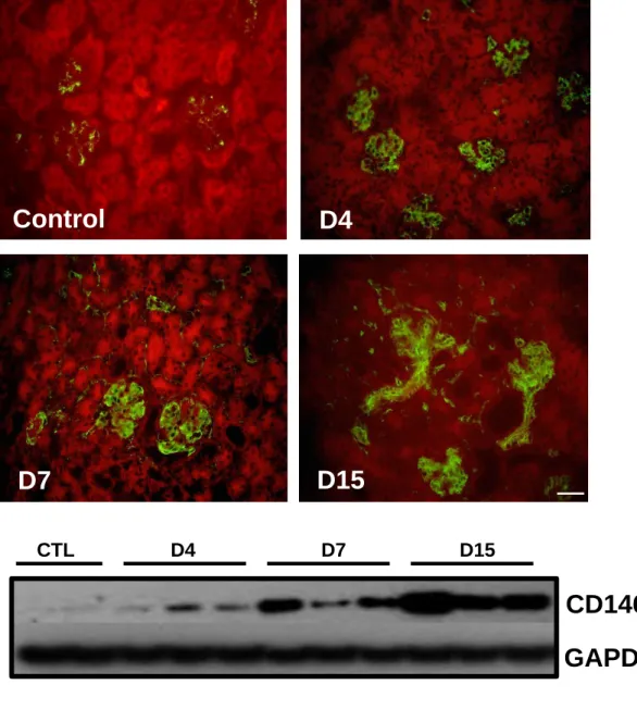
CD146 is induced in damaged glomeruli
To assess the expression of CD146 during the progression of glomerular disease, we induced a passive nephrotoxic serum glomerulonephritis (NTS-GN) in wild type mice. Animals were sacrificed at 4, 7 and 15 days post NTS administration. Immunofluorescence showed a weak expression of CD146 in glomeruli of healthy animals (Fig1A). CD146 expression progressively increased from day 4 to day 15 in peritubular capillaries and mainly within damaged glomeruli. This upregulation was confirmed by western blotting of renal tissues (Fig1B and 1C). The specificity of the CD146 antibodies has been checked by using renal tissues from CD146 KO mice (FigS1). Thus, CD146 expression was highly and continuously increased with the progression of NTS-GN.
For Hypertension Peer Review. Do not distribute.
Destroy after use.
10
CD146 knock-out mice are protected from nephrotoxic serum-induced
glomerulonephritis
To assess the role of CD146 in GN, we injected NTS in both CD146 WT and KO mice. Mice were sacrificed 15 days post NTS administration. NTS-GN resulted, as expected, in a significant body weight increase in WT animals (Fig2A). In accordance, renal function was altered since both proteinuria and blood urea nitrogen (BUN) were highly increased (Fig2B
and 2C). In contrast, in CD146 KO mice, body weight and BUN increase were blunted.
Furthermore, elevation of proteinuria was significantly lower in these mice. Quantification of histological lesions by Masson’s Trichrome coloration showed that NTS injection induced less damages in CD146 KO mice (Fig2D). Indeed, a significant structural protection in CD146 KO animals was demonstrated by a significant decrease of both crescent formation and tubular dilation compared to WT ones (Fig2E and 2F). Thus, CD146 KO animals were protected from glomerular and tubular lesions induced by NTS-GN, which preserved renal function and blunted proteinuria.
CD146 KO mice show reduced inflammation and renal fibrosis after NTS injection
Evaluation of inflammatory markers in kidneys by qPCR showed that the induction of IL-1β, TNF-α and ICAM-1 mRNAs was blunted in CD146 KO mice, 15 days after NTS injection (Fig3A, 3B and 3C). Accordingly, F4/80 immunostaining showed less macrophage infiltration in the renal cortex of CD146 KO mice (Fig3D and 3E). Furthermore, these animals displayed reduced CD3 cell infiltrates on day 15 (Fig3F and 3G). Consequently, renal fibrosis was limited in mice lacking CD146. Indeed, mRNA expression of fibrotic markers such as TGF-β and collagen I was blunted in CD146 KO mice (Fig3H and 3I). Quantification of Sirius Red coloration confirmed a reduction of renal interstitial fibrosis in these animals (Fig3J).
For Hypertension Peer Review. Do not distribute.
Destroy after use.
11
Altogether, these data demonstrate that lack of CD146 markedly restricted renal inflammation and renal interstitial fibrosis in NTS-GN.
CD146 is increased in the glomerular endothelium during the progression of NTS-GN
To identify the cell type overexpressing CD146 within damaged glomeruli after aggression, we performed colocalization experiments using appropriate markers of the glomerular components, at day 7 post NTS administration. CD146 was slightly expressed at basal conditions in the vascular tuft (Fig1A). After induction of NTS-GN, CD146 overexpression colocalized mainly with the endothelial marker CD31/PECAM (Fig4A). In contrast, CD146 was not detected in podocytes, as there was no colocalization with nestin (Fig4B). It was neither detected in parietal cells, as we did not detect any colocalization of CD146 with Claudin-1 (Fig4C), nor in mesangial cells (Fig4D). Thus, our data confirmed that CD146 was highly overexpressed mainly within injured glomerular endothelial cells after the induction of the disease.
CD146 endothelial-specific deletion protects mice against NTS-GN
To estimate the impact of the endothelial CD146 overexpression in the progression of NTS-GN, we generated the CD146-EC-del mouse strain. CD146 was specifically deleted in the vascular endothelium by interbreeding mice harboring a tamoxifen-inducible VE-cadherin Cre-recombinase23 with CD146flox-ZsGreen mice (Fig5A). The specificity of CD146 deletion in endothelial cells was verified by immunofluoresence, after tamoxifen injection and 15 days post NTS administration (Fig5B) and further confirmed by qPCR (Fig5C). CD146-EC-del mice showed limited increase of body weight, proteinuria and BUN (Fig6A, 6B and C). This functional protection was accompanied by an overall preservation of renal structure (Fig6D), as number of crescents (Fig6E) and tubular damage (Fig6F) were both dramatically reduced compared to respective controls 15 days after the induction of NTS-GN. Furthermore, F4/80
For Hypertension Peer Review. Do not distribute.
Destroy after use.
12
immunostaining showed reduced monocyte infiltration in the renal cortex of CD146-EC-del mice (Fig7A). In accordance, qPCR experiments showed that upregulation of both VCAM-1 and MCP1 were fully abolished (Fig7B and 7C). Subsequently, TGF-β1 and Collagen I mRNAs increased expression was blunted in CD146-EC-del mice compared to control kidneys (Fig7D and 7E). These data demonstrate that endothelial overexpression of CD146 promotes the progression of experimental GN in mice.
CD146 transcript numbers are increased in human glomerulopathies
CD146 mRNA expression was analyzed in microdissected glomeruli from renal biopsies of patients suffering from diabetic nephropathy (DN, n=7), minimal change disease (MCD, n=5), IgA nephropathy (IgA, n=27), focal segmental glomerulosclerosis (FSGS, n=10), membranous glomerulonephritis (MGN, n=21), lupus nephritis (SLE, n=32) and rapidly progressive glomerulonephritis (RPGN, n=23). Compared to control biopsies (n=6), damaged glomeruli showed a pronounced transcriptional upregulation of CD146 transcripts (Fig8).
Discussion
A variety of chronic inflammatory diseases is associated with disruptions of the inter-endothelial function and junction integrity28. Defects in the organization of endothelial junctions occur also during the progression of chronic renal diseases. Indeed, in biopsies from patients with nephropathies of several etiologies, we have previously reported that high expression of endothelial CD146 was associated with endocapillary proliferation, proteinuria and inflammation22. Moreover, increased levels of CD146 were consistent with changes in renal morphometry during the progression of chronic renal failure suggesting that the junctional function supported by CD146 may be altered in these pathological settings. However the pathological significance of this overexpression remained unclear. Interestingly in our study,
For Hypertension Peer Review. Do not distribute.
Destroy after use.
13
increased expression of CD146 seemed to promote renal disease since its deletion protected kidney from immune aggression and prevent severe renal dysfunction. Thus CD146 may represent a novel therapeutic target against the initiation and the progression of glomerulonephritis.
In our study, CD146 was overexpressed in renal microcirculation, mainly within injured glomeruli and already since the early stages of the disease. CD146 was also found to be overexpressed within proximal tubules at later time points (Fig1A). This is consistent with previous observations that reported CD146 expression in renal tubular cells in vivo and in vitro under pathological conditions22,29. In our experimental model, where the starting point of the disease is the glomerular compartment, CD146 KO mice were protected against the development of renal inflammation, proteinuria and weight gain. CD146 was also reported to be expressed in myeloid cells in mice30, but we showed that CD146 was mainly overexpressed within injured endothelium thanks to colocalization experiments. Thus, we generated an endothelial specific KO of CD146 to discern the specific role of endothelial CD146 in NTS-GN. Similar to the global CD146 KO, CD146-EC-del mice were also preserved after the induction of GN. This clearly demonstrates a major role of the endothelial cells in controlling glomerular damage but also interstitial inflammation. These results are in accordance with our previous study demonstrating that endothelial CD146 is involved in the regulation of monocyte transendothelial migration in vitro8. Indeed, upon tumor necrosis factor-α stimulation, CD146 expression was increased at both cellular junctions and apical membrane of HUVEC and further contributed to monocyte transmigration through the endothelial monolayer. Similar results were more recently reported in a mouse model of multiple sclerosis, in which targeting endothelial CD146 decreased neuroinflammation by limiting lymphocyte extravasation on the blood brain barrier31. Considering these data, it would be of major interest to further delineate
For Hypertension Peer Review. Do not distribute.
Destroy after use.
14
the role of CD146 in glomerular and interstitial inflammation in the pathogenesis of renal diseases.
Another interesting finding of our study is that renal fibrosis was blunted in both CD146 KO and CD146-EC-del mice. Even if this observation is reported in a model of crescentic GN, which is a rare disease in humans, this is of particular interest as renal fibrosis is the common evolution of all kidney diseases and is highly predictive of the progression of CKD32. The following hypotheses can be proposed to explain the reduction of fibrosis burden. First, the diminution of fibrosis could be a consequence of reduced inflammation in the injured kidney. Second, endothelial CD146 could play a key role in the control of the fibrotic process in response to the activation of endothelial cells. Indeed, endothelial to mesenchymal transition (Endo-MT) has been proposed to contribute to the accumulation of activated fibroblasts and myofibroblasts in fibrotic kidneys, in different models of experimental nephropathy in mice33. In this line, the reduction of fibrosis could be partially explained by a reduction in the Endo-MT when CD146 is down-expressed. However, more recent studies demonstrated that the contribution of the Endo-MT program to the emergence of renal fibrosis was moderate34. Another hypothesis could be the role of CD146 to induce cell activation as it has been described in the epithelial to mesenchymal transition phenomenon in other tissues, a major event in the metastatic dissemination35,36. Indeed, as CD146 can be shed at the cell membrane to generate soluble CD146, sCD146 released by endothelial cells, could stimulate fibroblast proliferation and transition to a myofibroblast phenotype, leading to renal extracellular matrix deposition and fibrosis. Consequently, another important question to explore will be to determine the CD146 isoform involved in the observed effects. We have described two different isoforms, long and short, which differ by their intracellular part. Whereas the long isoform is present at the junction of endothelial cells, and plays a major role in monocyte transmigration, the short isoform is rather present at the apical membrane and could be shed to
For Hypertension Peer Review. Do not distribute.
Destroy after use.
15
generate the soluble form of CD146 that will bind and stimulate monocytes16,18. Whether one or both isoforms are modulated in NTS-induced GN and whether they are able to cooperate in the phenomenon will have to be explored.
GN is recognized as the second most frequent cause of end-stage renal disease (ESRD) worldwide37. Despite aggressive immunosuppressive therapies, rapidly progressive GN can lead to irreversible renal damage with CKD and frequently ESRD. Considering the need for therapeutic targets to withhold glomerular damage and tubulo-interstitial inflammation and fibrosis in GN, we used a powerful experimental tool sharing some features with human glomerulopathies, to investigate the role of CD146 in renal inflammatory injury. Indeed, injection of sheep serum rich in immunoglobulins against glomerular antigens induces an immediate inflammatory response characterized by renal infiltration of cells of the immune system, followed by severe glomerular damage and rapid loss of renal function27,38,39. This is a robust model of rapidly progressive renal disease as mice are dying the latest 4 weeks after the induction of the disease27,40. Thus, this model of GN can be used to delineate the pathogenic processes leading to immune nephritis, over a quick time frame and allowed to identify several factors that potentially dictate disease severity. This has important clinical implications, both from the perspective of genetic susceptibility as well as clinical therapeutics41. Indeed, several mediators such as cytokines, chemokines, adhesion molecules, surface receptors and extracellular matrix proteins have been shown to play a crucial role in mediating the inflammatory response in CKD42-52. Thus, a major finding of our study is that CD146 deletion is beneficial in this model of GN since in both CD146 KO and CD146-EC-del mice the progression of the disease was blunted. Consequently, it is of interest to develop specific inhibitory devices against CD146. This is reinforced by the exaggerated expression of this junctional molecule within glomeruli in biopsies of patients suffering from different type of GN in comparison with healthy controls (Fig8). Interestingly, our data are also in accordance with
For Hypertension Peer Review. Do not distribute.
Destroy after use.
16
publically available data obtained by the NephroSeq consortium showing overexpression of CD146 in glomeruli from patients with various glomerular diseases (https://www.nephroseq.org/resource/login.htm). Further experiments using additional and non-glomerular experimental models of renal disease, will be necessary to investigate whether the beneficial effects of CD146 deletion are model-dependant, or can be considered as a more common protective approach against renal inflammation and fibrosis.
Nevertheless, our results support a major role of the endothelium in kidney to control the aggression towards all the compartments, glomeruli but also tubules. We demonstrated that endothelial CD146 plays a major role in the progression of GN. Thus, targeting this junctional molecule may represent a good therapeutic strategy to prevent the progression of GN.
Acknowledgments
Support for this study was provided by the French National Research Agency ( ANR-11-BSV1-0019 to FDG, CEC) and the Inserm institute (CEC). AA is doctoral fellow of the French Ministry of National Education (Ecole Doctorale de Physiologie&Physiopathologie, ED 394). We also thank Drs MichelAurrand-Lions and Christos Chatziantoniou for helpful discussions and the Imaging and Cytometry Facility of Tenon, UMR-S1155 Inserm and Andreia de Sousa Jorge for excellent technical assistance.
Competing financial interests
For Hypertension Peer Review. Do not distribute.
Destroy after use.
17
References
1. Mathieson, P.W. Glomerulonephritis. SeminImmunopathol. 2007;29:315-316
2. Moeller MJ, Smeets B. Novel target in the treatment of RPGN: the activated parietal cell. Nephrol Dial Transplant. 2013;28:489-492
3. George F, Poncelet P, Laurent JC, Massot O, Arnoux D, Lequeux N, Ambrosi P, Chicheportiche C, Sampol J. Cytofluorometric detection of human endothelial cells in whole blood using S-Endo 1 monoclonal antibody. J Immunol Methods. 1991;139:65-75
4. Bardin N, Anfosso F, Masse JM, Cramer E, Sabatier F, Le Bivic A, Sampol J, Dignat-George F. Identification of CD146 as a component of the endothelial junction involved in the control of cell-cell cohesion. Blood 2001;98:3677-3684
5. Anfosso F, Bardin N, Vivier E, Sabatier F, Sampol J, Dignat-George F. Outside-in signaling pathway linked to CD146 engagement in human endothelial cells. J Biol Chem. 2001;276:1564-1569
6. Solovey AN, Gui L, Chang L, Enenstein J, Browne PV, Hebbel RP. Identification and functional assessment of endothelial P1H12. J Lab Clin Med. 2001;138:322-31
7. Jouve N, Bachelier R, Despoix N, Blin MG, Matinzadeh MK, Poitevin S, Aurrand-Lions M, Fallague K, Bardin N, Blot-Chabaud M, Vely F, Dignat-George F, Leroyer AS.CD146 mediates VEGF-induced melanoma cell extravasation through FAK activation. Int J Cancer. 2015;137:50-60
8. Bardin N, Blot-Chabaud M, Despoix N, Kebir A, Harhouri K, Arsanto JP, Espinosa L, Perrin P, Robert S, Vely F, Sabatier F, Le Bivic A, Kaplanski G, Sampol J, Dignat-George F. CD146 and its soluble form regulate monocyte transendothelial migration. Arteriosclerosis,
For Hypertension Peer Review. Do not distribute.
Destroy after use.
18
9. Yan X, Lin Y, Yang D, Shen Y, Yuan M, Zhang Z, Li P, Xia H, Li L, Luo D, Liu Q, Mann K, Bader BL. A novel anti-CD146 monoclonal antibody, AA98, inhibits angiogenesis and tumor growth. Blood 2003;102:184-191
10. Chan B, Sinha S, Cho D, Ramchandran R, Sukhatme VP. Critical roles of CD146 in zebrafish vascular development. Dev Dyn. 2005;232:232-244
11. Harhouri K, Kebir A, Guillet B, Foucault-Bertaud A, Voytenko S, Piercecchi-Marti MD, Berenguer C, Lamy E, Vely F, Pisano P, Ouafik L, Sabatier F, Sampol J, Bardin N, Dignat-George F, Blot-Chabaud M. Soluble CD146 displays angiogenic properties and promotes neovascularization in experimental hind-limb ischemia. Blood 2010;115:3843-3851 12. Halt KJ, Pärssinen HE, Junttila SM, Saarela U, Sims-Lucas S, Koivunen P, Myllyharju J, Quaggin S, Skovorodkin IN, Vainio SJ. CD146(+) cells are essential for kidney vasculature development. Kidney Int. 2016;90:311-324
13. Taira E, Nagino T, Taniura H, Takaha N, Kim CH, Kuo CH, Li BS, Higuchi H, Miki N. Expression and functional analysis of a novel isoform of gicerin, an immunoglobulin superfamily cell adhesion molecule. Journal Biol Chem 1995;270:28681-28687
14. Vainio O, Dunon D, Aissi F, Dangy JP, McNagny KM, Imhof BA. HEMCAM, an adhesion molecule expressed by c-kit+ hemopoietic progenitors. J Cell Biol 1996;135:1655-1668
15. Alais S, Allioli N, Pujades C, Duband JL, Vainio O, Imhof BA, Dunon D. HEMCAM/CD146 downregulates cell surface expression of beta1 integrins. Journal of cell
science 2001;114:1847-1859
16. Kebir, A., Harhouri, K., Guillet, B., Liu, J.W., Foucault-Bertaud, A., Lamy, E., Kaspi, E., Elganfoud, N., Vely, F., Sabatier, F., Sampol, J., Pisano, P., Kruithof, E.K., Bardin, N., Dignat-George, F., Blot-Chabaud, M.CD146 short isoform increases the proangiogenic potential of endothelial progenitor cells in vitro and in vivo. Circ Res. 2010;107:66-75
For Hypertension Peer Review. Do not distribute.
Destroy after use.
19
17. Guezguez B, Vigneron P, Lamerant N, Kieda C, Jaffredo T, Dunon D. Dual role of melanoma cell adhesion molecule (MCAM)/CD146 in lymphocyte endothelium interaction: MCAM/CD146 promotes rolling via microvilli induction in lymphocyte and is an endothelial adhesion receptor. J Immunol 2007;179:6673-6685
18. Bardin N, Moal V, Anfosso F, Daniel L, Brunet P, Sampol J, Dignat-George F. Soluble CD146, a novel endothelial marker, is increased in physiopathological settings linked to endothelial junctional alteration. Thromb Haemost 2003;90:915-920
19. Boneberg EM, Illges H, Legler DF, Furstenberger G. Soluble CD146 is generated by ectodomain shedding of membrane CD146 in a calcium-induced, matrix metalloprotease-dependent process. Microvascular research 2009; 78:325-331
20. Bardin N, Reumaux D, Geboes K, Colombel JF, Blot-Chabaud M, Sampol J, Duthilleul P, Dignat-George F.Increased expression of CD146, a new marker of the endothelial junction in active inflammatory bowel disease. Inflammatory bowel diseases 2006;12:16-21
21. Pasquier E, Bardin N, De Saint Martin L, Le Martelot MT, Bohec C, Roche S, Mottier D, Dignat-George F. The first assessment of soluble CD146 in women with unexplained pregnancy loss. A new insight? Thromb Haemost 2005;94:1280-1284
22. Daniel L, Bardin N, Moal V, Dignat-George F, Berland Y, Figarella-Branger D. Tubular CD146 expression in nephropathies is related to chronic renal failure. Nephron Exp Nephrol 2005;99:e105-111
23. Wang Y, Nakayama M, Pitulescu ME, Schmidt T.S, Bochenek ML, Sakakibara A, Adams S, Davy A, Deutsch U, Lüthi U, Barberis A, Benjamin LE, Mäkinen T, Nobes CD, Adams RH. E Ephrin-B2 controls VEGF-induced angiogenesis and lymphangiogenesis. Nature 2010;465:483-866
For Hypertension Peer Review. Do not distribute.
Destroy after use.
20
24. Djudjaj S, Chatziantoniou C, Raffetseder U, Guerrot, D, Dussaule JC, Boor P, Kerroch M, Hanssen L, Brandt S, Dittrich A, Ostendorf T, Floege J, Zhu C, Lindenmeyer M, Cohen CD, Mertens PR. Notch-3 receptor activation drives inflammation and fibrosis following tubulointerstitial kidney injury. J Pathol 2012;228:286-299
25. Cohen CD, Lindenmeyer MT, Eichinger F, Hahn A, Seifert M, Moll AG, Schmid H, Kiss E, Gröne E, Gröne HJ, Kretzler M, Werner T, Nelson PJ.. Improved elucidation of biological processes linked to diabetic nephropathy bysingle probe-based microarray data analysis. PLoS
One 2008;3:e2937
26. Mesnard L, Keller AC, Michel ML, Vandermeersch S, Rafat C, Letavernier E, Tillet Y, Rondeau E, Leite-de-Moraes MC. Invariant natural killer T cells and TGF-beta attenuate anti-GBM glomerulonephritis. J Am Soc Nephrol. 2009;20:1282-1292
27. Kerroch M, Guerrot D, Vandermeersch S, Placier S, Mesnard L, Jouanneau C, Rondeau E, Ronco P, Boffa JJ, Chatziantoniou C, Dussaule JC. Genetic inhibition of discoidin domain receptor 1 protects mice against crescentic glomerulonephritis. FASEB J. 2012;26:4079-4091 28. Komarova YA, Kruse K, Mehta D, Malik AB. Protein Interactions at Endothelial Junctions and Signaling Mechanisms Regulating Endothelial Permeability. Circ Res. 2017;120:179-206 29. Wang N, Fan Y, Ni P, Wang F, Gao X, Xue Q, Tang L. High glucose effect on the role of CD146 in human proximal tubular epithelial cells in vitro. J Nephrol 2008;21:931-940 30. Despoix N, Walzer T, Jouve N, Blot-Chabaud M., Bardin N, Paul P, Lyonnet L, Vivier E, Dignat-George F, Vély F. Mouse CD146/MCAM is a marker of natural killer cell maturation.
For Hypertension Peer Review. Do not distribute.
Destroy after use.
21
31. Duan H, Xing S, Luo Y, Feng L, Gramaglia I, Zhang Y, Lu D, Zeng Q, Fan K, Feng J, Yang D, Qin Z, Couraud PO, Romero IA, Weksler B, Yan X. Targeting endothelial CD146 attenuates neuroinflammation by limiting lymphocyte extravasation to the CNS. Sci Rep. 2013;3:1687
32. Rockey DC, Bell PD, Hill JA. Fibrosis--a common pathway to organ injury and failure. N Engl J Med. 2015;372:1138-1149
33. Zeisberg EM, Potenta SE, Sugimoto H, Zeisberg M, Kalluri R. Fibroblasts in kidney fibrosis emerge via endothelial-to-mesenchymal transition. J Am Soc Nephrol. 2008;19:2282-2287
34. LeBleu VS, Taduri G, O'Connell J, Teng Y, Cooke VG, Woda C, Sugimoto H, Kalluri R. Origin and function of myofibroblasts in kidney fibrosis. Nat Med. 2013;19:1047-1053
35. Zeng Q, Li W, Lu D, Wu Z, Duan H, Luo Y, Feng J, Yang D, Fu L, Yan X. CD146, an epithelial-mesenchymal transition inducer, is associated with triple-negative breast cancer.
Proc Natl Acad Sci U S A. 2012;109:1127-1132
36. Liang YK, Zeng Xiao YS, Wu Y, Ouyang YX, Chen M, Li YC, Lin HY, Wei XL, Zhang YQ, Kruyt FA, Zhang GJ. MCAM/CD146 promotes tamoxifen resistance in breast cancer cells through induction of epithelial-mesenchymal transition, decreased ERα expression and AKT activation. Cancer Lett. 2017;386:65-76
37. Chadban SJ, Atkins RC. Glomerulonephritis. Lancet. 2005;365:1797-1806
38. Toubas J, Beck S, Pageaud AL, Huby AC, Mael-Ainin M, Dussaule JC, Chatziantoniou C, Chadjichristos CE.. Alteration of connexin expression is an early signal for chronic kidney disease. Am J Physiol Renal Physiol. 2011;301:F24-32
39. Mesnard L, Cathelin D, Vandermeersch S, Rafat C, Luque Y, Sohier J, Nochy D, Garcon L, Callard P, Jouanneau C, Verpont MC, Tharaux PL, Hertig A, Rondeau E. Genetic background-dependent thrombotic microangiopathy is related to vascular endothelial growth factor receptor
For Hypertension Peer Review. Do not distribute.
Destroy after use.
22
2 signaling during anti-glomerular basement membrane glomerulonephritis in mice. Am J
Pathol. 2014;184:2438-2449
40. Kavvadas P, Abed A, Poulain C, Authier F, Labéjof LP, Calmont A, Alfieri C, Prakoura N, Dussaule JC, Chatziantoniou C, Chadjichristos CE. Decreased Expression of Connexin 43 Blunts the Progression of Experimental GN. J Am Soc Nephrol. 2017;28:2915-2930
41. Fu Y, Du Y, Mohan C. Experimental anti-GBM disease as a tool for studying spontaneous lupus nephritis. Clin Immunol. 2007;124:109-118
42. El Machhour F, Keuylian Z, Kavvadas P, Dussaule JC, Chatziantoniou C.. Activation of Notch3 in Glomeruli Promotes the Development of Rapidly Progressive Renal Disease. J Am
Soc Nephrol. 2015;26:1561-75
43. Luque Y, Cathelin D, Vandermeersch S, Xu X, Sohier J, Placier S, Xu-Dubois YC, Louis K, Hertig A, Bories JC, Vasseur F, Campagne F, Di Santo JP, Vosshenrich C, Rondeau E, Mesnard L. Glomerular common gamma chain confers B- and T-cell-independent protection against glomerulonephritis. Kidney Int. 2017;91:1146-1158
44. Tang WW, Feng L, Vannice JL, Wilson CB. Interleukin-1 receptor antagonist ameliorates experimental anti-glomerular basement membrane antibody-associated glomerulonephritis. J
Clin Invest. 1994;93:273-279
45. Le Hir M, Haas C, Marino M, Ryffel B. Prevention of crescentic glomerulonephritis induced by anti-glomerular membrane antibody in tumor necrosis factor-deficient mice. Lab
Invest. 1998;78:1625-1631
46. Schwarting A, Wada T, Kinoshita K, Tesch G, Kelley VR. IFN-gamma receptor signaling is essential for the initiation, acceleration, and destruction of autoimmune kidney disease in MRL-Fas(lpr) mice. J Immunol. 1998;161:494-503
For Hypertension Peer Review. Do not distribute.
Destroy after use.
23
47. Kitching AR, Holdsworth SR, Tipping PG. IFN-gamma mediates crescent formation and cell-mediated immune injury in murine glomerulonephritis. J Am Soc Nephrol. 1999;10:752-759
48. Karkar AM, Smith J, Pusey CD. Prevention and treatment of experimental crescentic glomerulonephritis by blocking tumour necrosis factor-alpha. Nephrol Dial Transplant. 2001;16:518-524
49. Kishimoto T. Interleukin-6: from basic science to medicine--40 years in immunology. Annu
Rev Immunol. 2005;23:1-21
50. Henique C, Bollee G, Lenoir O, Dhaun N, Camus M, Chipont A, Flosseau K, Mandet C, Yamamoto M, Karras A, Thervet E, Bruneval P, Nochy D, Mesnard L, Tharaux PL.. Nuclear Factor Erythroid 2-Related Factor 2 Drives Podocyte-Specific Expression of Peroxisome Proliferator-Activated Receptor γ Essential for Resistance to Crescentic GN. J Am Soc Nephrol. 2016;27:172-88.
51. Prakoura N, Kavvadas P, Kormann R, Dussaule JC, Chadjichristos CE, Chatziantoniou C. NFκB-Induced Periostin Activates Integrin-β3 Signaling to Promote Renal Injury in GN. J Am
Soc Nephrol. 2017;28:1475-1490
52. Henique C, Bollée G, Loyer X, Grahammer F, Dhaun N, Camus M, Vernerey J, Guyonnet L, Gaillard F, Lazareth H, Meyer C, Bensaada I, Legrès L, Satoh T, Akira S, Bruneval P, Dimmeler S, Tedgui A, Karras A, Thervet E, Nochy D, Huber TB, Mesnard L, Lenoir O, Tharaux PL. Genetic and pharmacological inhibition of microRNA-92a maintains podocyte cell cycle quiescence and limits crescentic glomerulonephritis. Nat Commun. 2017; 28;8:1829
For Hypertension Peer Review. Do not distribute.
Destroy after use.
24
Novelty and Significance
What is new?
The expression of CD146/MCAM is highly increased in humans and rodents in glomerular diseases.
CD146 KO mice showed preservation of renal structure and function after the induction of experimental glomerulonephritis, compared to wild type animals.
Similar protective effects were observed when CD146 was deleted specifically in the vascular endothelium, as both functional and structural renal parameters were close to healthy animals.
What is relevant?
While defects in the organization of endothelial junctions have been reported to participate to chronic renal disease, this study provides evidence that the expression CD146/MCAM is highly increased within injured glomerular endothelium, leading to severe structural tissue damage promoting thus the progression of renal disease.
Summary
CD146/MCAM can promote renal inflammation and further fibrosis during the progression of glomerular diseases. Because this protein expression is elevated during the progression of glomerulonephritis in rodents and in humans, our findings may have therapeutic relevance in chronic renal disease.
For Hypertension Peer Review. Do not distribute.
Destroy after use.
25
Figure Legends
Figure 1. CD146 is induced in experimental GN in mice
CD146 (in green) is highly upregulated within damaged kidneys progressively from day 4 to day 15 post NTS administration in mice and mainly localized within the injured glomerulus (A). Renal cryosections were counterstained with Evans blue (red). This upregulation was confirmed by western blot (B, C). *, P < 0.05; **, P < 0.01, ***, P < 0.001. (* NTS versus CTL, Scale bar 50μm).
Figure 2. CD146 knock-out mice are protected against NTS-induced glomerulonephritis.
Body weight increase in mice after the induction of the disease (A). Renal function was evaluated by proteinuria, expressed as grams of protein/mmol of creatinine (B) and BUN (C). All three parameters reveal a slower progression of the disease in CD146 knock-out animals. Mason trichrome staining of kidney slices (D) shows that renal structure was preserved in CD146 knock-out since they developed less glomerular crescents (E) and tubular dilation (F). Crescents are expressed as the percentage (%) of glomeruli presenting cellular crescents. *, P < 0.05; **, P < 0.01, ***, P<0.001. (* NTS versus CTL,# KO NTS versus NTS, Scale bars 50μm for large microphotograph and 20μm for zoomed-in area).
Figure 3. CD146 knock-out mice develop less renal inflammation and fibrosis.
QPCR for Il-1β (A), TNF-α (B) and ICAM-1 (C) showed restricted inflammatory response in kidneys of CD146 KO mice 15 days after the induction of NTS-GN. Immunohistochemistry for F4/80 (D, E) and CD3 (F, G), confirmed respectively limited monocyte and lymphocyte adhesion in CD146 KO mice. At the same time point QPCR for TGFβ (H), collagen III (I) and quantification of Sirius red staining (J), showed restriction of the fibrotic response in CD146
For Hypertension Peer Review. Do not distribute.
Destroy after use.
26
KO mice.*, P < 0.05; **, P < 0.01; ***, P < 0.001. (* NTS versus CTL,# KO NTS versus NTS, Scale bar 50μm).
Figure 4. CD146 is strongly induced in glomerular endothelial cells after NTS administration.
Double immunofluorescense in renal cortical slices for CD146 and various markers of glomerular components such as endothelial cells (CD31/PECAM1) (A), podocytes (Nestin) (B), parietal (Claudin-1) (C) and mesangial cells (α-SMA) (D), showed that CD146 expression was induced mainly in glomerular endothelial cells, 7 days post NTS-GN. (Scale bar 20μm).
Figure 5. Generation of CD146-EC-del mice.
CD146-floxed mice were first crossed with the B6.Cg-Gt(ROSA)26Sortm6(CAG-ZsGreen1)Hze/J mice (Jackson laboratories) which express the fluorescent protein ZsGreen1 as a reporter for CRE-recombinase activity. Then, mice with endothelial cell-specific deletion of the CD146 gene were generated by further crossing CD146 flox-Zs Green animals with the Cdh5 (PAC)-CreERT2 mouse strain established by Ralf Adams23 (A). The deletion of the CD146 gene was induced by I.P. injections of tamoxifen diluted in corn oil (10 mg/ml solution, 1 mg tamoxifen/injection) and administered on 3 consecutive days.
Immunofluorescence for CD146 confirmed its deletion from the vascular endothelium (B). In accordance, CD146 mRNA expression was also decreased in the kidneys of the CD146-EC-del mice (C). ***, P < 0.01; #, P< 0.05 (* NTS versus CTL,# KO NTS versus NTS, Scale bar 20μm).
Figure 6. Endothelial-specific deletion of CD146 protected renal function and structure after the induction of NTS-GN
In CD146-EC-del mice the NTS-induced increase of body weight (A), proteinuria (B) and BUN (C) were all blunted. Furthermore, Masson’s trichrome staining in renal cortical slices (D) reveals that glomerular crescents (E) and tubular dilation (F) were highly reduced 15 days
For Hypertension Peer Review. Do not distribute.
Destroy after use.
27
post NTS injection. *, P < 0.05; **, P < 0.01 (NTS versus CD146-EC-del); #, P < 0.05; ##, P < 0.01 (NTS versus CTL-PBS), (Scale bar 50μm).
Figure 7. CD146 endothelial-specific deletion protected mice against both inflammation and renal interstitial fibrosis
F4/80 staining showed that monocyte infiltration was blunted in CD146-EC-del mice 15 days post NTS injection (A). In accordance, QPCR for VCAM-1 (B) and MCP-1 (C) showed that upregulation of those inflammatory markers was blunted. Furthermore, QPCR for TGF-β1 (D) and Col1a1 (E) showed similar results. *, P < 0.05; **, P < 0.01, #, P < 0.05; ##, P < 0.01. (* NTS versus CTL,# NTS versus CD146-EC-del, Scale bar 50μm).
Figure 8. Expression of CD146 transcripts in glomerular diseases in humans
Gene expression data obtained from isolated glomeruli of patients with diabetic nephropathies (DN), minimal change disease (MCD), IgA nephropathy (IgA), focal segmental glomerulosclerosis (FSGS), membranous glomerulonephritis (MGN), lupus nephritis (SLE), rapidly progressive glomerulonephritis (RPGN) and controls (pre-transplant allograft biopsies). CD146 expression was significantly and differentially regulated in glomerulopathies compared with controls.
For Hypertension Peer Review. Do not
distribute. Destroy after use.
Figure 1
Control
D4
D7
D15
CD146
GAPDH
0 0.2 0.4 0.6 0.8 1 1.2 1.4 1.6 1.8**
***
CTL D4 D7 D15 CD 14 6/GAPD H*
A
B
C
CTL D4 D7 D15For Hypertension Peer Review. Do not
distribute. Destroy after use.
CTRL KO CTRL NTS KO NTS 0 20 40 60 80 0 2 4 6 8 10 12 14 D0 D4 D7 D15
*
-10 -5 0 5 10 15 20 25 D0 D4 D7 D15 WT CTL KO CTL WT NTS KO NTS**
%
weight
gain
Proteinuria (g/mmol) BUN (mmol/l)
Figure 2
WT NTS KO NTSA
B
D
E
C
F
CTRL KO CTRL NTS KO NTS 0 1 2 3 T ubular dilat ion index ### Cres cen ts (% ) ### CTRL KO CTRL NTS KO NTS 0 20 40 60 80 # * *** ***For Hypertension Peer Review. Do not
distribute. Destroy after use.
CTRL KO CTRL NTS KO NTS 0 2 4 6 8 CTRL KO CTRL NTS KO NTS 0 2 4 6 8 10 CTRL KO CTRL NTS KO NTS 0 10 20 30 40 CTRL KO CTRL NTS KO NTS 0 2 4 6 8 CTRL KO CTRL NTS KO NTS 0.0 0.5 1.0 1.5 2.0 CTRL KO CTRL NTS KO NTS 0 20 40 60Figure 3
IL -1 β mRNA (a. u. ) WT NTS KO NTS WT NTS KO NTS TNF -α mRNA (a. u .) ICA M -1 mRNA (a. u .) F4/80 im mun osta ini ng (% of corte x surf ac e ) CD3 immuno sta in in g (% of corte x surfa ce ) TGF β mR N A (a. u .) Col3 mR N A (a. u .) Siriu s Red (% of c ortex surfa ce )A
D
F
B
C
E
G
H
I
J
***
###***
###**
**
**
##*
**
#**
##*
**
#***
# CTRL KO CTRL NTS KO NTS 0 1 2 3 4***
###For Hypertension Peer Review. Do not
distribute. Destroy after use.
Figure 4
Claudi
n
-1
A
B
C
D
αSMa
CD31
Nesti
n
CD146
merge
For Hypertension Peer Review. Do not
distribute. Destroy after use.
CTL
Figure 5
CD 146 mRN A (a. u.)A
B
C
CD146-endothelial specific KO mouse Ex 1 Ex 2 to 7 Ex 8 to 16 Cre Ex 1 Ex 2 to 7 Ex 8 to 16 X X CD146-Flox mouse B6.CgGt(ROSA)26Sortm6(CAG-ZsGreen1)Hze/J mouseCD146-Flox-Zs Green mouse
Cdh5 (APC)-CreERT2 mouse CTL-NTS CD146-EC-Del-NTS CTL PBS CTL NTS CD146-EC-Del NTS 0 1 2 3 4 ##
***
For Hypertension Peer Review. Do not
distribute. Destroy after use.
CTL NTS CD146-EC-Del NTS 0 1 2 3 4 CTL NTS CD146-EC-Del NTS 0 20 40 60 80 CTL PBS CTL NTS CD146-EC-Del NTS 0 10 20 30 40 50 0 1 2 3 4 D0 D4 D8 D12 D15 g
ΔBody weight
CTL PBS CTL NTS CD146-EC-Del NTS # #**
**
0 5 10 15 20 D0 D4 D8 D12 D15 Proteinuria (g/mmol) CTL PBS CTL NTS CD146-EC-Del NTS*
**
**
**
**
**
**
## # # # CTL NTS CD146-EC-Del NTS T ubular dilat ion index Cres cen ts (% )Figure 6
A
B
D
E
F
## ## BUN (mmol/l) #**
For Hypertension Peer Review. Do not
distribute. Destroy after use.
CTL PBS CTL NTS CD146-EC-Del NTS 0 2 4 6 8 10 CTL PBS CTL NTS CD146-EC-Del NTS 0 1 2 3 4 CTL PBS CTL NTS CD146-EC-Del NTS 0 10 20 30 40 CTL PBS CTL NTS CD146-EC-Del NTS 0 10 20 30 CTL NTS CD146-EC-Del NTS VC AM1 mRN A (a. u.) MCP1 mRN A (a. u.) TGF β 1 mRN A (a. u.) Col1a1 mRN A (a. u.)
Figure 7
A
B
C
D
E
###***
*
#*
#*
#For Hypertension Peer Review. Do not
distribute. Destroy after use.
Disease
DN
MCD
IgA
FSGS
MGN
SLE
RPGN
Average fold
increase 1,91 1,28 1,29 1,64 1,31 1,37 1,45