Publisher’s version / Version de l'éditeur:
Immunity (Cambridge, Mass.), 23, December 6, pp. 587-598, 2005
READ THESE TERMS AND CONDITIONS CAREFULLY BEFORE USING THIS WEBSITE.
https://nrc-publications.canada.ca/eng/copyright
Vous avez des questions? Nous pouvons vous aider. Pour communiquer directement avec un auteur, consultez la
première page de la revue dans laquelle son article a été publié afin de trouver ses coordonnées. Si vous n’arrivez
pas à les repérer, communiquez avec nous à PublicationsArchive-ArchivesPublications@nrc-cnrc.gc.ca.
Questions? Contact the NRC Publications Archive team at
PublicationsArchive-ArchivesPublications@nrc-cnrc.gc.ca. If you wish to email the authors directly, please see the
first page of the publication for their contact information.
This publication could be one of several versions: author’s original, accepted manuscript or the publisher’s version. /
La version de cette publication peut être l’une des suivantes : la version prépublication de l’auteur, la version
acceptée du manuscrit ou la version de l’éditeur.
For the publisher’s version, please access the DOI link below./ Pour consulter la version de l’éditeur, utilisez le lien
DOI ci-dessous.
https://doi.org/10.1016/j.immuni.2005.10.003
Access and use of this website and the material on it are subject to the Terms and Conditions set forth at
A poxvirus-encoded pyrin domain protein interacts with ASC-1 to
inhibit host inflammatory and apoptotic responses to infection
Johnston, James B.; Barrett, John W.; Nazarian, Steven H.; Goodwin,
Megan; Ricuttio, Dan; Wang, Gen; McFadden, Grant
https://publications-cnrc.canada.ca/fra/droits
L’accès à ce site Web et l’utilisation de son contenu sont assujettis aux conditions présentées dans le site
LISEZ CES CONDITIONS ATTENTIVEMENT AVANT D’UTILISER CE SITE WEB.
NRC Publications Record / Notice d'Archives des publications de CNRC:
https://nrc-publications.canada.ca/eng/view/object/?id=17bb19e6-dd1f-4954-a956-3e32ef91eaf2
https://publications-cnrc.canada.ca/fra/voir/objet/?id=17bb19e6-dd1f-4954-a956-3e32ef91eaf2
with ASC-1 to Inhibit Host Inflammatory
and Apoptotic Responses to Infection
James B. Johnston,
1,3John W. Barrett,
1Steven H. Nazarian,
1,2Megan Goodwin,
2Dan Ricuttio,
2Gen Wang,
1,3and Grant McFadden
1,2,*
1BioTherapeutics Research Group
Robarts Research Institute
1400 Western Road
London, Ontario N6G 2V4
Canada
2
Department of Microbiology and Immunology
University of Western Ontario
London, Ontario N6A 5C1
Canada
Summary
Proinflammatory caspases play an essential role in
in-nate immune responses to infection by regulating the
cleavage and activation of proinflammatory cytokines.
Activation of these enzymes requires the assembly of
an intracellular molecular platform, termed the
inflam-masome, which is comprised of members of the pyrin
domain (PYD)-containing superfamily of apoptosis
and inflammation-regulatory proteins. We report here
the identification and characterization of a
poxvirus-encoded PYD-containing protein that interacts with
the ASC-1 component of the inflammasome and
inhib-its caspase-1 activation and the processing of IL-1b
and IL-18 induced by diverse stimuli. Knockout viruses
that do not express this protein are unable to
produc-tively infect monocytes and lymphocytes due to an
abortive phenotype and are markedly attenuated in
susceptible hosts due to decreased virus
dissemina-tion and enhanced inflammatory responses at sites
of infection. Thus, modulation of inflammasome
func-tion constitutes an important immunomodulatory
strategy employed by poxviruses to circumvent host
antiviral responses.
Introduction
In any given host-pathogen interaction, a fine and
deli-cate balance exists between the ability of the host to
clear the pathogen and the ability of the pathogen to
evade or suppress host defenses. One consequence
of this coevolution is that pathogens encode highly
de-veloped and specific proteins to counteract the versatile
innate and adaptive host immune responses to
infec-tion. Poxviruses, in particular, encode an expansive
rep-ertoire of immunomodulatory proteins (
Seet et al., 2003
).
Although many of these proteins are dispensable for
replication in culture, they are often critical to productive
infection of an immunocompetent host. Principal among
these viral proteins are those that target innate
pro-cesses instrumental to limiting and resolving infections,
such as apoptotic and inflammatory response
path-ways. So effective are these strategies that several
poxvirus-encoded immunomodulators have been
pro-posed for use as therapeutic agents in the treatment of
inflammatory diseases (
Lucas and McFadden, 2004
).
Myxoma virus (MV) is a rabbit-specific poxvirus that is
the causative agent of myxomatosis, a generalized
dis-seminated infection characterized by the formation of an
extensive fulminating lesion at the primary site of
infec-tion and a viremia that spreads the infecinfec-tion through the
host lymphoreticular system to secondary organs and
tissues (
Fenner and Ratcliffe, 1965
). Death of the host
rapidly follows MV infection due, in part, to supervening
bacterial infections that accompany suppression of the
host immune system. This immunosuppression reflects
the concerted activity of the many immunomodulatory
proteins encoded by MV (
Barrett et al., 2001
). Among
these proteins is the product of the M13L gene, a
puta-tive immunomodulator possessing the PYRIN domain
(PYD) that is the defining feature of the PYD superfamily
of apoptosis and inflammatory regulators (
Fairbrother
et al., 2001; Stehlik and Reed, 2004
).
The PYD is structurally similar to the
death-domain-fold (
Fairbrother et al., 2001
), a protein-protein
interac-tion motif found in death domains (DD), death effector
domains (DED), and caspase activation recruitment
do-mains (CARD). Like these dodo-mains, the PYD likely
medi-ates homotypic protein-protein interactions between
components of signaling pathways involved in the
regu-lation of apoptosis, NF-kB activation, and
proinflam-matory cytokine production (
Fairbrother et al., 2001
).
Although the functions of many PYD proteins are still
be-ing determined, approximately 20 members have been
identified in humans (
Reed et al., 2003
). Moreover, their
potential to regulate apoptotic and inflammatory
signal-ing pathways is supported by evidence implicatsignal-ing
sev-eral PYD family members in the etiologies of hereditary
autoinflammatory diseases that share the symptom of
persistent systemic inflammation, including familial
Med-iterranean fever (FMF), familial cold autoinflammatory
syndrome (FCAS), Muckle-Wells syndrome, and chronic
infantile neurological cutaneous and articular syndrome
(
Centola et al., 1998; Hoffman et al., 2001; Hull et al., 2003;
McDermott and Aksentijevich, 2002
).
For some PYD family members, such as the pyrin-only
protein (POP)-1, the PYD is the sole functional motif
within the protein. Many others, however, possess
effec-tor domains in addition to the N-terminal PYD, such as
the CARD of ASC-1 (apoptosis-associated speck-like
protein containing CARD-1) or the NACHT domain of
cryopyrin (
Reed et al., 2003
). Recently, a model for
pro-caspase-1 activation has been proposed whereby
in-flammatory stimuli promote the formation of the
inflam-masome, a molecular scaffold comprised of ASC-1,
NALP-1, and caspases-1 and -5 (
Martinon et al., 2002
).
In this model, PYD-mediated interactions between
ASC-1 and NALP-1 expose distinct CARD motifs within
each protein that recruit and activate procaspase-1,
leading to the production of interleukin (IL)-1b and
IL-18. This process is modulated by other PYD family
*Correspondence:mcfadden@robarts.ca
3Present address: Institute for Nutrisciences and Health, National
Research Council of Canada, 93 Mount Edward Road, Charlotte-town, Prince Edward Island C1A 5T1, Canada.
members, including pyrin, cryopyrin, and POP-1, which
interact with the inflammasome via their PYD domains
to promote or inhibit inflammasome activity (
Richards
et al., 2001; Stehlik et al., 2003; Yu et al., 2005
).
We report here the identification and characterization
of the M13L gene product, M13L-PYD, a
poxvirus-encoded protein that contains an N-terminal PYD
do-main. Deletion of M13L attenuates MV virulence in
rab-bits, producing a phenotype in which myxomatosis
does not develop and the infection is rapidly resolved
in the absence of mortality despite an enhanced acute
in-flammatory response to infection. This loss of virulence
correlates with decreased virus spread and
dissemina-tion from primary sites of infecdissemina-tion due to an abortive
in-fection of monocytes and lymphocytes. We further
dem-onstrate that M13L-PYD colocalizes and interacts with
a cellular PYD protein, ASC-1, to modulate caspase-1
activity and processing of IL-1b and IL-18. Thus,
M13L-PYD is a virus-encoded member of the M13L-PYD superfamily
and a component of a potentially novel poxvirus
anti-inflammatory strategy.
Results
The MV M13L Gene Encodes a PYD Protein
The product of the MV M13L open reading frame (ORF)
was initially identified as a 13 kDa, nonsecreted, early
vi-ral protein that shared similarity with the p200 family of
IFN-inducible (IFI) proteins (
Cameron et al., 1999
). Based
on the lack of signal sequences for either secretion or
nuclear localization, it was classified as a cytosolic
pro-tein of indeterminate function. More recent analyses
have revealed an 81 residue domain at the amino
termi-nus (N-term) of the M13L product that shares 69%
amino acid similarity (42% identity) with the PYD (
Figure
1
A). Designated M13L-PYD, the protein lacked other
identifiable functional domains and appeared to be a
virus-encoded member of the PYD superfamily.
More-over, PYD-containing proteins were identified in the
ge-nomes of other poxviruses, including the closely related
Leporipoxvirus, Shope fibroma virus (SFV, gp013L), the
Yatapoxvirus, Yaba-like disease virus (YLDV, ORF18L),
and the Suipoxvirus, swinepox virus (SPV, ORF014L)
(
Figure 1
B). Thus, poxvirus PYD proteins were
con-served among diverse poxvirus genera.
Deletion of M13L Attenuates MV and Impedes Virus
Dissemination In Vivo
To determine the role of M13L-PYD in MV pathogenesis,
recombinant M13L knockout (KO) viruses were
con-structed by inserting a green fluorescence protein (GFP)
cassette under the control of a synthetic early/late
pox-virus promoter either within (vMyxgfpDPYD) or
down-stream of (vMyxgfpDCterm) the PYD (
Figure 1
C). To
en-sure the absence of adverse recombination events,
revertant viruses (vMyxrevPYD) were generated by
re-storing the intact M13L ORF. A recombinant virus in
which GFP was inserted into a noncoding site (vMyxgfp)
served as wild-type (wt) control. New Zealand White
(NZW) rabbits were then infected by subdermal flank
in-jection of each virus, and disease progression was
eval-uated daily over a 2 week period. Clinical results are
sum-marized in
Figure 2
A and described in detail in
Table 1
.
Rabbits infected with wt or revertant viruses exhibited
clinical symptoms consistent with myxomatosis, and
two-thirds of the animals in each group (4/6) were
eutha-nized within 10 days postinfection (PI) (
Figure 2
A). In
contrast, infection with vMyxgfpDPYD was nonlethal,
and all animals (4/4) survived after exhibiting
compara-tively mild symptoms that were resolved by day 9 PI
(
Figure 2
A). The second KO virus, vMyxgfpDCterm,
was also attenuated relative to wt virus, but infections
were slower to resolve and half of the animals (2/4)
were euthanized by day 13 PI (
Figure 2
A).
The characteristic clinical feature of myxomatosis is
the formation of necrotic lesions, termed myxomas, at
both the initial (primary lesion) and distal (secondary
le-sions) sites of infection (
Fenner and Ratcliffe, 1965
).
Dis-semination of MV to secondary sites likely involves
in-fected leukocytes traveling via the lymphatic system.
Consistent with vigorous virus spread, secondary
le-sions were abundant on the noses, ears, and eyelids of
rabbits infected with wt or revertant viruses (
Table 1
).
GFP-positive cells, from which infectious virus could
be isolated, were also prevalent in peripheral blood
col-lected from these animals (
Figure 2
B). Similarly,
second-ary lesions were evident in all animals infected with
vMyxgfpDCterm (
Table 1
) concurrent with circulating
in-fected leukocytes (
Figure 2
B). In comparison, small
sat-ellite and secondary lesions that rapidly resolved were
found in only half of the vMyxgfpDPYD-infected rabbits
(
Table 1
), while GFP-positive cells were detected
spo-radically in peripheral blood (
Figure 2
B). Thus, loss of
M13L-PYD function appeared to impair dissemination
of MV by infected peripheral leukocytes.
Differences were also observed at the site of
inocula-tion. Primary lesions progressed more rapidly in rabbits
infected with vMyxgfpDPYD compared to the other
viruses, becoming raised and inflamed by day five PI
(
Table 1
). Active virus replication was indicated by the
presence of strong GFP fluorescence, but these lesions
did not spread and rapidly ulcerated and regressed. In
contrast, the primary lesions induced by wt and revertant
viruses were slower to develop, continued to spread,
and became necrotic (
Table 1
). Primary lesions in
vMyxgfpDCterm-infected rabbits developed at a rate
comparable to wt and revertant viruses, but regressed
more quickly once recovery began (
Table 1
). RT-PCR
analysis revealed that proinflammatory markers, such
as IL-1b, IL-6, and macrophage chemoattractant protein,
were barely detectable in the primary lesions induced by
wt and revertant viruses (
Figure 2
C). These cytokines
were markedly upregulated in primary lesions tissue
from vMyxgfpDPYD-infected rabbits, however,
suggest-ing that acute inflammatory responses at the primary site
of infection were elevated in the absence of M13L-PYD.
The M13L KO Virus Is Attenuated due to Impaired
Infection of Leukocytes
To further investigate the possibility that dissemination
of vMyxgfpDPYD was impaired by the inability to
pro-ductively infect leukocytes, growth curves were
gener-ated for KO and control viruses after infection of rabbit
RK13 fibroblasts and RL5 lymphocytes. All viruses
rep-licated equally well in RK13 cells, forming foci indicative
of virus spread and reaching similar maximum titers
(
Figure 3
A). However, vMyxgfpDPYD was attenuated in
RL5 cells and replicated to lower titers than the other
viruses (
Figure 3
B). Similar results were observed using
primary rabbit peripheral blood lymphocytes (PBL) and
monocytes (PBM). Titers obtained from cells infected
with vMyxgfp and vMyxrevPYD were much higher than
those obtained with vMyxgfpDPYD (
Figure 3
C). Thus,
deletion of M13L-PYD was associated with a decreased
ability to productively infect cells of lymphoid and
mye-loid lineages.
Expression and Subcellular Localization
of M13L-PYD
PYD proteins associated with inflammasome formation
(
Masumoto et al., 1999
) and their regulators (
Mansfield
et al., 2001
) are typically expressed within the cytosol;
therefore, we next investigated the subcellular
localiza-tion of M13L-PYD. Fluorescence microscopy of human
293T fibroblasts transiently transfected with a plasmid
expressing GFP alone revealed fluorescence that was
diffuse and not associated with specific subcellular
structures (
Figure 4
A). In contrast, GFP-tagged
M13L-PYD (M13L-Ngfp) initially exhibited a discrete
perinu-clear distribution after transfection in 293T cells, but
was dispersed throughout the cytosol as aggregates
or punctate bodies by 72 hr (
Figure 4
A). A similar
expres-sion pattern was observed in transfected human THP-1
monocytes (
Figure 4
A) and in rabbit fibroblasts in the
context of infection (
Figure 4
A). This pattern did not
re-flect the GFP tag since M13L-PYD-Myc/his fusion
pro-teins also localized to discrete cytosolic bodies (data
not shown), and an N-term GFP tag has been shown
not to affect the localization or function of other PYD
proteins (
Mansfield et al., 2001; Masumoto et al.,
2001
). A comparable perinuclear expression profile has
been described for pyrin and was erroneously reported
to represent localization to the Golgi complex (
Chen
et al., 2000
). Similarly, M13L-Ngfp expressed in 293T
cells did not colocalize with fluorescent markers specific
for either the Golgi (
Figure 4
B) or the endoplasmic
retic-ulum (ER). A fluorescent endolysosomal marker did
co-localize with perinuclear M13L-Ngfp, but not with the
late-forming cytosolic aggregates of M13L-PYD (
Figure
4
B).
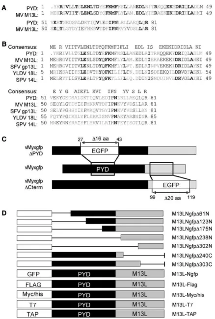
Figure 1. Bioinformatic Analyses and Manip-ulation of the M13L ORF
(A) CLUSTAL alignment of the 81 residue N-terminal PYD from M13L and the NCBI sequence for the PYD domain (PAAD/DAPIN, pfam02758.11). Residues are color-coded as having identity (bold black), similarity (black), or no similarity (gray).
(B) Partial alignment of PYD proteins from MV and other poxviruses, including SFV (NP051902), YLDV (NP073403), and SPV (NP570174). The consensus sequence for this alignment is shown.
(C) M13L KO viruses were constructed by tar-geted disruption of the M13L ORF and inser-tion of a GFP cassette (EGFP) driven by a synthetic early/late viral promoter. The GFP cassette was inserted either within the PYD starting at aa 27 (vMyxgfpDPYD) or down-stream of the PYD starting at aa 99 (vMyxgfpDCterm).
(D) Summary of the epitope tags used to con-struct M13L-PYD fusion proteins. Deletion mutants of M13L-Ngfp were generated by progressive truncation of sequences from either the amino (N) or the carboxyl (C) termi-nus at the indicated residues by PCR primer walking.
The subcellular distribution of PYD proteins is
deter-mined largely by their PYD even when other functional
domains are encoded (
Mansfield et al., 2001; Richards
et al., 2001
). Thus, M13L GFP fusion proteins were
gen-erated by targeted deletion of sequences both within
and outside of the PYD (
Figure 1
D) and expressed
in 293T cells by transfection. As illustrated with
M13LD61D, from which the first 22 amino acids of the
PYD had been removed, disruption of the M13L PYD
re-sulted in a loss of both the early perinuclear distribution
(data not shown) and the late cytosolic dispersion (
Figure
4
C). In contrast, deletions outside of the PYD did not alter
this profile significantly, and even a fusion protein
com-prised of the M13L PYD alone (M13LD240C) continued
to localize in cytosolic bodies (
Figure 4
C). Taken
to-gether, these findings indicated that M13L-PYD followed
an expression profile similar to known cytosolic PYD
proteins that was dependent on an intact PYD.
ASC-1 Is a Binding Partner of M13L-PYD
PYD family members that interact with ASC-1 and
mod-ulate its activity have been shown to colocalize with
ASC-1 in perinuclear and cytosolic bodies in a
PYD-dependent manner (
Masumoto et al., 1999; Richards
et al., 2001
). Given the expression profile of
M13L-PYD, we next assessed whether M13L-PYD also
colo-calized with ASC-1. Following expression in 293T cells,
M13L-Ngfp and Myc/his-tagged ASC-1 were found to
colocalize within both the perinuclear bodies at 48 hr
(
Figure 4
D, insets) and the cytosolic aggregates at
72 hr (
Figure 4
D). Subsequent studies suggested that
this colocalization reflected a specific interaction
be-tween ASC-1 and M13L-PYD. Binding of ASC-1 to
M13L was assessed by coimmunoprecipitation and
Western blot in 293T cells cotransfected with plasmids
expressing M13L-PYD and ASC-1 tagged with Flag
or Myc/his epitopes. Consistent with an interaction
be-tween these proteins, Myc/his-tagged ASC-1 was
de-tected in elutants following immunoprecipitation of
M13L-Flag, but not in cells transfected with a control
vector that did not express M13L-PYD (
Figure 4
E). This
finding was supported by the reciprocal experiment in
which Myc/his-tagged M13L-PYD was detected by
Western blot in elutants following ASC-Flag pull-downs
(
Figure 4
E).
An in vitro binding assay was then used to determine
whether this interaction occurred under biologically
rel-evant conditions. Lysates from 293T cells transfected
with M13L-PYD tagged with the tandem affinity
purifica-tion (TAP) epitope were incubated with lysates from
ac-tivated THP-1 cells, and binding partners were isolated
using the TAP method (
Wang et al., 2004
). Following
separation by SDS-PAGE, silver staining revealed
a prominent band at a molecular weight of 22 kDa that
Figure 2. Disruption of the M13L ORF Attenuates MV
Adult NZW rabbits were infected in each flank by intradermal injection of wt MV (vMyxgfp), M13L KO viruses (vMyxgfpDPYD, vMyxgfpDCterm), or a revertant virus (vMyxrevPYD).
(A) Summary of the mean clinical scores (+SD) assigned to each group after daily rankings according to a graduated scale. Larger values indicate poorer clinical outcomes due to greater disease severity. Clinical scores are not available after day 10 PI for rabbits infected with vMyxgfp or vMyxrevPYD due to poor survival rates.
(B) Fluorescence microscopy detection of MV-infected cells in PBMC isolated at day 7 PI from representative animals in each infected group. Values are expressed as a percentage of total cells isolated and represent the average number of GFP-positive cells in four samples 6 SD. (C) Primary lesion tissue was harvested at day 5 PI from two rabbits in each group, and the expression of host molecules upregulated during inflammation was assessed by semiquantitative RT-PCR via primers specific for rabbit IL-1b, IL-6, and MCP-1. Dermal tissue from an uninfected rabbit served as a control (no virus). Band intensities were determined by densitometry and values are indicated in arbitrary units following cor-rection for the intensity of GAPDH.
was subsequently confirmed to be ASC-1 by mass
spectrometric analysis (
Figure 4
F). A similar interaction
between M13L-PYD and other members of the PYD
su-perfamily has not been observed by mass spectrometry
when M13L-PYD is used as bait. Under the experimental
conditions used, only ASC-1 has been detected in
elu-tants. Taken together, these findings indicated that
M13L-PYD interacted with ASC-1 and establish the
po-tential for the viral protein to impact on the regulation of
inflammation and apoptosis after infection.
Expression of M13L-PYD Inhibits Caspase-1
Activation in Monocytes
The phenotype of the M13L-PYD KO virus implied a role
for the protein in suppressing host apoptotic or
inflam-matory responses to infection. Since caspase-1
partici-pates in both pathways and is tightly regulated by ASC-1
and other components of the inflammasome, we next
investigated the impact of M13L-PYD on caspase-1
ac-tivity. Untransfected THP-1 cells and cells expressing
M13L-Ngfp, M13L-NgfpD61N, or GFP alone were
sub-jected to mechanical disruption or incubated in the
pres-ence or abspres-ence of LPS, and caspase-1 activation was
assessed by Western blot detection of the cleavage of
procaspa1 to its active form. THP-1 cells were
se-lected because they constitutively express PYD proteins
that are activated by mechanical disruption and LPS
treatment (
Martinon et al., 2002
) and because
M13L-PYD can be expressed in these cells by high-efficiency
transfection (
Figure 4
A) or by direct infection with MV
(unpublished data). The cleaved, activated isoform of
caspase-1 was readily detectable in untransfected cells
or cells expressing GFP alone after mechanical
disrup-tion or stimuladisrup-tion with LPS, but only minimal caspase-1
cleavage was evident in THP-1 cells expressing
M13L-PYD (
Figure 5
A). As demonstrated after expression of
M13L-NgfpD61N, disruption of the M13L-PYD restored
inhibition of caspase-1 activation to wt levels (
Figure
5
A). These findings were confirmed using a colorimetric
caspase-1 activity assay in which expression of intact,
but not truncated, M13L-PYD reduced caspase-1
activ-ity by approximately 50% under the same experimental
conditions (
Figure 5
B). We next determined whether
expression of M13L-PYD could also inhibit the
biologi-cal consequences of caspase-1 activation by detecting
processing of the proinflammatory cytokine, IL-1b.
Western blot analyses confirmed that LPS treatment
or mechanical disruption induced abundant IL-1b
pro-cessing in untransfected cells and cells expressing
GFP or NgfpD61N, but that expression of
M13L-Ngfp inhibited IL-1b processing after either stimulus
(
Figure 5
C). Similarly, levels of secreted IL-1b in CM from
M13L-Ngfp-transfected cells were decreased by
ap-proximately 30% compared to levels in control cultures
when a quantitative sandwich ELISA was used (
Figure
5
D).
The impact of M13L-PYD on caspase activation in the
context of poxvirus infection was also investigated.
In-fection of THP-1 cells with vMyxgfpDPYD was itself
suf-ficient to induce caspase-1 processing that was largely
absent in uninfected cells or cells infected with wt virus
(
Figure 6
A). However, it should be noted that Western
blot analysis revealed additional bands following
detec-tion of caspase-1 in MV-infected cells that were not
present in transiently transfected cells. Whether these
observations are nonspecific or represent additional
processing of caspase-1 induced by active infection is
unclear. Of interest, no differences were observed in
the activation of caspase-3, a primary component of the
intrinsic apoptosis pathway, after infection with either wt
Table 1. Pathogenicity of vMyxgfp, vMyxrevPYD, vMyxgfpDPYD, and vMyxgfpDCterm
Day PI vMyxgfp vMyxrevPYDa vMyxgfpDPYD vMyxgfpDCterm
3 primary (1 cm) primary (1 cm) primary (1 cm) primary (1 cm) 5 primary raised (2.5 cm) primary raised (2 cm) primary raised (3.5 cm);
inflamed; crown flattened
primary raised (1.6 cm) no satellites some satellites decreased food and
water intake
7 primary larger (4.0 cm) primary larger (3.0 cm) healing primary larger (2.7 cm) numerous satellites (3–15) numerous satellites (4–10) ulcerating, cavitating
primary
small satellites secondary lesions on nose,
ears, eyelids
secondary lesions on nose and eyelids
small satellites and secondary lesions on 2 rabbits
small secondary lesions on ears
swollen head ano-genital inflammation
ano-genital inflammation
9 primary large, inflamed primary large (4 cm) healing primary larger but ulcerating satellites too numerous
to discern
numerous satellites >10 primary and secondary lesions regressing
secondary lesions on lip ears and head swollen;
fur ruffled
secondary lesions on ears, nose, and eyelids (2–10)
animals recovering small secondary lesions on ears and lips numerous secondary
lesions
discharge from eyes animals recovering swollen eyelids, multiple
lesions, discharge
nose and head swollen, fur ruffled
ano-genital inflammation ano-genital inflammation
14 33% survivalb 33% survival 100% survival 50% survival
a
Revertant virus constructed in the vMyxgfpDPYD background.
MV or the M13L-PYD KO virus (
Figure 6
A). Compared to
mock-infected cultures, only minimal caspase-3
pro-cessing was observed in cells infected with either virus.
The inhibitory effect of M13L-PYD was not limited to
events induced by infection since caspase-1 activation
was also decreased in THP-1 cells that were infected
with vMyxgfp prior to stimulation with LPS or
mechani-cal disruption (
Figure 6
B). In contrast, cells infected
with vMyxgfpDPYD exhibited higher levels of caspase-1
activity in response to either stimulus (
Figure 6
B),
indi-cating that expression of M13L-PYD in infected cells
also altered responses to exogenous stimuli.
Consistent with the results obtained in
Figure 5
,
acti-vation of caspase-1 was associated with increased
pro-cessing of IL-1b. Levels of secreted, active IL-1b were
elevated in CM from THP-1 cells infected with wt or
KO virus relative to mock-infected cells (
Figure 6
C).
However, only minimal increases were observed in
vMyxgfp-infected cells, whereas IL-1b concentrations
in CM from cells infected with vMyxgfpDPYD were
in-creased by more than 3-fold during the 48 hr time
course. Similar results were observed with IL-18,
an-other proinflammatory cytokine that undergoes
pro-cessing by caspase-1 (
Akita et al., 1997
). Although levels
of secreted, active IL-18 were increased in CM from
THP-1 cultures infected with either wt or KO virus
rela-tive to mock-infected cells at 12 hr PI, IL-18 was
2.3-fold more abundant in THP-1 cells infected with
vMyxgfpDPYD (
Figure 6
C). Moreover, IL-18 levels in
vMyxgfp-infected THP-1 cultures decreased below
that of mock-infected cells as infection progressed,
while IL-18 remained elevated in KO virus-infected
cultures. Thus, expression of M13L-PYD during MV
in-fection was associated with increased caspase-1
acti-vation and concurrent processing and secretion of
pro-inflammatory cytokines.
Discussion
Poxviruses employ a diverse array of proteins to
down-regulate innate or acquired host immune responses by
targeting such processes as apoptosis; the production
of interferons, chemokines, and inflammatory cytokines;
and the activity of cytotoxic T cells, natural killer cells,
complement, and antibody (
Seet et al., 2003
). The
obvi-ous sequence similarity between some poxvirus genes
and the cDNA versions of cellular counterparts suggests
that they are the product of ancestral capture,
recombi-nation, and reassortment events that occurred during
coevolution of the virus and its vertebrate hosts (
McLy-saght et al., 2003
). Consistent with that hypothesis, we
have described the initial characterization of a new class
of poxvirus immunomodulatory proteins typified by the
product of the MV M13L ORF, M13L-PYD. Structurally
and functionally, M13L-PYD closely resembles
mem-bers of the PYD superfamily of proteins that regulate
ap-optotic and inflammatory signaling pathways in myeloid
cells (
Bertin and DiStefano, 2000; Gumucio et al., 2002
).
Here, we report the ability of M13L-PYD to interfere with
the host inflammasome complex and thus block this
as-pect of the cellular proinflammatory responses to the
vi-rus infection.
The case for membership in this family is multifold.
From a structural viewpoint, M13L-PYD possesses the
Figure 3. Deletion of the M13L ORF Restricts MV Replication in Leu-kocytes
Multistep growth curves from rabbit RK13 fibroblasts (A) and RL5 lymphocytes (B) after infection at moi = 0.01 and titration of progeny virus on BGMK cells. Values represent log (mean titer) from triplicate wells 6 SD.
(C) Primary rabbit PBM and PBL were infected at moi = 1 and viral titers were measured at 48 hr PI.
N-term PYD that defines members of the PYD
superfam-ily (
Fairbrother et al., 2001
). In fact, like PYD proteins
such as POP-1, the M13L gene product consists almost
entirely of a single PYD and encodes no other functional
domain or motif. Moreover, the M13L PYD shares a high
degree of similarity with the classical PYD sequence and
it is a critical determinant of the properties of M13L-PYD.
From a functional perspective, M13L-PYD exhibited the
capacity to modulate caspase-1 activation and
inflam-matory cytokine processing pathways in myeloid cells
that have previously been shown to be regulated
by PYD family members, such as ASC-1 (
Srinivasula
et al., 2002
). As with other PYD proteins, the PYD of
M13L was implicated as a potential determinant of this
function. Consistent with the role of the PYD as a
homo-typic protein-protein interaction motif, M13L-PYD
colo-calized in the cytosol with ASC-1 and was found to
inter-act with this protein in binding assays. Taken together,
these findings constitute strong evidence that
M13L-PYD is in fact a functional viral M13L-PYD protein.
Poxvirus immunomodulatory proteins can be divided
by function into three strategic classes: virostealth
pro-teins, viromimetics (virokines and viroreceptors), and
virotransducers (
Seet et al., 2003
). M13L-PYD appears
to be a member of this latter group, which act
intracellu-larly to micromanipulate host-cell signal transduction
Figure 4. Expression and Subcellular Localization of M13L-PYD
(A) M13L-Ngfp was expressed by transfection in human 293T fibroblasts (i–vi) and differentiated THP-1 monocytes (vii and viii), and localization was assessed at 48 or 72 hr by fluorescence microscopy. Nuclei are shown in blue after DAPI counterstaining. T7-tagged M13L-PYD under the control of an early/late VV promoter was expressed in rabbit RK13 cells that were infected with vMyxgfpDPYD (ix and x). Expression of M13L-PYD was detected by indirect immunofluorescence with antibody against the T7 epitope and a TRITC-conjugated secondary antibody (original magnification, 6403).
(B) Subcellular localization of M13L-Ngfp in 293T cells was assessed at 48 hr by fluorescent stains specific for the Golgi complex (Golgitracker, blue, i–iii) or lysosomes (lysotracker, red, iv–vi) (original magnification, 6403).
(C) Localization of representative GFP fusion proteins generated by successive truncation of M13L-PYD within (M13LNgfpD61N, ii) or outside of (M13LNgfpD240C, iii) the PYD. Proteins were expressed in 293T and compared to full-length M13L-Ngfp (i) (original magnification, 4003). (D) Colocalization of M13L-Ngfp (i) and Myc/his-tagged ASC-1 (ii) was assessed 48 hr (insets) and 72 hr after cotransfection in 293T cells. ASC-1 expression was detected by indirect immunofluorescence with TRITC-conjugated secondary antibody to detect the Myc epitope.
(E) Representative Western blots showing the interaction between M13L-PYD and human ASC-1 expressed in 293T cells by cotransfection. Left: Flag-tagged M13L was immunoprecipitated by antibody specific for the Flag epitope (IP, anti-Flag), and bound Myc/his-tagged ASC-1 was de-tected by immunoblot with antibody against the Myc epitope (IB, anti-Myc) in elutants from cells transfected with plasmid expressing M13L-Flag but not the Flag epitope alone (pcDNA3-Flag). Right: The reciprocal experiment with Flag-tagged ASC-1 and Myc/his-tagged M13L. (F) Lysates from 293T cells transfected with TAP-tagged M13L were incubated with lysates from activated THP-1 cells and bound proteins iso-lated with the TAP method. A representative silver-stained gel following separation of bound proteins by SDS-PAGE is shown with arrows in-dicating a prominent band with a molecular weight of the 22 kDa that was determined to be ASC-1. This band was not evident when lysates ob-tained from 293T cells transfected with empty plasmid (pcDNA3) or plasmid expressing the TAP epitope alone (pcDNA3-TAP) were analyzed.
machinery and disconnect innate antiviral pathways
from their biological effect. MV encodes several
viro-transducers that target apoptosis and caspase activity,
including the antiapoptotic serine protease inhibitor
(serpin), Serp-2, and the apoptotic regulators encoded
by the MV M11L, M-T4, and M-T5 genes (
Everett and
McFadden, 1999
). Of these, only the MV Serp-2 protein
has been shown to weakly inhibit caspase-1 activity
and modulate inflammatory and apoptotic responses
in lymphoid cells (
Messud-Petit et al., 1998
). However,
unlike M13L-PYD in this study, Serp2 has limited effect
on the activity of human caspase-1 (
Messud-Petit et al.,
1998
) and is unlikely to have significantly influenced the
results reported here. Thus, M13L-PYD may represent
the primary modulator of caspase-1 activation encoded
by MV.
When comparing the properties of M13L-PYD with
those of PYD proteins that function as regulators of
the activity of other PYD proteins, such as pyrin,
numer-ous parallels are evident. M13L-PYD exhibits a high
de-gree of similarity with pyrin, particularly within the first 50
residues of the PYD. Like pyrin (
Mansfield et al., 2001;
Richards et al., 2001
), M13L-PYD exhibits a distinctive
cytosolic and punctate perinuclear distribution that
was dependent on sequences within the PYD. For pyrin,
this pattern also reflected colocalization with ASC-1 (
Ri-chards et al., 2001
). Thus, at first blush it may seem
sur-prising that expression of M13L-PYD is associated with
decreased caspase-1 activation, whereas the
interac-tion between pyrin and ASC-1 is a proinflammatory
event (
Yu et al., 2005
). Since the only functional domain
encoded by M13L is the PYD, it is conceivable that
M13L-PYD binds to ASC-1 via a homotypic interaction
mediated by the PYD and inhibits its activation similar
to POP-1 (
Stehlik et al., 2003
). However, M13L-PYD
shares little sequence similarity with POP-1 (data not
shown). Alternatively, M13L-PYD may bind to ASC-1
and acts as a competitive inhibitor of pyrin to prevent
ASC-1 activation. This model is consistent with the
mechanism of action of other poxvirus
immunomodula-tory proteins that are homologs of cellular proteins (
Seet
et al., 2003
). Although we have yet to find evidence of
a potential interaction between M13L-PYD and other
PYD family members, the high degree of similarity
be-tween the PYDs of M13L and IFI proteins that are
ex-pressed in the cytosol, such as myeloid nuclear
differen-tiation antigen (MNDA) (
Asefa et al., 2004
), may indicate
alternate functions for M13L-PYD.
The sequences of more than two dozen poxvirus
ge-nomes have been determined and immunomodulatory
proteins targeting all facets of innate and adaptive
immune responses have been reported (
Seet et al.,
2003
). In fact, so diverse are these genes that a single
immunomodulatory protein that is common to all
poxvi-ruses has yet to be identified. However, genes predicted
to encode PYD proteins that closely align with
M13L-PYD are also present in the genomes of poxviruses
from other Leporipoxviridae, as well as representatives
from the Suipoxviridae (
Afonso et al., 2002
) and the
Ya-tapoxviridae (
Lee et al., 2001
). Phylogenetic analyses
have demonstrated that, together with the
Capripoxvir-idae, these three genera cluster to form the largest
sub-grouping of Chordopoxviruses (
Gubser et al., 2004
),
suggesting a common evolutionary ancestor that may
have initially acquired a host PYD protein. However,
no gene analogous to M13L has been identified in the
genomes of the capripoxvirus species that have been
sequenced to date (
Tulman et al., 2001, 2002
), possibly
indicating an earlier evolutionary divergence from other
viruses in this group. Of particular interest is the fact that
orthopoxviruses, such as vaccinia virus (VV) (
Calderara
et al., 2001
), and the molluscipoxvirus, molluscum
con-tagiosum (
Xiang and Moss, 2003
), encode secreted
pro-teins that act as molecular scavengers to bind and
se-quester IL-1b, whereas comparable orthologs are
absent in members of the Capripoxvirus,
Leporipoxvi-rus, SuipoxviLeporipoxvi-rus, and Yatapoxvirus genera (
Barry and
McFadden, 1997
). Thus, poxvirus-encoded PYD
pro-teins capable of disrupting the intracellular signaling
pathways leading to IL-1b processing and secretion
may serve a similar role.
Advances in the field of viral immunomodulation
con-tinue to be made with increasing frequency, providing
important clues about the selective pressures that drive
the coevolution of virus and host. Moreover, greater
un-derstanding of the mechanisms by which poxviruses
Figure 5. Expression of M13L-PYD Inhibits Caspase-1 Activation and IL-1b Processing Full-length Ngfp or truncated M13L-NgfpD61N were expressed in differentiated human THP-1 cells by transient transfection. (A and C) Caspase-1 and IL-1b activation was induced by mechanical disruption of mem-brane integrity or stimulation with LPS (600 mg/ml for 6 hr). Activation of caspase-1 (A) and IL-1b (C) was assessed by Western blot detection of the active, cleaved isoform of each enzyme. Detection of the housekeeping gene, actin, ensured equal loading. (B) Caspase-1 activity in parallel cultures was quantified by spectrophotometric detection of pNA-YVAD cleavage and changes were ex-pressed as a percentage (6SD) of values ob-tained for untreated controls (100%). (D) IL-1b levels in conditioned media from LPS-treated cells were quantified by sand-wich ELISA and were expressed as pg of cy-tokine/ml of CM 6 SD.
modulate key components of the immune system has
in-creased the potential for viral immunomodulatory
pro-teins to be used as therapeutic agents (
Lucas and
McFadden, 2004
). Deregulated IL-1b production and
chronic inflammation are hallmarks of many
autoim-mune diseases that present both systemically and
within the central nervous system, including rheumatoid
arthritis and multiple sclerosis (
Kinne et al., 2000;
Mina-gar et al., 2002
). Thus, poxvirus immunomodulatory
pro-teins that target monocyte activity and regulation of
IL-1b may have the potential to be exploited in the
de-sign of novel anti-inflammatory therapies for the
treat-ment of these diseases.
Experimental Procedures Cell Culture
BGMK, RK13, and 293T cells were cultured in DMEM (GIBCO-BRL) completed with 10% fetal bovine serum (FBS) and antibiotics. THP-1 monocytes and rabbit RL5 lymphocytes were cultured in complete
RPMI 1640 medium (GIBCO-BRL). For differentiation, THP-1 cells were stimulated for 24 hr with 50 ng/ml phorbol-12-myristate-13-acetate (PMA, Sigma). PBMC used in infection studies were ob-tained from healthy NZW rabbits and isolated by density-gradient centrifugation, as described previously (Johnston and Power, 1999). Blood-derived PBM were separated from PBL by adherence on polystyrene flasks for 1 hr and maintained in complete RPMI. Nonadherent PBL were collected and cultured in complete RPMI supplemented with recombinant IL-2 (100 IU/ml, Sigma).
Viruses
Recombinant and KO viruses were constructed in a MV (strain Lau-sanne) background and propagated and titrated using the methods described previously (Opgenorth et al., 1992). Wild-type vMyxgfp contained a GFP cassette driven by a synthetic VV early/late pro-moter that was inserted between ORFs M135R and M136R ( John-ston et al., 2003). Two KO viruses deficient for M13L-PYD expression were similarly constructed by selectively deleting specific sequen-ces within the M13L ORF and inserting a GFP cassette by homolo-gous recombination: vMyxgfpDPYD, for which nucleotides 14852 to 14902 within the M13L-PYD were deleted, and vMyxgfpDCterm, for which nucleotides 14633 to 14690 near the C terminus of M13L Figure 6. Deletion of M13L-PYD Results in Increased Caspase-1 Activity
Differentiated THP-1 cells were mock-infected or mock-infected with vMyxgfp and vMyxgfpDPYD, and cells and CM were har-vested at the indicated times PI.
(A) Expression of caspase-1 and caspase-3 was assessed by Western blot.
(B) Caspase-1 activity induced in mock- or MV-infected THP-1 cells by LPS stimulation (600 mg/ml for 6 hr) or mechanical disruption was measured via a pNA-YVAD cleavage as-say and changes were expressed as a per-centage (6SD) of values obtained for mock-infected controls (100%).
(C) Levels of secreted IL-1b and IL-18 were determined by ELISA and expressed as pg of cytokine/ml of CM 6 SD.
were removed (Figure 1C). Disruption of the M13L ORF was con-firmed by PCR and gel shift analyses. To ensure that the phenotype of each KO virus did not reflect adverse mutations introduced into other genes during recombination, a revertant virus was constructed (vMyxrevPYD) to restore the intact M13L ORF. Infections were per-formed and growth curves constructed as previously described (Johnston et al., 2005)
Animal Studies
NZW rabbits were housed in level 2 containment facilities at the Uni-versity of Western Ontario Animal Care Facility according to the guidelines of the Canadian Council on Animal Care. Experiments were conducted on two separate dates using different preparations of each virus. Injections were performed intradermally in each thigh using 1000 focus forming units (ffu) of vMyxgfp (n = 9), vMyxrevPYD (n = 4), vMyxgfpDPYD (n = 7), or vMyxgfpDCterm (n = 4) per site. Two rabbits injected with PBS served as controls. Disease progression was monitored daily for symptoms consistent with Myxomatosis (Fenner and Ratcliffe, 1965) and scored on a graduated scale ac-cording to disease severity by animal care technicians unaware of the status of the animal. A clinical score between 0 (complete ab-sence of disease) and 20 (moribund) was assigned to each rabbit, with moribund animals sacrificed using euthanyl administered intra-venously after anesthesia. Animals from each group were also sac-rificed at 4, 7, and 10 days PI, and a complete postmortem examina-tion was performed. Tissue, including the primary and secondary lesions, spleen, and lymph nodes, was collected and divided into two samples that were either snap frozen in liquid nitrogen for isola-tion of RNA and protein or embedded in paraffin for analysis by im-munohistochemistry. To detect circulating MV-infected cells, PBMC were isolated from blood collected by intravenous puncture at day 3 and 7 PI and immediately visualized by fluorescence microscopy to detect the GFP expression indicative of MV infection.
Fusion Proteins and Epitope-Specific Antibodies
The complete M13L ORF, and M13L variants in which specific re-gions of the M13L ORF were selectively deleted, were PCR amplified from genomic viral DNA and subcloned into expression vectors using restriction sites introduced by the PCR primers (Figure 1D). Constructs included fusion proteins expressing TAP (cDNA3.6-TAP,Wang et al. [2004]), Myc/his (pcDNA3.1myc-his, Invitrogen), and GFP (phrGFP-N, Stratagene) epitopes. Flag and T7 tags were in-troduced during amplification of M13L with PCR primers containing sequences encoding the epitope. Plasmid encoding untagged hu-man ASC-1 was kindly provided by J. Reed (The Burnham Institute, La Jolla, CA) (Stehlik et al., 2002). Flag and Myc/his epitope-tagged ASC-1 were generated by subcloning into the appropriate vector. The identities of all clones were confirmed by sequence analysis, and expression of fusion proteins was confirmed by fluorescence microscopy or Western analyses via mouse monoclonal antibodies specific for Myc (Invitrogen), T7 (Novagen), Flag (Sigma), or GFP (Clontech) epitopes. Gel shift analysis was used to confirm that the fluorescence observed represented a GFP fusion product. A rab-bit peroxidase-anti-peroxidase antiserum (anti-PAP; Sigma) recog-nizing the protein A sequence was utilized for detecting the TAP tag.
Subcellular Localization of M13L-PYD
Human 293T cells were grown to 90% confluence and transiently transfected with plasmids encoding full-length or truncated M13L fusion proteins using Lipofectamine 2000 reagent (Invitrogen) in accordance with the manufacturer’s instruction. After 24 hr, trans-fected cells were harvested, adhered to poly-L-lysine-coated cover-slips, and cultured as indicated. PMA-differentiated THP-1 cells were adhered to coverslips at 50% confluence and transfected us-ing ExGen 500 Reagent (Fermentas, Inc.). Untransfected cells and cells transfected with the appropriate plasmid lacking exogenous sequences (vector alone) served as controls. At 48 hr and 72 hr post-transfection, cells were mounted on glass slides in Prolong Antifade medium (Molecular Probes) and assessed by fluorescence micros-copy. To detect subcellular localization, transfected cells were counterstained with fluorescent markers (Molecular Probes) target-ing the Golgi complex (BODIPY FL C5-ceramide, 1:100),
endoplas-mic reticulum (ER-Tracker Blue-White DPX, 1:750), mitochondria (MitoFluor Red 589, 1:5000), actin (Texas red-X phalloidin, 1:20),
lysosomes (LysoTracker Red DND-99, 1:13000), or the nucleus (DAPI, 1:330).
To determine M13L-PYD localization in the context of MV infec-tion, RK13 cells were initially transfected with pSC65 plasmid en-coding M13L-T7 under the control of the VV early/late promoter. Transfected cells were infected after 24 hr with vMyxgfpDPYD at moi = 1 and cultured for an additional 24 or 48 hr. M13L expression was assessed by indirect immunofluorescence by the use of mono-clonal antibody specific for the T7 epitope (1:100) and a TRITC-conjugated goat-anti-mouse secondary antibody (1:500, Jackson ImmunoResearch). Isotype control antibodies were used to ensure the lack of nonspecific antibody binding.
Immunoprecipitation and TAP Purification
293T cells were cotransfected with plasmid encoding FLAG-tagged M13L or Myc/his-tagged ASC-1 and cultured for 72 hr. Transfected cells were lysed in CellLytic-M reagent (Sigma) containing protease inhibitors and cleared lysates incubated overnight with anti-FLAG agarose beads (Sigma). Beads were pelleted by centrifugation, washed twice, and incubated at 65ºC for 10 min in SDS-PAGE sam-ple buffer containing 100 mM DTT. The eluted comsam-plexes were re-solved by SDS-PAGE and then analyzed by immunoblotting with antibodies against the Myc/his epitope. The reciprocal experiment was conducted in the same manner with M13L tagged with the Myc/his epitope and FLAG-tagged ASC-1. For TAP isolations, 293T cells transfected with M13L conjugated to either the TAP or Myc/his epitope were lysed in 2% CHAPS buffer containing prote-ase inhibitors, and the lysates were cleared by centrifugation. Ly-sates from approximately 107THP-1 cells were prepared by
pas-sage through a 22 gauge needle as described, combined with the 293T cell lysates and incubated overnight at 4ºC. The M13L-TAP fu-sion protein and associated proteins were then isolated using the TAP method described previously (Wang et al., 2004), concentrated to an appropriate volume, and resolved by SDS-PAGE. Separated proteins were visualized by silver staining (Bio-Rad).
Immunoblot Analyses
Protein levels in cleared cell lysates were quantified with a protein assay kit (Bio-Rad), and Western blot analyses were performed as previously described (Johnston et al., 2003). Polyclonal antibodies included those specific for the proenzyme and cleaved forms of caspase-1 (1:1000, Upstate Biotechnology), IL-1b (1:1000, Cell Sig-naling Technology), and caspase-3 (1:1000, Cell SigSig-naling Technol-ogy). Equal loading of each sample was confirmed by detection of the housekeeping gene, actin, by use of monoclonal antibodies ob-tained from Santa Cruz Biotechnology at a dilution of 1:3000. Horse-radish peroxidase (HRP)-conjugated anti-mouse and goat-anti-rabbit secondary antibodies were obtained from Jackson ImmunoResearch Laboratories.
RT-PCR
Necropsied tissue was ground under liquid nitrogen and passed through a QiaShredder (Qiagen) column to disrupt cells and connec-tive tissue. Total cellular RNA was then isolated using a RNAeasy Mini Kit (Qiagen) and DNase treated for 30 min at 37ºC. cDNA was prepared using Superscript II RT (Invitrogen) and amplified with pri-mers specific for rabbit IL-1b, IL-6, or MCP-1. Loading of equal amounts of template cDNA was assessed by amplification of GAPDH. Amplification of DNase-treated RNA obtained from each sample prior to reverse transcription served as controls to ensure the lack of contaminating DNA. PCR products were separated by agarose gel electrophoresis and visualized by staining with ethidium bromide.
Detection of Caspase-1 Activity and Cytokine Processing Differentiated THP-1 cells were mock-infected or infected with wt or KO virus at an moi = 5, and cells and conditioned media (CM) were harvested at various time points PI. Activation of caspase-1 and subsequent processing of IL-1b and IL-18 was assessed by Western blot detection of the cleaved isoforms of each protein in cell lysates. Secreted IL-1b and IL-18 was detected in CM with quantitative hu-man IL-1b and IL-18 sandwich ELISA Kits (R&D Systems) according to manufacturer’s instructions. Levels of IL-1b and IL-18 in triplicate samples were quantified by referencing a standard curve generated
in parallel and expressed as pg cytokine/ml of CM. Caspase-1 activ-ity was quantified using a colorimetric assay based on spectropho-tometric detection of caspase-1 cleavage of the chromophore p-nitroaniline (pNA) from substrate containing the YVAD recognition site (Calbiochem). Mean values of triplicate samples (corrected for background) were expressed relative to untreated controls. Activa-tion of caspase-1 and cytokine processing in response to exoge-nous stimuli was similarly assessed in cells that were mock infected or infected with wt or KO virus and cells that were transiently trans-fected with plasmid encoding full-length or truncated M13L. Stimuli included treatment with lipopolysaccharide (LPS, 600 mg, Sigma) for 1 hr or mechanical disruption of membrane integrity. To disrupt membrane integrity, cells were harvested in cold PBS, incubated for 15 min on ice in hypotonic buffer, and then passaged repeatedly through a 22 gauge needle.
Bioinformatic Analyses
The M13L sequence (accession number AF170726) was used as ini-tial query in a BLASTp (Altschul et al., 1997) search of the public do-main database to identify similarity to other proteins. A construction of multiple alignments with various PYD-containing proteins was carried out using the CLUSTAL W Multiple Sequence Alignment soft-ware (Thompson et al., 1994). Cell sorting predictions for M13L-PYD was carried out using PSORT II (Nakai and Horton, 1999), which de-termines the likelihood of the presence of a signal peptide and sub-cellular localization. The protein domain search tool Prosite (Appel et al., 1994) was used to identify putative patterns, profiles, and/or motifs present within the M13L amino acid sequence.
Acknowledgments
This work was supported by grants from the Canadian Institutes for Health Research (CIHR). G.M. holds a Canada Research Chair in Mo-lecular Virology. J.B.J. held a CIHR Fellowship during completion of this work. We thank J. Reed for valuable reagents and helpful com-ments on this manuscript. We would like to dedicate this work to the memory of Frederique Messud-Petit.
Received: October 15, 2004 Revised: September 23, 2005 Accepted: October 7, 2005 Published: December 13, 2005
References
Afonso, C.L., Tulman, E.R., Lu, Z., Zsak, L., Osorio, F.A., Balinsky, C., Kutish, G.F., and Rock, D.L. (2002). The genome of swinepox virus. J. Virol. 76, 783–790.
Akita, K., Ohtsuki, T., Nukada, Y., Tanimoto, T., Namba, M., Okura, T., Takakura-Yamamoto, R., Torigoe, K., Gu, Y., Su, M.S., et al. (1997). Involvement of caspase-1 and caspase-3 in the production and processing of mature human interleukin 18 in monocytic THP.1 cells. J. Biol. Chem. 272, 26595–26603.
Altschul, S.F., Madden, T.L., Schaffer, A.A., Zhang, J., Zhang, Z., Miller, W., and Lipman, D.J. (1997). Gapped BLAST and PSI-BLAST: a new generation of protein database search programs. Nucleic Acids Res. 25, 3389–3402.
Appel, R.D., Bairoch, A., and Hochstrasser, D.F. (1994). A new gen-eration of information retrieval tools for biologists: the example of the ExPASy WWW server. Trends Biochem. Sci. 19, 258–260. Asefa, B., Klarmann, K.D., Copeland, N.G., Gilbert, D.J., Jenkins, N.A., and Keller, J.R. (2004). The interferon-inducible p200 family of proteins: a perspective on their roles in cell cycle regulation and differentiation. Blood Cells Mol. Dis. 32, 155–167.
Barrett, J.W., Cao, J.X., Hota-Mitchell, S., and McFadden, G. (2001). Immunomodulatory proteins of myxoma virus. Semin. Immunol. 13, 73–84.
Barry, M., and McFadden, G. (1997). Virus encoded cytokines and cytokine receptors. Parasitology 115 (Suppl), S89–S100.
Bertin, J., and DiStefano, P.S. (2000). The PYRIN domain: a novel motif found in apoptosis and inflammation proteins. Cell Death Dif-fer. 7, 1273–1274.
Calderara, S., Xiang, Y., and Moss, B. (2001). Orthopoxvirus IL-18 binding proteins: affinities and antagonist activities. Virology 279, 22–26.
Cameron, C., Hota-Mitchell, S., Chen, L., Barrett, J., Cao, J.X., Mac-aulay, C., Willer, D., Evans, D., and McFadden, G. (1999). The com-plete DNA sequence of myxoma virus. Virology 264, 298–318. Centola, M., Aksentijevich, I., and Kastner, D.L. (1998). The heredi-tary periodic fever syndromes: molecular analysis of a new family of inflammatory diseases. Hum. Mol. Genet. 7, 1581–1588. Chen, X., Bykhovskaya, Y., Tidow, N., Hamon, M., Bercovitz, Z., Spi-rina, O., and Fischel-Ghodsian, N. (2000). The familial mediterranean fever protein interacts and colocalizes with a putative Golgi trans-porter. Proc. Soc. Exp. Biol. Med. 224, 32–40.
Everett, H., and McFadden, G. (1999). Apoptosis: an innate immune response to virus infection. Trends Microbiol. 7, 160–165. Fairbrother, W.J., Gordon, N.C., Humke, E.W., O’Rourke, K.M., Star-ovasnik, M.A., Yin, J.P., and Dixit, V.M. (2001). The PYRIN domain: a member of the death domain-fold superfamily. Protein Sci. 10, 1911–1918.
Fenner, F., and Ratcliffe, F. (1965). Myxomatosis (Cambridge, UK: Cambridge University Press).
Gubser, C., Hue, S., Kellam, P., and Smith, G.L. (2004). Poxvirus ge-nomes: a phylogenetic analysis. J. Gen. Virol. 85, 105–117. Gumucio, D.L., Diaz, A., Schaner, P., Richards, N., Babcock, C., Schaller, M., and Cesena, T. (2002). Fire and ICE: the role of pyrin do-main-containing proteins in inflammation and apoptosis. Clin. Exp. Rheumatol. 20, S45–S53.
Hoffman, H.M., Mueller, J.L., Broide, D.H., Wanderer, A.A., and Ko-lodner, R.D. (2001). Mutation of a new gene encoding a putative pyrin-like protein causes familial cold autoinflammatory syndrome and Muckle-Wells syndrome. Nat. Genet. 29, 301–305.
Hull, K.M., Shoham, N., Chae, J.J., Aksentijevich, I., and Kastner, D.L. (2003). The expanding spectrum of systemic autoinflammatory disorders and their rheumatic manifestations. Curr. Opin. Rheuma-tol. 15, 61–69.
Johnston, J., and Power, C. (1999). Productive infection of human peripheral blood mononuclear cells by feline immunodeficiency vi-rus: implications for vector development. J. Virol. 73, 2491–2498. Johnston, J.B., Barrett, J.W., Chang, W., Chung, C.S., Zeng, W., Masters, J., Mann, M., Wang, F., Cao, J., and McFadden, G. (2003). Role of the serine-threonine kinase PAK-1 in myxoma virus replication. J. Virol. 77, 5877–5888.
Johnston, J.B., Nazarian, S.H., Natale, R., and McFadden, G. (2005). Myxoma virus infection of primary human fibroblasts varies with cel-lular age and is regulated by host interferon responses. Virology 332, 235–248.
Kinne, R.W., Brauer, R., Stuhlmuller, B., Palombo-Kinne, E., and Burmester, G.R. (2000). Macrophages in rheumatoid arthritis. Arthri-tis Res. 2, 189–202.
Lee, H.J., Essani, K., and Smith, G.L. (2001). The genome sequence of Yaba-like disease virus, a yatapoxvirus. Virology 281, 170–192. Lucas, A., and McFadden, G. (2004). Secreted immunomodulatory viral proteins as novel biotheraputics. J. Immunol. 173, 4765–4774. Mansfield, E., Chae, J.J., Komarow, H.D., Brotz, T.M., Frucht, D.M., Aksentijevich, I., and Kastner, D.L. (2001). The familial Mediterra-nean fever protein, pyrin, associates with microtubules and colocal-izes with actin filaments. Blood 98, 851–859.
Martinon, F., Burns, K., and Tschopp, J. (2002). The inflammasome: a molecular platform triggering activation of inflammatory caspases and processing of proIL-beta. Mol. Cell 10, 417–426.
Masumoto, J., Taniguchi, S., Ayukawa, K., Sarvotham, H., Kishino, T., Niikawa, N., Hidaka, E., Katsuyama, T., Higuchi, T., and Sagara, J. (1999). ASC, a novel 22-kDa protein, aggregates during apoptosis of human promyelocytic leukemia HL-60 cells. J. Biol. Chem. 274, 33835–33838.
Masumoto, J., Taniguchi, S., Nakayama, J., Shiohara, M., Hidaka, E., Katsuyama, T., Murase, S., and Sagara, J. (2001). Expression of apoptosis-associated speck-like protein containing a caspase re-cruitment domain, a pyrin N-terminal homology domain-containing
protein, in normal human tissues. J. Histochem. Cytochem. 49, 1269–1275.
McDermott, M.F., and Aksentijevich, I. (2002). The autoinflammatory syndromes. Curr. Opin. Allergy Clin. Immunol. 2, 511–516. McLysaght, A., Baldi, P.F., and Gaut, B.S. (2003). Extensive gene gain associated with adaptive evolution of poxviruses. Proc. Natl. Acad. Sci. USA 100, 15655–15660.
Messud-Petit, F., Gelfi, J., Delverdier, M., Amardeilh, M.F., Py, R., Sutter, G., and Bertagnoli, S. (1998). Serp2, an inhibitor of the inter-leukin-1beta-converting enzyme, is critical in the pathobiology of myxoma virus. J. Virol. 72, 7830–7839.
Minagar, A., Shapshak, P., Fujimura, R., Ownby, R., Heyes, M., and Eisdorfer, C. (2002). The role of macrophage/microglia and astrocytes in the pathogenesis of three neurologic disorders: HIV-associated dementia, Alzheimer disease, and multiple scle-rosis. J. Neurol. Sci. 202, 13–23.
Nakai, K., and Horton, P. (1999). PSORT: a program for detecting sorting signals in proteins and predicting their subcellular localiza-tion. Trends Biochem. Sci. 24, 34–36.
Opgenorth, A., Graham, K., Nation, N., Strayer, D., and McFadden, G. (1992). Deletion analysis of two tandemly arranged virulence genes in myxoma virus, M11L and myxoma growth factor. J. Virol. 66, 4720–4731.
Reed, J.C., Doctor, K., Rojas, A., Zapata, J.M., Stehlik, C., Fiorentino, L., Damiano, J., Roth, W., Matsuzawa, S., Newman, R., et al. (2003). Comparative analysis of apoptosis and inflammation genes of mice and humans. Genome Res. 13, 1376–1388.
Richards, N., Schaner, P., Diaz, A., Stuckey, J., Shelden, E., Wadhwa, A., and Gumucio, D.L. (2001). Interaction between pyrin and the apoptotic speck protein (ASC) modulates ASC-induced apoptosis. J. Biol. Chem. 276, 39320–39329.
Seet, B.T., Johnston, J.B., Brunetti, C.R., Barrett, J.W., Everett, H., Cameron, C., Sypula, J., Nazarian, S.H., Lucas, A., and McFadden, G. (2003). Poxviruses and immune evasion. Annu. Rev. Immunol. 21, 377–423.
Srinivasula, S.M., Poyet, J.L., Razmara, M., Datta, P., Zhang, Z., and Alnemri, E.S. (2002). The PYRIN-CARD protein ASC is an activating adaptor for caspase-1. J. Biol. Chem. 277, 21119–21122. Stehlik, C., and Reed, J.C. (2004). The PYRIN connection: novel play-ers in innate immunity and inflammation. J. Exp. Med. 200, 551–558. Stehlik, C., Fiorentino, L., Dorfleutner, A., Bruey, J.M., Ariza, E.M., Sagara, J., and Reed, J.C. (2002). The PAAD/PYRIN-family protein ASC is a dual regulator of a conserved step in nuclear factor kappaB activation pathways. J. Exp. Med. 196, 1605–1615.
Stehlik, C., Krajewska, M., Welsh, K., Krajewski, S., Godzik, A., and Reed, J.C. (2003). The PAAD/PYRIN-only protein POP1/ASC2 is a modulator of ASC-mediated nuclear-factor-kappa B and pro-caspase-1 regulation. Biochem. J. 373, 101–113.
Thompson, J.D., Higgins, D.G., and Gibson, T.J. (1994). CLUSTAL W: improving the sensitivity of progressive multiple sequence align-ment through sequence weighting, position-specific gap penalties and weight matrix choice. Nucleic Acids Res. 22, 4673–4680. Tulman, E.R., Afonso, C.L., Lu, Z., Zsak, L., Kutish, G.F., and Rock, D.L. (2001). Genome of lumpy skin disease virus. J. Virol. 75, 7122– 7130.
Tulman, E.R., Afonso, C.L., Lu, Z., Zsak, L., Sur, J.H., Sandybaev, N.T., Kerembekova, U.Z., Zaitsev, V.L., Kutish, G.F., and Rock, D.L. (2002). The genomes of sheeppox and goatpox viruses. J. Virol. 76, 6054–6061.
Wang, G., Barrett, J.W., Nazarian, S.H., Everett, H., Gao, X., Bleack-ley, C., Colwill, K., Moran, M.F., and McFadden, G. (2004). Myxoma virus M11L prevents apoptosis through constitutive interaction with Bak. J. Virol. 78, 7097–7111.
Xiang, Y., and Moss, B. (2003). Molluscum contagiosum virus inter-leukin-18 (IL-18) binding protein is secreted as a full-length form that binds cell surface glycosaminoglycans through the C-terminal tail and a furin-cleaved form with only the IL-18 binding domain. J. Virol. 77, 2623–2630.
Yu, J.W., Wu, J., Zhang, Z., Datta, P., Ibrahimi, I., Taniguchi, S., Sa-gara, J., Fernandes-Alnemri, T., and Alnemri, E.S. (2005). Cryopyrin
and pyrin activate caspase-1, but not NF-kappaB, via ASC oligomer-ization. Cell Death Differ., in press.