HAL Id: hal-02401568
https://hal.archives-ouvertes.fr/hal-02401568
Submitted on 10 Dec 2019HAL is a multi-disciplinary open access archive for the deposit and dissemination of sci-entific research documents, whether they are pub-lished or not. The documents may come from teaching and research institutions in France or abroad, or from public or private research centers.
L’archive ouverte pluridisciplinaire HAL, est destinée au dépôt et à la diffusion de documents scientifiques de niveau recherche, publiés ou non, émanant des établissements d’enseignement et de recherche français ou étrangers, des laboratoires publics ou privés.
Antibiotic Containing Poly Lactic Acid/Hydroxyapatite
Biocomposite Coatings for Dental Implant Applications
Ipek Karacan, Innocent Jacob Macha, Gina Choi, Sophie Cazalbou, Besim
Ben-Nissan
To cite this version:
Ipek Karacan, Innocent Jacob Macha, Gina Choi, Sophie Cazalbou, Besim Ben-Nissan. Antibiotic Containing Poly Lactic Acid/Hydroxyapatite Biocomposite Coatings for Dental Implant Applications. Key Engineering Materials, 2017, 758, pp.120-125. �10.4028/www.scientific.net/KEM.758.120�. �hal-02401568�
OATAO is an open access repository that collects the work of Toulouse researchers and makes it freely available over the web where possible
Any correspondence concerning this service should be sent
to the repository administrator: tech-oatao@listes-diff.inp-toulouse.fr
This is an author’s version published in: http://oatao.univ-toulouse.fr/24478
To cite this version:
Karacan, Ipek and Macha, Innocent Jacob and Choi, Gina and Cazalbou, Sophie and Ben-Nissan, Besim Antibiotic Containing Poly Lactic
Acid/Hydroxyapatite Biocomposite Coatings for Dental Implant Applications.
(2017) Key Engineering Materials, 758. 120-125. ISSN 1662-9795
Antibiotic Containing Poly Lactic Acid/Hydroxyapatite Biocomposite
Coatings for Dental Implant Applications
Ipek Karacan
1,a, Innocent Macha
2,b, Gina Choi
1, Sophie Cazalbou
3,cand B. Ben-Nissan
11University of Technology Sydney, School of Life Sciences, Sydney, Australia 2Department of Mechanical and Industrial Engineering, University of Dar es Salaam,
P.O Box 35131, Dar es Salaam, Tanzania
3CIRIMAT Carnot Institute, CNRS-INPT-UPS, Faculty of Pharmacie, University of Toulouse, France
aIpek.Karacan@student.uts.edu.au France, bimacha@udsm.ac.tz, csophie.cazalbou@univ-tlse3.fr
Keywords: PLA thin film composites, hydroxyapatite, hard coral, drug release studies, coating dental implants
Abstract. The biodegradable and biocompatible antibiotic containing thin film composites are very appropriate biomaterials as coating materials for dental implants because of their adjustable drug loading and release rates for the prevention of implant related infections. Coralline hydroxyapatite (HAp) was loaded with gentamicin antibiotics and combined with a biodegradable polylactic acid (PLA) to form thin film composites. PLA-HAp, PLA-Gentamicin (GM) and PLA-HAp-GM composites were produced, and their dissolution studies were carried out in phosphate buffered saline under SINK conditions. It was observed that the coatings could be efficiently applied to titanium dental implants and the drug release rates can be efficiently controlled.
1. Introduction
Despite the considerable evolution in biomaterials, the bone repair processes in dental and orthopedic implantology are too often compromised due to post-operative complications. In general, when the implant-related infections are observed, the worst scenario is to replace with the new implant which is very painful and sometimes complicated. Another solution is to apply different conventional antibiotic therapies which are not always successful treatments [1, 2]. This is the reason a large number of researches currently focuses on the use of new multifunctional bio-active coated implants for in situ drug delivery to prevent post-operative complications or treat certain bone diseases such as osteomyelitis which can avoid treatments. Especially, using drug release composites for implant coating are effective on the prevention and treating of these infections and diseases [3, 4].
Marine invertebrates are one of the potential and utility sources which can be converted to calcium phosphate bioceramics as a drug carrier in the drug delivery systems. For instance, foraminifera whose unique structure of exoskeleton has interconnected porous network are very attractive marine species [1, 5]. In order to provide a wide range for drug delivery systems, biodegradable and biocompatible thin film composites are generally used. Biodegradable thin film composites are easily applicable materials in clinical application because they allow controlling drug dissolution rate [6].
The main aim of this investigation was to apply Gentamicin containing converted coralline microspheres embedded in PLA matrix for sustained and efficient drug release coatings on titanium dental implants.
2. Experimental 2.1. Materials
Coral samples were obtained from the Great Barrier Reef, QLD, Australia. All chemicals were obtained from Sigma Aldrich, Australia.
2.2. Methods
In this research microspheres of coral were converted into HAp using hydrothermal technique and incorporated within biodegradable thin poly-lactic acid (PLA) to form thin film composites. Treatment of the coral and the conversion methods were covered in our previous publications [8].
2.2.1. Hydrothermal Synthesis of Calcium Phosphate
The coral samples were crushed then cleaned with 2% (v/v) NaClO, and then ground within an aluminium oxide ball mill (46 rpm, 2 h), sieved with 100 μm sieve, then cleaned again with 2% (v/v) NaClO and then dried at 100 oC for 2 hours before use. Cleaned coral were converted to hydroxyapatite (HAp) by hydrothermal conversion method which was described in Ben-Nissan procedures [7]. Briefly, the coral powder was mixed with the required amount of diammonium hydrogen phosphate (NH4)2HPO4 which is calculated according to Ca/P ratio of HAp to obtain HAp. This chemical reaction was occurred under high temperature and pressure. Therefore, cleaned coral powder is converted with (NH4)2HPO4 in a Parr reactor at 250 oC and 8.0MPa pressure for three hours.
2.2.2. Production of PLA composites and Drug Loading
The solvent casting technique which was identified in [4] was used to prepare PLA thin films and their composites which were denited as PLA with gentamicin (PLA-GM), PLA with HAp (PLA-HAp) and PLA with GM loaded HAp (PLA-HAp-GM), respectively. The drug loading and composites preparation were illustrated in Fig. 1 [8]. This method allows us to preserve the nano and meso-porous structure as appropriate drug carrier systems for sustained release of antibiotics (gentamicin (GM)) on titanium dental implants. The characterization of PLA composites were carried out by SEM (ZEISS Supra55VP, Zeiss, Germany) and phase analysis of the products were carried out by X-ray powder diffraction using D8 ADVANCE (BRUKER), radiation Cu K(alpha)1+2 = 0.15418 nm, no monochromator, from 20 to 80° (2 theta), steps of 0.02°, 2 seconds/step, divergence slit 0.3°, Sollers slit 2.5°, detector linear: LynxEye (2.73°). Dissolution studies were carried out with phosphate buffer saline (PBS) for gentamicin under the constant speed of 100 rpm in a temperature controlled water bath shaker at 37oC. Gentamicin concentration was measured by Cary 100 UV-Vis Spectrophotometer (Agilent Technologies, Victoria, Australia, Cary Series UV-Vis Spectrophotometer) at 332 nm.
Fig. 1 Schematic illustration of the drug loading and dissolution procedure [8].
2.2.3. Coating of Titanium Plate with PLA composites
PLA thin film composite coatings were carried out on the titanium (Ti) plate and dental implants by spin and dip coating respectively. The spin coating method was given in previous publications [9], however, the method was modified in this study. The coating was carried out at 3000, 5000 and 10000 rpm speeds on titanium flat plates using Headway rotary spinner (Headway, USA). A range of coatings was tried: PLA film alone, PLA-HAp, PLA-GM and PLA-HAp-GM. Films on titanium dental implants were carried out and characterized.
3. Results and Discussion
PLA thin film composites which are PLA-HAp, PLA-GM, and PLA-HAp-GM, respectively were produced successfully.
The release kinetics of drugs was studied in order to evaluate the effectiveness of the drug loaded HA particles on the drug release behavior.
Fig. 2 The Gentamicin release rate changes during 8 weeks in PBS solution.
A number of different stages were observed during the gentamicin release in PBS during 8 weeks as shown in Fig. 2. The first stage is an initial burst of surface bound drug release which can be referred as the direct dissolution of drug from the film PLA surface, the rest of the stages dependent on the particle size and amount and their dissociation rates. It is thought that the order of dissolution progresses according to a specific dissolution under the experimental environmental condition but it might involve: (i) surface drug release from the PLA coating, (ii) PLA matrix dissolution, and release of the drug from the matrix (iii) drug release from the particle surface and particle dissolution and break down further drug release, (iv) combination of all at once after break down of the particles. As stated above the drug release rates depends on the number of material and environmental factors, but it was observed that the initial stage was very fast for PLA-GM composite than PLA-HAp-GM composite. Knowing the particle size and composition effect on the dissolution this is not surprising.
Fig. 3 The SEM images of PLA composites during dissolution study at the start of the testing and after 3 weeks period.
Drug particles were entrapped within the nano and meso porous structure of coralline HAp, so HAp provided to control of gentamicin release rate in PLA composite according to its dissolution rate. It has been shown that PLA used in this study starts to degrade after one to two weeks. There is no strong affinity between drug and HAp only secondary forces and the effect of interconnected pores. Also PLA control the release by its degradation rate through its molecular weight which can
be adjusted to give a specific dissolution rate, so the higher the molecular weight, the lower the degradation rate. The release mechanism is by diffusion; however, degradation of PLA and dissociation of HAp particle triggers the dissolution and influence the diffusion rates of the drug.
Because of the complex structure or the drug release study, the dissolution data were analyzed statistically, and they were fitted by Korsmeyer-Peppas method with a mathematical expression of F = ktn in this study. According to results of the statistical calculations, the drug might be released by diffusion in PLA-GM (n< 0.5) while PLA-HAp-GM (n> 1) had mixed transport mechanism. Therefore, the drug release mechanism of PLA-HAp-GM can be considered more complex than PLA-GM.
During the dissolution studies, the release of GM highlights a burst followed by a sustained release period extending for three weeks. The morphological changes of PLA-GM, PLA-HAp-GM and PLA-HAp composites and the degradation of the PLA film in samples were observed during 3 weeks by SEM at the Fig. 3. End of the 3rd week, the PLA film degradation was seen clearly in PLA-HAp films. The progression of film degradation for PLA-GM and PLA-HAp-GM was also shown in Fig. 3. By week 8 most of the drug encapsulated within the polymer matrix is expected to be released. The film degradation and correlate are well with drug dissolution. All drug releasing and kinetic studies described in detail at the research paper of Macha et al.[10]. According to results of this research, when the drugs are associated with HAp particles the release is substantially delayed on dental implants under experimental conditions. It can be postulated that we have succeeded to control the release rate of GM in the PBS solution by the micro-porous structure of coralline HAp, so the devices could be used for a relative slow drug release applications and the release rate could be tailored so as to increase the release time. Indeed, the release of the drug from the coated film not only depends on the polymeric matrix but by the accessibility of the drug positioned within the porous network of the particles and by interactions within the immediate physiologic environment.
Fig. 4 The SEM images of coated Ti plates with low (A) and high (B) magnification of HAp-PLA thin film, the black arrows refer to HAp particles, and low (C) and high (D) magnification of
GM-HAp-PLA thin film, the white arrows refer to GM loaded HAp particles, and low (E) and high (F) magnification of PLA film.
Morphological analysis of coating titanium flat plates with the composites was studied by SEM. Fig. 4-A and B show the surface morphology of Ti plates coated with HAp-PLA while the morphology of Ti plates coated with GM-HAp-PLA was seen in Fig. 4-C and D. Finally, the porous structure of PLA film which was coated on Ti plate was obtained at low and high magnifications in Fig. 4-E and F, respectively. HAp particles in Fig.4-B and GM loaded HAp particles in Fig. 4-D on PLA film was illustrated at high magnification, so there is no chemical interaction between polymer and bioceramic material-drug particles. The results suggest that GM loaded HAp particles have more smooth morphology than the HAp particles according to SEM images.
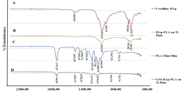
Fig. 5 The Spectra of FT-IR for PLA thin film, Coralline HAp, HAp-PLA coated on Ti plate, and GM-HAp-PLA coated on Ti plates.
Figure 5 shows FTIR spectra of HAp and PLA together with their thin film composites on Ti plates. The peaks of 10% HAp coated samples were matched with the peaks of coralline HAp in Fig. 5-A and B. The FT-IR spectra of Ti coated with GM-HAp-PLA in Fig. 5-D which was produced 10% GM loaded HAp coincides with the spectra of PLA thin film which was illustrated in Fig. 5-C. In this study, the FT-IR analysis suggested that HAp and GM drug particles did not interact chemically with PLA when mixed together and used as a coating material on Ti plate, so PLA matrix only acts as a barrier during the drug releasing process.
4. Conclusions
The results from this study demonstrated that dental implants could be coated with biodegradable polymer such poly lactic acid that contains antibiotic loaded HAp microspheres that can be produced and used to reduce or in certain instances even eliminate bacterial infection. Further characterization on in vitro and in vivo studies is needed to confirm this more accurately. 5. Acknowledgement
The authors would like to thank the Australian Academy of Science and the Horizon 2020 European Commission Grants for this collaborative research.
References
[1] M. Aviv, I. Berdicevsky, M. Zilberman: J Biomed Mater Res A., Vol. 83, (2007), p. 10-9. [2] P. Wu, D.W. Grainger: Biomaterials, Vol. 27, (2006), p. 2450-67.
[3] J. Chou, B. Ben-Nissan, D.W. Green, S.M. Valenzuela, L. Kohan: Advanced Engineering Materials, Vol. 13, (2011), p. 93-9.
[4] I.J. Macha, B. Ben-Nissan, B. Milthorpe: Current Nanoscience, Vol. 10, (2014), p. 200-4. [5] J.Chou, S. Valenzuela, D.W. Green, L. Kohan, B. Milthorpe, M.Otsuka, B. Ben-Nissan:
Nanomedicine Vol. 9, (2014), p. 1131–1139.
[6] I.J. Macha, S. Cazalbou, R. Shimmon, B. Ben-Nissan, B. Milthorpe: J Tissue Eng Regen Med., (2015) doi: 10.1002/term.2066.
[8] B. Ben-Nissan, I.J. Macha, S. Cazalbou: Nanomedicine , Vol 11, (2016), p. 531-544.
[9] R. Roest, B.A. Latella, G. Heness, B. Ben-Nissan: Surface and Coatings Technology Vol. 205, (2011), p. 3520-3529.
[10] I.J. Macha, B. Ben-Nissan, W. Müller: Kinetics and the theoretical aspects of drug dissolution from PLA/ HAp thin films.
![Fig. 1 Schematic illustration of the drug loading and dissolution procedure [8].](https://thumb-eu.123doks.com/thumbv2/123doknet/14244088.487276/4.892.210.689.832.1012/fig-schematic-illustration-drug-loading-dissolution-procedure.webp)