Alternative Isoform Regulation in Myotonic
Dystrophy
by
Eric T. Wang
B.A., Harvard University (2004)
Submitted to the Harvard-MIT Division of Health Sciences and
Technology
in partial fulfillment of the requirements for the degree of
Doctor of Philosophy in Medical Engineering and Medical Physics
at the
MASSACHUSETTS INSTITUTE OF TECHNOLOGY
February 2012
©
Massachusetts Institute of Technology 2012. All rights reserved.
A u th o r ...
Harvard-MIT Division of Health Sciences and Technology
January 13, 2012
C ertified b y ... ...
Christopher B. Burge
Professor
Thesis Supervisor
Certified by...
David E. Housman
Professor
Thesis Supervisor
A ccepted by .
... ...
... .... ...
Ram Sasikekharan
Director, Harvard-MIT Division of Health Sciences and Technology
Alternative Isoform Regulation in Myotonic Dystrophy
by
Eric T. Wang
Abstract
Myotonic dystrophy (DM) is the most common form of adult onset muscular dys-trophy, affecting more than 1 in 8000 individuals globally. The symptoms of DM are multi-systemic and include myotonia, severe muscle wasting, cardiac arrhyth-mias, cataracts, gastrointestinal dysfunction, and cognitive deficits. DM is caused by the expansion of CTG or CCTG repeat sequences expressed in noncoding portions of RNA, which sequester or activate RNA splicing factor proteins, leading to widespread deleterious changes in transcriptome isoform usage.
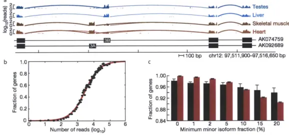
We developed a method for studying transcriptomes, RNAseq, which provides a high resolution, digital inventory of gene and isoform expression. By applying RNAseq to human tissues and cell lines, we discovered that essentially 92-94% of all human genes are alternatively spliced, 86% of them with a minor isoform frequency
15% or more. We found that the majority of alternative splicing and alternative
polyadenylation and cleavage events are tissue-regulated, and that patterns of these RNA processing events are strongly correlated across tissues, implicating protein factors that may regulate both types of events.
We applied this method towards the goal of identifying transcriptome changes occurring in DM, focusing on the Muscleblind-like (MBNL) family of RNA bind-ing proteins, which are functionally inactivated by CUG or CCUG repeats. Usbind-ing RNAseq to profile tissues and cells depleted of MBNLs, we found that MBNL1 and MBNL2 co-regulate hundreds of redundant targets. MBNL1 UV cross-linking and immunoprecipitation, followed by sequencing (CLIPseq), was used to identify the in
vivo transcriptome-wide binding locations of MBNL1, and facilitated the
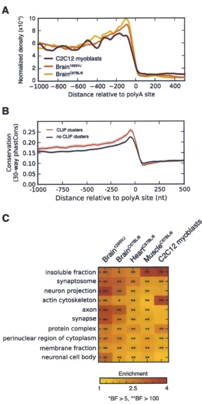
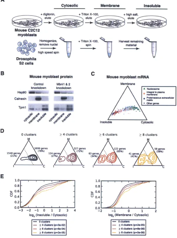
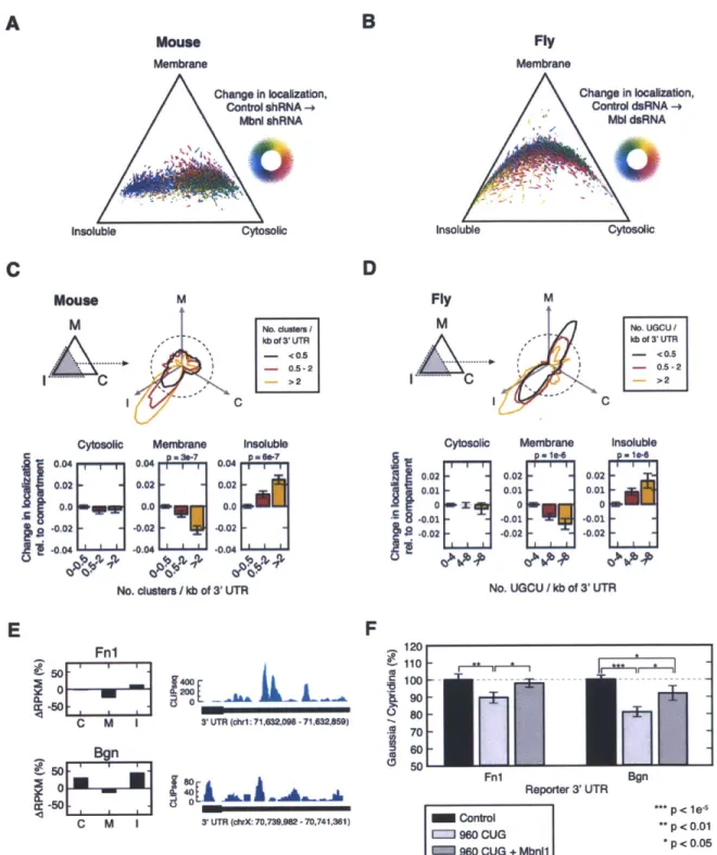
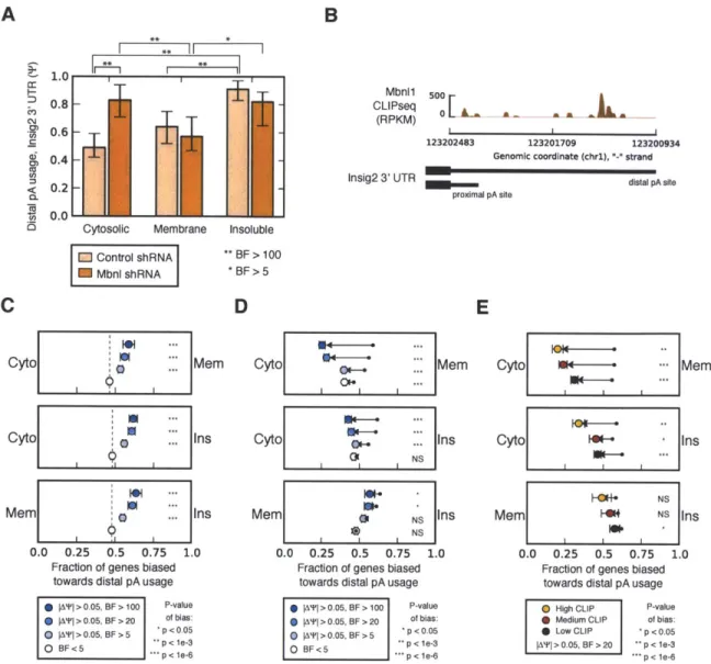
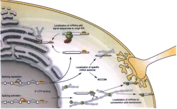
construc-tion of a context-dependent RNA map for MBNL1 splicing regulaconstruc-tion. Extensive 3' UTR binding of MBNL1 was found to localize mRNAs to membrane compartments of mouse myoblasts, suggesting a new global function for MBNLs, and additional mechanisms by which MBNL depletion can lead to DM symptoms.
Thesis Supervisor: Christopher B. Burge Title: Professor
Thesis Supervisor: David E. Housman Title: Professor
Acknowledgments
The journey that this thesis represents would not have been possible without the encouragement, assistance, and well-wishes of many individuals. Firstly, I must thank my PhD advisors, Chris Burge and David Housman, who have both been available, supportive, and instructive over the past 5 years. They have cultivated both depth and breadth in my approach to solving scientific problems within the context of a global community, and have helped integrate me within that community. The unique laboratory environments they create have provided a fantastic place to learn, explore, and make progress.
Many thanks to MIT for the Poitras Pre-doctoral Fellowship, and NIH for MEMP-BIG training grant funding, which in combination gave me the freedom to pursue research of my choosing over virtually my entire graduate career.
I thank my first research mentor, Guillermo Garcia-Cardefia, for instilling in me
an excitement about science, and guiding me towards HST. Thanks to Ben Larman and Kush Parmar for serving and continuing to serve as personal and scientific in-spirations, and for convincing me to champion research topics about which I am passionate.
I thank Charles Thornton for giving me the confidence to find a laboratory in
Boston to perform myotonic dystrophy research, and Tom Cooper for advice and discussions, seemingly instantly on demand over email. Thanks to David Brook and Andy Berglund for two scientifically and socially stimulating summers.
I thank my thesis committee members, Phil Sharp and Bob Brown, for their
availability, thoughtful insights, and support over the years. Thanks to all members of the Burge and Housman laboratories, for their scholarship, support, and collegiality.
I have truly enjoyed the friendship and great times we have had in Cambridge, and
look forward to many more nights of everyone enjoying my BBQ chicken and lobster rolls.
I must thank my family for more than I can describe. I thank my best friend and
wife, Beth, without whose support this work would not be possible. Her optimism, encouragement, and patience have been felt not only by me, but as a result, by all of those with whom I interact. I thank Thomas for always being there, and moving to Cambridge so that we can see each other daily, and work together on something in which we both believe. Finally, I thank my parents for raising and loving me, and helping me feel like I can accomplish anything. I thank Mom for teaching and showing me how to work hard, be persistent, and achieve the impossible; and Dad, for teaching me to think critically, to remain calm and collected during challenging times, and throughout life, but particularly before 7th grade science fair, that good science begins with a good question. Mom and Dad, to you I dedicate this thesis.
Contents
1 Introduction 9
1.1 O verview . . . . 9
1.2 B ackground . . . . 10
1.2.1 Myotonic dystrophy is an RNA gain-of-function disease . . . . 10
1.2.2 Regulation of mRNA in the transcriptome by a cis-trans code 15 1.2.3 Methods to assay the transcriptome . . . . 20
1.3 Goals and Organization... . . . . . . . . 23
1.4 References... . . . . . . . . 25
2 Alt. Isoform Regulation in Tissues 33 2.1 Introduction... . . . . . . . . 35 2.2 R esults . . . . 36 2.3 Discusssion... . . . . . . . . 46 2.4 Methods summary . . . . 46 2.5 References.. . . . . . . . 47 2.6 Figure Legends . . . . 51 2.7 F igures . . . . 54 3 Transcriptome Regulation by MBNLs 59 3.1 Introduction . . . . 62 3.2 R esults . . . . 63 5
. . . . 7 5 3.4 M ethods . . . . 3.5 References... 3.6 Figure Legends 3.7 Figures. . . . . 4 Conclusion 4.1 Summary . . . . . 4.2 Future Directions 4.3 References... . . . . . . . . . . . . . . . . . . . . . . . . . . . . 1 0 3 105 . . . . 107
A Abundance of Ubiquitously Expressed Genes I
B Splice site strength-dependent activity and genetic buffering by
poly-G runs I
C Analysis and design of RNA sequencing experiments for identifying
isoform regulation
D Global regulation of alternative splicing during myogenic
differenti-ation
[09
[21
131
143
E Supplementary Information for Alt. Isoform Regulation in Tissues 159
F Supplementary Information for Transcriptome Regulation by MBNLs187
. . . . 77 .. . . . . . . . 8 6 . . . . 9 0 . . . . 95 103 3.3 Discussion . . . .
List of Figures
2-1 Frequency and relative abundance of alternative splicing isoforms in
hum an genes. . . . . 54
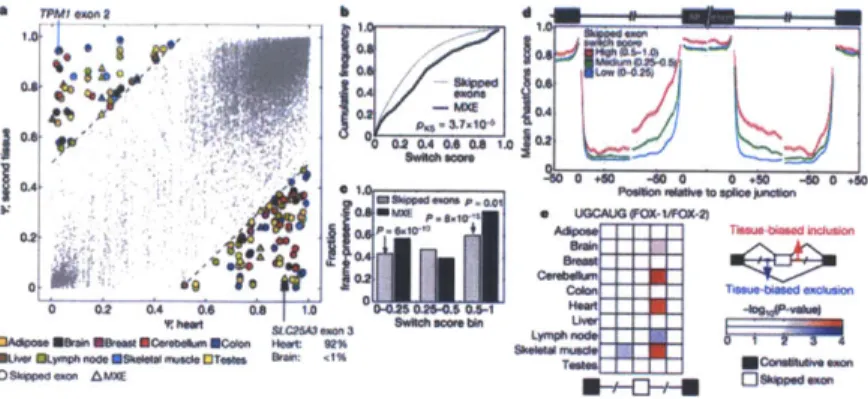
2-2 Pervasive tissue-specific regulation of alternative mRNA isoforms. . . 55
2-3 The extent of individual-specific differences in alternative isoform
ex-pression . . . . 56
2-4 Conservation and function of switch-like alternative splicing exons.. 57
2-5 Evidence for coordination between splicing and polyadenylation. . . 58
3-1 Dependence of splicing changes on total MBNL levels by RNA-seq
analysis.. . . . . . . . . 95
3-2 MBNL binding is conserved and associated with context-dependent
splicing activity. . . . . . 96
3-3 An RNA map of MBNL activity at nucleotide resolution. . . . . 97
3-4 MBNL binding in 3' UTRs is common and associated with increased
conservation and specific cellular compartments. . . . . 98
3-5 mRNA localization is associated with functional binding by MBNL. . 99
3-6 MBNL binding is associated with regulation of mRNA localization in
mouse and fly, and may contribute to protein secretion. . . . 100
3-7 MBNL regulates isoform-specific mRNA localization via binding to
alternative 3 UTRs. . . . . 101
F-i Read coverage at the MBNL1 exon 3 locus by RNA-seq. . . . . 188
F-2 Cassette exon T in tissues and myoblasts depleted of MBNLs. . .. . 189
F-3 Correlation in AT as estimated by RNA-seq and splicing microarray. 190
F-4 MBNL protein levels in myoblasts depleted of MBNLs. . . . . 191
F-5 Correlation between extent of MBNL depletion and splicing changes
or gene expression changes. . . . . 192
F-6 Western blot analysis of marker proteins in fly S2 cells. . . . . 193 F-7 Proportion of CLIP-seq reads mapping to regions of the transcriptome. 194
Chapter 1
Introduction
1.1
Overview
Myotonic Dystrophy (DM) is the most common form of adult onset muscular
dystro-phy and affects over 1 in 8000 people globally. The symptoms of DM include myotonia,
severe muscle wasting, cardiac arrhythmias, cataracts, gastrointestinal dysfunction, and cognitive deficits (Harper 1979). The autosomal dominant disease is caused by either CTG repeat expansions in the 3' UTR of the DMPK gene (DM1) (Brook et al.
1992; Mahadevan et al. 1992), or CCTG repeat expansions in the first intron of the CNBP gene (DM2) (Liquori et al. 2001). A notable feature of DM is that it is an RNA
gain-of-function disease; while forming intranuclear foci, RNA repeats sequester and activate splicing factors in trans, and cause global defects in RNA processing (Cooper et al. 2009). Within this thesis, I describe my efforts towards establishing a system
by which these changes in RNA processing can be accurately measured, and its use
in understanding the functions of the Muscleblind-like (MBNL) family of splicing factors, a set of proteins that are functionally depleted in DM and responsible for the regulation of a number of molecular events perturbed in DM.
1.2. Background
1.2
Background
1.2.1
Myotonic dystrophy is an RNA gain-of-function
dis-ease
First formally described in 1909 by Hans Steinert, myotonic dystrophy, also known as Steinert's Disease, has captured the attention of neurologists and geneticists through-out history because the disease possesses several interesting yet puzzling features (Harper 1979). It is a progressive, multi-systemic disease, and is dominantly inher-ited. The hallmark symptom of DM is myotonia, or the inability to relax muscles after contraction, and is often one of the first symptoms experienced by patients during the first phenotypic onset of symptoms. Other symptoms include muscle wasting and weakening, cardiac arrhythmias, smooth muscle dysfunction, cataracts, insulin resis-tance, cognitive abnormalities, and hypersomnia. However, these symptoms occur with such variable frequency among different patients that DM has been described as "the most variable disease known to mankind". Perhaps the most consistently observed feature of DM is the characteristic manner in which it affects younger gen-erations of a DM-affected family more severely than older gengen-erations (Ashizawa et al. 1992). This phenomena of genetic anticipation often produces a picture in which parents and grandparents, having experienced minor DM symptoms, are formally di-agnosed only after their children, in whom the repeat has expanded further, are born with severe developmental defects. The repeat length-dependent age of onset (Harper 1979), also observed in other diseases such as Huntington's disease (Snell et al. 1993), presented a mystery for disease pathogenesis, and was not explained until several years after discovery of the causative disease locus.
The causative locus for type 1 DM was successfully mapped in 1992 by several different research groups (Brook et al. 1992; Fu et al. 1992; Mahadevan et al. 1992); the trinucleotide repeats were found to reside in the 3' untranslated region of a gene
on chromosome 19q13. This gene was named DMPK and studied for possible clues that might reveal the molecular events leading to the myriad phenotypes observed in DM patients. It was observed that protein levels of DMPK were reduced to ap-proximately 50% of normal levels in DM patients, and therefore for several years it was hypothesized that the cause of the DM phenotype was partly due to haploinsuf-ficiency of the DMPK protein. DMPK null mice were created to test this hypothesis, but while they exhibited a minor cardiac phenotype late in life, in general they did not exhibit phenotypes characteristic of DM such as myotonia or muscle wasting (Reddy et al. 1996). Studies investigating the molecular basis for DMPK haploinsufficiency revealed important clues about DM pathogenesis. In particular, it was found that measured levels of DMPK RNA depended critically on the procedure used to isolate RNA; typical guanidium-based methods resulted in DMPK measurements ~2-fold less than those obtained if treating RNA with proteinase K or ultracentrifugated through a cesium chloride cushion (Davis et al. 1997). This finding was consistent with the observation that the expanded repeats in DMPK formed insoluble nuclear foci, visible upon in situ hybridization against CUG RNA (Taneja et al. 1995).
The discovery of a second type of DM, DM type 2, provided important evidence that the pathological effects of the expanded repeats occur in trans rather than in cis. DM2 patients were found to exhibit many symptoms of DM1, including myotonia, cataracts, frontal balding, and insulin resistance. However, in contrast to DM1, the pattern of skeletal muscle wasting in DM2 was found to proceed in a proximal to dis-tal fashion. The causative locus in DM2 was found to be an expanded CCTG repeat tract in the first intron of a completely different gene, CNBP, found on chromosome
3q21, suggesting that RNA expression of CTG-containing repeat sequence, the
com-monality between DM1 and DM2, could be causative of disease pathology (Liquori et al. 2001). To formally test the hypothesis that expanded CUG repeats were causative of DM, a mouse model was generated in which 250 CTG repeats were placed in the
3' UTR of an unrelated gene, and driven by the human skeletal actin promoter, such
that it was expressed primarily in skeletal muscle (Mankodi et al. 2000). This mouse (Human Skeletal Actin-Long Repeat, HSALR), indeed exhibited several DM pheno-types, including myotonia, centralized muscle nuclei, and heterogeneous muscle fiber cross sectional area. In parallel, proteins which bound the expanded repeat RNA in a repeat length-dependent manner were biochemically purified and identified, us-ing ultraviolet cross-linkus-ing and mass spectrometry approaches (Miller et al. 2000). These proteins were found to be homologous to the highly conserved Muscleblind-like (MBNL) family of RNA binding proteins, first identified in Drosophila (Begemann et al. 1997). With the information that expanded CUG repeat expression is sufficient to cause DM pathology, and that MBNL proteins bind CUG repeats, experiments were performed to test the hypothesis that nuclear sequestration of MBNLs contributes to DM phenotypes. An MBNL1 loss of function mouse was created, in which the
exon containing the translation initiation codon was deleted (Kanadia et al. 2003). This mouse was found to exhibit myotonia, cataracts, and various muscle defects, confirming that functional depletion of MBNL1 could lead to at least a subset of DM phenotypes. Furthermore, at the molecular level, the alternative splicing patterns of several mRNAs were found to be aberrantly regulated in mice lacking MBNL1, sug-gesting that the cellular pool of MBNL1 protein available to regulate these targets was perturbed by expression of expanded CUG repeats. To confirm that sequestration of MBNLs in DM was responsible for its functional depletion, exogenous over-expression of MBNL1 was attempted in the HSALR mouse model. Adenoviral-mediated over-expression of MBNL1 protein -2-fold above endogenous protein levels was sufficient to partially rescue myotonia (Kanadia et al. 2006). Over-expression of MBNL1 was also sufficient to rescue aberrant alternative splicing patterns that were observed in the CUG repeat-expressing mice, confirming that the regulation of these targets is sensitive to the intracellular concentration of free MBNL1. These three major ex-1.2. Background
Chapter 1. Introduction
periments, the creation and characterization of the HSALR mouse, the creation and characterization of the MBNL1 knockout mouse, and over-expression of MBNL1 in
HSALR complete a loop of biochemical and genetic logic stating that a major portion
of DM pathology is caused by expanded CUG repeats which sequester MBNL proteins in trans, leading to their functional depletion in cells and tissues, resulting in down-stream molecular changes causative of DM symptoms. The repeat length-dependent age of onset for DM could also be explained by MBNL depletion; greater numbers of repeats, further expanded somatically during the lifetime of an individual (Wong et al. 1995), lead to increased sequestration of MBNL, and a more severe phenotype occurring at an earlier age.
In a separate line of investigation, it was observed that over-expression of CUG repeats leads to the stabilization of another RNA-binding protein, CUGBP and ETR-like 1 (CELFI) (Timchenko et al. 1996; Wang et al. 2007). CELF1 was previously discovered to regulate a number of pre-mRNAs also observed to be aberrantly spliced in DM, including cardiac troponin T exon 5 and insulin receptor exon 11 (Philips et al.
1998; Savkur et al. 2001). DM patient tissues and myoblasts contain -2-fold elevated
levels of CELF1 protein as compared to normal tissues, suggesting that CELF1 activa-tion may lead to a cascade of MBNL1-independent cellular events responsible for other DM symptoms. Using a combination of pharmacological and genetic approaches, it was demonstrated that the stabilization of CELF1 protein in DM1 occurs through hyperphosphorylation by members of the protein kinase C (PKC) family of signaling molecules; these modifications to CELF1 lead to increased half-life, particularly in the nucleus as compared to the cytoplasm (Kuyumcu-Martinez et al. 2007; Orengo et al. 2008). Through unknown mechanisms, one or several isozymes of PKC are phos-phorylated and activated by expression of the expanded CTG-containing DMPK 3' UTR. Pharmacological inhibition of PKC successfully blocked the over-expression of
CELF1 in a separate mouse model of DM in which the 3' end of DMPK, with
Chapter 1. Introduction 1.2. Background
panded CTG repeats, was over-expressed in heart (Wang et al. 2009). Furthermore,
while the MBNL knockout mouse does not exhibit muscle wasting, over-expression of CELFI in skeletal muscle of mice does (Ward et al. 2010), and suggests that this characteristic DM symptom is downstream of CELFI activation and independent of MBNL1 depletion. This series of experiments suggested that hyperphosphorylation of CELFI could be a critical event responsible for an MBNL-independent arm of DM cellular pathology, and that CELF1-dependent RNA processing events may lie proximal to the skeletal muscle wasting phenotype.
While sequestration of MBNLs and over-expression of CELFs have received the greatest amount of attention from investigators in the DM field as pathways most likely to be relevant to DM pathogenesis, a number of additional hypotheses for how expanded repeats lead to DM symptoms have been proposed and investigated over the past 19 years. These hypotheses include the epigenetic silencing of genes adjacent to DMPK, including DMAHP and SIX5, via altered DNA structure (Thornton et al.
1997; Klesert et al. 1997), the sequestration of other RNA binding proteins (Kim
et al. 2005; Paul et al. 2006), transcription factor leaching (Ebralidze et al. 2004), dysregulation of RNA stability and localization (Du et al. 2010; Adereth et al. 2005), and aberrant translation of trinucleotide repeats (Zu et al. 2011). Some or all of these pathways could be activated in DM patients; unclear is the extent to which each pathway leads to the phenotypic consequences, and how it does so at the molecular level. The complexity of DM pathogenesis motivates global, unbiased evaluations of all the molecular changes that occur in response to expression of expanded repeats. Several studies using this type of approach have already revealed valuable insights. For example, the majority of splicing changes in the mouse expressing expanded CUG repeats in skeletal muscle is reproduced in MBNL1 knockout mouse muscle, suggest-ing that MBNL1 depletion accounts for most of the transcriptome changes observed in the HSALR model (Du et al. 2010). While previous studies have primarily focused 1.2. Background
on splicing, additional types of RNA regulation, such as alternative cleavage and polyadenylation, and regulation of RNA localization, may be perturbed in DM. Fur-ther global studies which address these oFur-ther areas and utilize oFur-ther mouse models and DM patient tissue samples should yield additional valuable insights which will help identify the relevant pathways and molecular changes that cause DM pathogen-esis, facilitating a greater understanding of DM and the development of therapeutic interventions to treat its symptoms.
1.2.2
Regulation of mRNA in the transcriptome by a
cis-trans code
Normal function of numerous biological processes requires the proper regulation of
mRNA. This regulation is often achieved through interactions of cis elements found
in primary, secondary, or tertiary RNA sequence, with other trans-acting complexes composed of protein and/or RNA components. These interactions can change the abundance, sequence, and locations of mRNA in the cell, which in turn dictate whether and where the mRNA is degraded, translated, or stored. The types of reg-ulation most relevant to studies discussed in this thesis include pre-mRNA splicing, cleavage and polyadenylation, message stability, and regulation of RNA localization. Each of these processes involves a number of core and auxiliary factors, some of which are only active in specific pre-mRNA or mRNA contexts and can exert varying functions depending on how they interact with the RNA substrate.
Pre-mRNA splicing PmRNA splicing is the process by which introns are re-moved and exons are joined, via trans-esterification and ligation reactions, respec-tively (Berget et al. 1977; Chow et al. 1977). For each intron being spliced, the core spliceosomal factors U1 and U2 snRNPs, together with mBBP, bind to the 5' and 3' splice sites of the intron (Lerner et al. 1980; Rogers and Wall 1980). The 2'-hydroxyl
of the branch point attacks the phosphodiester bond of the 5' splice site, separating the previous exon from a newly formed lariat structure. The free hydroxyl at the 3' end of 5' splice site then attacks the phosphodiester bond of the 3' splice site, joining the 5' splice site to the 3' splice site, and releasing the lariat (Wahl et al. 2009).
While this process occurs with high fidelity for constitutively spliced exons and introns, the information found at true splice sites is insufficient for directing splicing machinery to function at the appropriate locations (Lim and Burge 2001). Addi-tional cis elements, exonic splicing silencers (ESS), exonic splicing enhancers (ESE), intronic splicing silencers (ISS), and intronic splicing enhancers (ISE) perform the important role of either enhancing weaker splice sites and suppressing decoy splice sites (Black 1995; Fairbrother et al. 2004) These elements have been identified using both computational and biochemical methods, and often form the binding sites for the serine/arginine-rich (SR) proteins and heterogeneous nuclear ribonucleoproteins (hnRNPs), which bind preferentially in exons and introns, respectively. These trans factors bind to specific ESS, ESE, ISE, and ISS motifs to recruit or block core
spliceo-somal components, and their binding in different spatial orientation relative to splice sites can produce different types of activity (Matlin et al. 2005).
Alternative splicing differs from constitutive splicing in that different pairs of splice sites can be joined together in different tissues, cell types, or contexts, resulting in the production of mRNAs with different exonic sequences. The different mRNA isoforms produced by alternative splicing provide a means by which multiple mRNAs and proteins with different properties can be produced from the same gene locus, and are thought to significantly contribute to the diversity of the transcriptome and proteome (Blencowe 2006). Alternative splicing can lead to exon skipping, alternative
3' or 5' splice site usage, the use of adjacent exons in a mutually exclusive manner, or
the use of distinct first or last exons. Like constitutive splicing, alternative splicing is regulated by a complex cis-trans code, in which regulatory sequences or structures 1.2. Background
Chapter 1. Introduction
are bound by protein factors that can differ in their expression or activity in different contexts (Matlin et al. 2005). Understanding the rules by which these protein factors exert their functions is a fundamental goal in the study of RNA splicing, and RNA biology.
Regulation of alternative splicing is particularly relevant in myotonic dystrophy because the MBNL family of alternative splicing regulators is sequestered by expanded RNA repeats, and functionally inactivated (Cooper et al. 2009). For example, MBNL1 normally represses the inclusion of exon 7a of the chloride channel 1 (CLCN1) pre-mRNA, preventing the inclusion of a premature stop codon-containing exon (Mankodi et al. 2002). This allows full length CLCN1 mRNA to be translated, leading to CLCN1 protein production and expression at the sarcolemma. Loss of MBNL1 activity leads to exon 7a inclusion, degradation of CLCN1 mRNA by nonsense-mediated decay, and loss of CLCN1 protein expression, leading to defective chloride ion transport and myotonia symptoms. In contrast, MBNL1 normally activates the inclusion of insulin receptor (IR) exon 11, but in myotonic dystrophy cells, exon 11 is aberrantly skipped, leading to the increased production of a protein isoform of IR that is less responsive to changes in glucose concentration (Savkur et al. 2001). From these two examples, it is clear that both splicing repression and activation can be mediated by the interactions between pre-mRNA and RNA binding proteins.
Regulation of mRNAs via 3' end processing A subset of alternative splicing
regulators have also been demonstrated to regulate processes occurring at the 3' ends of messages (Maniatis and Reed 2002). Cis-elements encoding both stabilizing and de-stabilizing elements recruit various RNA binding proteins, which can exert various activities through binding to 3' UTRs. For example, CELF1 has been demonstrated to destabilize messages via binding to GU-rich sequences in 3' UTRs, in the tumor necrosis factor 3' UTR (Zhang et al. 2008; Lee et al. 2010) and perhaps many other 3'
1.2. Background
UTRs containing these sequences (Vlasova et al. 2008). While the precise mechanism
by which CELF1 binding leads to decreased mRNA half-life, one possibility is that
it recruits deadenylases that degrade the polyA tail, which normally protects the mRNA from degradation by exonucleases. Other de-stabilizing elements include AU-rich elements (Barreau et al. 2005), first discovered in 3' UTRs of cytokine mRNAs (Caput et al. 1986), and microRNA sites, which serve as cis-elements that can recruit
~23 nucleotide-long mature microRNAs, products of cleavage by proteins Drosha (Lee
et al. 2003) and Dicer, in a manner dependent on base complementarity (Bartel 2004). The microRNA-mRNA interaction leads to Argonaute protein-dependent recruitment of the RNA-induced silencing complex, and subsequent degradation of the mRNA via enhanced deadenylation (Lim et al. 2005), and/or reduced translation (Guo et al. 2010; Hendrickson et al. 2009; Selbach et al. 2008; Baek et al. 2008).
Regulation of other processes occurring at 3' ends of mRNAs, for example cleavage and polyadenylation, can also influence the rate at which mature mRNA is produced and the levels at which it is expressed. During cleavage and polyadenylation, cleavage and polyadenylation specificity factor (CPSF) recognizes the consensus AAUAAA (or similar) poly(A) signal sequence, and cleavage stimulating factor (CstF) recognizes a
U- or GU-rich downstream element. CPSF catalyzes the cleavage of pre-mRNA, and
collaborates with poly(A) polymerase and poly(A) binding protein to subsequently adds a tail of adenosines to the cleaved substrate (Mandel et al. 2008). Enhanced or decreased rates of assembly of these complexes can affect the efficiency of mRNA maturation, and therefore gene expression levels for those mRNAs. For example, the RNA binding protein hnRNP H has been demonstrated to enhance the efficiency of of cleavage and polyadenylation through recruitment of poly(A) polymerase to 3' UTRs to which it binds (Alkan et al. 2006).
Over half of all human genes contain multiple cleavage and polyadenylation sites (Zhang et al. 2005), allowing the production of diverse 3' UTRs, which may con-Chapter 1. Introduction
fer distinct properties to their mRNAs. The selection of particular cleavage and
polyadenylation sites can be mediated by trans factors in a tissue-specific manner,
as has been shown for the RNA binding factor Nova2 (Licatalosi et al. 2008). When binding within 30 nucleotides of the poly(A) signal sequence, Nova2 inhibits usage of the site, likely through preventing formation of a functional 3' processing com-plex. In contrast, when binding to distal elements, Nova2 enhances the usage of the associated poly(A) site, potentially through antagonizing the action of other repres-sive factors. Several dynamic biological processes, for example T-cell activation or cancer progression and metastases, exhibit global changes in alternative cleavage and polyadenylation (Sandberg et al. 2008; Mayr and Bartel 2009). During each of these transformations, the majority of 3' UTRs are shortened, and it has been proposed that this provides a means for these transcripts to avoid targeting by intracellular degradative machinery, for example microRNAs, to enhance protein production ca-pacity in conditions of high metabolic demand.
Subcellular localization of mRNA Yet another mode of regulation that can
occurs through the interaction of RNA binding proteins, often with 3' ends, is that of RNA localization (Lecuyer et al. 2009). While the composition of mRNA is not directly altered via differential localization, the decay or storage of the mRNA, and subcellular locations of subsequently translated proteins can be drastically affected
by where the mRNA is transported. Localized RNA was first described for actin
mRNA in ascidian embryos (Jeffery et al. 1983), and since that time, proper mRNA localization has been associated with cellular function in E. coli (Nevo-Dinur et al. 2011), S. cerevisiae (Bertrand et al. 1998), Drosophila (Gavis and Lehmann 1994; Kugler and Lasko 2009), and mammalian systems (Lawrence and Singer 1986). ASHI mRNA is localized to the budding tip in dividing yeast, and its mis-localization leads to a loss of mate type switching. Localization of bicoid, oskar, and nanos mRNAs in
Drosophila are critical for the establishment of polarity during embryo development
(Gavis and Lehmann 1994; Kugler and Lasko 2009). The proper localization of
/--actin to the leading edge of motile fibroblasts is required for efficient synthesis of F-actin at the cell periphery, responsible for generating the forces necessary to propel a cell forward along a surface (Shestakova et al. 2001). A recent global study of over six thousand Drosophila mRNAs in embryos suggest that the majority of mRNAs are distributed in a non-uniform pattern, and that these patterns are linked to function (Lecuyer et al. 2007).
RNA localization has been demonstrated to be mediated through 3' UTR se-quences or structures that allow their recognition by trans factors, which act as adaptors to facilitate their transport along cytoskeletal filaments by molecular motors (Kislauskis et al. 1994). For example,
#-actin
mRNA contains a CACCC "zipcode" sequence, which is recognized by Zipcode-Binding Protein (ZBP), and subsequently interacts with an actin-myosin motor complex which carries cargo to the periphery of the cell (Ross et al. 1997). The role of RNA binding proteins in the regulation of RNA localization still largely remains to be explored, and the elucidation of a cis-trans code for RNA localization will require the integrated use of molecular, biochemical, cellular, and image-based techniques.1.2.3
Methods to assay the transcriptome
Similar to the DM field, approaches to assess the transcriptome have undergone rapid evolution over the past 19 years. Around the same time that the gene for DM1 was successfully identified, dbEST, a database for expressed sequence tags (ESTs), was established as a division of GenBank at the National Center for Biotechnology Infor-mation (NCBI) (Adams et al. 1991). These tags, short subclones of cDNA sequence isolated from biological material, have been used to identify gene transcripts and gene structures. When mapped to particular loci in in sufficient quantities, ESTs 1.2. Background
have been used to make quantitative inferences about transcript isoform abundances. Similarly, the first use of Serial Analysis of Gene Expression (SAGE) was published in 1995 (Velculescu et al. 1995); SAGE also employs census methods to quantitate the number of transcript fragments sampled, sequenced, and therefore present in a biological sample. The primary difference between SAGE and EST approaches is the length of RNA insert sequenced; SAGE tags are typically created by concatemerizing fragmented RNA and therefore are much shorter, allowing for increased sequencing depth of overall shorter tags. Commonly used for cancer samples, SAGE provided a quantitative metric for transcript abundance, and foreshadowed the advent of deep sequencing approaches for assaying transcriptomes.
Both SAGE and EST sequencing approaches were limited in depth by cost consid-erations, and a technology that was pioneered much earlier but not popularized until
SAGE was established was complementary cDNA microarrays (Schena et al. 1995).
Comprised of spotted DNA clusters on a glass slide, microarrays facilitated the bind-ing of RNA or cDNA to individual spots whose identities could be recorded and mapped back to specific transcripts or transcript isoforms. The extent of binding of dye-labeled RNA or cDNA to particular spots was demonstrated to be proportional to the starting amount present in a biological sample, and therefore quantitative, transcriptome-scale measurements were made routinely using this technology. Sig-nificant efforts were put forth to standardize the production, use, and interpretation of transcriptome measurements using microarrays, and as a result this technology has partly fueled a revolution in biology and its relationship to computation. While microarrays catalyzed a cultural transition in the psyche of many biologists, they suf-fered from a number of technical drawbacks, including cross-hybridization of probes with similar sequence and poor dynamic range. A decade or so of widespread "ana-log" microarray usage by many laboratories around the world paved the way for a digital revolution, in which sequencing census methods reminiscent of EST and
SAGE approaches provided high resolution information at greater throughput and
lower cost.
The digital revolution in transcriptomics has been made possible by a Moore's Law-like phenomenon in which the cost of sequencing has decreased exponentially at least over the past decade (Mardis 2008), accelerating in the past 5 years or so with the advent of so-called "next-generation" sequencing technologies. Miniaturization of sequencing reactions has allowed millions of assays in parallel, using quantities of enzymes and substrates similar to before, but harnessing advances primarily in imaging and optics. High throughput sequencing of DNA fragments, from DNA samples or RNA samples converted to DNA libraries, has made possible the true application of digital census methods in the characterization and quantitation of transcript and isoform abundancies. Its application to specific biological problems is discussed extensively in this thesis, particularly in Chapter 2.
High throughput sequencing has thus been applied not only to studying the
tran-scriptome, but to studying trans factors which interact with the transcriptome.
Bio-chemical techniques can be used to purify subsets of nucleic acid which interact with
DNA- or RNA- binding proteins, with which deep sequencing libraries can be
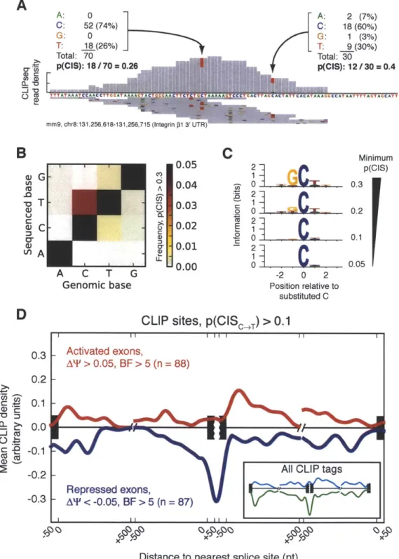
pre-pared. One technique that will be described in Chapter 3, Appendix A, and Appendix B is ultraviolet (UV) cross-linking and immunoprecipitation (CLIP), a procedure which relies on the ability of UV irradiation to cause covalent cross-linking between protein and nucleic acid interacting within Angstrom distances (Ule et al. 2005). UV cross-linking of these interactions in live cells, followed by cell lysis and immuno-capture of proteins of interest, enables the preparation of deep sequencing libraries which reveal the transcriptome-wide binding locations of an RNA binding protein, and valuable clues about its function (Licatalosi et al. 2008).
1.2. Background
Chapter 1. Introduction 1.3. Goals and Organization
1.3
Goals and Organization
My goals for this thesis were to develop a method for assaying the transcriptome with
high resolution and accuracy, and apply it towards questions about myotonic
dystro-phy. Chapter 2 describes work in which I characterized the use of high-throughput
sequencing for assaying transcriptomes (RNAseq), and demonstrates its utility in studying the regulation of alternative isoforms between various human tissues and cell lines. Chapter 3 describes work in which RNAseq and other sequencing approaches were used to characterize the cellular activities of the Muscleblind-like family of RNA binding proteins, the major protein family whose activity is perturbed in myotonic dystrophy. The approaches I describe in this thesis embody a model I have chosen in the larger endeavor to comprehensively survey the DM transcriptome and under-stand the rules by which proteins dysregulated in DM perform their normal cellular functions. The completion of this goal is beyond the scope of this thesis, but in the conclusion chapter, I describe next steps that must be taken to fulfill that goal in its entirety.
Appendix A contains a publication building on the findings described in Chapter 2, and further characterizes differences between different tissue transcriptomes and their implications for the organization of biological systems. Appendix B contains a publication describing the context-dependent functions of the RNA-binding protein hnRNP H, in which my first efforts with CLIP produced data informative about the binding locations and function of hnRNP H. Appendix C contains a publication in which a method for computing posterior distributions of alternative isoform usage under a Bayesian mixture model was developed, and describes additional context-dependent functions of hnRNP H in regulating alternative cleavage and polyadenyla-tion, and also describes our experiments characterizing the strengths and weaknesses of paired-end RNAseq libraries for analysis of the transcriptome. Appendix D con-tains a publication in which a subset of RNA processing events regulated during the
Chapter 1. Introduction 1.3. Goals and Organization
differentiation of myoblasts into myotubes is studied, to characterize their temporal
behavior and identify trans factors that may mediate their regulation. Appendix E
contains supplementary information for Chapter 2, and Appendix F contains supple-mentary information for Chapter 3.
1.4
References
Adams, MD, JM Kelley, JD Gocayne, M Dubnick, MH Polymeropoulos, H Xiao, CR Merril, A Wu, B Olde, and RF Moreno (June 1991). "Complementary DNA sequencing: expressed sequence tags and human genome project." In: Science
252.5013, pp. 1651-6 (cit. on p. 20).
Adereth, Y, V Dammai, N Kose, R Li, and T Hsu (Dec. 2005). "RNA-dependent inte-grin alpha3 protein localization regulated by the Muscleblind-like protein MLP1." In: Nat Cell Biol 7.12, pp. 1240-7. DOI: 10. 1038/ncb1335 (cit. on p. 14).
Alkan, SA, K Martincic, and C Milcarek (Jan. 2006). "The hnRNPs F and H2 bind to similar sequences to influence gene expression." In: Biochem J 393.Pt 1, pp.
361-71. DOI: 10. 1042/BJ20050538 (cit. on p. 18).
Ashizawa, T, JR Dubel, PW Dunne, CJ Dunne, YH Fu, A Pizzuti, CT Caskey,
E Boerwinkle, MB Perryman, and HF Epstein (Oct. 1992). "Anticipation in
my-otonic dystrophy. II. Complex relationships between clinical findings and structure of the GCT repeat." In: Neurology 42.10, pp. 1877 83 (cit. on p. 10).
Baek, D, J Villen, C Shin, FD Camargo, SP Gygi, and DP Bartel (Sept. 2008). "The impact of microRNAs on protein output." In: Nature 455.7209, pp. 64-71. DOI:
10. 1038/nature07242 (cit. on p. 18).
Barreau, C, L Paillard, and HB Osborne (2005). "AU-rich elements and associated factors: are there unifying principles?" In: Nucleic Acids Res 33.22, pp. 7138-50.
DOI: 10. 1093/nar/gki1012 (cit. on p. 18).
Bartel, DP (Jan. 2004). "MicroRNAs: genomics, biogenesis, mechanism, and func-tion." In: Cell 116.2, pp. 281-97 (cit. on p. 18).
Begemann, G, N Paricio, R Artero, I Kiss, M P6rez-Alonso, and M Mlodzik (Nov.
1997). "muscleblind, a gene required for photoreceptor differentiation in Drosophila,
encodes novel nuclear Cys3His-type zinc-finger-containing proteins." In:
Develop-ment 124.21, pp. 4321-31 (cit. on p. 12).
Berget, SM, C Moore, and PA Sharp (Aug. 1977). "Spliced segments at the 5' terminus of adenovirus 2 late mRNA." In: Proc Natl Acad Sci U S A 74.8, pp. 3171-5 (cit. on p. 15).
Bertrand, E, P Chartrand, M Schaefer, SM Shenoy, RH Singer, and RM Long (Oct.
1998). "Localization of ASHI mRNA particles in living yeast." In: Mol Cell 2.4, pp. 437-45 (cit. on p. 19).
Black, DL (Oct. 1995). "Finding splice sites within a wilderness of RNA." In: RNA
1.8, pp. 763-71 (cit. on p. 16).
Blencowe, BJ (July 2006). "Alternative splicing: new insights from global analyses." In: Cell 126.1, pp. 37-47. DOI: 10 .1016/j .cell.2006.06.023 (cit. on p. 16). Brook, JD, ME McCurrach, HG Harley, AJ Buckler, D Church, H Aburatani, K
Hunter, VP Stanton, JP Thirion, and T Hudson (Feb. 1992). "Molecular basis of myotonic dystrophy: expansion of a trinucleotide (CTG) repeat at the 3' end of a transcript encoding a protein kinase family member." In: Cell 68.4, pp. 799-808 (cit. on pp. 9, 10).
Caput, D, B Beutler, K Hartog, R Thayer, S Brown-Shimer, and A Cerami (Mar.
1986). "Identification of a common nucleotide sequence in the 3'-untranslated
1.4. References
region of mRNA molecules specifying inflammatory mediators." In: Proc Natl
Acad Sci U S A 83.6, pp. 1670-4 (cit. on p. 18).
Chow, LT, RE. Gelinas, TR Broker, and RJ Roberts (Sept. 1977). "An amazing se-quence arrangement at the 5' ends of adenovirus 2 messenger RNA." In: Cell 12.1,
pp. 1-8 (cit. on p. 15).
Cooper, TA, L Wan, and G Dreyfuss (Feb. 2009). "RNA and disease." In: Cell 136.4, pp. 777-93. DOI: 10.1016/j. cell.2009.02.011 (cit. on pp. 9, 17).
Davis, BM, ME McCurrach, KL Taneja, RH Singer, and DE Housman (July 1997). "Expansion of a CUG trinucleotide repeat in the 3' untranslated region of my-otonic dystrophy protein kinase transcripts results in nuclear retention of tran-scripts." In: Proc Natl Acad Sci U S A 94.14, pp. 7388-93 (cit. on p. 11).
Du, H, MS Cline, RJ Osborne, DL Tuttle, TA Clark, JP Donohue, MP Hall, L Shiue,
MS Swanson, CA Thornton, and M Ares Jr (Feb. 2010). "Aberrant alternative
splicing and extracellular matrix gene expression in mouse models of myotonic dystrophy." In: Nat Struct Mol Biol 17.2, pp. 187-93. DOI: 10. 1038/nsmb. 1720
(cit. on p. 14).
Ebralidze, A, Y Wang, V Petkova, K Ebralidse, and RP Junghans (Jan. 2004). "RNA leaching of transcription factors disrupts transcription in myotonic dystrophy." In:
Science 303.5656, pp. 383-7. DOI: 10.1126/science.1088679 (cit. on p. 14).
Fairbrother, WG, GW Yeo, R Yeh, P Goldstein, M Mawson, PA Sharp, and CB Burge (July 2004). "RESCUE-ESE identifies candidate exonic splicing enhancers in vertebrate exons." In: Nucleic Acids Res 32.Web Server issue, W187-90. DOI:
10.1093/nar/gkh393 (cit. on p. 16).
Fu, YH, A Pizzuti, RG Fenwick Jr, J King, S Rajnarayan, PW Dunne, J Dubel, GA Nasser, T Ashizawa, and P de Jong (Mar. 1992). "An unstable triplet repeat in a gene related to myotonic muscular dystrophy." In: Science 255.5049, pp. 1256-8
(cit. on p. 10).
Gavis, ER and R Lehmann (May 1994). "Translational regulation of nanos by RNA
localization." In: Nature 369.6478, pp. 315-8. DOI: 10. 1038/369315a0 (cit. on
pp. 19, 20).
Guo, H, NT Ingolia, JS Weissman, and DP Bartel (Aug. 2010). "Mammalian
microR-NAs predominantly act to decrease target mRNA levels." In: Nature 466.7308,
pp. 835-40. DOI: 10. 1038/nature09267 (cit. on p. 18).
Harper, PS (1979). Myotonic dystrophy. Vol. v. 9. Philadelphia: Saunders. ISBN:
0721645275 (cit. on pp. 9, 10).
Hendrickson, DG, DJ Hogan, HL McCullough, JW Myers, D Herschlag, JE Ferrell, and PO Brown (Nov. 2009). "Concordant regulation of translation and mRNA abundance for hundreds of targets of a human microRNA." In: PLoS Biol 7.11, e1000238. DOI: 10. 1371/j ournal. pbio. 1000238 (cit. on p. 18).
Jeffery, WR, CR Tomlinson, and RD Brodeur (Oct. 1983). "Localization of actin messenger RNA during early ascidian development." In: Dev Biol 99.2, pp.
408-17 (cit. on p. 19).
Kanadia, RN, KA Johnstone, A Mankodi, C Lungu, CA Thornton, D Esson, AM Timmers, WW Hauswirth, and MS Swanson (Dec. 2003). "A muscleblind knock-Chapter 1. Introduction
out model for myotonic dystrophy." In: Science 302.5652, pp. 1978-80. DOI: 10. 1126/science.1088583 (cit. on p. 12).
Kanadia, RN, J Shin, Y Yuan, SG Beattie, TM Wheeler, CA Thornton, and MS Swanson (Aug. 2006). "Reversal of RNA missplicing and myotonia after muscle-blind overexpression in a mouse poly(CUG) model for myotonic dystrophy." In:
Proc Natl Acad Sci U S A 103.31, pp. 11748-53. DOI: 10. 1073/pnas. 0604970103
(cit. on p. 12).
Kim, DH, MA Langlois, KB Lee, AD Riggs, J Puymirat, and JJ Rossi (2005). "Hn-RNP H inhibits nuclear export of mRNA containing expanded CUG repeats and a distal branch point sequence." In: Nucleic Acids Res 33.12, pp. 3866-74. DOI:
10. 1093/nar/gki698 (cit. on p. 14).
Kislauskis, EH, X Zhu, and RH Singer (Oct. 1994). "Sequences responsible for intra-cellular localization of beta-actin messenger RNA also affect cell phenotype." In:
J Cell Biol 127.2, pp. 441 51 (cit. on p. 20).
Klesert, TR, AD Otten, TD Bird, and SJ Tapscott (Aug. 1997). "Trinucleotide repeat expansion at the myotonic dystrophy locus reduces expression of DMAHP." In:
Nat Genet 16.4, pp. 402-6. DOI: 10. 1038/ng0897-402 (cit. on p. 14).
Kugler, JM and P Lasko (2009). "Localization, anchoring and translational control of oskar, gurken, bicoid and nanos mRNA during Drosophila oogenesis." In: Fly
(Austin) 3.1, pp. 15-28 (cit. on pp. 19, 20).
Kuyumcu-Martinez, NM, GS Wang, and TA Cooper (Oct. 2007). "Increased steady-state levels of CUGBP1 in myotonic dystrophy 1 are due to PKC-mediated hy-perphosphorylation." In: Mol Cell 28.1, pp. 68-78. DOI: 10. 1016/j .molcel.200
7.07.027 (cit. on p. 13).
Lawrence, JB and RH Singer (May 1986). "Intracellular localization of messenger RNAs for cytoskeletal proteins." In: Cell 45.3, pp. 407-15 (cit. on p. 19).
Lecuyer, E, H Yoshida, and HM Krause (June 2009). "Global implications of mRNA localization pathways in cellular organization." In: Curr Opin Cell Biol 21.3,
pp. 409-15. DOI: 10. 1016/j . ceb. 2009. 01. 027 (cit. on p. 19).
L6cuyer, E, H Yoshida, N Parthasarathy, C Alm, T Babak, T Cerovina, TR Hughes, P Tomancak, and HM Krause (Oct. 2007). "Global analysis of mRNA localization reveals a prominent role in organizing cellular architecture and function." In: Cell
131.1, pp. 174-87. DOI: 10.1016/j. cell.2007.08.003 (cit. on p. 20).
Lee, JE, JY Lee, J Wilusz, B Tian, and CJ Wilusz (2010). "Systematic analysis of cis-elements in unstable mRNAs demonstrates that CUGBP1 is a key regulator of mRNA decay in muscle cells." In: PLoS One 5.6, e11201. DOI: 10. 1371/journ al. pone.0011201 (cit. on p. 17).
Lee, Y, C Ahn, J Han, H Choi, J Kim, J Yim, J Lee, P Provost, 0 Ridmark, S Kim, and VN Kim (Sept. 2003). "The nuclear RNase III Drosha initiates microRNA processing." In: Nature 425.6956, pp. 415-9. DOI: 10. 1038/nature01957 (cit. on
p. 18).
Lerner, MR, JA Boyle, SM Mount, SL Wolin, and JA Steitz (Jan. 1980). "Are snRNPs involved in splicing?" In: Nature 283.5743, pp. 220-4 (cit. on p. 15).
Licatalosi, DD, A Mele, JJ Fak, J Ule, M Kayikci, SW Chi, TA Clark, AC Schweitzer,
JE Blume, X Wang, JC Darnell, and RB Darnell (Nov. 2008). "HITS-CLIP yields
Chapter 1. Introduction 1.4. References
genome-wide insights into brain alternative RNA processing." In: Nature 456.7221,
pp. 464-9. DOI: 10 . 1038/nature07488 (cit. on pp. 19, 22).
Lim, LP, NC Lau, P Garrett-Engele, A Grimson, JM Schelter, J Castle, DP Bartel,
PS Linsley, and JM Johnson (Feb. 2005). "Microarray analysis shows that some
microRNAs downregulate large numbers of target mRNAs." In: Nature 433.7027,
pp. 769-73. DOI: 10. 1038/nature03315 (cit. on p. 18).
Lim, LP and CB Burge (Sept. 2001). "A computational analysis of sequence features involved in recognition of short introns." In: Proc Natl Acad Sci U S A 98.20,
pp. 11193-8. DOI: 10. 1073/pnas. 201407298 (cit. on p. 16).
Liquori, CL, K Ricker, ML Moseley, JF Jacobsen, W Kress, SL Naylor, JW Day, and LP Ranum (Aug. 2001). "Myotonic dystrophy type 2 caused by a CCTG expansion in intron 1 of ZNF9." In: Science 293.5531, pp. 864-7. DOI: 10. 112 6/science. 1062125 (cit. on pp. 9, 11).
Mahadevan, M, C Tsilfidis, L Sabourin, G Shutler, C Amemiya, G Jansen, C Neville, M Narang, J Barcel6, and K O'Hoy (Mar. 1992). "Myotonic dystrophy mutation: an unstable CTG repeat in the 3' untranslated region of the gene." In: Science
255.5049, pp. 1253-5 (cit. on pp. 9, 10).
Mandel, CR, Y Bai, and L Tong (Apr. 2008). "Protein factors in pre-mRNA 3'-end processing." In: Cell Mol Life Sci 65.7-8, pp. 1099-122. DOI: 10. 1007/s00018-0
07-7474-3 (cit. on p. 18).
Maniatis, T and R Reed (Apr. 2002). "An extensive network of coupling among gene expression machines." In: Nature 416.6880, pp. 499-506. DOI: 10. 1038/416499a
(cit. on p. 17).
Mankodi, A, MP Takahashi, H Jiang, CL Beck, WJ Bowers, RT Moxley, SC Cannon, and CA Thornton (July 2002). "Expanded CUG repeats trigger aberrant splicing of ClC-1 chloride channel pre-mRNA and hyperexcitability of skeletal muscle in myotonic dystrophy." In: Mol Cell 10.1, pp. 35-44 (cit. on p. 17).
Mankodi, A, E Logigian, L Callahan, C McClain, R White, D Henderson, M Krym, and CA Thornton (Sept. 2000). "Myotonic dystrophy in transgenic mice express-ing an expanded CUG repeat." In: Science 289.5485, pp. 1769-73 (cit. on p. 12). Mardis, ER (2008). "Next-generation DNA sequencing methods." In: Annu Rev
Ge-nomics Hum Genet 9, pp. 387-402. DOI: 10. 1146/annurev. genom. 9. 081307. 16
4359 (cit. on p. 22).
Matlin, AJ, F Clark, and CWJ Smith (May 2005). "Understanding alternative splic-ing: towards a cellular code." In: Nat Rev Mol Cell Biol 6.5, pp. 386-98. DOI:
10. 1038/nrm1645 (cit. on pp. 16, 17).
Mayr, C and DP Bartel (Aug. 2009). "Widespread shortening of 3'UTRs by alter-native cleavage and polyadenylation activates oncogenes in cancer cells." In: Cell 138.4, pp. 673-84. DOI: 10 .1016/j . cell.2009.06.01 6 (cit. on p. 19).
Miller, JW, CR Urbinati, P Teng-Umnuay, MG Stenberg, BJ Byrne, CA Thornton, and MS Swanson (Sept. 2000). "Recruitment of human muscleblind proteins to (CUG)(n) expansions associated with myotonic dystrophy." In: EMBO J 19.17, pp. 4439-48. DOI: 10.1093/emboj/19.17.4439 (cit. on p. 12).
Nevo-Dinur, K, A Nussbaum-Shochat, S Ben-Yehuda, and 0 Amster-Choder (Feb. 2011). "Translation-independent localization of mRNA in E. coli." In: Science 331.6020, pp. 1081-4. DOI: 10.1126/science.1195691 (cit. on p. 19).
Orengo, JP, P Chambon, D Metzger, DR Mosier, GJ Snipes, and TA Cooper (Feb.
2008). "Expanded CTG repeats within the DMPK 3' UTR causes severe skeletal
muscle wasting in an inducible mouse model for myotonic dystrophy." In: Proc
Natl Acad Sci U S A 105.7, pp. 2646-51. DOI: 10. 1073/pnas .0708519105 (cit. on
p. 13).
Paul, S, W Dansithong, D Kim, J Rossi, NJG Webster, L Comai, and S Reddy (Sept.
2006). "Interaction of muscleblind, CUG-BP1 and hnRNP H proteins in
DM1-associated aberrant IR splicing." In: EMBO J 25.18, pp. 4271-83. DOI: 10. 103
8/sj .emboj .7601296 (cit. on p. 14).
Philips, AV, LT Timchenko, and TA Cooper (May 1998). "Disruption of splicing regulated by a CUG-binding protein in myotonic dystrophy." In: Science 280.5364,
pp. 737-41 (cit. on p. 13).
Reddy, S, DB Smith, MM Rich, JM Leferovich, P Reilly, BM Davis, K Tran, H Ray-burn, R Bronson, D Cros, RJ Balice-Gordon, and D Housman (July 1996). "Mice lacking the myotonic dystrophy protein kinase develop a late onset progressive myopathy." In: Nat Genet 13.3,. pp. 325-35. DOI: 10. 1038/ng0796-325 (cit. on
p. 11).
Rogers, J and R Wall (Apr. 1980). "A mechanism for RNA splicing." In: Proc Natl
Acad Sci U S A 77.4, pp. 1877-9 (cit. on p. 15).
Ross, AF, Y Oleynikov, EH Kislauskis, KL Taneja, and RH Singer (Apr. 1997). "Characterization of a beta-actin mRNA zipcode-binding protein." In: Mol Cell
Biol 17.4, pp. 2158-65 (cit. on p. 20).
Sandberg, R, JR Neilson, A Sarma, PA Sharp, and CB Burge (June 2008). "Prolifer-ating cells express mRNAs with shortened 3' untranslated regions and fewer mi-croRNA target sites." In: Science 320.5883, pp. 1643-7. DOI: 10. 1126/science.
1155390 (cit. on p. 19).
Savkur, RS, AV Philips, and TA Cooper (Sept. 2001). "Aberrant regulation of in-sulin receptor alternative splicing is associated with inin-sulin resistance in myotonic dystrophy." In: Nat Genet 29.1, pp. 40-7. DOI: 10. 1038/ng704 (cit. on pp. 13,
17).
Schena, M, D Shalon, RW Davis, and PO Brown (Oct. 1995). "Quantitative moni-toring of gene expression patterns with a complementary DNA microarray." In:
Science 270.5235, pp. 467-70 (cit. on p. 21).
Selbach, M, B Schwanhdusser, N Thierfelder, Z Fang, R Khanin, and N Rajewsky (Sept. 2008). "Widespread changes in protein synthesis induced by microRNAs." In: Nature 455.7209, pp. 58-63. DOI: 10. 1038/nature07228 (cit. on p. 18). Shestakova, EA, RH Singer, and J Condeelis (June 2001). "The physiological
signifi-cance of beta -actin mRNA localization in determining cell polarity and directional motility." In: Proc Natl Acad Sci U S A 98.13, pp. 7045-50. DOI: 10. 1073/pnas.
121146098 (cit. on p. 20).
Snell, RG, JC MacMillan, JP Cheadle, I Fenton, LP Lazarou, P Davies, ME Mac-Donald, JF Gusella, PS Harper, and DJ Shaw (Aug. 1993). "Relationship between
Chapter 1. Introduction 1.4. References
trinucleotide repeat expansion and phenotypic variation in Huntington's disease." In: Nat Genet 4.4, pp. 393-7. DOI: 10.1038/ng0893-393 (cit. on p. 10).
Taneja, KL, M McCurrach, M Schalling, D Housman, and RH Singer (Mar. 1995). "Foci of trinucleotide repeat transcripts in nuclei of myotonic dystrophy cells and tissues." In: J Cell Biol 128.6, pp. 995-1002 (cit. on p. 11).
Thornton, CA, JP Wymer, Z Simmons, C McClain, and RT Moxley 3rd (Aug. 1997). "Expansion of the myotonic dystrophy CTG repeat reduces expression of the
flanking DMAHP gene." In: Nat Genet 16.4, pp. 407-9. DOI: 10. 1038/ng0897-407
(cit. on p. 14).
Timchenko, LT, JW Miller, NA Timchenko, DR DeVore, KV Datar, L Lin, R Roberts,
CT Caskey, and MS Swanson (Nov. 1996). "Identification of a (CUG)n triplet
re-peat RNA-binding protein and its expression in myotonic dystrophy." In: Nucleic
Acids Res 24.22, pp. 4407 14 (cit. on p. 13).
Ule, J, K Jensen, A Mele, and RB Darnell (Dec. 2005). "CLIP: a method for identi-fying protein-RNA interaction sites in living cells." In: Methods 37.4, pp. 376-86.
DOI: 10.1016/j .ymeth.2005.07.018 (cit. on p. 22).
Velculescu, VE, L Zhang, B Vogelstein, and KW Kinzler (Oct. 1995). "Serial analysis of gene expression." In: Science 270.5235, pp. 484-7 (cit. on p. 21).
Vlasova, IA, NM Tahoe, D Fan, 0 Larsson, B Rattenbacher, JR Sternjohn, J Vas-dewani, G Karypis, CS Reilly, PB Bitterman, and PR Bohjanen (Feb. 2008). "Conserved GU-rich elements mediate mRNA decay by binding to CUG-binding
protein 1." In: Mol Cell 29.2, pp. 263-70. DOI: 10.1016/j.molcel.2007.11.024
(cit. on p. 18).
Wahl, MC, CL Will, and R Lihrmann (Feb. 2009). "The spliceosome: design prin-ciples of a dynamic RNP machine." In: Cell 136.4, pp. 701-18. DOI: 10 . 101
6/j. cell.2009.02.009 (cit. on p. 16).
Wang, GS, DL Kearney, M De Biasi, G Taffet, and TA Cooper (Oct. 2007). "Elevation of RNA-binding protein CUGBP1 is an early event in an inducible heart-specific mouse model of myotonic dystrophy." In: J Clin Invest 117.10, pp. 2802-11. DOI:
10.1172/JCI32308 (cit. on p. 13).
Wang, GS, MN Kuyumcu-Martinez, S Sarma, N Mathur, XHT Wehrens, and TA Cooper (Dec. 2009). "PKC inhibition ameliorates the cardiac phenotype in a mouse model of myotonic dystrophy type 1." In: J Clin Invest 119.12, pp.
3797-806. DOI: 10.1172/JCI37976 (cit. on p. 14).
Ward, AJ, M Rimer, JM Killian, JJ Dowling, and TA Cooper (Sept. 2010). "CUGBP1 overexpression in mouse skeletal muscle reproduces features of myotonic dystrophy type 1." In: Hum Mol Genet 19.18, pp. 3614-22. DOI: 10. 1093/hmg/ddq277 (cit. on p. 14).
Wong, LJ, T Ashizawa, DG Monckton, CT Caskey, and CS Richards (Jan. 1995). "Somatic heterogeneity of the CTG repeat in myotonic dystrophy is age and size dependent." In: Am J Hum Genet 56.1, pp. 114-22 (cit. on p. 13).
Zhang, H, JY Lee, and B Tian (2005). "Biased alternative polyadenylation in human tissues." In: Genome Biol 6.12, R100. DOI: 10. 1186/gb-2005-6-12-rlOO (cit. on
Chapter 1. Introduction 1.4. References
Zhang, L, JE Lee, J Wilusz, and CJ Wilusz (Aug. 2008). "The RNA-binding protein
CUGBP1 regulates stability of tumor necrosis factor mRNA in muscle cells:
im-plications for myotonic dystrophy." In: J Biol Chem 283.33, pp. 22457-63. DOI:
10. 1074/j bc. M802803200 (cit. on p. 17).
Zu, T, B Gibbens, NS Doty, M Gomes-Pereira, A Huguet, MD Stone, J Margolis, M
Peterson, TW Markowski, MAC Ingram, Z Nan, C Forster, WC Low, B Schoser,
NV Somia, HB Clark, S Schmechel, PB Bitterman, G Gourdon, MS Swanson, M
Moseley, and LPW Ranum (Jan. 2011). "Non-ATG-initiated translation directed
by microsatellite expansions." In: Proc Natl Acad Sci U S A 108.1, pp. 260-5. DOI: 10. 1073/pnas. 1013343108 (cit. on p. 14).