Lasers Med Sci (2007) 22: 10–14 DOI 10.1007/s10103-006-0411-0
O R I G I N A L A RT I C L E
Mihai A. Constantinescu . Alex Alfieri . George Mihalache . Florian Stuker .
Angélique Ducray . Rolf W. Seiler . Martin Frenz . Michael Reinert
Effect of laser soldering irradiation on covalent bonds
of pure collagen
Received: 20 July 2006 / Accepted: 1 September 2006 / Published online: 7 November 2006 # Springer-Verlag London Limited 2006
Abstract Laser tissue welding and soldering is being increasingly used in the clinical setting for defined surgical procedures. The exact induced changes responsible for tensile strength are not yet fully investigated. To further improve the strength of the bonding, a better understanding of the laser impact at the subcellular level is necessary. The goal of this study was to analyze whether the effect of laser irradiation on covalent bonding in pure collagen using irradiances typically applied for tissue soldering. Pure rabbit and equine type I collagen were subjected to laser irradiation. In the first part of the study, rabbit and equine collagen were compared using identical laser and irradia-tion settings. In the second part of the study, equine collagen was irradiated at increasing laser powers. Changes in covalent bonding were studied indirectly using the sodium dodecylsulfate polyacrylamide gel electrophoresis (SDS-PAGE) technique. Tensile strengths of soldered membranes were measured with a calibrated tensile force gauge. In the first experiment, no differences between the species-specific collagen bands were noted, and no changes in banding were found on SDS-PAGE after laser irradiation. In the second experiment, increasing laser irradiation power showed no effect on collagen banding in SDS-PAGE. Finally, the laser tissue soldering of pure collagen membranes showed virtually no determinable tensile strength. Laser irradiation of pure collagen at typical power settings and exposure times generally used in laser
tissue soldering does not induce covalent bonding between collagen molecules. This is true for both rabbit and equine collagen proveniences. Furthermore, soldering of pure collagen membranes without additional cellular components does not achieve the typical tensile strength reported in native, cell-rich tissues. This study is a first step in a better understanding of laser impact at the molecular level and might prove useful in engineering of combined collagen-soldering matrix membranes for special laser soldering applications.
Keywords Laser tissue soldering . Pure collagen . SDS-PAGE . Optic microscopy . Electron microscopy
Introduction
Laser-assisted tissue welding has been subjected to investigations for surgical applications over the last three decades. Improvements in the quality of tissue welding were obtained by adding a tissue solder usually containing a dye such as indocyanine green (ICG) for light absorption and proteins, mostly serum albumin. Successful laser tissue welding and soldering is based on thermal tissue alteration, wherefore the quality of tissue fusion depends on the extent of the thermal injury [1–4]. Optimal tissue soldering with laser would allow in an ideal case a sufficient surgical tensile strength with only minimal immediate and long-term tissue denaturation. To achieve this goal and improve laser soldering towards clinical application, a better understanding of the nature of the bonding is necessary [5]. This knowledge would allow further improvement in solder contents and consistency, thus avoiding detrimental inhomogeneous solder deposition [6]. To date, the laser-induced conformational alterations of collagen molecules have been believed to be the main effect of laser irradiation on tissues, leading to the observed tensile strengths. Two mechanisms have so far been discussed [7–9]: interdigita-tion of collagen fibrils as seen on electron microscopy studies, suggesting changes in the 3-D conformation of the molecules, and the induction of new covalent bonds M. A. Constantinescu
Plastic Surgery Department, Inselspital, University of Bern, 3010 Bern, Switzerland
A. Alfieri . G. Mihalache . A. Ducray . R. W. Seiler . M. Reinert (*)
Neurosurgical Department, Inselspital, University of Bern, 3010 Bern, Switzerland
e-mail: [email protected] Tel.: +41-31-6322486
Fax: +41-31-3822414 F. Stuker . M. Frenz
Biomedical Photonics Department,
Institute of Applied Physics, University of Bern, 3012 Bern, Switzerland
between collagen molecules. The studies analyzing the covalent forces after laser irradiation using the sodium dodecylsulfate polyacrylamide gel electrophoresis (SDS-PAGE) technique were performed with collagen-rich substrates containing more or less cellular components. The reported results have been so far controversial [8,10, 11], most likely due to the simultaneous use of protein solder and complex multicellular tissues. The aim of the present study was to analyze the impact of laser irradiation on 100% pure cell-free collagen molecules, using ICG for light absorption without albumin solder.
Materials and methods
Collagen types
For analysis, purified rabbit collagen type I (Sigma Aldrich, Buchs, SG, Switzerland) and pure equine collagen type 1 membrane (Baxter Germany, Heidelberg, Germany) were used.
Laser irradiation system and laser dye absorber
An 808-nm gallium–aluminum–arsenic diode laser with a maximal output power of 50 W (Fisba Optik, St. Gallen, Switzerland) was used. Light delivery was done using a multimode low-OH quartz fiber, with a core diameter of 400μm. As laser light absorber, a 0.1% ICG liquid solution provided by Acros Chemie Janssen Pharmaceuticals (Geel, Belgium) was used. The laser irradiation time was 20 s for all experiments performed in this study unless otherwise specified.
Thermal measurements
To measure the maximum temperature obtained during welding, a calibrated thermal camera—Radiance HS (Raytheon, Massachusetts, USA)—was used. Camera control and data recording were done with Image Desk II software. For further data processing and analysis, we used an IDL 6.2.
Experimental design
In a first series of SDS-PAGE (SDS-PAGE 1), pure rabbit collagen type I and pure equine collagen type I were studied before and after laser irradiation and for inter-species variation. The collagen was natively imbued into the ICG solution for 2 h and then subjected to the laser irradiation (Table1). Four samples were thus obtained and processed according to the SDS-PAGE protocol.
In a second series of SDS-PAGE (SDS-PAGE 2), pure equine collagen was imbued in the ICG solution for 2 h and thereafter subjected to various irradiation with increasing laser power (Table2).
SDS-PAGE protocol
Collagen samples (1 mg) were dissolved in 3 ml sodium acetate 0.5 M, in a water bath at 37°C under stirring. The SDS-PAGE procedure was performed according to the method of Laemmli [12]. Briefly, a separating gel consisting of a polymerization of acrylamide 8%, Tris– HCl 1.5 M, pH 8.8, 120 μl ammonium persulfate (APS) 10% and 10μl tetramethylethylenediamine (TEMED) was used. The stacking gel consisted of a solution of acrylamide 5%, Tris–HCl 0.5 M, pH 6.8, 120 μl APS 10% and 10 μl TEMED.
Once the gels were cast, an amount of 20μl of collagen solution was mixed in a ratio of 2:1 with bromphenol blue solution and heated in a water bath at 95°C for 10–20 min. The samples were loaded into the gel slots and the migration started, initially at 75 V for 10 min and subsequently at 150 V for 90 min. At the end of the migration (when 50 kDa standard protein reached the bottom of the mini-gel), the SDS-PAGE-separated proteins were stained for 4 h by using a Coomassie Blue solution containing 0.025% Coomassie Brilliant Blue R250, 50% methanol, 7.5% acetic acid and subsequently destained with a solution containing 7.5% acetic acid and 10% absolute ethanol, under gentle translational movements at room temperature.
Table 1 The parameters used for irradiation of pure collagen for SDS-PAGE 1 Sample type Laser wavelength (nm) Irradiance (W/cm2) Area of irradiation (cm2) Maximum temperature (°C) Rabbit collagen 808 6 0.2 85.7±0.2 Rabbit collagen 808 6 0.2 85.1±0.2
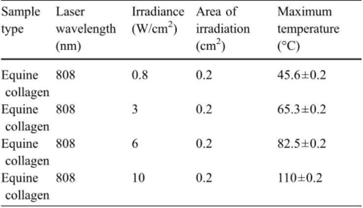
Table 2 The parameters used for the irradiation of pure type I equine for SDS-PAGE 2
Sample type Laser wavelength (nm) Irradiance (W/cm2) Area of irradiation (cm2) Maximum temperature (°C) Equine collagen 808 0.8 0.2 45.6±0.2 Equine collagen 808 3 0.2 65.3±0.2 Equine collagen 808 6 0.2 82.5±0.2 Equine collagen 808 10 0.2 110±0.2
Welding experiments and tensile strength measurements
The welding experiments were performed using two pure collagen membranes of the size of 1×0.5 cm each. The area of interaction was 0.5×0.5 cm. For welding, the liquid ICG solder without and with albumin was used. The irradiation area corresponded to 0.2 cm2. Temperature was measured from above at the irradiation area.
To measure the tensile strength, a test stand equipped with a fixed force gauge (BFG50, Mecmesin, GB) and a moving table was used to pull the two welded and soldered tissue samples apart. The maximum force was measured in Newtons during the pulling process.
Results
SDS-PAGE 1
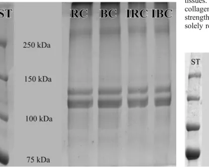
No changes were observed in the distribution of the bands when comparing the non-irradiated with the laser-irradiated pure rabbit type I and pure equine type I collagen (Fig. 1). Two most pronounced bands were observed between 100 and 150 kDa as compared to the standard in all studied samples. No new bands appeared after laser irradiation.
SDS-PAGE 2
No changes were observed in the distribution of the bands when comparing the non-irradiated with the
laser-irradiated pure equine collagen using increasing laser power irradiances (Fig. 2). Again, the two most pro-nounced bands appeared between 100 and 150 kDa.
Tensile strength
Virtually no adherence of the pure collagen layers was observed after laser irradiation. The tensile strength of the laser-welded pure equine collagen membranes was practi-cally inexistent irrespective of the applied laser powers and irradiation duration. Adding albumin had no effect on tensile strength.
Discussion
Although laser tissue welding was first described as early as 1964 [13] and has since been intensely investigated, the exact mechanism of tensile strength remains unclear. The impact of laser irradiation on collagen—believed to be mechanically involved—is controversial to date. However, in order to further optimize the laser soldering technique and to bring it in reach of new clinical applications, the role of the collagenous membranes and especially their role in tensile strength induction necessitate further investigations. The basic question whether collagen is or is not by itself responsible for the induction of the laser-induced tensile bonding was addressed in this study. In reviewing the literature, various hypotheses on the role of collagen can be found. In previous studies, the collagen structures analyzed were impure in that the collagen was embedded between multiple cellular structures, and its contributing percentage in these combinations varied depending on the chosen tissues. To date, it remains unclear to what extent the collagen molecules are involved in the gain of mechanical strength, and whether covalent bonding alterations are solely responsible for laser tissue soldering.
Fig. 1 SDS-PAGE of pure rabbit collagen (RC), pure equine collagen (BC) and SDS-PAGE after laser irradiation of pure rabbit (IRC) and pure equine collagen (IBC). There are no changes in banding after laser irradiation of collagen. The standard (ST) shows the molecular weight in kilodaltons
Fig. 2 SDS-PAGE of pure equine collagen at different laser output powers at 0.16, 0.61, 1.11 and 1.78 W, corresponding to 0.8, 3, 6, 10 W/cm2, respectively. No changes in banding occurred despite the increase in laser power output. The standard (ST) shows the molecular weight in kilodaltons
Covalent versus non-covalent bonding theories
In a SDS-PAGE study of non-pure collagen tissue, Bass et al. [8] reported no changes in band distribution after laser irradiation. In contradiction to these findings, Murray et al. [10] reported the appearance of a new band in the SDS-PAGE of argon-laser-welded guinea pig skin. This band had to result from a newly formed high-molecular-weight protein, enforcing the hypothesis on covalent bonding as the mechanical basis of laser welding and soldering. These contradictory findings might have a simple explanation: Both Bass et al. and Murray et al. did not study pure collagen molecules. Their findings therefore do not reflect the sole interaction of laser with pure collagen but with various combined, cellular, collagen-rich tissues. Although collagen is responsible for the cross-linking in living, unirradiated tissues, the results of the current study raised questions about the nature of the binding forces induced by laser welding or soldering. These results challenge further the postulated (and so far unproven) role of collagen in successful tensile strength induction after laser-induced tissue connection. Finally, the SDS-PAGE findings after thermal denaturation of pure collagen show no changes in the collagen structure, thus allowing the assumption that forces other than the covalent bonding of collagen molecules are responsible for successful laser tissue welding.
3-D conformation changes of collagen molecules
Different study groups have analyzed the 3-D conforma-tion of laser-irradiated tissues, reporting either a loosening and some sort of interaction between collagen strands [9] or an unraveling of collagen bundles and subsequent partial interdigitation [14]. Although undoubtedly existent, the reported changes as well as the loss of the birefringence in the phase contrast light microscopy remain descriptive in nature and cannot be used on their own to quantify the responsible forces in laser tissue welding and soldering. The presented results from the SDS-PAGE on pure laser-irradiated collagen show no such molecular breakdown or molecular neo-formation, which would be reflected in a new band formation.
Tensile strength in laser collagen welding and soldering
The observations made in this study by the SDS-PAGE lead to the conclusion that pure collagen alone is not responsible for tensile strength induction in laser tissue welding and soldering. These findings parallel other experimental results in which laser tissue welding and soldering was less successful when used in tissue with low cellular/matrix ratio like tendons [8, 11, 15]. The tensile strength measurements performed in the current study support the above literature findings, in that pure collagen
membrane fails to reveal any adherence at all when connecting with laser.
On the other hand, laser tissue welding and soldering with high cellular/matrix ratio in tissues like skin, blood vessels, intestinal submucosa and kidneys demonstrated a superior tensile strength upon laser tissue welding [4,5,9, 10,15–20]. These observations raise the question whether intracellular cytoplasm components might be the one delivering the necessary“ingredients” for tensile strength induction in thermal tissue connection.
The role of albumin in tensile strength augmentation Although albumin has been used as an adjunct for improving tensile strength, it failed to show any benefit in the soldering of pure collagen membranes. Histological analyses of tissues after laser soldering in combination with albumin showed albumin to penetrate the cell-rich vessel layers into the media interdigitating with the tissues.
Superior laser tissue soldering results are thus achieved in highly cellular tissues possibly because of a more lax structure of the extracellular matrix, allowing a better interdigitation with the solder. Cell-rich tissues can additionally offer the assumed advantage of additional intracellular proteins that could create non-covalent bonds with the solder and increase the post-irradiation tensile strength.
Conclusion
The results of this study suggest that laser irradiation of pure collagen at irradiation parameters used for laser tissue welding and soldering does not influence covalent binding in the irradiated collagen molecules.
Pure collagen can neither generate alone nor in combination with albumin solder the tensile strength observed in combined cell-rich tissues upon welding or soldering.
Whereas the collagen matrix may have a scaffolding role for the actual binding forces, a cellular component appears to be indispensable for successful laser tissue soldering. Further investigations with engineered and controlled tissue seeding on pure collagen matrices will possibly allow the optimization of further solder materials for clinical applications.
Acknowledgment This study was funded by the Swiss National Foundation Grant 3200B0-107611, Department of Clinical Research, Inselspital, University of Bern, Bern, Switzerland, Novartis Stiftung für Biologische und Medizinische Forschung.
References
1. McNally-Heitzelmann KM, Welch AJ (2003) Laser tissue welding. In: Vo-Dinh T (ed) Biomed photonics handbook, vol 39. CRC, London
2. Bass LS, Treat MR (1995) Laser tissue welding: a compre-hensive review of current and future clinical applications. Lasers Surg Med 17(4):315–349
3. Foyt D, Johnson JP, Kirsch AJ, Bruce JN, Wazen JJ (1996) Dural closure with laser tissue welding. Otolaryngol Head Neck Surg 115(6):513–518
4. Fried NM, Walsh JT Jr (2000) Laser skin welding: in vivo tensile strength and wound healing results. Lasers Surg Med 27(1):55–65
5. McNally KM, Sorg BS, Chan EK, Welch AJ, Dawes JM, Owen ER (1999) Optimal parameters for laser tissue soldering. Part I: tensile strength and scanning electron microscopy analysis. Lasers Surg Med 24(5):319–331
6. Ott B, Constantinescu MA, Erni D, Banic A, Schaffner T, Frenz M (2004) Intraluminal laser light source and external solder: in vivo evaluation of a new technique for microvascular anasto-mosis. Lasers Surg Med 35(4):312–316
7. McNally KM, Sorg BS, Welch AJ, Dawes JM, Owen ER (1999) Photothermal effects of laser tissue soldering. Phys Med Biol 44(4):983–1002; discussion 1002 pages follow
8. Bass LS, Moazami N, Pocsidio J, Oz MC, LoGerfo P, Treat MR (1992) Changes in type I collagen following laser welding. Lasers Surg Med 12(5):500–505
9. Schober R, Ulrich F, Sander T, Durselen H, Hessel S (1986) Laser-induced alteration of collagen substructure allows microsurgical tissue welding. Science 232(4756):1421–1422 10. Murray LW, Su L, Kopchok GE, White RA (1989)
Cross-linking of extracellular matrix proteins: a preliminary report on a possible mechanism of argon laser welding. Lasers Surg Med 9(5):490–496
11. Helmsworth TF, Wright CB, Scheffter SM, Schlemm DJ, Keller SJ (1990) Molecular surgery of the basement membrane by the argon laser. Lasers Surg Med 10(6):576–583
12. Laemmli U (1970) Cleavage of structural proteins during the assembly of the head of bacteriophageT4. Nature 227(5259):680–685
13. Yahr WZ, Strully KJ, Hurwitt ES (1964) Non-occlusive small arterial anastomosis with a neodymium laser. Surg Forum 15:224–226
14. Anderson NJ, Edelhauser HF, Sharara N, Thompson KP, Rubinfeld RS, Devaney DM, L’Hernault N, Grossniklaus HE (2002) Histologic and ultrastructural findings in human corneas after successful laser in situ keratomileusis. Arch Ophthalmol 120(3):288–293
15. Kilkelly FX, Choma TJ, Popovic N, Miller DW, Sweet DE (1996) Tendon repair by laser welding: a histologic and biomechanical comparison and suture repair with CO2 and
argon lasers. Lasers Surg Med 19(4):487–491
16. Ware MH, Buckley CA (2003) The study of a light-activated albumin protein solder to bond layers of porcine small intestinal submucosa. Biomed Sci Instrum 39:1–5
17. Ogan K, Jacomides L, Saboorian H, Koeneman K, Li Y, Napper C, Hoopman J, Pearle MS, Cadeddu JA (2003) Sutureless laparoscopic heminephrectomy using laser tissue soldering. J Endourol 17(5):295–300
18. Mendoza GA, Acuna E, Allen M, Arroyo J, Quintero RA (1999) In vitro laser welding of amniotic membranes. Lasers Surg Med 24(5):315–318
19. Maitz PK, Trickett RI, Dekker P, Tos P, Dawes JM, Piper JA, Lanzetta M, Owen ER (1999) Sutureless microvascular anastomoses by a biodegradable laser-activated solid protein solder. Plast Reconstr Surg 104(6):1726–1731
20. Li ZR, Chi YL, Ke RC (2000) Sutureless end-to-end bowel anastomosis in rabbit using low-power CO(2) laser. World J Gastroenterol 6(4):557–560