Brain glucose utilization in systemic lupus erythematosus
with neuropsychiatric symptoms: a controlled positron
emission tomography study
A. Otte 1,3, S.M. Weiner 2, H.H. Peter 2, J. Mueller-Brand 1, M. Goetze 1, E. Moser 3, J. Gutfleisch 2, S. Hoegerle 3, RD. Juengling 3, E.U. Nitzsche 3
1 Institute of Nuclear Medicine, University Hospital, Basel, Switzerland
2 Department of Rheumatology and Immunology, University Hospital Freiburg, Germany 3 Department of Nuclear Medicine, University Hospital Freiburg, Germany
Received 28 February and in revised form 2 April 1997
Abstract.
In contrast to morphological imaging [such as magnetic resonance imaging (MRI) or computed tomog- raphy], functional imaging may be of advantage in the detection of brain abnormalities in cases of neuropsyehi- atric systemic lupus erythematosus (SLE). Therefore, we studied 13 patients (aged 40_+14 years, 11 female, 2 male) with neuropsychiatric SLE who met four of the American Rheumatism Association criteria for the clas- sification of SLE. Ten clinically and neurologically heal- thy volunteers served as controls (aged 40+12 years, 5 female, 5 male). Both groups were investigated using fluorine-18-1abelled fluorodeoxyglucose brain positron emission tomography (PET) and cranial MRI. The nor- mal controls and 11 of the 13 patients showed normal MRI scans. However, PET scan was abnormal in all 13 SLE patients. Significant group-to-group differences in the glucose metabolic index (GMI=region of interest up- take/global uptake at the level of the basal ganglia and thalamus) were found in the parieto-occipital region on both sides: the GMI of the parieto-oceipital region on the right side was 0.922+0.045 in patients and 1.066+0.081 in controls (P<0.0001, Mann Whitney U test), while on the left side it was 0.892+0.060 in pa- tients and 1.034+0.051 in controls (P=0.0002). Parieto- occipital hypometabolism is a conspicuous finding in mainly MRl-negative neuropsychiatric SLE. As the pari- eto-occipital region is located at the boundary of blood supply of all three major arteries, it could be the most vulnerable zone of the cerebrum and may be affected at an early stage of the cerebrovascular disease.Key words: Neuropsychiatric systemic lupus erythema- tosus - Positron emission tomography - Fluorine-18 flu- orodeoxyglucose - Parieto-occipital brain hypometabo- lism
Correspondence to: A. Otte, Institute of Nuclear Medicine, De- partment of Radiological Science, University Hospital, School of Medicine, Petersgraben 4, CH-4031 Basel, Switzerland
Eur J Nucl Med (1997) 24:787-791
Introduction
Studies with positron emission tomography (PET) and single-photon emission tomography (SPET) have shown that in certain brain disorders functional imaging is more sensitive than morphological imaging such as magnetic resonance imaging (MRI) and X-ray computed tomogra- phy. This applies to traumatic brain injury [1-8], whip- lash injury [9, 10], migraine [11], or Alzheimer's disease [12, 13], and also neuropsychiatric systemic lupus ery- thematosus (SLE) [14-16]. Neuropsychiatric SLE has in most cases been studied in late stages of the disease; lit- tle is known of early neuropsychiatric SLE. There are conflicting reports regarding the regions involved in neu- ropsychiatric SLE: using azetazolamide-enhanced SPET in an SLE patient with preclinical central nervous system (CNS) involvement, GdJnwald et al. [14] showed a marked reduction in the cortical perfusion reserve, par- ticularly in both frontal lobes, whereas Kovacs et al. [17] showed various other abnormal regions such as the oc- cipital cortex or the basal ganglia in cases of active neu- ropsychiatric SLE. Szer et al. [18] reported on a girl with a concurrent ischaemic event involving both pari- etal lobes. Therefore, we wondered whether there is a typical pattern of metabolism in neuropsychiatric SLE, reflecting the vulnerability of the different zones in the cerebrum.
Materials and methods
Each study participant signed an informed consent form, explain- ing the investigative nature of the study, its risks, and its merits. Study population. Ten normal controls (mean age 40___12 years, 5 female, 5 male) and 13 patients (mean age 40_+14 years, 11 re-
male, 2 male) were studied. All patients met four of the American Rheumatism Association criteria for the classification of SLE [19]. All study participants were interviewed with regard to head- ache, vertigo, visual symptoms (blurred vision, scintillating scoto- ma, oscillopsia), impaired memory and/or concentration, depres- sion, sleep disturbances, anxiety and instability [20].
None of the study participants exhibited anamnestic hints of neurovascular or neurodegenerative diseases. Secondary causes such as infection, uraemia, hypertension, drugs and metabolic ab- normalities were excluded. Eleven of the 13 neuropsychiatric SLE patients received corticosteroid treatment and three received im- munosuppression with cyclophosphamide or azathioprine. As re- gards the control group, only participants with normal clinical and neurological findings were included in the study. For the patient group, only those patients were included who showed clinical and neurological findings indicating CNS involvement by the disease. Neuropsychiatric manifestation due to SLE was defined as an ab- normality of neurological function identified by current history and physical examination and noted as a change from a prior state, if this abnormality could not be attributed to any cause other than the disease process itself. The CNS manifestations were classified into "major" (seizures, focal motor or sensory deficits, generalized disturbances, psychosis, organic brain syndrome) and "minor" signs (paraesthesia or clumsiness without objective findings, headache, cognitive disorders) according to the classification of How et al. [21]. Nine patients with "initial" CNS SLE had shown neuropsychiatric signs for no longer than 1 year, whereas four pa- tients with "manifest" CNS disease had been symptomatic for more than 1 year. All patients diagnosed as having manifest CNS SLE (n=4) had one or more major signs, and two of them showed pathological findings on MRI. Patients in the initial CNS SLE group had one major sign (n=5) or one to three minor signs (n
=4).
MRI of the brain was also undertaken in all patients and con- trois.
The following neuropsychiatric symptoms were observed in the SLE patients:
- Impairment of memory and of intelligent function: n=6 patients
- Psychosis: n=4 - Seizure: n = l
- Focal motor deficits: n=2
- Persistent headache: n=4 - Paraesthesia: n=2 - Vertigo: n=3
- Impairment of orientation and perception: n=l
- Visual symptoms: n=4 - Recurrent apoplexy: n= 1
The following concomitant clinical and serological parameters were observed in the SLE patients:
- Positive antinuclear antibodies: n = 13 patients - Positive anti-ds DNA: n=7
- Positive antibodies against extractable nuclear antigen: n= 10 - Phospholipid antibodies: n=6 - Cutaneous lesions: n=7 Lupus nephritis: n=8 - Arthralgia: n=6 - Myalgia: n=4 - Sicca complex: n=4 - Cytopenia: n=4
Radiopharmaceuticals. All participants were injected with 370 MBq fluorine-I 8 fluorodeoxyglucose (FDG) into a peripheral
vein. FDG was synthesized and prepared as reported previously [22].
PET study protocol. Before the start of the PET procedure, each study participant had to fast for at least 12 h before the study and to empty the bladder. Then, during the 10-rain waiting period pri- or to injection, patients and controls were placed on a bed in the preparation area in a quiet room with dim lights, remaining awake, but with closed eyes. After injection, patients and controls remained on the bed for another 30 min. The head of each subject was then positioned parallel to the orbitomeatal line with the aid of a laser beam, after which it was placed in a moulded thermo- plastic head support to minimize head movements during the scanning procedure. All subjects were studied at rest. The acquisi- tion started about 35 min p.i. PET imaging was performed on a Siemens CTI ECAT EXACT tomograph. This device records 31 planes simultaneously, which encompass a 10.8-cm field of view. The spatial resolution is 6.0 mm full-width at half-maximum. Photon attenuation was automatically calculated using Siemens- CTI standard software [23]. A total of six frames, each of 5 rain duration, were acquired. Images were reconstructed using filtered back-projection (Shepp-Logan filter, cut-off 0.3 cycles/pixel). For data analysis, summed images were used (summing the dynami- cally acquired frames 1-6 into one image).
Data analysis. Standardized cortical and subcortical elliptical re- gions of interest (ROIs) were assigned to three adjacent transaxial slices of the following regions by consensus of two independent investigators: 1. Frontal 2. Parietal 3. Occipito-frontal 4. Temporo-lateral 5. Temporo-mesial 6. Parieto-occipital (P-O) 7. Thalamus 8. Putamen 9. Caudate nucleus
10. Cerebellar (mean of lateral and dorsal cerebellum) 11. Brain stem
12. Global at the level of the thalamus and basal ganglia
The shape and size of ROIs were the same in all patients and controls, since the ROI templates were copied onto the corre- sponding planes of each study. The shape and size of global ROIs were fitted to actual head form by manipulation. The position of all ROIs was adjusted to the corresponding anatomical localiza- tion. In each ROI the average uptake per pixel per time was deter- mined.
ROI analysis was performed using a SUN sparc station and SUNVIEW software. To allow inter-individual comparisons, re- gional cerebral metabolism was normalized by global glucose me- tabolism (glucose metabolic index, GMI; GMI=ROI uptake/global uptake) determined from the global brain ROI at the level of the thalamus and basal ganglia.
Statistical analysis. All data are expressed as mean _+1 SD. Group- to-group differences were tested using the Mann Whitney U test (M.W.) and the software StatView 4.10.
Results
MRI of the brain showed no pathological findings in any of the controls or in 11 of the 13 patients. The two pa- tients with pathological MRI findings had had organic neuropsychiatric symptoms for more than 1 year. In con-
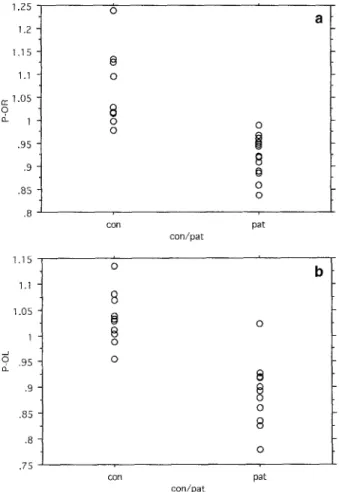
1,25 1.2 1.15 1.1 cc 1.05 2 1 .95 .9 .85 .8 1.15 1.1 ] iO 5 1 .95 .9 .85 .8 ,75 O O
8
o O COIl con/pat pat 0 con pat con/patFig. 1. Scattergrams of" GMI values of P-O on the right (a) and left (b) sides, in the control group (con, n=10) and the patient group
(pat, n= 13)
trast, the patient group revealed a significant hypometab- olism in the P-O regions [on the right (R) and left (L) side] as compared to the control group (Fig. 1):
- GMI P-O R: controls 1.066+0.081, patients 0.922_+0.045; P<0.0001
- GMI P-O L: controls 1.034___0.051, patients 0.892___0.060; P=0.0002
Group-to-group differences in regions other than the P-O were not statistically significant. However, careful evaluation of the individual metabolic pattern did addi- tionally reveal, in six of the 13 patients (46%), GMI val- ues which were more than one or two standard devia- tions below normal control values in regions other than the P-O. In one patient (no. 5), P-O hypometabolism was combined with a patchy uptake pattern over the whole brain, while in another (no. 1) it was combined with a relative hypermetabolism in the basal ganglia and thala- mi (Table 1 ).
Furthermore, when the SLE patient group was divid- ed into patients who showed CNS involvement for the first time at admission (group 1, n=9) and patients with organic neuropsychiatric symptoms for more than 1 year (group 2, n=4), no significant difference was found be- tween groups 1 and 2 with regard to glucose utilization in any of the analysed brain regions.
Discussion
Our study indicates that ueuropsychiatric SLE - irre- spective of the stage of CNS involvement (i.e. initial or non-initial) - has a typical pattern of hypometabolism in the P-O region, although non-P-O regions may also be affected. Since the P-O region is located at the boundary of supply of all three major arteries, it is thought to be the most vulnerable zone of the cerebrum [24]. There-
Table 1. Brain PET findings in patients
Pat. P-o,R P-o,L ER EL E R F,L O-ER O-F,L CN,B PUT, B TH,B P-P
1 - - 0 O 0 0 0 0 ++ ++ ++ No 2 . . . 0 0 0 0 No 3 -- 0 0 ~ 0 0 0 0 0 O No 4 . . . . 0 0 0 0 0 0 0 0 0 No 5 . . . . 0 0 0 0 0 O 0 0 O Yes 6 -- 0 0 0 0 O 0 O 0 0 No 7 - 0 0 0 0 0 0 0 No 8 0 0 O 0 0 0 0 0 0 0 No 9 . . . O 0 0 0 0 0 0 0 No 10 . . . 0 0 0 0 0 0 0 0 No 11 -- 0 0 0 0 0 0 0 0 No 12 . . . . O 0 0 0 + 0 0 No 13 -- O 0 0 0 0 O 0 O O No
P-O, Parieto-occipital; P, parietal; F, frontal; O-F, occipito-frontal; CN, caudate nucleus; PUT, putamen; TH, thalamus; P-P, patchy uptake pattern; R, right brain side; L, left brain side; B, both sides. -, GMI>I SD below normal controls; - -, >2 SD; +, GMI>I SD above normal controls; ++, >2 SD; Q, GMI within normal limits.
fore, we think that this region is frequently affected at an early stage of SLE with suspected CNS involvement. MRI findings are inconspicuous in early neuropsychiat- ric SLE, in contrast to the well-known M R I findings in manifest and chronic CNS SLE [25-27]; against this background the information provided by our PET data might have important diagnostic and therapeutic impli- cations at an early stage of the disease. However, the question of whether vasculitis or infarctions and haem- orrhages in this P-O zone are the consequence or a cause of the SLE process remains open.
Abnormalities in the P-O region are not pathogno- monic for neuropsychiatric SLE: they can also be found, for example, in the late whiplash syndrome [9, 10, 28], in dementia of the Alzheimer type [12], in the elderly [13], in sleep apnea syndrome [13] and following cere- bral hypoxia [13]. However, after careful clinical and se- rological classification of our neuropsychiatric SLE pa- tients, these differential diagnoses could be excluded.
In studies on the pathology of cerebral lupus, vascu- lopathy, infarctions and haemorrhages are often ob- served, whereas vasculitis is rare [29]. Furthermore, the concept of hypercoagulability in SLE patients has divert- ed the direction of therapy from immunosuppression [30] (as in the majority of our patients) towards antico- agulation [31]. Randomized trials have commenced in order to determine the optimal mode of therapy for SLE patients under strict inclusion criteria, based on well-de- fined patient categories. Functional imaging like PET, or SPET, could be a powerful tool for such an enterprise.
References
1. Bavetta S, Nimmon CC, White J, McCabe J, Huneidi AH, Bomanji J, Birkenfeld B, Charlesworth M, Britton KE, Green- wood RJ. A prospective study comparing SPET with MRI and CT as prognostic indicators following severe closed head inju- ry. NucI Med Commun 1994; 15: 961-968.
2. Masdeu JC, VanHerretum RL, Kleiman A, Anselmi G, Kis- sane K, Horng J, Yudd A, Luck D, Grundman M. Early single photon emission computed tomography in mild head trauma, a controlled study. J Neuroimag 1994; 4:177-181.
3. Prayer L, Wimberger D, Oder W, Kramer J, Schindler E, Podreka I, Imhof H. Cranial MR imaging and cerebral 99mTc- HMPAO SPECT in patients with subacute or chronic severe closed head injury and normal CT examinations. Acta Radiol 1993; 34: 593-599.
4. Newton MR, Greenwood RJ, Britton KE. A study comparing SPECT with CT and MRI after closed head injury. J Neurol Neulvsurg Psychiatry 1992; 55: 92-94.
5. Roper SN, Mena I, King WA, Schweitzer J, Garrett K, Me- hninger CM, McBride D. An analysis of cerebral blood flow in acute closed head injury using technetium-99m-HMPAO SPECT and computed tomography. J Nucl Med 1991; 32: 1684-1687 (erratum 2070).
6. Nedd K, Sfakianakis G, Ganz W, Uricchio B, Vernberg D, Vil- lanueva R Jabir AM, Bartlett J, Keena J. 99mTc-HMPAO SPECT of the brain in mild to moderate traumatic brain injury patients compared with CT - a prospective study. Brain Inj 1993; 7: 469-479.
7. Gray BG, Ichise M, Chung DG, Kirsh JC, Franks W. Techne- tium-99m-HMPAO SPECT in the evaluation of patients with a remote history of traumatic brain injury: a comparison with x- ray computed tomography. J Nucl Med 1992; 33: 52-58. 8. Yamakami I, Yamaura A, Isobe K. Types of traumatic brain
injury and regional cerebral blood flow assessed by 99mTc- HMPAO SPECT. Neurol Med Chir 1993; 33: 7-12.
9. Otte A, Mueller-Brand J, Fierz L. Brain SPECT findings in late whiplash syndrome. Lancet 1995; 345:1513-1514. 10. Otte A, Ettlin TM, Fierz L, Mueller-Brand J. Parieto-occipital
hypoperfusion in late whiplash syndrome: first quantitative SPECT study using Tc-99m-bicisate (ECD). Eur J Nucl Med 1996; 23: 72-74.
11. Friberg L. Cerebral blood flow changes in migraine: methods, observations and hypotheses. J Neurol 1991; 238 Suppl 1: S12-S17.
l 2. Waldemar G, Bruhn R Kristensen M, Johnsen A, Paulson O, Las- sen NA. Heterogeneity of neocortical cerebral blood flow deficits in dementia of the Alzheimer type: a 99mTc-HMPAO-SPECT study J Neurol Neurosurg Psychiatry 1994; 57: 285-295.
13. Miller BL, Mena I, Daly J, Gombetti RJ, Goldberg MA, Less- er I, Garetti K, Villanueva-Meyer J, Liu CK. Temporo-parietal hypoperfusion with single photon emission computerized to- mography in conditions other than Alzheimer's disease. De- mentia 1990; 1: 41-45.
14. Gr~nwald F, Schomburg A, Badali A, Ruhlmann J, Pavics L, Biersack HJ. lSFDG PET and azetaolamide-enhanced 99mTc- HMPAO SPET in systemic lupus erythematosus. Eur J Nucl Med 1995; 22: 1073-1077.
15. Sibbitt WL, Sibbitt RR. Magnetic resonance spectroscopy and positron emission tomography scanning in neuropsychiatric systemic lupus erythematosus. Rheum Dis Clin North Am 1993; 19: 851-868.
16. Stoppe G, Wildhagen K, Meyer GJ, Schober O. Use of fluoro- deoxyglucose PET in the diagnosis of central nervous system lupus erythematosus and a comparison with CT and MRI. Nu- klearmedizin 1989; 28: 187-192.
17. Kovacs JA, Urowitz MB, Gladman DD, Zeman R. The use of single photon emission computerized tomography in neuro- psychiatric SLE: a pilot study. J Rheumatol 1995; 22: 1247-1253.
18. Szer IS, Miller JH, Rawlings D, Shaham B, Bernstein B. Ce- rebral perfusion abnormalities in children with central nervous system manifestations of lupus by single photon emission computed tomography. J Rheumatol 1993; 20:2143-2148. 19. Tan EM, Cohen AS, Fried JF, et al. The 1982 revised criteria
for the classification of systemic lupus erythematosus (SLE). Arthritis Rheum 1982; 25: 1271-1277.
20. Ettlin T, Kischka U, Reichmann S, Radt~ EW, Heim S, a Wen- gen D, Benson FD. Cerebral symptoms after whiplash injury of the neck: a prospective clinical and neuropsychological study of whiplash injury. J Neurol Neulvsurg Psychiatry 1992; 55: 943-948.
21. How A, Dent PB, Liao SK, Denburg JA. Antineuronal anti- bodies in neuropsychiatric systemic lupus erythematosus. Ar- thritis Rheum 1985; 28: 789--795.
22. Hamacher K, Coenen HH, St0cklin G. Efficient stereospecific synthesis of no-carrier added 2-[18F]-fluoro-2-deoxy-D-glu - cose using aminopolyether supported nucleophilic substitu- tion. JNucl Med 1986; 27: 235-238.
23. Huang SC, Carson R, Phelps M, Hoffman E, Schelbert H, Kuhl D. A boundary method for attenuation correction in pos- itron emission tomography. 1EEE Trans Nucl Sci 1981; 22: 627-637.
24. Graham DG, Briefly JB. Vascular disorders of the central ner- vous system. In: Adams J, ed. NeuropathoIogy. London: Ed- ward Arnold; 1984: 125-207.
25. Isshi K, Hirohata S, Hashimoto T, Miyashita H. Systemic lu- pus erythematosus presenting with diffuse low density lesions in the cerebral white matter on computed axial tomography scans: its implication in the pathogenesis of diffuse central nervous system lupus. J Rheumatol 1994; 21:1758 1762. 26. Gonzalez-Crespo MR, Blanco FJ, Ramos A, Ciruelo E, Mateo
I, Lopez-Pino MA, Gomez-Reino JJ. Magnetic resonance im- aging of the brain in systemic lupus erythematosus. Br J Rheumatol 1995; 34: 1055-1060.
27. Baum KA, Hopf U, Nehrig C, Stover M, Schorner W. System- ic lupus erythematosus: neuropsychiatric signs and symptoms related to cerebral MRI findings. Clin Neurol Neurosurg 1993; 95: 29-34.
28. Otte A, Ettlin TM, Fierz L, Kischka U, Muerner J, Mueller- Brand J. Brain perfusioin patterns in 136 patients with chronic symptoms after distorsion of the cervical spine using single photon emission computed tomography, technetium-99m- HMPAO and technetium-99m-ECD: a controlled study. J Vasc lnvest 1997; 3: 1-5.
29. vanDam AR Diagnosis of pathogenesis of CNS lupus. Rheu- matollnt 1991; l l : 1-11.
30. McInness PM, Schuttinga J, Sanslone WR, Stark SR Klippel JH. The economic impact of treatment of severe lupus nephri- tis with prednisolone and cyclophosphamide. Arthritis Rheum 1994; 37: 1000-1006.
31. Bruyn GA. Controversies in lupus: nervous system involve- ment. Ann Rheum Dis 1995; 54: 159-167.