Cell Tissue Res (2005) 322: 237–244 DOI 10.1007/s00441-005-0005-3
R E G U L A R A RT I C L E
Detlev Grabs . Mathias Bergmann
Differential appearance of dynamin in constitutive and regulated
exo-endocytosis: a single-cell multiplex RT-PCR study
Received: 16 February 2005 / Accepted: 4 May 2005 / Published online: 19 July 2005 # Springer-Verlag 2005
Abstract Neurons in the central nervous system establish, via their axons and dendrites, an extended network that allows synaptic transmission. During developmental mat-uration and process outgrowth, membrane turnover is nec-essary for the enlargement and subsequent growth of axons and dendrites from the perikarya to the target cell (con-stitutive exocytosis/endocytosis). After targeting and syn-apse formation, small synaptic vesicles are needed for the quantal release of neurotransmitters from the presynaptic terminal with subsequent recycling by regulated exocyto-sis/endocytosis. An investigation of the onset of the ap-pearance of mRNA and protein in dissociated cultures of neurons from mouse hippocampus or from chick retina has shown an early abundance of proteins involved in exocy-tosis, such as syntaxin 1, SNAP-25, and synaptotagmin 1, whereas dynamin 1, a protein necessary for clathrin-me-diated endocytosis, can be detected only after neurons have established contacts with neighboring cells. The results re-veal that constitutive membrane incorporation and regu-lated synaptic transmitter release is mediated by the same neuronal proteins. Moreover, the data exclude that dy-namin 1 takes part in constitutive recycling before synapse formation, but dynamin 2 is present at this stage. Thus, dynamin 2 may be the constitutive counterpart of dynamin 1 in growing neurons. Synapse establishment is linked to an upregulation of dynamin 1 and thereby represents the beginning of the regulated recycling of membranes back into the presynaptic terminal.
Keywords Neuronal development . Exocytosis . Endocytosis . Presynaptic proteins . Dynamin . Chick (White Leghorn) . Mouse
Introduction
In mature neurons, synaptic vesicles continuously recycle within the presynaptic nerve terminal. This cycle includes the fusion of synaptic vesicles with the plasma membrane and transmitter release by exocytosis followed by endo-cytic membrane internalization. The regulated exo-/endo-cytosis is thought to be mediated by presynaptic proteins. These proteins include the members of SNARE proteins involved in exocytic events (for a review, see Bruns and Jahn 2002) and a group of proteins including dynamin, amphiphysin, and endophilin involved in endocytic events (Herskovits et al. 1994; McPherson et al. 1996; Higgins and McMahon2002; Reutens and Begley2002).
However, membrane expansion and retrieval have to oc-cur during neuronal development along the processes, at the tip of growing processes, and during the establishment of synapses. These mechanisms underlying diverse altera-tions within neuronal growth cones have been the subject of intense investigation. Many of these investigations have centered on the cytoskeleton, which displays significant changes during growth cone elongation (Letourneau1996). Other studies have focused on the involvement of recep-tor proteins in ion channels (Contestabile2000), adhesion molecules (Walsh and Doherty 1997), guidance molecules (Cook et al.1998), or neurotrophins (Huang and Reichardt
2001). Additionally, the removal, translocation, and addi-tion of membrane (and their proteins) must be in the transformation of the growth cone into a neuronal process and, later on, into a mature synaptic terminal. Although key proteins for both pathways have been named (Jahn et al.2003), the exact mechanisms of membrane extension (exocytosis) and retrieval (endocytosis) in living neurons are still poorly understood in developing neurons.
In-situ and in-vitro studies have been shown that pre-synaptic proteins involved in exocytosis are expressed during neuronal development and undergo extensive re-distribution prior to and during synapse formation (e.g., Bergmann et al. 1991; Catsicas et al. 1994; Grabs et al.
1994; Grosse et al. 1998) and are transported together as distinct packets (Ahmari et al.2000). Several proteins have D. Grabs (*) . M. Bergmann Department of Medicine/Anatomy, University Fribourg, Rte A. Gockel 1, 1700 Fribourg, Switzerland e-mail: [email protected] Tel.: +41-26-3008547 Fax: +41-26-3009733
been shown to mediate membrane expansion during out-growth (Steiner et al.2002). The cleavage of other proteins by clostridial neurotoxins inhibits axonal (Osen-Sand et al.
1993; Igarashi et al. 1996) and dendritic (Grosse et al.
1999) growth. Moreover, constitutive membrane recycling is reported not to be restricted to the nerve terminal but to occur in cycles of exo-/endocytosis over the whole axonal surface (Matteoli et al. 1992; Kraszewski et al. 1995). However, conflicting data from neuroendocrine pheochro-mocytoma cell lines (PC12) indicate that neurite extension can occur, although known members of the exocytic machinery are absent from specialized strains of these cells (Leoni et al. 1999; Grundschober et al. 2002). Additionally, data from null-mutant mice suggest that, in the absence of either VAMP2 (synaptobrevin 2; Schoch et al.2001) or SNAP-25 (Washbourne et al.2002), synaptic transmission is decreased, although normal axonal out-growth and synaptic targeting still occurs.
Whereas the main focus of the previous investigations has been on the exocytic machinery, little is known about the involvement of soluble presynaptic proteins used in endocytosis, such as dynamin, amphiphysin, and endophi-lin, in dendrite and axon outgrowth and synapse formation. Although dynamin and amphiphysin are established text-book members of clathrin-mediated endocytosis, endophi-lin (Ringstad et al.1997) is thought to be necessary for lipid modification at the plasma membrane during endocytosis (Huttner and Schmidt 2000), and the blockage of en-dophilin has been shown to interfere with clathrin “un-coating” (Gad et al.2000) but not with neurotransmission (Verstreken et al. 2002). However, multiple isoforms of dynamin and endophilin are differentially expressed in the central nervous system (Cook et al. 1994; Nakata et al.
1993; Ringstad et al. 2001). In-situ expression data for dynamin and amphiphysin in chick retinotectal neurons have suggested a delay between the upregulation of these proteins compared with presynaptic proteins involved in exocytosis (Bergmann et al. 1999; Grabs et al.2000). In-terestingly, a delayed appearance of dynamin has also been found during motor neuron development and neuromus-cular synaptogenesis (Noakes et al. 1999). The growth cones of neurons treated with amphiphysin antisense-probes collapse (Mundigl et al. 1998). Nevertheless, the specific functions of these endocytic proteins in imma-ture neurons and during synapse formation remains to be determined.
Materials and methods
Animals
All experiments were carried out in accordance with the guidelines published by the Swiss Academy of Medical Sciences) and the Swiss Academy of Natural Sciences regarding the use of animals for experimental procedures. White Leghorn chicks (embryonic day 10, ED10) and mouse embryos (ED17) were used. Chicks were taken from termed eggs, whereas mother mice were anesthetized
with Vetanarcol (Pentobarbital 100 mg/kg body weight), and their embryos were removed by Caesarian section.
Cell culture
Primary cultures were established from the hippocampi of ED17 mice or from retinae of ED10 White Leghorn chicks. Single-cell suspensions were prepared by mechanical and enzymatic dissociation in Neurobasal medium supplement-ed with B27 solution (Invitrogen, Basel, Switzerland). The cells were suspended at a density between 100,000 and 500,000 cells/cm2 in poly-D-lysine-coated plastic dishes (Nunc, Wiesbaden, Germany) and kept for up to 21 days in culture.
Single-cell reverse transcription/polymerase chain reaction
Cultivated cells were chosen under visual control under a Leica DM-IRBE microscope and picked up with a 15-μm glass capillary (WPI, Aston, UK). Single cells were trans-ferred into a proteinase/SDS/RNase inhibitor solution and heated for 5 min at 95°C for lysis and enzyme inactivation. Reverse transcription (RT) and polymerase chain reaction (PCR) were performed in single tubes by using the One-Step RT-PCR system (Qiagen, Basel, Switzerland). There-after, specific first-round primers for all tested protein sequences were added to the solution. A second run of PCR with nested primers used only one pair of primers for a specific protein in each tube.
First-round primers for the mouse hippocampus were as given below.
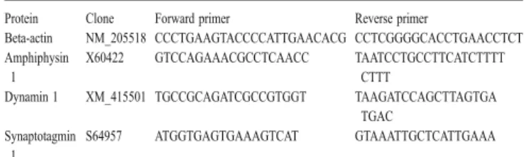
Protein Clone Forward primer Reverse primer
Beta-actin NM_007393 CCCTGAAGTACCCCATTGAACATG CCTCAGGGCATCGGAACCGCT Amphiphysin
1
NM_175007 GTATGGACGGGAAGATGTAAAGAT CTGGGCAGGGGAGAAGG Dynamin 1 NM_010065 TGCCGCAGATCGCCGTGGT TCAGGTCCAACTTGGTGA
TGAC
Alpha-adaptin X14971 TCCGGCTCATCAACAACGCTATCA GCCGCCCGCTGCCTCAC Endophilin 2 U58885 GGGGCCGAAGGGACCAAACT GCCGTGCAGCATCCCCTC
ATAC
Syntaxin 1a D45208 ACGGCCAAGGACAGCGATGAC TGGTACTTGACGGCCTTCTT GGTG
Synaptotagmin 1
NM_009306 GCTGAACTGCCCGCCCTGGAC GCTCTGCGCCGGTGCTGTT GTAG
Nested primers for the mouse hippocampus were as follows.
Protein Clone Forward primer Nested primer
Beta-actin NM_007393 GATCTGGCACCACACCTTCTAC GCACAGCTTCTCTTTGATG Amphiphysin 1 NM_175007 CTAAGCGCAGCAGGAAG TGACGTAAAAGCCAACTCGA Dynamin 1 NM_010065 GGCGTCCCCTGGTCCTG GGTGCGCTGACCCTGGG Alpha-adaptin X14971 GTGCCGCCCTATGCCTACTG ACACTCCACTAACCGCCCTT
TCAC
Endophilin 2 U58885 CATGGCAAGGAACTAGGTGGAGA GGGGCCGGGGCTTGAACT Syntaxin 1a D45208 TCCCCGAACCCCGATGAGA AGAGGCAAAGATGGCAGGG
TTCC
First-round primers for the chick retina were as below. Protein Clone Forward primer Reverse primer
Beta-actin NM_205518 CCCTGAAGTACCCCATTGAACACG CCTCGGGGCACCTGAACCTCT Amphiphysin
1
X60422 GTCCAGAAACGCCTCAACC TAATCCTGCCTTCATCTTTT CTTT
Dynamin 1 XM_415501 TGCCGCAGATCGCCGTGGT TAAGATCCAGCTTAGTGA TGAC
Synaptotagmin 1
S64957 ATGGTGAGTGAAAGTCAT GTAAATTGCTCATTGAAA
Nested primers for the chick retina were as follows. Protein Clone Forward primer Nested primer
Beta-actin NM_205518 GATCTGGCACCACACTTTCTAC GCACAGCTTCTCCTTGATG Amphiphysin 1 X60422 ACATGAAGACGGGCATC CATCACATTTCTCACCAA Dynamin 1 XM_415501 GGCGTCCGCTGGTCCTG GGTGCGTTGCCCTTGAG Synaptotagmin 1 S64957 GGAGGAGGAGGAAAAGAAGAT GAATAATTCCAACCAGAAGC
To compare dynamin 1 and dynamin 2 in the mouse hippocampus different primers were used. First-round prim-ers were as given below.
Protein Clone Forward primer Reverse primer Dynamin 1 NM_010065 CCCGGCGTCCCCTGGTCCTG ACGTCCCGCGCATCTGTG Dynamin 2 NM_007871 TACCCCGAGAAGGACCAGGCAGAG TGGCGCAGAGAAGGGGTCGT TGTT
Nested primers for the mouse hippocampus were as follows.
Protein Clone Forward primer Nested primer
Dynamin 1 NM_010065 CACCGGCACCAACAAGGGCATTTC CCGAGTTGGCAGGGGACACAGC Dynamin 2 NM_007871 CAGGCTCAGCGGCGGGACGAC GGCGCTGTGGAGTGGGGCTGTG
Immunostaining of primary cultures
For immunocytochemical detection, cell cultures were fixed for 30 min with 4% paraformaldehyde in 0.1 M phosphate buffer saline (pH 7,4). Fixed cultures were per-meated with 0.1% Triton X-100 for 30 min. For immu-nofluorescence, mouse monoclonal or rabbit polyclonal antibodies recognizing dynamin 1 (Transduction Labora-tories), dynamin 1 (DG1, kindly provided by P. DeCamilli; see Butler et al.1997), synaptotagmin 1 (clone 41.1, kindly provided by R. Jahn; see Brose et al.1992), and SNAP-25 (SMI-81, Sternberger Monoclonals) were diluted 1:100 in goat serum dilution buffer (20 mM NaPO4 pH 7.4, 15% normal goat serum, 450 mM NaCl) and incubated overnight at 4°C. Antigen/antibody complexes were visualized by using Alexa 488- or Alexa 546-conjugated goat anti-rabbit or goat anti-mouse IgGs 1:100 for 2 h (for further details, see Grabs et al.1994; Grosse et al.1998,1999). The cul-tures were examined and photographed by using a Leitz DM-IRBE microscope equipped with epifluorescence.
For ultrastructural analysis, cultures were fixed with 2% paraformaldehyde and 0.5% glutaraldehyde in 0.1 M cacodylate buffer (pH 7.2) for 30 min. Cultures were
pro-cessed for pre-embedding staining by the avidin-biotin-complex technique (for further details, see Grosse et al.
1998). Sections were postfixed with 1% OsO4, counter-stained with 4% uranyl acetate and 0.2% lead citrate, and examined in a Zeiss EM10 electron microscope.
Results
Low density cultures were established either from embry-onic mouse hippocampus (ED17) or from embryembry-onic chick retina (ED10). For investigation of the developmental appearance of the mRNA of synaptic proteins, single cells were kept under controlled in-vitro conditions. Two dif-ferent stages of neuronal development were compared: 1. neurons grown in isolation with no connections to
neighboring neurons (Fig.1a) and
2. neurons with established connections to one or more adjacent neurons (Fig.1b).
Fig. 1 Selection of single cells. Single neurons either grown in isolation (a) or integrated in a neuronal network (b) were chosen as described. By means of micropipettes with a 15-μm tip containing phosphate-buffered saline (PBS), target cells were localized (c), picked up by negative pressure in the pipette (d), and transferred into Eppendorf tubes filled with RNase protection medium
All cells were taken between day-in-vitro 5 (DIV5) and 16 (DIV16) from the culture system. In order to withdraw single cells out of the culture, fine capillaries were drawn out to give tips with a diameter of 15μm. Capillaries were placed under microscopical control beside the chosen neu-rons (Fig.1c). Single cell bodies were picked up by neg-ative pressure by using an UltraMicroPump II (WPI) and immediately transferred into RNase protection medium (Fig.1d).
mRNA was extracted from these single cells and reverse-transcribed. cDNA was then amplified by two rounds of multiplex RT-PCR in the same cell for beta-actin (control), for proteins involved in endocytosis (amphiphysin 1, dyna-min 1, alpha-adaptin, endophilin 2), and for proteins in-volved in exocytosis (syntaxin 1, synaptotagmin 1). In the chick retina, we tested for beta-actin, amphiphysin 1, dynamin 1, and synaptotagmin 1.
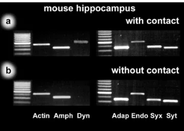
In the mouse hippocampus, all cells with established contacts exhibited mRNAs for all tested endo- and exocytic proteins (Fig.2a). The results for cells grown in isolation were similar for most of the protein mRNAs, which were abundant in these cells. In contrast, in isolated neurons without a neuronal network, we were never able to detect a signal for dynamin 1 (Fig.2b, lane 4).
The results from cultures of chick retina cells were identical to those from the mouse hippocampus. Cells with established contacts revealed signals for all tested mRNAs (Fig. 3a), whereas cells lacking contacts to neighboring cells were devoid only of dynamin 1 mRNA (Fig.3b, lane 4).
In order to determine the differential expression of dynamin isoforms 1 and 2, we investigated their presence in the mouse hippocampal system. Interestingly, we found that dynamin 2 was expressed in neurons with and without synaptic contact (Fig.4, lanes 3, 4), whereas the data for dynamin 1 was identical to that described above (Fig. 4, lanes 1, 2).
To verify the data obtained by RT-PCR at the protein level, we investigated the expression of proteins by using the immunocytochemical double-fluorescence technique. The analysis of the distribution for synaptotagmin 1 and dynamin 1 in the mouse hippocampus revealed that the pattern of immunostaining was strictly stage-dependent. Early developmental stages (DIV5) revealed an immuno-signal for synaptotagmin 1 in the cell body and neuronal Fig. 3 Single-cell RT-PCR from chick retina (lane 1 size marker).
As found in the hippocampus, we could detect beta-actin (Actin), amphiphysin 1 (Amph), and synaptotagmin 1 (Syt) in cells with con-tacts (a) and in cells without concon-tacts (b). Dynamin 1 (Dyn) could be found in neurons connected to neighboring neurons (a, lane 4) but was missing in cells without contacts (b, lane 4)
Fig. 2 Single-cell RT-PCR from mouse hippocampus (lanes 1, 5 size markers). Identified pyramidal cells from mouse hippocampal cultures were used in order to detect differences in the mRNA content between neurons with and without established contacts to neighboring neurons. Although transcripts were found for beta-actin (Actin), amphiphysin 1 (Amph), alpha-adaptin (Adap), endophilin 2 (Endo), syntaxin 1 (Syx), and synaptotagmin 1 (Syt) in cells with contacts (a) and in cells without contacts (b), dynamin 1 (Dyn) transcripts were detected in cells with neuronal contacts (a, lane 4) but were missing in cells without contacts (b, lane 4)
Fig. 4 Differential appearance of dynamin isoforms in mouse hip-pocampus (lane 1 size marker). Mouse hippocampal cultures were used to detect differences in mRNA expression between dynamin isoforms 1 (Dyn1) and 2 (Dyn2). Neurons with established contacts to neighboring neurons (+) expressed both mRNAs (lanes 2, 4). Cells without contacts (-) exhibited no transcripts for dynamin 1 (Dyn1-, lane 3), whereas transcripts for dynamin 2 (Dyn2-) were detectable (lane 5)
processes, whereas dynamin 1 was not detectable (Fig.5a, b). If cells were kept in culture for more then 2 weeks (DIV16) and developed a neuronal network, double-stain-ing showed a similar expression pattern for synaptotagmin 1 and dynamin 1 in the cell bodies and neuronal processes (Fig. 5c, d). In agreement with the data from the mouse hippocampus, we found a stage-dependent expression pat-tern of SNAP-25 and dynamin 1 in the chick retina. Again, early developmental stages (DIV6) were immunostained for SNAP-25, a protein known as t-SNARE in the pre-synaptic terminal, and were devoid of dynamin 1 (Fig.6a, b). Cells that were grown in culture for longer times (DIV15) and that had visible contacts to adjacent cells
showed equivalent immunoreactivity for SNAP-25 and dynamin 1 in the same neurons (Fig.6c, d). At the ultra-structural level, we found dynamin 1 localized throughout the presynaptic terminal in DIV 13 neurons with staining of synaptic vesicles and the plasma membrane (Fig.7).
Taken together, our results reveal that exocytic protein mRNA is present in all investigated cells at all stages. Data from the chick retina and the mouse hippocampus show that mRNA transcripts are also present for endocytic pro-teins (chick: amphiphysin 1; mouse: amphiphysin 1, alpha-adaptin, and endophilin 2) in all preparations. Dynamin 1 has however only been detected in cells with established neuronal contacts, whereas dynamin 2 has been found in Fig. 5 Immunostaining of
mouse hippocampal cultures. Mouse hippocampal cultures express synaptotagmin 1 (a) in the cell body and processes from early stages of development (DIV5 day-in-vitro 5), but no dynamin 1 (b) can be detected at this stage. Later stages (DIV16) exhibit a similar immunostain-ing for synaptotagmin 1 (c) and for dynamin 1 (d). Bars 20 μm
all the investigated cells from our mouse hippocampal cultures.
Discussion
The data from this study reveal two main aspects of neu-ronal development. On the one hand, single-cell multiplex RT-PCR has detected that all tested mRNAs, with the exception of dynamin 1, are abundant even at early stages of neuronal development. On the other hand, dynamin 1 has only been detected once neurons have established connections to neighboring cells, whereas the ubiquitously
expressed dynamin isoform 2 is detectable before synapse formation.
In addition to the active reconstruction of microfilaments (Letourneau1996; Tseng and Wirtz2004), early stages of neuronal maturation and process outgrowth are character-ized by the incorporation of newly synthescharacter-ized membrane (Ahmari et al.2000; Zakharenko and Popov2000), which is transported as so-called constitutive vesicles to the growth cone. It remains a matter of discussion whether vesicles for the enlargement of the membrane surface are identical to the synaptic vesicles that are found in mature synapses for the release of transmitters (Leoni et al.1999; Grundschober et al.2002). Our results are of relevance to Fig. 6 Immunostaining of chick
retinal cultures. Cultures taken from chick retina express SNAP-25 (a) in the cell body and processes from early stages of development (DIV6). Only faint staining of dynamin 1 (b) can be detected in cell clusters (arrow) but none in isolated neurons or growth cones at this stage. Later stages (DIV15) ex-hibit similar immunostaining for SNAP-25 (c) and for dynamin 1 (d). Bars 50 μm
this debate and to findings that have revealed the existence of spontaneous exocytosis during neurite extension along the processes (Gao and Van den Pol2000). We have found that most of the tested proteins, including the plasma membrane anchors (t-SNAREs) syntaxin 1 and SNAP-25, are expressed at these early stages, and thus it is tempting to speculate that constitutive and synaptic vesicles share a similar set of membrane proteins.
We have also found that dynamin 1 mRNA and protein are not detectable in any of the tested cells from the two studied culture systems before the establishment of synap-tic contacts. Interestingly, the results are strictly stage-re-lated (contacts to other neurons being present or absent), depending on the density of neurons, and are not time-dependent. As we have shown previously in the retino-tectal system of the chick in situ, a significant delay occurs between the onset of the expression of exocytic proteins and that of endocytic proteins (Bergmann et al.1999,2000; Grabs et al.2000). Shortly after synapse formation, how-ever, endocytic proteins are upregulated to fulfill their role in the recycling of membrane from the plasma membrane back to the presynaptic terminal (Südhof2004). We have
found, by studying single cells, that amphiphysin 1 mRNA is available in isolated neurons, but that dynamin 1 mRNA is first detectable after synaptogenesis with neighboring cells. Previous studies have shown that the blocking of dynamin by mutant isoforms or by peptides leads to an arrest in endocytosis (Shupliakov et al. 1997; Damke et al. 2001), but that membrane recycling occurs earlier (Matteoli et al.1992; Kraszewski et al.1995). Our data thus exclude that dynamin 1 is part of the machinery of mem-brane recycling during neurite growth. However, dynamin 2 is expressed before synapse formation.
The appearance of dynamin 1 mRNA and the presence of known components of endocytosis thus mark a time-point at which neurites stop growing and start recycling their plasma membrane. Our data have revealed that dy-namin 1 is specific for the regulated recycling of synaptic vesicles after synapse formation, whereas dynamin 2 may act as the constitutive counterpart of dynamin 1.
Acknowledgements The authors thank R. Jahn (Göttingen, Ger-many) and P. DeCamilli (New Haven, USA) for the generous supply of antibodies, and L. Clement, M. Kaczorowski, and C. Weber for their excellent technical assistance.
References
Ahmari SE, Buchanan J, Smith SJ (2000) Assembly of presynaptic active zones from cytoplasmic transport packets. Nat Neurosci 3:445–451
Bergmann M, Lahr G, Mayerhofer A, Gratzl M (1991) Expression of synaptophysin during the prenatal development of the rat spinal cord: correlation with basic differentiation processes of neurons. Neuroscience 42:569–582
Bergmann M, Grabs D, Rager G (1999) Developmental expression of dynamin in the chick retinotectal system. J Histochem Cyto-chem 47:1297–1306
Bergmann M, Grabs D, Rager G (2000) Expression of presynaptic proteins is closely correlated with the chronotopic pattern of axons in the retinotectal system of the chick. J Comp Neurol 418:361–372
Brose N, Petrenko AG, Südhof TC, Jahn R (1992) Synaptotagmin: a calcium sensor on the synaptic vesicle surface. Science 256: 1021–1025
Bruns D, Jahn R (2002) Molecular determinants of exocytosis. Pflügers Arch Eur J Physiol 443:333–338
Butler MH, David C, Ochoa G-C, Freyberg Z, Daniell L, Grabs D, Cremona O, De Camilli P (1997) Amphiphysin II (SH3P9; BIN1), a member of the amphiphysin/Rvs family, is concen-trated in the cortical cytomatrix of axon initial segments and nodes of Ranvier in brain and around T tubules in skeletal muscle. J Cell Biol 137:1355–1367
Catsicas S, Grenningloh G, Pich EM (1994) Nerve-terminal pro-teins: to fuse to learn. Trends Neurosci 17:368–373
Contestabile A (2000) Roles of NMDA receptor activity and nitric oxide production in brain development. Brain Res Brain Res Rev 32:476–509
Cook TA, Urrutia R, McNiven MA (1994) Identification of dynamin 2, an isoform ubiquitously expressed in rat tissues. Proc Natl Acad Sci U S A 91:644–648
Cook G, Tannahill D, Keynes R (1998) Axon guidance to and from choice points. Curr Opin Neurobiol 8:64–72
Damke H, Binns DD, Ueda H, Schmid SL, Baba T (2001) Dynamin GTPase domain mutants block endocytic vesicle formation at morphologically distinct stages. Mol Biol Cell 12:2578–2589 Fig. 7 Ultrastructural distribution of dynamin 1 in chick retinal
cul-tures. Immunoelectron microscopy reveals that dynamin 1 is lo-calized throughtout the presynaptic terminal in DIV13 neurons. Synaptic vesicles and the plasma membrane are heavily stained for dynamin 1. Bar 1 μm
Gad H, Ringstad N, Löw P, Kjaerulff O, Gustafsson J, Wenk MR, Di Paolo G, Nemoto Y, Crum J, Ellisman MH, De Camilli P, Shupliakov O, Brodin L (2000) Fission and uncoating of syn-aptic clathrin-coated vesicles are perturbed by disruption of interactions with the SH3 domain of endophilin. Neuron 27: 301–312
Gao XB, Van den Pol AN (2000) GABA release from mouse axonal growth cones. J Physiol (Lond) 523:629–637
Grabs D, Bergmann M, Schuster T, Fox PA, Brich M, Gratzl M (1994) Differential expression of synaptophysin and synapto-porin during pre- and postnatal development of the rat hip-pocampal network. Eur J Neurosci 6:1765–1771
Grabs D, Bergmann M, Rager G (2000) Developmental expression of amphiphysin in the retinotectal system of the chick: from mRNA to protein. Eur J Neurosci 12:1545–1553
Grosse G, Tapp R, Wartenberg M, Sauer H, Fox PA, Grosse J, Gratzl M, Bergmann M (1998) Prenatal hippocampal granule cells in primary cell culture form mossy fiber boutons at pyramidal cell dendrites. J Neurosci Res 51:602–611
Grosse G, Grosse J, Tapp R, Kuchinke J, Gorsleben M, Fetter I, Höhne-Zell B, Gratzl M, Bergmann M (1999) SNAP-25 re-quirement for dendritic growth of hippocampal neurons. J Neurosci Res 56:539–546
Grundschober C, Malosio ML, Astolfi L, Giordano T, Nef P, Meldolesi J (2002) Neurosecretion competence: a comprehen-sive gene expression program identified in PC12 cells. J Biol Chem 277:36715–36724
Herskovits JS, Burgess CC, Obar RA, Vallee RB (1994) Effects of mutant rat dynamin on endocytosis. J Cell Biol 122:565–578 Higgins MK, McMahon HT (2002) Snap-shots of clathrin-mediated
endocytosis. Trends Biochem Sci 27:257–263
Huang EJ, Reichardt LF (2001) Neurotrophins: roles in neuronal development and function. Annu Rev Neurosci 24:677–736 Huttner WB, Schmidt A (2000) Lipids, lipid modification and
lipid-protein interaction in membrane budding and fission—insights from the roles of endophilin A1 and synaptophysin in synaptic vesicle endocytosis. Curr Opin Neurobiol 10:543–551 Igarashi M, Kozaki S, Terakawa S, Kawano S, Ide CF, Komiya Y
(1996) Growth cone collapse and inhibition of neurite growth by Botulinum neurotoxin C1: a t-SNARE is involved in axonal growth. J Cell Biol 134:205–215
Jahn R, Lang T, Südhof TC (2003) Membrane fusion. Cell 112:519– 533
Kraszewski K, Mundigl O, Daniell L, Verderio C, Matteoli M, De Camilli P (1995) Synaptic vesicle dynamics in living cultured hippocampal neurons visualized with CY3-conjugated anti-bodies directed against the lumenal domain of synaptotagmin. J Neurosci 15:4328–4342
Leoni C, Menegon A, Benfenati F, Toniolo D, Pennuto M, Valtorta F (1999) Neurite extension occurs in the absence of regulated exocytosis in PC12 subclones. Mol Biol Cell 10:2919–2931 Letourneau PC (1996) The cytoskeleton in nerve growth cone
mo-tility and axonal pathfinding. Perspect Dev Neurobiol 4:111– 123
Matteoli M, Takei K, Perin MS, Südhof TC, De Camilli P (1992) Exo-endocytotic recycling of synaptic vesicles in developing processes of cultured hippocampal neurons. J Cell Biol 117: 849–861
McPherson PS, Garcia EP, Slepnev VI, David C, Zhang XM, Grabs D, Sossin WS, Bauerfeind R, Nemoto Y, De Camilli P (1996) A presynaptic inositol-5-phosphatase. Nature 379:353–357 Mundigl O, Ochoa G-C, David C, Slepnev VI, Kabanov AV, De
Camilli P (1998) Amphiphysin I antisense oligonucleotides inhibit neurite outgrowth in cultured hippocampal neurons. J Neurosci 18:93–103
Nakata T, Takemura R, Hirokawa N (1993) A novel member of dynamin family of GTP-binding proteins is expressed specifi-cally in the testis. J Cell Sci 105:1–5
Noakes PG, Chin D, Kim SS, Liang S, Phillips WD (1999) Ex-pression and localization of dynamin and syntaxin during neu-ral development and neuromuscular synapse formation. J Comp Neurol 410:531–540
Osen-Sand A, Catsicas M, Staple JK, Jones KA, Ayala G, Knowles J, Grenningloh G, Catsicas S (1993) Inhibition of axonal growth by SNAP-25 antisense oligonucleotides in vitro and in vivo. Nature 364:445–448
Reutens AT, Begley CG (2002) Endophilin-1: a multifunctional protein. Int J Biochem Cell Biol 34:1173–1177
Ringstad N, Nemoto Y, De Camilli P (1997) The SH3p4/Sh3p8/ SH3p13 protein family: binding partners for synaptojanin and dynamin via a Grb2-like Src homology 3 domain. Proc Natl Acad Sci U S A 94:8569–8574
Ringstad N, Nemoto Y, De Camilli P (2001) Differential expression of endophilin 1 and 2 dimers at central nervous system syn-apses. J Biol Chem 276:40424–40430
Schoch S, Deák F, Königstorfer A, Mozhayeva M, Sara Y, Südhof TC, Kavalali ET (2001) SNARE function analyzed in sy-naptobrevin/VAMP knockout mice. Science 294:1117–1122 Shupliakov O, Löw P, Grabs D, Gad H, Chen H, David C, Takei K,
De Camilli P, Brodin L (1997) Synaptic vesicle endocytosis impaired by disruption of dynamin-SH3 domain interactions. Science 276:259–263
Steiner P, Sarria J, Huni B, Marsault R, Catsicas S, Hirling H (2002) Overexpression of neuronal Sec1 enhances axonal branching in hippocampal neurons. Neuroscience 113:893
Südhof TC (2004) The synaptic vesicle cycle. Annu Rev Neurosci 27:509–547
Tseng Y, Wirtz D (2004) Dendritic branching and homogenization of actin networks mediated by arp2/3 complex. Phys Rev Lett 93:258104
Verstreken P, Kjaerulff O, Lloyd TE, Atkinson R, Zhou Y, Meinertzhagen IA, Bellen HJ (2002) Endophilin mutations block clathrin-mediated endocytosis but not neurotransmitter release. Cell 109:101–112
Walsh FS, Doherty P (1997) Neural cell adhesion molecules of the immunoglobulin superfamily: role in axon growth and guid-ance. Annu Rev Cell Dev Biol 13:425–456
Washbourne P, Thompson PM, Carta M, Costa ET, Mathews JR, Lopez-Bendito G, Molnar Z, Becher MW, Valenzuela CF, Partridge LD, Wilson MC (2002) Genetic ablation of the t-SNARE SNAP-25 distinguishes mechanisms of neuroexocy-tosis. Nat Neurosci 5:19–26
Zakharenko S, Popov S (2000) Plasma membrane recycling and flow in growing neurites. Neuroscience 97:185–194