HAL Id: hal-01280758
https://hal.archives-ouvertes.fr/hal-01280758
Submitted on 1 Mar 2016HAL is a multi-disciplinary open access archive for the deposit and dissemination of sci-entific research documents, whether they are pub-lished or not. The documents may come from teaching and research institutions in France or abroad, or from public or private research centers.
L’archive ouverte pluridisciplinaire HAL, est destinée au dépôt et à la diffusion de documents scientifiques de niveau recherche, publiés ou non, émanant des établissements d’enseignement et de recherche français ou étrangers, des laboratoires publics ou privés.
Pseudolysogeny and sequential mutations build
multiresistance to virulent bacteriophages in
Pseudomonas aeruginosa
Libera Latino, Cédric Midoux, Yolande Hauck, Gilles Vergnaud, Christine
Pourcel
To cite this version:
Libera Latino, Cédric Midoux, Yolande Hauck, Gilles Vergnaud, Christine Pourcel. Pseudolysogeny and sequential mutations build multiresistance to virulent bacteriophages in Pseudomonas aeruginosa. Microbiology, Microbiology Society, 2016, �10.1099/mic.0.000263�. �hal-01280758�
Pseudolysogeny and sequential mutations build multiresistance to virulent bacteriophages in 1
Pseudomonas aeruginosa
2
3
Libera Latino1, Cédric Midoux1, Yolande Hauck1, Gilles Vergnaud1, Christine Pourcel1# 4
1Institute for Integrative Biology (I2BC), CEA, CNRS, Univ. Paris-Sud, Université Paris-Saclay, 5
91198 Gif-sur-Yvette cedex, France 6
7
Running title: Pseudolysogeny and bacterial evolution 8
Subject category: Environmental biology 9
10
#Address correspondence to: Christine Pourcel 11
I2BC, Bât 400, Université Paris-Sud, 91405 Orsay cedex, France 12 [email protected]; [email protected] 13 Tel: +33 1 69 15 30 01 14 15
Keywords: Pseudolysogen, virulent phages, mutations, phase variation, carrier state 16
Number of words in abstract: 233 17
Number of words in main text: 7526 18
Number of tables and figures: 4 Tables, 7 figures 19
Footnote: PAO1Or has been deposited in the EMBL-EBI database under accession number 20
LN871187 21
2 22
Abstract 23
Coevolution between bacteriophages and their prey is the result of mutualistic interactions. Here we 24
show that pseudolysogeny is a frequent outcome of infection by virulent phages of Pseudomonas 25
aeruginosa, and that selection of resistant bacterial mutants is favored by continuous production of
26
phages. We investigated the frequency and characteristics of P. aeruginosa strain PAO1 variants 27
resisting infection by different combinations of virulent phages belonging to four genera. The 28
frequency of resistant bacteria was 10-5 for single phage infection and 10-6 for infections with 29
combinations of two or four phages. The genome of 27 variants was sequenced and the comparison 30
with the genome of the parental PAO1 strain allowed the identification of point mutations or small 31
indels. Four additional variants were characterized by a candidate gene approach. In total, 27 32
independent mutations were observed affecting 14 genes and a regulatory region. The mutations 33
affected genes involved in biosynthesis of type IV pilus, alginate, LPS and O-antigen. Half of the 34
variants possessed changes in homopolymer tracts responsible for frameshift mutations, and these 35
phase variation mutants were shown to be unstable. Eleven double mutants were detected. The 36
presence of free phage DNA was observed in association with exclusion of superinfection in half of 37
the variants, and in three of them no chromosomal mutation could be found. Upon further growth of 38
these pseudolysogens, some variants with new chromosomal mutations were recovered presumably 39
due to continuous evolutionary pressure. 40
INTRODUCTION 41
Pseudomonas aeruginosa is a bacterium frequently found in the environment and often associated
42
with human infections in clinical settings. This species displays an important genome plasticity due 43
in large part to horizontal gene transfer of genomic islands and mobile elements, but also to de novo 44
mutations (Spencer et al., 2003). Bacteriophages are key actors in diversification of P. aeruginosa 45
by selecting for resistant mutants, and in turn adapting to new bacterial genotypes in a coevolution 46
arm race (Brockhurst et al., 2005; Dennehy, 2012). A large variety of P. aeruginosa bacteriophages 47
has been isolated, some showing a wide host range. However, several studies have illustrated that 48
from 6 to 10% of genetically different clinical P. aeruginosa strains were not lysed by currently 49
known phages (Essoh et al., 2013). Spontaneous mutations responsible for phage resistance are 50
frequently related to alterations in the phage receptor (Hyman & Abedon, 2010; Labrie et al., 2010). 51
In P. aeruginosa, adsorption mutants are principally affected in type IV pili, often used as receptors 52
by podoviruses (-KMV) (Chibeu et al., 2009) and siphoviruses, or in lipopolysaccharide (LPS), a 53
major virulence factor (King et al., 2009; Lam et al., 2011) involved in the binding of myoviruses. 54
We and others showed that large chromosomal deletions could be selected in bacteria resistant to 55
single or multiple phages. Such deletions encompassed genes involved in fimbriae, outer membrane 56
proteins or LPS components (Latino et al., 2014; Le et al., 2014; Tanji et al., 2008). Resistance to 57
phages due to modified type IV pili has consequences on bacterial motility by affecting twitching, a 58
form of solid surface translocation (Chiang & Burrows, 2003). Type IV pili allow the adherence of 59
P. aeruginosa cells to the host epithelium, and also play a role in biofilm formation (Bucior et al.,
60
2012; Klausen et al., 2003; O'Toole & Kolter, 1998). The mechanism of resistance associated with 61
the loss of pili may drive an increase in P. aeruginosa diversity and strongly reduce infectivity 62
(Brockhurst et al., 2005; Hahn, 1997). Scanlan et al. showed that coevolution of phages and 63
bacteria leads to the emergence of a complex population of cells with mutations that sometimes 64
increase bacterial fitness but also constrain evolution (Scanlan et al., 2015). 65
It is generally accepted that the outcome of virulent bacteriophage infection is a lytic cycle leading 66
to bacterial death, whereas temperate phages can either perform a lytic cycle or lysogenize their 67
host. Pseudolysogeny is a third state, most frequently described for temperate phages as an 68
intermediate between the lytic cycle and lysogeny, allowing the bacteria to survive infection (Ripp 69
& Miller, 1997; Ripp & Miller, 1998). Pseudolysogeny was first described as an unstable 70
interaction which is not productive and eventually resolves into true lysogeny or virulent growth 71
4 (Baess, 1971). Los et al. (Los et al., 2003) demonstrated that Escherichia coli phage T4 can form 72
pseudolysogens in starved, slowly growing cells. They showed that superinfection of the host by 73
another T-even phage was responsible for lysis inhibition (LIN) (Bode, 1967) caused by the T4rI 74
gene product (Bode, 1967). Later, pseudolysogeny was defined as a stage in the bacteriophage 75
development, without multiplication of the genome, allowing subsequent restart and resumption of 76
the virus cycle (Los & Wegrzyn, 2012). In P. aeruginosa, pseudolysogeny was documented in 77
slowly growing cells with two phages responsible for generalized transduction, F116 a temperate 78
phage and UT1 a virulent phage (Ripp & Miller, 1997; Ripp & Miller, 1998). The role played by 79
pseudolysogeny in the emergence of bacterial mutants has not been demonstrated. Early work by 80
Demerec and Fano described mutants of E. coli obtained on agar medium following infection by 81
seven different phages (T1 to T7) (Demerec & Fano, 1945). The authors noted that phages were 82
present for a long time after they re-isolated resistant colonies, and finally obtained mutants 83
showing different patterns of cross-resistance. A high frequency of what were likely double-mutants 84
was observed, but the authors were not able at that time to identify the genetic changes that 85
conferred the heritable cross-resistance. 86
We wished to go further in the analysis of phage-driven P. aeruginosa evolution and investigated 87
the mechanisms by which P. aeruginosa survives infection by one or a mixture of virulent 88
bacteriophages belonging to different genera. We characterized mutations selected by phages and 89
showed that maintenance of phage DNA in pseudolysogens over many colony-purification steps 90
was a major factor in allowing selection of additional mutations. 91
METHODS 92
Bacterial strains and phages. A single colony of P. aeruginosa PAO1, a reference strain 93
originating from a patient (Stover et al., 2000) and propagated in the laboratory for several years, 94
was cultivated for storage at -80°C and for genome extraction and sequencing. This representative, 95
thereafter called PAO1Or (where Or stands for Orsay), was used to isolate phage-resistant mutants. 96
Two podoviruses, vB_PaeP_PAO1_Ab05 (Ab05) and vB_PaeP_C2-10_Ab09 (Ab09), and two 97
myoviruses, vB_PaeM_PAO1_Ab17 (Ab17) and vB_PaeM_PAO1_Ab27 (Ab27) representing four 98
different genera were used in this study, alone, or combining a podovirus with a myovirus, or in a 99
cocktail of all four phages. These phages, isolated in Abidjan (Côte d’Ivoire), have been described 100
in detail in (Essoh et al., 2015). PAO1 LPS and type IV pilus transposon mutants were obtained 101
from “The P. aeruginosa Transposon Mutant Library” (grant #NIH P30DK089507), UW Genome 102
Sciences, USA. 103
Isolation of phage-resistant bacteria. Bacteria were inoculated at a 600 nm absorbance (A600) of 104
0.01 into glass vials with aeration, containing 5 ml of Luria broth (LB) medium, and grown (37°C, 105
shaking at 180 rotations per minute (rpm)) to an A600 of 0.2. Infections were performed at a 106
multiplicity of infection (MOI) of 0.1. Infections on solid medium used a 10 μl inoculum of the 107
bacterial culture (2 x 10⁶ colony forming units (CFU)) mixed with 10 μl of a suspension, containing 108
either a single phage genera, a cocktail of two, or a cocktail of all four phages (105 plaque forming 109
units (PFU) for each phage). Ten μl of SMG (saline magnesium gelatin) phage buffer (SMG 110
comprises NaCl at 5.8 g l-1, MgSO4 at 2 g l-1, 1 M Tris-HCl, and gelatin at 0.1 g l-1 [pH 8.0]) were 111
used in negative controls. The mixture was kept for 15 min at room temperature (RT), before being 112
poured on a fresh LB agar plate (1.5% wt/vol agar) with 4 ml of soft agar (0.7% wt/vol agar) and 113
incubated at 37°C for 3 days. Because no stable resistant variants were obtained with the solid assay 114
for phage Ab27, alone or associated with Ab05, liquid infection was also performed when using 115
Ab27. Bacteria were infected during the log phase (A600 of 0.6) at an MOI of 0.001 each 24 h for a 116
total of three infections. Thereafter the surviving bacteria were plated onto LB agar plates. 117
Calculation of the frequency of resistance. An overnight culture of P. aeruginosa PAO1Or was 118
used to inoculate fresh medium to an A600 of 0.1. Bacterial cultures in the late log phase (A600 of 119
about 1, equivalent to 109 bacteria per ml, determined by titrating the bacteria), were 10-fold 120
serially diluted. One hundred µl of each dilution were mixed with 10 µl (about 106 PFU) of a single 121
6 phage suspension or a mixture of two or four phages as described above. The samples were kept for 122
15 min at RT and then poured on fresh LB agar plates using 4 ml of soft agar. Plates were inverted 123
and incubated at 37°C for 24 h. The frequency of resistance was calculated considering that all the 124
colonies growing on the plates after 24 h of incubation were resistant to phages used for the 125
infection. The divisor was the number of plated bacteria. 126
Phage susceptibility assay. Aliquots (500 µl) from the liquid culture of variants (A600 of 0.8 to 1.2) 127
were mixed with 6 ml of 0.7% wt/vol LB agar and poured onto a square LB 1.5% wt/vol agar plate. 128
Five dilutions (1010, 109, 108, 107, 106 PFU ml-1) from a progenitor stock of each phage were spotted 129
(10 µl) onto the soft agar layer, incubated at 37°C overnight, and inspected for plaque formation. 130
The resistance of the mutants against the phage was expressed as EOP (efficiency of plating) using 131
PAO1Or as a control. 132
Virucide assay. The protocol described by de Siqueira et al. (de Siqueira et al., 2006) was used to 133
prepare a virucide solution from Chinese black tea leaves. The phage-containing bacteria were 134
treated for 10 min with 3 volumes of virucide, followed by centrifugation, washing with phosphate-135
buffered-saline (PBS) and incubation at 37°C for 30 min with 50 µg ml-1 DNaseI. Then total 136
bacterial DNA was purified. 137
Adsorption assay. An overnight bacterial culture was diluted to an A600 of 0.1-0.6 and left to 138
equilibrate at 37°C. Approximately 106 phages were added to 1 ml of the diluted bacterial culture 139
(1x10⁸ to 6x10⁸ bacteria). At a fixed time point, 50 µl of the mixture were transferred to a 1.5 ml 140
conical centrifuge tube containing 940 µl of LB medium and 10 µl of chloroform. The suspension 141
was vortexed for 5 sec and centrifuged in order to pellet the phages adsorbed on the bacterial 142
surface. Then, 10 µl of the unadsorbed phage suspension was titrated. Phage adsorption was 143
expressed as the percentage of the initial amount of phage employed for the infection that did not 144
adsorb to the bacterial surface after 16 min (time necessary for adsorption of the four phages). 145
Phenotypic assays. A planktonic culture of strain P. aeruginosa PAO1Or prepared from a single 146
colony of a fresh LB agar plate was used as a reference in all experiments. To test for hemolytic 147
activity, 10 µl of an overnight culture of phage-resistant mutants (A600 of 2) were spotted onto 148
Sheep blood (5% wt/vol) agar, and plates were incubated for 24 h at 37°C. For twitching motility 149
assessment, one µl of an overnight bacterial culture (A600 of 2) was inoculated between the agar and 150
the plastic surface of LB 1.5% wt/vol agar plates. The diameter of the motility zone around the 151
inoculation site was measured after 24 h incubation at 37°C. Lipopolysaccharides (LPS) were 152
purified using the method of Hitchcock and Brown (Hitchcock & Brown, 1983). In order to 153
normalize the samples for the subsequent gel analysis, a similar amount of lyophilized bacteria was 154
disrupted in lysis buffer (Tris 1M, 2% SDS, 4% β-mercaptoethanol and 10% glycerol), prior to LPS 155
extraction. The LPS were resolved by electrophoresis on a 15% SDS-polyacrylamide gel, and the 156
band pattern was visualized using the silver staining method (Fomsgaard et al., 1990). 157
Biofilm formation. 96-wells microtiter plates (Greiner) containing LB were inoculated with an 158
overnight bacterial culture (A595 of ≈ 0.1) and incubated at 37 °C for 48 h. Before proceeding with 159
biofilm quantification, the A595 was recorded. The wells were washed three times with PBS, 200 µl 160
of 0.1% wt/vol crystal violet was added and the plate was kept for 30 min at RT. The unattached 161
crystal violet was washed three times with PBS and then the remaining biomass was quantified by 162
re-suspending it into 200 µl of absolute ethanol. The A595 was then divided by the A595 value 163
measured for planktonic bacteria in each well to account for the difference in growth rates of the 164
mutants. 165
Colony lift and hybridization. A circular Nylon N+ membrane (Nytran) was applied on the agar 166
plate on which fifty-two colonies had been plated. After 5 min, the membrane was lifted using 167
forceps and treated successively for 2 min with NaOH 0.4 N twice, Tris 1 M pH 7.5 twice, 2X 168
Saline Sodium Citrate (SSC) twice. After this, the membrane was dried on Whatman filter paper 169
and kept at 20°C until use. 170
8 Pre-hybridization was performed at 65°C for 4 h with 2 ml of hybridization buffer (Church & 171
Gilbert, 1984) per membrane. The probe was labeled using the MegaprimeTM kit (GE Healthcare 172
Amersham) and hybridization was performed overnight at 65°C in hybridization buffer. Washes 173
were done successively with 2XSSC and 0.1% wt/vol SDS, 0.5XSSC and 0.1% wt/vol SDS, 174
0.2XSSC and 0.1% wt/vol SDS. 175
DNA extraction, PCR and sequencing. PCR was performed on thermolysates or purified DNA 176
using oligonucleotides listed in Table S1. Thermolysates were produced by diluting 10 µl of 177
overnight culture in 200 µl of water and heating at 95°C for 5 min. For DNA purification, bacteria 178
were lysed in lysis buffer (Tris 10 mM, pH 7.8, EDTA 10 mM, NaCl 10 mM, SDS 0.5% wt/vol), 179
treated with proteinase K at 50 µg ml-1 for 2 h at 50°C, followed by one phenol and one chloroform 180
extraction, and ethanol precipitation. The isolates were verified for contamination from other P. 181
aeruginosa strains, commonly used in our laboratory, using PCR with oligonucleotides directed
182
against VNTRs ms216 and ms217 as previously described (Vu-Thien et al., 2007). The isolates 183
were also screened for the presence of phage DNA by PCR performed on thermolysates using the 184
specific phage oligonucleotides listed in Table S1. 185
Gene cloning and expression. PCR amplicons were cloned into the pUCP24 plasmid, a generous 186
gift of Dr. Schweizer (West et al., 1994). This is a shuttle vector which replicates in E. coli and in 187
P. aeruginosa, and contains a multiple cloning site downstream lacZ. The PAO1 mucA gene was
188
PCR-amplified using oligonucleotides mucA_Clon_F_Bam 189
5’TGGGATCCCGAGAAGCCTGACACAGC3’ and mucA_Clon_R_Hind 190
5’GAAAGCTTACCGCCATCAGGCTGCCA3’, which included restriction sites for BamHI and 191
HindIII. The amplicons were digested with BamHI and HindIII, ligated into the vector similarly
192
digested and transformed into E. coli, in which replication of pUCP24 is optimal (West et al., 193
1994). A selected recombinant was then used to transform P. aeruginosa strains by electroporation 194
using the fast protocol described by Choi et al. (Choi et al., 2006). Transformants were selected 195
using 10 µg ml-1 Gentamycin, and the presence of the plasmid was verified by PCR amplification 196
using a mucA forward oligonucleotide mucA-int_F5’ACGCAGGTAGATCGGCAGAC3’ and a 197
plasmid reverse oligonucleotide pUCP24_MCS_R 5’GGCCTCCTTCGCTATTACGCC3’. The 198
colony aspect was observed under the stereomicroscope. The transformants were then tested for 199
their susceptibility to the four bacteriophages. 200
Whole genome sequencing. Ten µg purified bacterial DNA was sent for draft whole genome 201
Illumina sequencing to the IMAGIF platform (CNRS, Gif sur Yvette, France). Libraries were made 202
from sheared fragments of DNA with a mean size of 900 bp, and 250 bp paired-end reads were 203
produced. One million up to 5 million reads were obtained corresponding to a 40-200 fold average 204
coverage. The mutations were identified by comparison with the genome of the PAO1Or sequence 205
using native GeneiousR9 tools default parameters (Biomatters, New Zealand). The Geneious 206
mapper with the “Medium-Low Sensitivity/Fast” parameter option was used to map the reads of 207
each variant against the PAO1Or genome. The “Find Variations/SNPs” analysis was used with the 208
parameter “Minimum Variant Frequency” set to 0.25. When a SNP or an indel was identified, 209
sequencing reads mapping in the mutated gene plus 1 kb on both sides were recovered, reassembled 210
and the contig was aligned with the PAO1Or genome. This allowed the precise localisation of short 211
deletions. Mutations were confirmed by PCR amplification of the affected gene and Sanger 212
sequencing (Beckman-Cogenics). 213
De novo assembly of phage reads was done with GeneiousR9 native assembler using the
Medium-214
Low Sensitivity/Fast parameter. 215
Nucleotide sequence accession number. The DNA sequence of the PAO1 strain representative 216
PAO1Or has been deposited in the EMBL-EBI database under accession number LN871187, 217
available from the European Nucleotide Archives (ENA) browser at 218
http://www.ebi.ac.uk/ena/data/view/ project PRJEB9838. 219
RESULTS 220
10 Phage-tolerant bacteria show a variety of phenotypes and phage susceptibility patterns.
221
Our goal was to evaluate the frequency and diversity of PAO1Or mutants emerging from infection 222
with phages belonging to different genera, used alone or in cocktails. We hypothesized that each 223
phage may select for specific mutations. Four different virulent phages displaying various host 224
ranges (Essoh et al., 2015) were used, alone or in combination of two or four. On PAO1Or, Ab05 225
(KMV-like phage), Ab09 (N4-like phage) and Ab17 (KPP10-like phage) produced clear plaques, 226
whereas Ab27 (PB1-like phage) produced tiny, turbid plaques. First we investigated the nature of 227
the four phages primary receptor by testing the susceptibility of two PAO1 transposon mutants, 228
affected in type IV pili (pilA mutant) or LPS-O antigen (algC mutant) synthesis genes. Ab05 was 229
not capable of growing on a type IV pilus mutant, as previously reported for most KMV-like 230
phage (Ceyssens et al., 2011) whereas growth of Ab09, Ab17 and Ab27 was restricted on the LPS 231
defective mutant. Then we designed an experimental procedure to allow for phage amplification 232
and isolation of independent resistant mutants. For this purpose, PAO1Or was infected at an MOI of 233
0.1 (one phage for ten bacteria) by single phage or cocktails, before plating the bacteria in soft agar 234
on LB solid medium. The cocktails consisted of a 1:1 mixture of Ab09 and Ab17 or Ab05 and 235
Ab27, and a 1:1:1:1 mixture of Ab09, Ab17, Ab05 and Ab27. Complete lysis was obtained in 8 h, 236
with the exception of dispersed insensitive bacteria which, after 72 h, produced colonies with 237
different morphologies (Fig. 1a). We calculated the frequency of surviving cells to be 3.2x10⁻⁵ for 238
single phage infection, 4x10⁻⁶ for double infection and 3.8x10⁻⁶ for multiple infections. Colonies of 239
variable shape, size and appearance were picked from seven independent experiments and were 240
purified by three re-isolation steps, in order to ensure that a pure population was obtained. A single 241
colony was recovered after the third re-isolation step (P3) and used to inoculate an overnight culture 242
which was then stored at -80°C in glycerol. This stock was later used for genomic DNA purification 243
and to perform further tests (Fig. 1a). The majority of PAO1Or variants recovered after Ab05, Ab09 244
and Ab17 single or multiple infections were “tolerant” to at least one phage. Tolerance is defined as 245
the capacity to survive the phage infection, whether this was due to a lack of receptor or to any 246
other mechanism. In contrast, none of the surviving bacteria recovered from infection with Ab27 247
alone turned out to be stably tolerant to Ab27. A similar observation was made by Hosseinidoust et 248
al. (Hosseinidoust et al., 2013a) who failed to isolate bacteria resistant to phage E79, another PB1-249
like phage. 250
In total, thirty-two PAO1Or variants were retained and tentatively distributed into five groups 251
according to their phage susceptibility pattern, evaluated by the efficiency of plating (Table 1 and 252
Table S2). The thirteen Group 1 variants displayed normal susceptibility only to phage Ab05. The 253
six Group 2 variants showed intermediate susceptibility patterns to the different phages. The five 254
Group 3 variants were resistant only to phage Ab05. The four Group 4 variants displayed full 255
resistance to phages Ab05 and Ab27, and reduced susceptibility to phage Ab09 and Ab17, 256
characterized by the production of small plaques instead of large, clear ones. Four variants resisting 257
all four phages constituted Group 5. In Group 2, a mucoid phenotype was stably observed for 258
PAO1-02, PAO1-06 and PAO1-13, whereas PAO1-17 continuously produced two types of colonies 259
on solid LB media, some with a smooth appearance as seen for the control PAO1Or strain, and 260
others surrounded by an irregular transparent edge (Fig. 1b). This phenotype may be related to an 261
observed decrease in swarming capacity (data not shown). In addition, mucoid colonies appeared 262
after several days of growth. When replated, the PAO1-17 colonies surrounded by a transparent 263
edge again produced both types of colonies, whereas the others stably maintained their phenotype. 264
Growth on Sheep blood agar plates showed the existence of new phenotypic characteristics for 265
some variants as compared to the parental PAO1Or strain (Fig. S1). Variants PAO1-02, PAO1-13 266
and PAO1-17 lacked the hemolytic ability displayed by the wild-type PAO1Or but, interestingly, the 267
colonies of PAO1-13 presented zones of reversion to the wild-type phenotype. 268
Unexpectedly, PCR amplification showed that phage DNA could still be detected at the P3 re-269
isolation step in fifteen of the 32 variants (Table 1). We checked whether the phage DNA was 270
12 inside the bacteria or adsorbed on the surface by treating two of the variants with a virucide (tea 271
decoction) and DNaseI digestion, followed by several washings of the bacteria pellet. Phage DNA 272
was still present in large amounts in the bacteria, as shown by semi-quantitative PCR reaction (Fig. 273
S2 shows PA01-20 and PAO1-32), suggesting that the phage genome was maintained in an 274
episomal state: lysogeny was not likely as these phages are believed to be strictly lytic, based on 275
their genome characteristics, and because the amount of phage DNA appears to be in large excess 276
over that of the bacterial DNA (see dedicated paragraph below). The results obtained suggest that 277
some bacterial cells might contain in the order of 100 phage genomes. 278
An adsorption assay was performed with the sixteen variants devoid of phage DNA showing that 279
resistance was linked to absence of phage binding to the bacterial surface (Fig. 2). In order to 280
identify the mutations conferring resistance, and to investigate in more details the variants 281
containing phage DNA, whole genome sequencing was performed on DNA extracted from 23 282
variants selected into the different groups, at the P3 purification step. 283
A wide range of chromosomal mutations is selected by phages 284
To identify de novo mutations, it was necessary to sequence the genome of the parental PAO1Or 285
strain, prepared from the culture used to derive phage-tolerant variants. The PAO1Or sequencing 286
reads were mapped against the sequence of the reference PAO1 (NC_002516) strains, allowing the 287
assembly of the full genome and identification of differences (Fig. S3 and Table S3). These 288
differences included a large inversion between rRNA sequences (positions 727 255 to 4 788 575), 289
the presence of a copy of filamentous Pf1 prophage in PAO1Or at position 5 242 103 to 5 254 164, 290
and 63 SNPs or short indels events. As expected, some of these differences, including the inversion 291
and the Pf1 prophage plus a number of the SNPs and indels were previously reported by 292
Klockgether et al. (Klockgether et al., 2010). Others were specific to the PAO1Or sub line. 293
The sequencing reads from each of the 23 whole-genome sequenced phage-tolerant variants were 294
mapped against the PAO1Or genome showing a uniform distribution with a mean coverage of 40-295
200 fold and only a few places with low coverage and relatively poor quality sequence, common to 296
all variants. In variants PAO1-30, PAO1-32 and PAO1-34, no chromosomal mutation could be 297
detected. In fourteen variants a single chromosomal mutation was identified. Six variants were 298
double mutants. Coverage at these genetic alterations was always at least 40, and up to 250 fold. 299
PAO1-02, with a mucoid phenotype, was analyzed by PCR using a candidate gene approach and 300
this allowed to find a frameshift mutation in the mucA gene. Table 2 displays the position and 301
nature of the mutations, as well as the percentage of sequencing reads containing a particular 302
mutation. In several variants, reads corresponding to both the wild-type (WT) and mutated sequence 303
could be found, indicating that the cell population was mixed. This indicates that an unstable 304
mutation has reverted back. Three different kinds of genetic alterations were found, phase variation, 305
deletions of 10 bp or more, and nucleotide substitutions. Fourteen genes were affected. The wzy 306
mutations A(7) to A(8) at position 1 976 849 as well as A(7) to A(6) at the same position, and G(6) 307
to G(5) at position 1 977 338, were found in six, one and other two mutants, respectively. Seven 308
different frameshift mutations due to a single nucleotide insertion or deletion in homopolymer tracts 309
resulted, in six cases, in early termination of protein synthesis. In the last case, PAO1-06, deletion 310
of a T in a stretch of four Ts in the mucA gene suppressed normal termination of translation 311
resulting in the production of a longer MucA protein fused with the beginning of MucB. Deletions 312
were found in three type IV pili structural genes, pilY1, pilQ and pilJ: pilY1 was missing ten bp in 313
PAO1-37 and 109 bp in PAO1-33, pilQ was missing 19 bp in PAO1-26 and 555 bp in PAO1-20, 314
whereas pilJ was missing 11 bp in PAO1-35. A 213 bp deletion was detected in the PAO1-22 algC 315
gene. The deletions either caused a frameshift and the creation of a premature stop codon, or deleted 316
an internal domain. Eight variants showed a single nucleotide substitution. All mutations were 317
confirmed by PCR amplification and Sanger sequencing. 318
The different mutations potentially affected the biosynthesis of membrane structures that participate 319
in binding of phages to their receptor. Mutations in the gene cluster regulating the production of 320
14 alginate were selected by Ab09, and could reduce the efficiency of infection of all the phages. The 321
wzy, wzz2 or wbpL genes are members of the heteropolymeric O-specific antigen (OSA)
322
biosynthesis cluster in PAO1 (Lam et al., 2011). Gene migA encodes a rhamnosyltransferase 323
involved in the LPS core capping (Poon et al., 2008), whereas wapH and dnpA are known to be 324
involved in the synthesis of LPS polysaccharide (Hansen et al., 2007; Liebens et al., 2014), and pgi 325
encodes a glycosyl transferase (Rocchetta et al., 1999). Mutations in algC affect the biosynthesis of 326
alginate, LPS and rhamnolipids, biosurfactants necessary for bacterial swarming motility and 327
biofilm formation (Olvera et al., 1999). Overall, the phage susceptibility pattern of each mutant 328
correlated well with the nature of the mutated genes. Infection with Ab09, Ab17 and Ab27 mainly 329
selected mutations in genes regulating LPS and O-antigen biosynthesis, while Ab05 selected 330
mutations in genes involved in type IV pilus synthesis. The number and variety of observed 331
mutations was very high confirming that the procedure used to isolate the variants allowed for 332
selection of independent events. 333
The observed mutations are responsible for modifying the phage receptor 334
To confirm that the observed mutations were responsible for affecting the bacteriophage receptor, 335
we investigated the phenotype of the three classes of mutants affected in type IV pilus, LPS and 336
alginate biosynthesis. The motility of the variants was evaluated by performing a twitching assay on 337
semisolid agar. Compared to the PAO1Or control, the diameter of the twitching zone was 338
significantly reduced in all variants, but the strongest effect was observed with those bearing a 339
mutation in Pil genes and/or resisting Ab05 infection (Fig. 3). PAO1-32 and PAO1-34 were also 340
affected in twitching although no Pil mutations could be observed, but this was likely related to a 341
continuous cell death due to phage production. Indeed upon culture in LB broth the cells lysed 342
totally after reaching an absorbance at 600 nm (A600) of 0.8. Inhibition of twitching was 343
accompanied by a decrease in biofilm formation, except for PAO1-20 and PAO1-06 (Fig. 4). This 344
may be due to the existence of a mixed population of bacteria in these variants. The LPS were 345
extracted in PAO1-04 altered in wzy and migA, and PAO1-07 altered in wzy, and were analysed by 346
polyacrylamide gel electrophoresis. Fig. 5 displays the banding profiles compared to that of 347
PAO1Or, showing absence of the A- and B- bands, as well as modifications in the proportion of core 348
and core +1 bands. PAO1-04 possessed only the core +1 oligosaccharide form. In contrast, variant 349
PAO1-07 possessed both bands in equal amounts, whereas PAO1Or had a small proportion of core + 350
1. Absence of core oligosaccharide in PAO1-04 was likely a consequence of the mutation in migA. 351
Finally to confirm that the observed mucA mutations were responsible for the mucoid phenotype, 352
we tested whether the mutants could be complemented by the WT gene. A full mucA amplicon was 353
cloned into an expression vector which was then introduced into 02, 06 and PAO1-354
13. In the three cases the transformants were no longer showing a mucoid appearance, whereas the 355
vector alone was not reversing the mucoid phenotype. In addition, the mucA transformants 356
recovered normal susceptibility to all phages. 357
Persistence of phage DNA in pseudolysogens 358
In eleven variants, phage DNA represented a proportion of 2 to 85% of sequencing reads. The very 359
high proportion of phage DNA in some samples could only be explained by the presence of free 360
phages, inside bacteria, and/or attached to cells. To confirm that phage DNA was present inside 361
bacteria, we performed another genome sequencing of PAO1-17 and PAO1-20 at the P3 362
purification step, after treatment of the bacterial pellet with DNaseI and the virucide, followed by 363
three washing steps. The results showed that 1.6% of reads still corresponded to Ab09 in PAO1-17, 364
and 52% to Ab17 in PAO1-20, similarly to what was observed in the first sequencing analysis 365
(Table 2). A search for hybrid reads between phage and bacteria genomes did not bring any 366
significant result, indicating that the phage DNA was not inserted into the bacterial chromosome, 367
and therefore we refer to these bacteria as pseudolysogens. Interestingly, the three variants in which 368
no chromosomal mutation could be found, PAO1-30, PAO1-32 and PAO1-34, possessed large 369
amounts of phage DNA: PAO1-30 immune to Ab05, Ab17 and Ab27 contained high levels of Ab05 370
16 DNA; PAO1-32 and PAO1-34, immune to Ab09, Ab17 and Ab27, contained Ab27 DNA. 371
Intermediate resistance profiles observed in some variants were correlated with the existence of a 372
mixed population of wild type and mutated bacteria, and with the presence of phage DNA. 373
In the sequenced samples in which a high proportion of sequence reads derived from phage DNA 374
were present, it was possible to assemble the full phage genome sequence. This led to the 375
identification of several single nucleotide differences in tail fiber genes, as compared to the parental 376
genotype. In three pseudolysogens obtained independently, an Ab05 tail fiber gene displayed two 377
SNPs. By PCR and sequencing, we could also observe these SNPs in a fraction of the phages used 378
to select for resistant bacteria (Fig. S4). Similarly a single SNP was observed in an Ab17 tail fiber 379
gene from variants PAO1-20 and PAO1-22, and in the ribosome binding site of an Ab27 tail gene 380
from variants PAO1-24, PAO1-32 and PAO1-34. This might reflect the selection of phage variants 381
by strain PAO1Or, possibly affecting the capacity of the bacteria to resist phages. However, we 382
could not see any differences in binding to the host or plating efficiency with these phage 383
genotypes, as compared to the parental genotype. 384
Stability of the pseudolysogen state 385
Viable phages were released by pseudolysogens, sometimes at high titers, during overnight culture 386
in LB medium. This suggested that a portion of the bacterial population could achieve a productive 387
viral cycle. To evaluate the dynamics inside pseudolysogen colonies, we measured the percentage 388
of bacteria containing phage DNA and producing viable phages, and evaluated for how long phage 389
DNA was maintained. For this purpose, 1 µl of some bacterial strains from the frozen P3 stock were 390
spread on LB agar (P30). Fifty-two colonies were picked and deposited successively on an LB agar 391
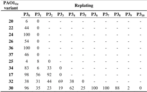
plate, then on an LB agar plate covered with a lawn of soft agar containing PAO1Or (see PAO1-30 392
replatings as an example in Fig. 6). After incubation at 37°C for 24 h, a lysis zone could be seen 393
around some colonies on the lawn of PAO1Or. One such phage-producing colony from the LB agar 394
plate was streaked onto a new LB plate and the procedure was repeated. The fraction of 395
pseudolysogenic cells contained in a single colony varies from 4 up to 100 percent. In PAO1-30 the 396
pseudolysogenic state was observed up to 10 replatings (Table 3). Hybridization with phage DNA 397
probes in a colony lift experiment confirmed that bacteria not releasing phages were devoid of 398
phage DNA, thus excluding the presence of colonies able to maintain phage DNA without releasing 399
functional phage particles (data not shown). 400
The presence of phage DNA and phage particles in important amounts up to ten colony replatings, 401
and of bacteria devoid of phages, is in agreement with a model of simultaneous and independent 402
lysis of some infected cells, random production of cured progeny from pseudolysogens, and further 403
amplification of phages by infection of these phage-free bacteria. Interestingly, PAO1-30, which 404
kept phage-producing cells for the longest time, showed peaks of phage abundance, reflecting a 405
classical equilibrium between phage production and bacteria predation (Table 3). 406
Continuous evolution of bacteria from pseudolysogens 407
A mixture of bacterial WT and mutant reads was clearly observed in PAO117, 22, 24, 26, 33, -408
37, after three purification steps, always accompanied by phage DNA (Table 2). This, added to the 409
high frequency of double mutants, suggested that immunity provided by the phage in a 410
pseudolysogenic state allowed survival and subsequent emergence of mutations. To investigate this 411
hypothesis, we tested whether new mutations would appear in response to the pressure imposed by 412
phages. We went back to the -80°C stocks of seven variants (P3), isolated new colonies, replated 413
them and tested for the presence of phage DNA by PCR, until a colony devoid of phage DNA was 414
obtained (Table 4). Susceptibility to the four phages was evaluated in these cured colonies, and the 415
mutations previously identified by whole genome sequencing were searched by PCR and Sanger 416
sequencing. Different situations existed when phage DNA was no longer present. The Group 3 417
PAO126 variant pilQ microdeletion was found in about two thirds of the colonies reisolated after -418
80°C storage, and it was associated with resistance to Ab05. Similarly, upon re-isolation of PAO1-419
37, about 50% of colonies were stable double wzy-pilY1 mutants, devoid of Ab05 and displaying 420
18 resistance to Ab05 and Ab27. In other variants, the phage susceptibility profile changed when 421
additional colony re-isolation steps were performed, and new mutations could be found upon 422
sequencing candidate genes (Table 4). In the mucoid variant PAO1-17_1 devoid of phage Ab09, a 423
new mucA frameshift mutation (a deletion of a single C in a stretch of five Cs present in the WT 424
strain) was identified in about half of the sequenced PCR products, resulting in superimposition of 425
two sequencing profiles (Fig. S5a). PAO1-20_1 and PAO1-22_1 acquired additional mutations in 426
wzy, providing resistance to LPS-dependent phages. PAO1-24_1, devoid of Ab27 DNA was shown
427
to resist all four phages whereas the PAO1-24 progenitor was susceptible to both Ab09 and Ab17 428
(Table 1). The original pilR mutation in PAO1-24 (Table 2) was confirmed through PCR and DNA 429
sequencing. Surprisingly, sequencing of a wzy PCR amplicon showed that the original insertion of 430
an additional A in a stretch of seven As residues in the WT wzy gene was replaced by a deletion of 431
one A, resulting in a frameshift and early stop. Similarly to the mucA mutation in PAO1-17_1, the 432
sequencing profile showed the superimposition of a wild type and mutated profile (Fig. S5b). 433
PAO1-25_1 and PAO1-36_1, devoid of Ab05,were sequenced, and mutations were found in pilR, 434
and in wzy and pilC, respectively. All the new mutations were confirmed by Sanger sequencing of 435
the PCR amplification products. 436
Colony re-isolation was also performed for the three pseudolysogens for which no chromosomal 437
mutation could be observed, PAO1-30 (Ab05 infection), -32, -34 (Ab05 and Ab27 co-infection) 438
(Table 1). PAO1-30_1 devoid of Ab05 still resisted Ab05. Three genes involved in type IV pilus 439
assembly were PCR-analysed in a candidate gene approach, and a new pilQ mutation was identified 440
showing a substitution of a T by a G causing a threonine to proline mutation (Table 4). In contrast, 441
PAO1-32_1 and PAO1-34_1, devoid of Ab27 DNA, recovered full susceptibility to all phages, and 442
Ab27-resistant mutants were not obtained. This confirmed that Ab27 conferred the observed 443
superinfection exclusion in the P3 variant and that it was not selecting mutants on both solid and 444
liquid media. 445
In summary, it appeared that pseudolysogenic colonies continuously evolved due to the production 446
of new functional phage particles that selected for new phage-resistant variants. Eventually, all 447
variants possessed mutations in one of the pilus type IV assembly genes, and, as expected, the 448
ability of phages to adsorb on their surface (Fig. S6) and the twitching motility of these variants 449
were defective when compared to the control PAO1Or (Fig. S7). 450
DISCUSSION 451
Pseudolysogeny is a major factor in selection of mutants 452
In our experimental model, pseudolysogeny appears to be a frequent outcome of infection by the 453
four virulent phages, providing immunity to the bacteria, and allowing emergence of mutations in 454
genes involved in receptor synthesis. In the present investigation, we might even underestimate the 455
frequency of pseudolysogeny as we started the analyses after three replatings for purification 456
purposes. The frequency of single mutants was on the order of one per 105 plated bacteria but, 457
surprisingly, we observed that double mutants could be recovered at a frequency of 10-6, which is 458
far higher than expected if these were present at the onset of infection. We show that selection of a 459
second mutation takes place in pseudolysogenic colonies that can constitute a reservoir for 460
bacteriophages exerting a permanent pressure on the bacteria. Many controlled studies have 461
demonstrated the role of starvation and slow growth in the establishment of pseudolysogeny. In 462
contrast, pseudolysogeny in rich medium is not understood (Los & Wegrzyn, 2012; Ripp & Miller, 463
1998). Being in the inner part of a colony might mimic starvation and slow growth conditions, 464
whereas cells in direct contact with the agar medium would be in a rich medium context. 465
We observe that pseudolysogeny is established in a situation when the large majority of bacteria has 466
been lysed and high amounts of phages are present, thus resembling the LIN control observed in T4. 467
We propose a model in which a pseudolysogenic cell, which may contain more than 100 phage 468
genome copies according to the phage burst size, forms, after several rounds of division, a colony 469
containing bacteria cured of the phage and bacteria in which the phage lytic cycle is resumed, 470
20 producing new phages (Fig. 7). The cured bacteria become prey for further amplification and 471
production of new pseudolysogens in which phage growth is stalled. This interaction between 472
phages and bacteria is reminiscent of the carrier state life cycle (CSLC) observed in different 473
systems (Siringan et al., 2014). However, in the present study the phage/host equilibrium is not 474
stable. The appearance of pseudolysogenic cells could occur when the amount of bacteriophages 475
and resistant mutants is higher than the total amount of WT susceptible bacteria allowing 476
bacteriophages to be protected against extinction. The relative efficiency of reactivation of the 477
phage cycle and production of cured bacteria determines the duration of the pseudolysogeny stage. 478
It will be interesting to perform in situ analyses to check whether the colony is a homogenous 479
population of cells or if there are sectors in which phage activation is favored, and to follow 480
fluctuations of free phage concentrations within a single colony. 481
A lack of immunity to superinfection, mediated by immunity genes in temperate phages is supposed 482
to differentiate true lysogeny from pseudolysogeny (Wommack & Colwell, 2000). The present 483
pseudolysogens demonstrate inhibition of superinfection by the same phage and, more interestingly, 484
by phages of different genera, which bind to different receptors. Immunity genes have been found 485
in E. coli T4 (imm) (Lu & Henning, 1989) and P1 (sim) (Maillou & Dreiseikelmann, 1990) 486
bacteriophages. The genes appear to be involved in the successful injection of phage DNA into the 487
cell. This mechanism could account for inhibition of phage infection by phages using different 488
receptors, but there is no evidence of such genes in our phages at the current time. Further 489
experiments are needed to understand at which stage phage multiplication is inhibited. 490
Red Queen dynamics/arm race coevolution 491
Studies performed in chemostats have addressed the coevolution dynamics of phage and bacteria in 492
controlled growth conditions (Betts et al., 2014; Buckling & Rainey, 2002). In some assays where 493
prey and predators are left to evolve for a long time two possible outcomes were described. In the 494
Arms race, the fittest genotype survives and this limits the diversity, whereas in the Red Queen 495
dynamics, frequency-dependent selection leads to constant production of new mutants, thus 496
maintaining diversity (Dennehy, 2012). In our assay, which takes place in a micro community, after 497
several rounds of coevolution, the population of free phages fluctuates, to the extent that they may 498
seem to almost disappear within the colony. A large diversity of resistant mutants is selected, and 499
eventually the colony will be phage-free. Reversion to WT phenotype is observed for alginate and 500
LPS mutants so that new preys will emerge. We observed, with three phages, the presence of new 501
phage genotypes in pseudolysogens, all three showing one or two SNPs in a tail fiber gene. The 502
mutations were present in a subpopulation of phages used to derive the resistant mutants, and may 503
have been selected during coevolution of phages and bacteria. No particular behavior of these 504
phages as compared to the parental ones could be demonstrated, such as plaque morphology, and 505
growth characteristics. However it is possible that these phages are capable of inducing a 506
pseudolysogenic stage at a higher frequency as compared to the ancestral phage. Our results 507
confirm that success in infection is not sufficient for phage survival, as phages are dependent upon 508
the survival of their host population (Chaturongakul & Ounjai, 2014), and therefore phage-host 509
relationships can be seen as not merely parasitic but as mutualistic (Williams, 2013). 510
Cross-resistance and reversibility of mutants 511
We showed that mutations selected by phages were often frameshift mutations known as phase 512
variation (Henderson et al., 1999). Frameshift mutations, due to variation in poly(A), poly(G), or 513
poly(T) stretches have been described in several bacterial genes as an adaptation mechanism to 514
different environmental conditions, and are reversible when the selective pressure is no longer 515
applied (Segura et al., 2004). Natural mutations of mucA observed in strains isolated from cystic 516
fibrosis patients were phase variation mutations (Spencer et al., 2003), or other frameshift mutations 517
(Pulcrano et al., 2012), also resulting in truncated proteins as seen in PAO1-02. 518
Interestingly, many of the mutations identified in this study occur in the wzy/wzx-dependent 519
pathway responsible for the synthesis of LPS O-antigen (Islam & Lam, 2014), and they are either 520
22 single nucleotide indels or mutations. LPS is composed of a lipid A membrane anchor, a core 521
oligosaccharide linker, and a distal polysaccharide termed O-antigen, in the form of A and B bands 522
(Taylor et al., 2013). Both WT and mutant forms of wzy and mucA genes were simultaneously 523
found in the presently described mutants, suggesting that the mutation can reverse at a high rate. 524
Constant variations in LPS and alginate biosynthesis pathways may help P. aeruginosa face 525
aggressions or environmental changes. This might be one explanation for the “colonial 526
dissociation” frequently observed with P. aeruginosa, characterized by colonial differences of a 527
single strain (Zierdt & Schmidt, 1964). 528
The different assays show that, depending on the bacteriophage used, the selected mutants, obtained 529
at a high frequency, display a large variety of phenotypic changes related to membrane permeability 530
and cell motility. Hosseinidoust et al. (Hosseinidoust et al., 2013a) described such phenotypes 531
induced by two phages which use type IV pilus and LPS as receptors, but they could not identify 532
the mutations. Phenotypic changes can alter bacterial virulence (Lyczak et al., 2000). Indeed, we 533
show that phage Ab09 often selects for mutants with a mucoid phenotype, probably related to an 534
increased capability to produce alginates. In the context of cystic fibrosis infection, mucoidy favors 535
the formation of protected colonies with increased resistance to opsonization, phagocytosis and 536
destruction by antibiotics (Pritt et al., 2007). It has been shown that alterations of a single band or 537
both bands of the O-antigen of P. aeruginosa PAO1 can give rise to mutants with increased 538
cytotoxicity mediated by the type III secretion system (TTSS) (Augustin et al., 2007). In addition, 539
changes in O-polysaccharide expression in PAO1 affects the size and protein content of outer 540
membrane vesicles and the formation of a robust biofilm (Murphy et al., 2014). 541
A total of 25 components are involved in the type IV pilus biogenesis (Kim et al., 2006). In the 542
present small scale investigation we observed ten mutations affecting five genes. Half of the 543
mutations are irreversible deletions which contrast with the high frequency of reversible phase 544
variation mutations in LPS. This suggests that the fitness cost of such mutants would rapidly lead to 545
their elimination, and that phages using type IV pili as receptors should be favored for phage 546
therapy. Several studies have investigated the effect of type IV pilus mutations and phage 547
resistance. Interestingly, phage F6, a dsRNA cystovirus of Pseudomonas syringae pathovar 548
phaseolicola selects for several types of mutants that differ in the number of type IV pili expressed
549
per cell, but none of the mutated genes were known to be directly involved in type IV pilus 550
expression (Sistrom et al., 2015). 551
Phage therapy is considered as a promising approach to fight against antibiotic resistant strains 552
(Abedon et al., 2011). Either readymade cocktails or ”sur- measure” phages will be used to treat 553
patients, similarly to what is still done in several countries of Eastern Europe. It is important to 554
investigate the risks linked to the use of phages, particularly in the selection of bacterial mutants 555
that could show deleterious characteristics (Hosseinidoust et al., 2013b), or drive the expression of 556
undesirable bacterial virulence factors (Olszak et al., 2015). In a mouse model of E. coli gut 557
infection, it was proposed that virulent phages remained inside bacteria in a pseudolysogenic state, 558
therefore becoming resistant to degradation and allowing persistence of bacteria (Maura & 559
Debarbieux, 2012; Maura et al., 2012). It would be interesting to know whether new variants 560
emerge in such experiments. On the other hand, some phages driving evolution toward loss of 561
virulence could be favored if they exist (Leon & Bastias, 2015). Another concern is the potential 562
role of bacteriophages in horizontal transfer, which could be favored by the long-term maintenance 563
of phage genomes inside the bacteria during pseudolysogeny. Additional experiments are needed to 564
further investigate the fate of the phages and bacteria in a pseudolysogen interaction. 565
ACKNOWLEDGEMENTS 566
The research was funded by a grant from Direction Générale de l’Armement (DGA) through 567
Agence Nationale de la Recherche (ANR, France) “Resisphage” ANR-13-ASTRID-0011-01. LL 568
was the recipient of a doctoral fellowship from DGA and IDEX, Paris-Saclay University. We are 569
24 grateful to Michael DuBow for fruitful discussions and valuable criticism of the manuscript. We 570
thank Hoang Vu-Thien for his advices as well as Marie-Agnès Petit for her helpful suggestions. We 571
thank Simone Séror and Barry Holland for their expert advices in the study of swarming. We are 572
very grateful to the anonymous reviewers for their comments which helped improving the 573
manuscript. We thank the LPS-BioSciences staff for help in analyzing LPS. This work has 574
benefited from facilities and expertise of the high throughput sequencing platform of I2BC. 575
REFERENCES 576
Abedon, S. T., Kuhl, S. J., Blasdel, B. G. & Kutter, E. M. (2011). Phage treatment of human infections. 577
Bacteriophage 1, 66‐85. 578
Augustin, D. K., Song, Y., Baek, M. S., Sawa, Y., Singh, G., Taylor, B., Rubio‐Mills, A., Flanagan, J. L., 579
Wiener‐Kronish, J. P. & other authors (2007). Presence or absence of lipopolysaccharide O antigens affects 580
type III secretion by Pseudomonas aeruginosa. J Bacteriol 189, 2203‐2209. 581
Baess, I. (1971). Report on a pseudolysogenic mycobacterium and a review of the literature concerning 582
pseudolysogeny. Acta Pathol Microbiol Scand B Microbiol Immunol 79, 428‐434. 583
Betts, A., Kaltz, O. & Hochberg, M. E. (2014). Contrasted coevolutionary dynamics between a bacterial 584 pathogen and its bacteriophages. Proc Natl Acad Sci U S A 111, 11109‐11114. 585 Bode, W. (1967). Lysis inhibition in Escherichia coli infected with bacteriophage T4. J Virol 1, 948‐955. 586 Brockhurst, M. A., Buckling, A. & Rainey, P. B. (2005). The effect of a bacteriophage on diversification of 587 the opportunistic bacterial pathogen, Pseudomonas aeruginosa. Proc Biol Sci 272, 1385‐1391. 588
Bucior, I., Pielage, J. F. & Engel, J. N. (2012). Pseudomonas aeruginosa pili and flagella mediate distinct 589
binding and signaling events at the apical and basolateral surface of airway epithelium. PLoS Pathog 8, 590
e1002616. 591
Buckling, A. & Rainey, P. B. (2002). Antagonistic coevolution between a bacterium and a bacteriophage. 592 Proc Biol Sci 269, 931‐936. 593 Ceyssens, P. J., Glonti, T., Kropinski, N. M., Lavigne, R., Chanishvili, N., Kulakov, L., Lashkhi, N., Tediashvili, 594 M. & Merabishvili, M. (2011). Phenotypic and genotypic variations within a single bacteriophage species. 595 Virol J 8, 134. 596
Chaturongakul, S. & Ounjai, P. (2014). Phage‐host interplay: examples from tailed phages and Gram‐ 597 negative bacterial pathogens. Front Microbiol 5, 442. 598 Chiang, P. & Burrows, L. L. (2003). Biofilm formation by hyperpiliated mutants of Pseudomonas aeruginosa. 599 J Bacteriol 185, 2374‐2378. 600 Chibeu, A., Ceyssens, P. J., Hertveldt, K., Volckaert, G., Cornelis, P., Matthijs, S. & Lavigne, R. (2009). The 601
adsorption of Pseudomonas aeruginosa bacteriophage phiKMV is dependent on expression regulation of 602
type IV pili genes. FEMS Microbiol Lett 296, 210‐218. 603
Choi, K. H., Kumar, A. & Schweizer, H. P. (2006). A 10‐min method for preparation of highly 604
electrocompetent Pseudomonas aeruginosa cells: application for DNA fragment transfer between 605 chromosomes and plasmid transformation. J Microbiol Methods 64, 391‐397. 606 Church, G. M. & Gilbert, W. (1984). Genomic sequencing. Proc Natl Acad Sci U S A 81, 1991‐1995. 607 de Siqueira, R. S., Dodd, C. E. & Rees, C. E. (2006). Evaluation of the natural virucidal activity of teas for use 608 in the phage amplification assay. Int J Food Microbiol 111, 259‐262. 609 Demerec, M. & Fano, U. (1945). Bacteriophage‐Resistant Mutants in Escherichia coli. Genetics 30, 119‐136. 610
Dennehy, J. J. (2012). What Can Phages Tell Us about Host‐Pathogen Coevolution? Int J Evol Biol 2012, 611
396165. 612
Essoh, C., Blouin, Y., Loukou, G., Cablanmian, A., Lathro, S., Kutter, E., Thien, H. V., Vergnaud, G. & 613
Pourcel, C. (2013). The Susceptibility of Pseudomonas aeruginosa Strains from Cystic Fibrosis Patients to 614
Bacteriophages. PLoS One 8, e60575. 615
Essoh, C., Latino, L., Midoux, C., Blouin, Y., Loukou, G., Nguetta, S. P., Lathro, S., Cablanmian, A., Kouassi, 616
A. K. & other authors (2015). Investigation of a Large Collection of Pseudomonas aeruginosa 617
Bacteriophages Collected from a Single Environmental Source in Abidjan, Cote d'Ivoire. PLoS One 10, 618 e0130548. 619 Fomsgaard, A., Freudenberg, M. A. & Galanos, C. (1990). Modification of the silver staining technique to 620 detect lipopolysaccharide in polyacrylamide gels. J Clin Microbiol 28, 2627‐2631. 621 Hahn, H. P. (1997). The type‐4 pilus is the major virulence‐associated adhesin of Pseudomonas aeruginosa‐‐ 622 a review. Gene 192, 99‐108. 623 Hansen, S. K., Haagensen, J. A., Gjermansen, M., Jorgensen, T. M., Tolker‐Nielsen, T. & Molin, S. (2007). 624
Characterization of a Pseudomonas putida rough variant evolved in a mixed‐species biofilm with 625 Acinetobacter sp. strain C6. J Bacteriol 189, 4932‐4943. 626 Henderson, I. R., Owen, P. & Nataro, J. P. (1999). Molecular switches‐‐the ON and OFF of bacterial phase 627 variation. Mol Microbiol 33, 919‐932. 628 Hitchcock, P. J. & Brown, T. M. (1983). Morphological heterogeneity among Salmonella lipopolysaccharide 629 chemotypes in silver‐stained polyacrylamide gels. J Bacteriol 154, 269‐277. 630 Hosseinidoust, Z., Tufenkji, N. & van de Ven, T. G. (2013a). Predation in homogeneous and heterogeneous 631
phage environments affects virulence determinants of Pseudomonas aeruginosa. Appl Environ Microbiol 632
79, 2862‐2871. 633
Hosseinidoust, Z., van de Ven, T. G. & Tufenkji, N. (2013b). Evolution of Pseudomonas aeruginosa 634 virulence as a result of phage predation. Appl Environ Microbiol 79, 6110‐6116. 635 Hyman, P. & Abedon, S. T. (2010). Bacteriophage host range and bacterial resistance. Adv Appl Microbiol 636 70, 217‐248. 637 Islam, S. T., Huszczynski, S. M., Nugent, T., Gold, A. C. & Lam, J. S. (2013). Conserved‐residue mutations in 638
Wzy affect O‐antigen polymerization and Wzz‐mediated chain‐length regulation in Pseudomonas 639
aeruginosa PAO1. Sci Rep 3, 3441. 640
Islam, S. T. & Lam, J. S. (2014). Synthesis of bacterial polysaccharides via the Wzx/Wzy‐dependent 641
pathway. Can J Microbiol 60, 697‐716. 642
Kaluzny, K., Abeyrathne, P. D. & Lam, J. S. (2007). Coexistence of two distinct versions of O‐antigen 643 polymerase, Wzy‐alpha and Wzy‐beta, in Pseudomonas aeruginosa serogroup O2 and their contributions to 644 cell surface diversity. J Bacteriol 189, 4141‐4152. 645 Kim, K., Oh, J., Han, D., Kim, E. E., Lee, B. & Kim, Y. (2006). Crystal structure of PilF: functional implication 646 in the type 4 pilus biogenesis in Pseudomonas aeruginosa. Biochem Biophys Res Commun 340, 1028‐1038. 647 King, J. D., Kocincova, D., Westman, E. L. & Lam, J. S. (2009). Review: Lipopolysaccharide biosynthesis in 648 Pseudomonas aeruginosa. Innate Immun 15, 261‐312. 649
Klausen, M., Heydorn, A., Ragas, P., Lambertsen, L., Aaes‐Jorgensen, A., Molin, S. & Tolker‐Nielsen, T. 650
(2003). Biofilm formation by Pseudomonas aeruginosa wild type, flagella and type IV pili mutants. Mol 651
Microbiol 48, 1511‐1524. 652
Klockgether, J., Munder, A., Neugebauer, J., Davenport, C. F., Stanke, F., Larbig, K. D., Heeb, S., Schock, 653
U., Pohl, T. M. & other authors (2010). Genome diversity of Pseudomonas aeruginosa PAO1 laboratory 654 strains. J Bacteriol 192, 1113‐1121. 655 Labrie, S. J., Samson, J. E. & Moineau, S. (2010). Bacteriophage resistance mechanisms. Nat Rev Microbiol 656 8, 317‐327. 657
Lam, J. S., Taylor, V. L., Islam, S. T., Hao, Y. & Kocincova, D. (2011). Genetic and Functional Diversity of 658
Pseudomonas aeruginosa Lipopolysaccharide. Front Microbiol 2, 118. 659
Latino, L., Essoh, C., Blouin, Y., Vu Thien, H. & Pourcel, C. (2014). A novel Pseudomonas aeruginosa 660
Bacteriophage, Ab31, a Chimera Formed from Temperate Phage PAJU2 and P. putida Lytic Phage AF: 661
Characteristics and Mechanism of Bacterial Resistance. PLoS One 9, e93777. 662
Le, S., Yao, X., Lu, S., Tan, Y., Rao, X., Li, M., Jin, X., Wang, J., Zhao, Y. & other authors (2014). 663
Chromosomal DNA deletion confers phage resistance to Pseudomonas aeruginosa. Sci Rep 4, 4738. 664