HAL Id: hal-01462139
https://hal.archives-ouvertes.fr/hal-01462139
Submitted on 8 Feb 2017
HAL is a multi-disciplinary open access
archive for the deposit and dissemination of
sci-entific research documents, whether they are
pub-lished or not. The documents may come from
teaching and research institutions in France or
abroad, or from public or private research centers.
L’archive ouverte pluridisciplinaire HAL, est
destinée au dépôt et à la diffusion de documents
scientifiques de niveau recherche, publiés ou non,
émanant des établissements d’enseignement et de
recherche français ou étrangers, des laboratoires
publics ou privés.
Sustainable low temperature preparation of
Mn3–xCoxO4 (0
≤ x < 3) spinel oxide colloidal
dispersions used for solar absorber thin films
Guillaume Salek, Pascal Dufour, Sophie Guillemet-Fritsch, Christophe
Tenailleau
To cite this version:
Guillaume Salek, Pascal Dufour, Sophie Guillemet-Fritsch, Christophe Tenailleau. Sustainable low
temperature preparation of Mn3–xCoxO4 (0
≤ x < 3) spinel oxide colloidal dispersions used for
solar absorber thin films. Materials Chemistry and Physics, Elsevier, 2015, vol. 162, pp. 252-262.
�10.1016/j.matchemphys.2015.05.065�. �hal-01462139�
To link to this article : DOI : 10.1016/j.matchemphys.2015.05.065
URL :
http://dx.doi.org/10.1016/j.matchemphys.2015.05.065
O
pen
A
rchive
T
OULOUSE
A
rchive
O
uverte (
OATAO
)
OATAO is an open access repository that collects the work of Toulouse researchers and
makes it freely available over the web where possible.
This is an author-deposited version published in :
http://oatao.univ-toulouse.fr/
Eprints ID : 16790
To cite this version :
Salek, Guillaume and Dufour, Pascal and
Guillemet-Fritsch, Sophie and Tenailleau, Christophe Sustainable
low temperature preparation of Mn3−xCoxO4 (0 ≤ x < 3) spinel
oxide colloidal dispersions used for solar absorber thin films.
(2015) Materials Chemistry and Physics, vol. 162. pp. 252-262.
ISSN 0254-0584
Any correspondence concerning this service should be sent to the repository
administrator:
staff-oatao@listes-diff.inp-toulouse.fr
Sustainable low temperature preparation of Mn
3!x
Co
x
O
4
(0 " x < 3)
spinel oxide colloidal dispersions used for solar absorber thin films
G. Salek, P. Dufour, S. Guillemet-Fritsch, C. Tenailleau
*Centre Interuniversitaire de Recherche et d'Ing!enierie des MAT!eriaux (CIRIMAT), UMR CNRS 5085, Universit!e de Toulouse e UPS, 118 Route de Narbonne, 31062 Toulouse Cedex 09, France
h i g h l i g h t s
g r a p h i c a l
a b s t r a c t
# Sustainable low temperature syn-thesis of oxide nanoparticles. # Stabilization of colloidal dispersions
free of organic precursors or surfactants.
# Influence of hydrodynamic parame-ters and physicochemical properties. # Thin films preparation by the dip-coating method and their optical properties.
# Promising method for enhanced ef-ficiency in the preparation and use of nanomaterials. Keywords: Oxides Precipitation Thin films Optical properties
a b s t r a c t
The preparation of pure crystalline oxide nanoparticles (with controlled composition, size and shape) and formation of stable suspensions free of complex organic precursors was developed and optimized at room temperature (or below 100$C). This reproducible water and ethanol synthesis and solution sta-bilization of oxide nanoparticles is based on Mn3!xCoxO4(0 " x < 3) composition materials. To our
knowledge, this is the first study on the complete MneCoeO spinel system synthesized at low tem-perature. The main hydrodynamic parameters, as well as the physical and chemical properties that control the oxide precipitation and nanoparticle size and morphology were characterized in detail for the family end member Mn3O4and used for the other compositions. X-ray diffraction and Scanning Electron
Microscopy images showed the influence of the alkaline solution concentration, pH, temperature and solvent on the nanoparticles properties. Neutron diffraction was used for determining the cationic dis-tribution in two compositions, i.e. CoMn2O4and MnCo2O4. While the tetrahedral site is mainly occupied
by Co2þ, four types of cations were determined for the octahedral sites. Zeta potential and rheological measurements were performed to determine the stability region of nanoparticles in aqueous solution. This innovative and low cost process was used to produce homogenous and crystalline metal oxide thin films that can be used as solar absorbers in various applications. Their optical properties were charac-terized. A second absorption edge, due to cobalt and observed in the visible region, is attributed to an intermediate band gap, which is a very important feature, especially for future solar cells. This sus-tainable synthesis of oxide nanoparticles and thin film preparation procedure is applicable to other oxide families.
* Corresponding author.
E-mail address:tenailleau@chimie.ups-tlse.fr(C. Tenailleau).
1. Introduction
Light absorber materials are the key components of systems that are capable of transforming solar energy into an useful energy for our modern needs. Photosynthesis, photochemistry, photocatalysis and photovoltaics are important research areas that study the interaction of light within a system in the aim of activating chemical reactions and/or producing electricity. Transition metal oxides, which crystallize with the spinel structure, are among the different families of light absorber materials. These have always been considered with a great attention thanks to their very inter-esting optical, electrical and magnetic properties related to the multi-valence cation. During the last decades, these were largely studied for various types of applications including photothermal conversion[1,2], pigments for paints[3,4], thermistors[5,6] catal-ysis[7]etc…Moreover, transition metals are naturally abundant and transition metal oxides are usually chemically stable with no adverse effect on the environment.
The initial motivation of this work was to produce homogenous and crystalline metal oxide thin films by a simple process that can be used as solar absorbers in solar cells. In particular, oxide thin films of spinel type can be incorporated in all-oxide photovoltaic cells, a new field in photovoltaics as evidenced by Rühle et al., in 2012[8]. Most of the components of optoelectronic systems should have no toxicity, improved capabilities and favor miniaturization. Thin layers of nanomaterials can be prepared by different tech-niques: physical vapor deposition[9,10], chemical vapor deposition
[11,12], serigraphy or electrochemistry [13,14], but most of them remain complicated or expensive to use. Tape casting, spin-coating and dip-coating are now very well developed techniques which enable the preparation of homogenous thin films that can be extended to a larger scale at low costs. These techniques require stable solutions of precursors. Nowadays, the solegel method is the most frequent process used for the elaboration of coatings at low temperature[15e17]. However, this method usually implies the use of non-environmentally friendly complex organic agents and sur-factants. Their removal also generally implies thermal decomposi-tion which can strongly deteriorate the thin films.
We developed a simple synthetic approach for preparing crys-tallized nanoparticles of solar light absorber oxides at room tem-perature by inorganic polycondensation, mainly in water. The main parameters responsible for the spinel oxide nanoparticle crystalli-zation in solution with controlled shape and size were studied in detail for the end member Mn3O4of the spinel MneCoeO family. A
meticulous work based on the understanding and control of the physical and chemical properties of particles in aqueous solution allowed us to extend the synthesis to the whole range of compo-sitions Mn3!xCoxO4 (0 < x < 3) and to obtain a full set of pure
manganese and cobalt spinel oxide stabilized sols. Oxide thin films were then prepared at ambient atmosphere by the dip-coating method and their optical properties studied over the UVeVis-IR regions.
2. Materials and methods
2.1. Preparations of oxide nanoparticles, colloidal dispersions and thin films
The precipitation method, which consists of mixing an aqueous solution of metal salts with an alkaline solution, is a simple, robust and low cost method. The condensation of cations in solution, depending on their nature and valence, as well as the aqueous medium conditions (Temperature and pH, essentially) can lead to the formation of oxide particles with tailored size and morphology that are directly crystallized in solution. Under our standard
synthesis conditions, a volume of 100 mL of metal sulfate salt precursors (99% MnSO4$H2O and 98% CoSO4.7H2O from Alfa Aesar)
with a concentration of 0.3 mol L!1were quickly introduced in the
middle of a dilute alkaline solution containing 66% molar excess of LiOH$H2O (>99% purity, purchased from Sigma) dissolved in
1400 mL of water (C ¼ 0.045 mol L!1) to obtain complete
precipi-tation and to ensure a strong alkaline solution. A constant stirring (300 rpm) was maintained during stirring for 30 min. Then three successive steps of centrifugation at 4000 rpm and washings with water were used for removing residual ions (Liþand SO
42!). The
supernatant conductivity, initially at 8 mS cm!1, decreases by 103
magnitudes of order after washings, close to the value of pure distilled water (5
m
S cm!1).A peptization stage at pH ¼ 6 with nitric acid (10!6M) was then
applied in order to stabilize colloidal suspensions of our spinel oxide nanoparticles. Again, three successive steps of centrifugation (8000 rpm) and washings here with absolute ethanol for a total volume of ~20 mL were performed. Finally, oxide nanoparticles were dispersed in an azeotrope solution containing 96% of absolute ethanol mixed with ultrapure water and sonicated at a frequency of 35 kHz for 5 min. The azeotrope solution can ensure a constant concentration of solvent during evaporation and the dielectric constant here obtained can improve the wettability of the sus-pension onto the substrate.
Oxide thin films were prepared at ambient pressure by the dip-coating technique using the previously made colloidal dispersion with a dip and retrieval speed of 200 mm min!1and the substrate
was immerged for 30 s. Commercial sodo-calcic glasses (5 mm in thickness) were used as substrates for spinel oxide optical prop-erties measurements.
2.2. Characterizations
Crystalline state and phase purity of the spinel oxides were characterized by X-ray diffraction (XRD) at room temperature with a Bruker D4 ENDEAVOR diffractometer using CuK
a
radiation (40 kV, 40 mA).Scanning Electron Microscopy (SEM) images used for deter-mining particles morphology and size, and for thin film surface were obtained with a JEOL JSM 6700F Microscope equipped with a Field Emission Gun.
Chemical analyses of the elements Mn, Co, Li and S were realized by Induced Coupled Plasma and Atomic Emission Spectrometry (ICP-AES) using a Jobin-Yvon 2000 instrument.
Neutron diffraction data were collected at room temperature on the Super D2B instrument available at the Institut Laue Langevin (ILL), Grenoble, France, in order to determine the cation distribu-tion. Around 3 g of sample powders were inserted into vanadium cylinders for measurements. Only two spinel oxides (CoMn2O4and
MnCo2O4) could be run through the easy access process available at
the ILL. The Super D2B instrument has a 135$take-off angle and 128 3He detectors. Powder diffraction patterns were recorded using a
1.5941 Å wavelength in the 10e160$2
q
range with a 0.05$step anda scan of 500,000 counts. All neutron data were analyzed for structural determination using the Rietveld method compiled with the FULLPROF program [18]. Typical structural parameters were refined, in order: scale factor, first terms of the background, zero shift, cell parameters, peak shape factors, atomic positions, thermal factors, absorption coefficient and site occupancies. Bond valence sum (BVS) calculations were also performed using the neutron data and the FULLPROF program considering two cations (Co2þ and
Mn2þ) for the tetrahedral sites and four cations (Co2þ, Co3þ, Mn3þ and Mn4þ) for the octahedral sites based on previous results
[19,20]. The cation concentrations on the octahedral site were calculated by determining the individual bond valence sums
obtained for two cations of a same element, which were averaged to the equivalent charge of the element.
Zeta Potential measurements were performed with a Zetasizer 3000 Malvern Instrument at a wavelength of 633 nm in order to determine the best pH areas for preparing colloidal suspensions. First, each oxide solution containing anionic flocculates was dispersed in distilled water to obtain a 0.001 volume fraction of particles. Each solution was then divided into ten equivalent parts that were studied under various pH conditions. Half were diluted with different quantities of 1 M nitric acid (pH < 7) and the other half with different amounts of potassium hydroxide (pH > 7). HNO3
and KOH were chosen since Kþand NO 3
!ions are hardly adsorbed at
the particle surface and do not modify the thickness of the ionic double layer. To our knowledge, isoelectric points of mixed spinel oxides from the MneCoeO ternary system have not yet been re-ported in the literature. We determined the electrophoretic mo-bilities and zeta potential values of the simple oxide Mn3O4. The
latter value was calculated based on the former according to Smoluchovski's equation and Henry's function or Huckel's function
[21]. The electrophoretic mobilities and zeta potential were very reproducible with error bars lower than 0.2
m
m cm V!1s!1and4 mV, respectively.
Particle granulometric distribution in colloidal dispersions was measured by Dynamic Light Scattering or DLS with a Zetasizer 3000 Malvern Instrument. In the case of a monodisperse distribution, the granulometric distribution is calculated by default using the cu-mulative method and indicated in percentage of intensity. The distribution in number is determined by Mie's theory from the intensity distribution. Each solution was diluted before DLS analysis but the pH and the salinity (or ionic strength) was the same as that for the concentrated original colloidal dispersion. In our study, three identical dilute dispersions were tested for each dispersion with an average of ten points for each dilute solution. A maximum uncertainty of ±5 nm was found over the whole range of measurements.
The rheological behavior of each colloidal dispersion was ob-tained for a spinel oxide solution concentration of 60 g L!1with an
Anton Parr MCR301 system by using a cone-plane type geometry (50 mm in diameter, 1$angle and 50
m
m separator).Optical transmittance and reflectance of oxide thin films deposited on 5 mm thick commercial glass substrates in the 300e1100 nm wavelength range were measured with a PV-300 from Bentham.
Topographic and roughness surface analyses were realized with a ZYGO instrument based on Mirau's interferometry and the Met-ropro software. This scanning white light interferometry technique provides a vertical scan range of up to 1 mm, and a lateral resolution of 0.5e1.2 microns (depending on the objective used ' 40 or ' 10, respectively).
3. Results & discussion
3.1. Influence of hydrodynamic parameters, physical and chemical properties for the precipitation of Mn3O4
A better understanding of the phenomena that control the synthesis of the nanoparticles with tailored size and morphology is important for their use as colloidal suspensions. The particle size strongly depends on the nucleation stage. Therefore, a study of the hydrodynamic conditions during the mixing of precursors and control of the physical and chemical characteristics of the envi-ronment (dilution, temperature, concentration, dielectric constant of the solution etc…) is essential to control the synthesis of oxide nanoparticles. This study first focused on Mn3O4and then extended
to a variety of manganese and cobalt spinel oxides in order to prove
the large potential of this technique and to adjust the optical properties of nanomaterials based on composition.
For a precipitation initiated by the mixing of reactants, the way of mixing and speed are essential parameters. The nucleation stage, which is the first part of the precipitation, occurs at the reactants interfaces. A fast agitation induces a turbulent mode which is more efficient for the interaction of reactants and dispersion in the me-dium. The turbulent mode chosen here was maintained by a double blade mixer, with a diameter covering half of the beaker, and a constant speed of 300 rpm to avoiding bubbles that formed at higher speeds.
The precipitation of hydroxides and oxides is due to deproto-nated water molecules coordinating to the hydrated cationic complex favored by the alkaline medium. A small pH variation is recommended in order to minimize the presence of heterogene-ities. Therefore, we decided to introduce the salts in a much larger volume of the buffered alkaline solution. In order to ensure com-plete dissolution of the solid phase and to avoid heterogeneous nucleation the concentration of the precursor salt solution was fixed to 0.3 mol L!1, which is around ten times larger than the salt solubility in distilled water at 20$C. A stirring time of 30 min was
chosen. During the aging period, a dissolution/reprecipitation phenomenon can occur, which is usually related to the precipitate solubility. This modifies the particle size and morphology with time. However, for very strong alkaline media just like in our pre-cipitation environment (pH > 12), hardly any dissolution/precipi-tation phenomenon occurs. The condensed phases formed after introducing the metal salts solution in the alkaline medium are thus thermodynamically stable.
3.1.1. Alkaline solution dilution and supersaturation
The driving force of the precipitation is the supersaturation (S) as it is a crucial parameter for the particles crystal phase, size and shape. S can be determined in different ways, especially as a function of the species concentrations in solution or the chemical potentials related to the species activities. The latter is more rele-vant when the precipitation process involving the kinetics of product formation is very fast. The determination of S is complex with Mn3O4as the cation presents two different oxidation states.
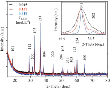
The solution supersaturation in our system was calculated for the Mn(OH)2intermediate phase at 25$C at three different volumes of
alkaline solutions (150, 460 and 1400 mL) corresponding to con-centrations of LiOH (C) equivalent to 0.419, 0.137 and 0.045 mol L!1,
respectively. At these conditions, S decreases from 6.6, 4.6 to 2.3 ('108), respectively. For high S values (>103) the nucleation rate
increases and the growing stage is limited. SEM images and asso-ciated histograms (Fig. 1) show monodisperse preparations of ox-ides. Particle shape is identical but the average particle size decreases almost linearly with dilution. For C ¼ 0.045 mol L!1, the
average particle size is 41 ± 12 nm. XRD patterns (Fig. 2) show that pure Mn3O4is formed for each synthesis. As the alkaline solution
concentration decreases, the average crystallite size, determined by the Debye-Scherrer formulae, decreases from 122 ± 59 to 30 ± 7 nm. So, when the dilution is increased, S and particle size both decrease. However, a lower S value should slow down the process of nucleation and favor the increase in the size of particles. A small variation in the very high S values, before and after dilution, will not influence the mechanisms of nucleation and growth, which are strongly related to the diffusion of the growing units. In con-ditions where S is high, the nucleation process is predominant and more homogeneous in the whole solution. Also, a dilute medium limits contacts between pre-existing nuclei and growing units which facilitates the generation of small particles.
3.1.2. Speed of reactants introduction and pH
According to Nielsen in 1964[22], the uniform homogenization of the medium for a precipitation by a mixture of solutions is closely related to the type, speed and duration of mixing. The way the salt solution is introduced into the alkaline solution induces a variation in pH and modifies the contact points between reactants. We tested two extreme conditions of mixing: a slow introduction of the salt solution (drop by drop) with a constant debit over a period
of time equivalent to 30 min, and a fast introduction (speed ¼ 5.5 L s!1) with the use of a large funnel. Note that the slow
introduction of salt and the stirring period end at the same time. In this case, the pH varies almost linearly with time (Supplementary Information 1). For a fast introduction, the initial pH value of 12.2 decreases abruptly by one unit in a minute then tends to stabilize for the next 6 min before decreasing again to a pH value of 11.2 (reached after 400 s) which remains constant until the end of the
Fig. 1. SEM images and corresponding particle size distribution histograms for Mn3O4after precipitation with (a) CLiOH¼ 0.419 mol L!1, (b) CLiOH¼ 0.137 mol L!1, (c) CLiOH¼ 0.045 mol L!1and fast introduction of metal salts at 25$C; (d) for a buffered alkaline solution (CLiOH¼ 0.045 mol L!1) with a slow introduction of metal salts and (e) ~constant pH still at 25$C; (f) fast introduction in LiOH (CLiOH¼ 0.045 mol L!1) at 50$C.
stirring sequence. Note that XRD analysis of the powder extracted from the solution after the first minute of fast introduction shows that crystallized Mn3O4 alone is observed therefore excluding a
dissolution-reprecipitation process (see Supplementary Information 1). The final pH is identical in both cases. SEM im-ages and associated histograms show that the only difference be-tween the oxide particles obtained after each path of salt introduction into the alkaline solution is their average sizes, d ¼ 27 ± 12 and 41 ± 12 nm for a slow and fast introduction, respectively (Fig. 1c and d). A similar crystallite size of 28 ± 7 nm is determined by the XRD patterns analysis of pure Mn3O4obtained in
both preparations. The slow introduction of a metal salt solution in an alkaline solution brings a constant amount of material without abrupt modification of the alkaline pH solution and creation of important heterogeneity zones. The nucleation process is therefore more homogeneous. However, a slow introduction can favor growth of particles. This is visibly hampered by the use of a very dilute medium. Knowing that an asymptote of the pH value is reached for a large amount of OH!ions, we did the synthesis with a
constant pH (pH ~ 12.3) for the whole mixing period by adding the salt solution to a large excess of LiOH powder ('1.6 the usual amount) dispersed in the same volume (1400 L) of alkaline solu-tion. SEM images (Fig. 1c and e) and XRD patterns show that oxide nanoparticles crystallize as single crystals of similar shape and size (d ¼ t ¼ 28 ± 6 nm) compared to a slow introduction process (Supplementary Information 2).
3.1.3. Dielectric constant of the solvent
A change in the dielectric constant of the solvent used for pre-paring the alkaline solution can modify the number of nuclei and shape of particles created during the precipitation process[23]. The species solubility in the solution is directly related to the dielectric constant and will modify the S value. Experiments were carried out under our standard conditions with three different water/ethanol volume ratios: 100/0, 80/20 or 60/40. A comparison between SEM images shows that the average particle size decreases (d ¼ 28, 26 and 20 ± 5 nm, respectively) when the dielectric constant of the medium decreases (Supplementary Information 3). This dielectric constant decreases from ~90 for pure distilled water to ~61 at 25$C
for a 60/40 volume ratio of water and ethanol. Crystallite sizes
extracted from the full width at half maximum (FWHM) of pure Mn3O4 XRD patterns (Supplementary Information 4) using the
Debye-Scherrer model are identical to the average particle sizes obtained from the SEM. The variation of the solution dielectric constant modifies the attractive forces between ions of opposite charges according to Coulomb's law. Ethanol gives a smaller dielectric constant than water. The use of ethanol decreases the solubility of the salt solution and the precipitating agent mixed together, which increases the medium supersaturation.
Also, the influence of the temperature on the synthesis was studied. The average particle size and distribution are larger at 50$C than at room temperature (see Fig. 1e andSupplementary
Information 5).
3.2. Mn3!xCoxO4(0 < x < 3) nanoparticles
Our synthesis method developed and optimized for Mn3O4was
applied to a larger range of compositions, with general formula Mn3!xCoxO4(0 < x < 3). CoMn2O4and MnCo2O4have already been
prepared by soft chemistry methods[24]. This is the first study known to the authors on the complete MneCoeO spinel system synthesized at low temperature. The preparation of pure spinel oxides over a wide composition range is not straightforward. For instance, Aukrust and Muan's phase diagram shows the complexity of preparing pure manganese-rich spinel oxides at low tempera-ture[25]. Such pure oxides can be obtained by usual solid state chemistry after quenching from high temperature or soft chemistry methods generally followed by thermal treatments. In the man-ganese and cobalt spinel oxide family, a structural transition is observed for a composition close to Mn1.4Co1.6O4at room
temper-ature. In our case, stoichiometric amounts of metal salts were mixed together in water before pouring into the alkaline solution following our standard conditions. Five intermediate compositions were studied in detail, namely Mn2.5Co0.5O4, Mn2.0Co1.0O4,
Mn1.5Co1.5O4, Mn1.0Co2.0O4 and Mn0.5Co2.5O4. ICP-AES analyses
confirmed the final mixed oxide compositions with a perfect stoi-chiometry match for each sample (within the 0.02 standard error of measurement). ICP-AES analyses also showed a very small con-centration of Liþ (lower than a few hundreds of ppm) and a
maximum sulfur quantity of 0.3 wt% which remained after each sample precipitation. This proves the efficiency of our synthetic method and washing steps for the removal of most of the residual elements from each solution. XRD measurements were performed on each sample after precipitation (Fig. 3). Each single phase Mn2.5Co0.5O4, Mn2.0Co1.0O4 and Mn1.5Co1.5O4 is tetragonal while
Mn1.0Co2.0O4and Mn0.5Co2.5O4 are cubic, in accordance with the
literature[6]. Note that for the cobalt-rich spinel phases, the solu-tion needs to be refluxed at 100$C for up to 2 h (seeFig. 3b and c).
The width and height of the (111) peak of the oxide phase indeed tend to indicate that a crystalline mixed oxy-hydroxyde phase is formed for Co-rich samples. The main intensity for the latter phase is observed at 21$ in 2-Theta, which corresponds to the (003)
reflection. This intermediate phase was also observed for the pure cobalt sample preparation[26]. This peak vanishes after refluxing for one and 2 h for Mn1.0Co2.0O4and Mn0.5Co2.5O4, respectively,
indicating that only the crystalline Co-rich spinel oxide phase is then present. Spinel oxide nanoparticles are spherical for all com-positions and the average diameter progressively decreases from 38 ± 14 nm (for Mn2.5Co0.5O4) to 23 ± 5 nm (for Mn0.5Co2.5O4) with
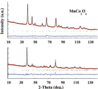
increasing cobalt. We performed neutron diffraction experiments on a Co-rich sample (Mn1.0Co2.0O4) of cubic symmetry and a
Mn-rich sample (Mn2.0Co1.0O4) of tetragonal symmetry in order to
determine their cation distributions (Fig. 4). Neutron diffraction can distinguish the manganese and cobalt elements due to distinct diffusion parameters. Rietveld refinements of the neutron data
Intensity (a
.u.)
2-Theta (deg.)
2-Theta (deg.) In te n si ty ( a. u .) 36.5 35.5 0.045 0.137 0.419 CLiOH (mol.L-1)Fig. 2. XRD patterns of Mn3O4after precipitation with (a) CLiOH¼ 0.419 mol L!1, (b) CLiOH¼ 0.137 mol L1, (c) CLiOH¼ 0.045 mol L!1. Inset shows (211) peak enlargement with the decrease of the alkaline solution concentration.
recorded at room temperature and the Bond Valence Sum (BVS) calculation indicate that the spinel oxide nanopowders contain mainly Co2þ on the tetrahedral site. Two couples of cations are determined on the octahedral site, for both phases, with a small quantity of Co3þand Mn4þfor the Mn-rich phase. The cationic
distributions can then be written as follows:
Similarly to the MnCo2O4 sintered ceramics, the cationic
dis-tribution as determined by the BVS calculations shows that our
nanopowders of the same composition contain close concentra-tions of Co2þand Co3þ, and Mn3þand Mn4þ. Our previous study
showed that a maximum of conductivity was obtained for a similar distribution, according to Verwey's law and the polaron hopping mechanism[6,24,27].
3.3. Colloidal dispersion stabilization of oxide nanoparticles Many techniques such as electrophoresis, doctor blade, serig-raphy, dip-coating or spin-coating were developed for the thin film
Fig. 3. XRD patterns of Mn3!xCoxO4with (top) tetragonal symmetry (x ¼ 0.5, 1.0 and 1.5 from top to bottom) and (bottom) cubic symmetry after precipitation (T0) for (left) x ¼ 2.0 and (right) x ¼ 2.5. T0þ x min indicates the time of reflux at 100$C after precipitation.
!Co2þ0:81Mn2þ0:19hCo2þ0:16Co3þ0:03Mn3þ1:76Mn4þ0:06iO4; for Mn2:0Co1:0O4;
preparation starting from a colloidal dispersion. Their advantages are numerous compared to the vapor deposition techniques: sim-ple, low cost, no vacuum is required, possible extension to large surfaces etc…We chose the dip-coating technique for our thin film preparation. The quality of the thin layer, which can be evaluated by a homogeneous distribution of nanoparticles on the substrate, thickness, density and presence of cracks…is highly dependent on the physical and chemical properties of the colloidal dispersion (degree of particles dispersion, solvent volatility, sol viscosity, substrate wettability…).
After synthesis and centrifugation, the spinel oxide particles are maintained together as anionic flocculates by different attractive forces, mainly Van der Waals forces and surface tension at the liquid/particle interface. Ultrasonication at 35 kHz for 5 min was performed to break apart the agglomerates of particles in aqueous solution. Surface charges of oxides are due to the polarization of the oxygen electronic density by the metal which lowers the hydroxyl groups at the surface. Depending on the cation nature and oxida-tion state close to the oxide surface, differences in the type and number of hydroxyls groups are observed. Zetametry measure-ments were undertaken for each spinel oxide to determine their IsoElectric Points (IEP) and domains of colloidal dispersion stability (Fig. 5). The IEP values progressively increase with the cobalt con-tent, varying from 7.0 for Mn3O4 to 10.5 for Mn0.5Co2.5O4. These
results indicate that the surface basicity of particles increase with the amount of cobalt. For all mixed spinel oxides, a large domain of constant zeta potential is observed for pH < 7. Zeta potential values of 40 ± 4 mV indicate a good stability of colloidal dispersions. A coloration of solution was observed for pH values lower than 6 due to partial dissolution of particles. Therefore, all preparations were peptized at pH ¼ 6 with nitric acid and stabilized with an azeotrope mixture containing 96 vol.% of absolute ethanol and 4 vol.% of ul-trapure water.
The granulometric distribution determined by the DLS method is given for each spinel oxide in Fig. 6. All solutions are mono-disperse with a narrow particle size distribution. The hydrody-namic particle average diameter decreases when the cobalt content increases in the mixed spinel oxide, with a maximum of 148 nm for
Mn3O4and 46 nm for Mn0.5Co2.5O4.
Fig. 6shows the viscosity variation as a function of the shear strain for each colloidal dispersion of spinel oxide. Two types of rheological behaviors can be identified: (i) for the Mn-rich phases with x " 1.5, which crystallize in the tetragonal structure, viscosity decreases with the constraints and a hysteresis is observed, indi-cating a rheofluidifying and thixotropic behavior while (ii) for the Co-rich phases of cubic symmetry with x > 1.5, viscosity remains constant over the whole measurement range which corresponds to a Newtonian behavior. Colloidal dispersions were stabilized for a few weeks up to six months and were used as such for the prep-aration of spinel oxide thin films at room temperature. A colloidal dispersion concentration of 60 g/L was chosen in order to obtain the thinnest possible homogeneous oxide layer on glass substrate (~300 nm). Note that this can also be achieved on quartz, metal and alloy substrates. Also, the azeotrope mixture containing essentially ethanol (96% in volume) allowed an excellent wettability of the substrate.
3.4. Spinel oxide thin films preparation by the dip-coating method and their optical properties
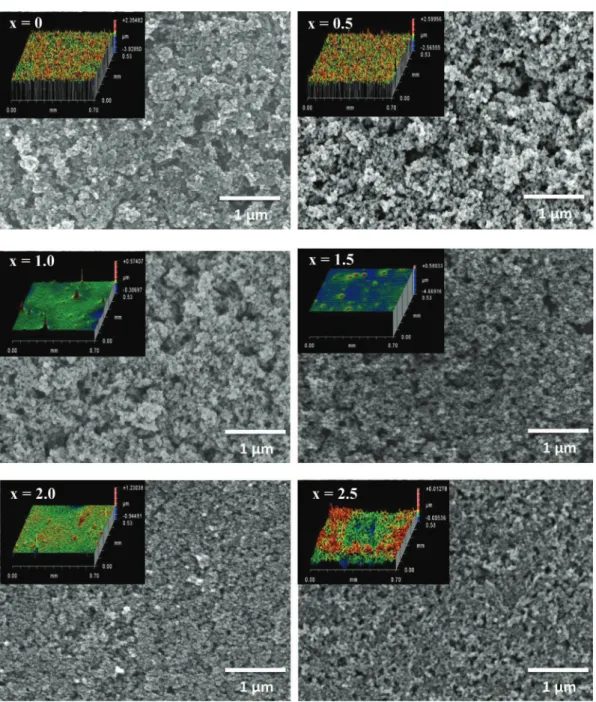
Oxide thin films of a thickness close to 300 nm were deposited with a NIMA DC small dip-coater from Lot-Oriel (withdrawal rate of 200 mm/min) on sodocalcic glasses (5 mm in thickness) which were preliminary cleaned with an alkaline detergent. FEG-SEM images show homogeneous layers and no cracks, although a bet-ter uniformity and compactness of the surface is noticed for the Co-rich phases, which contain smaller particles (Fig. 7). The surface profile of each oxide single layer was generated by white light optical interferometry measurements (see insets inFig. 7). The 3D reconstructions show small uncovered entities on the substrate for the two manganese-rich phases Mn3O4and Mn2.5Co0.5O4and the
average arithmetic roughness is 0.23 ± 0.06
m
m for both samples. In these small areas, the light goes straight through and is not re-flected by the glass substrate, which generates dips in the surface profile. These dips are not present for higher cobalt concentrations and the surface is much more regular, homogeneous and compact with an arithmetic average of roughness surface of 0.012 ± 0.003. Single layer porosity is related to the physical and chemical prop-erties of the dispersions, particularly to their stabilization by elec-trostatic repulsions. Particles rearrange to form a compact layer during the drying process. Xu and Anderson (1991) showed that the electrostatic repulsion forces were responsible for the film porosity[28]. The probability of particles to agglomerate increases with the
Fig. 4. Neutron diffraction patterns of (top) MnCo2O4and (bottom) Mn2CoO4 nano-powders recorded on D2B, ILL (l¼ 1.594 Å) after Rietveld refinements (observed ¼ red dots, calculated ¼ black lines, difference is in blue below). Reliability factors are: (top) Rp¼ 0.174, Rwp¼ 0.121, Rexp¼ 0.088,c2¼ 1.89, RB¼ 0.077, RF¼ 0.056 and (bottom) Rp¼ 0.357, Rwp¼ 0.270, Rexp¼ 0.127,c2¼ 4.49, RB¼ 0.120, RF¼ 0.106. (For inter-pretation of the references to color in this figure legend, the reader is referred to the web version of this article.)
Fig. 5. Zeta potential variation with pH for Mn3!xCoxO4(0 " x < 3). Nitric acid was used for pH values <7 and potassium hydroxide for pH > 7.
Fig. 6. (left) Percentage of intensity as a function of hydrodynamic particle diameter and (right) viscosity vs shearing stress for Mn3!xCoxO4(0 " x < 3).
lowering of the particles repulsion energy. In our case, the average zeta potential decreases at pH ¼ 6 when the cobalt content in-creases. During the retrieving stage of the substrate, the progressive solvent evaporation brings the particles closer to each other. For Mn-rich phases, minimizing the repulsive forces limits their reor-ganization and increases the film porosity.
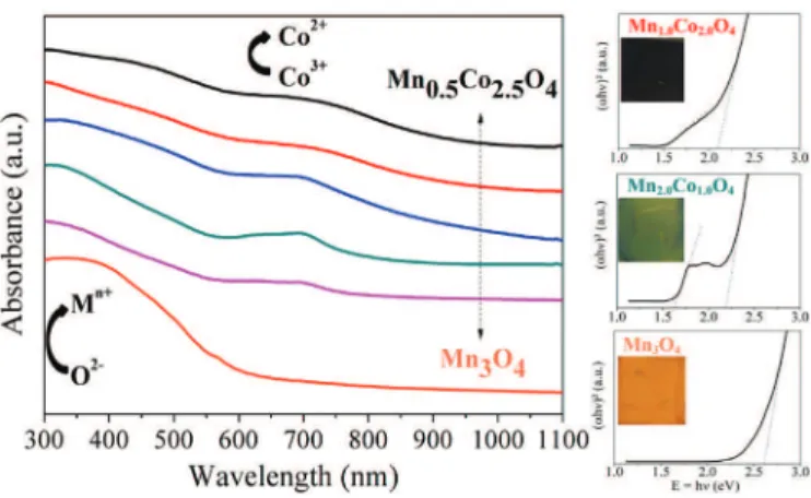
The optical properties of all our spinel oxide thin films were determined at room temperature after subtracting the influence of the glass substrate. Transmittance (T) and reflectance (R) were measured from the UV region to the IR region, i.e. 300e1100 nm. The Absorbance (A) spectra were deduced from the transmittance and reflectance measurements by using the simplified relation: A ¼ 1 ! T ! R (Fig. 8). Energy band gaps of spinel oxide semi-conductors were determined by using Tauc's relation[29]. The best fit of (
a
hy
)xvs hy
curve plotted for each sample was for x ¼ 2, whichcorresponds to a direct gap.
The absorbance spectrum for Mn3O4 shown in Fig. 8 is in
accordance with the literature[30]. Three main areas can be iso-lated: (1) in the UV region, below 400 nm, where the absorbance is maximum (~98%) and fairly constant; (2) from 400 to 600 nm where the absorbance decreases abruptly and almost linearly down to ~17%; and (3) above 600 nm where the decrease in absorbance is slow until it becomes negligible in the IR region. The haussmanite cation distribution is usually described with a direct spinel struc-ture as Mn2þ½Mn3þ
2)O4, where Mn2þ (3d5) is situated in the
tetrahedral site with a high spin state configuration such as all five orbitals contain only one electron (2e 3t2); the octahedral site is
occupied by Mn3þ(3d4) which exhibits a high spin state and (3t 2g 1e
g) configuration with a lifting of the egdegenerated levels due to
the Jahn-Teller effect. The insulating behavior usually observed in the direct spinel structure of Mn3O4is not necessarily in
contra-diction with the low energy band gap between the valence band and conduction band which characterizes a semi-conductor. In the first case, the large resistivity of the spinel oxide is due to the isolation of one type of cation on each crystallographic site, which hampers the hopping of polarons (and thus the electronic con-duction) otherwise observed when an element with two oxidation states (n and n þ 1) are on the same type of crystallographic site, preferentially the octahedral sites due to shortest distances. In the second case, the energy levels related to the atomic orbitals as defined by the band theory, are reasonably separated (Eg<3 eV) to create electron/hole pairs when under sufficient external energy,
for instance heat or solar absorbance. The maximum absorbance observed in the UV region is generally attributed to a charge transfer between the oxygen p orbitals towards the d orbitals of the Mn2þand Mn3þcations[30,31]. Small intra-atomic ded transitions between Mn3þ in octahedral crystal fields can occur at higher
wavelengths and the interactions become negligible near the IR region. An optical direct band gap of 2.6 eV was determined for Mn3O4(seeFig. 8), close to the value obtained by Xu et al., in 2006
(Eg¼ 2.54 eV) for Mn3O4thin films prepared by chemical
deposi-tion in soludeposi-tion[32]. A gap of 2.36 eV was also determined on thin films of Mn3O4but the authors attributed this lower value to the
presence of a residual hydroxyl phase[33]. Inversely, a gap value of 3.3 eV was measured for spherical nanoparticles of Mn3O4[34].
This gap shift towards higher energy values, associated with a blue shift, is due to a quantum confinement related to the small particle size and the separation of the continuous band into discrete levels. Such large optical band gaps are interesting for optoelectronic ap-plications with light emission in the low wavelength regions. On the contrary, low gap values show an interesting aptitude of the material for solar absorbance as required for example in photo-catalysis or photovoltaics.
The variations in the absorbance spectra for the mixed spinel oxide materials Mn3!xCoxO4(0 < x < 3) are similar to that of Mn3O4
with a general decrease of the absorbance with the increasing wavelength (see Fig. 8). However, the presence of cobalt in the spinel structure induces systematically a second absorption front near 700 nm in wavelength. This front is also observed in the end family member Co3O4 [35]. For stoichiometric Co3O4, the cation
distribution is Co2þ½Co3þ2)O4, where Co2þions are positioned on
the tetrahedral sites with a high spin state configuration 3d7(3e4t2)
while Co3þions occupy the octahedral sites with a low spin state
configuration 3d6(6t
2g0eg). A first absorption band in the UV region
is attributed to the inter-atomic charge transfer from the anion to the cation, in the case of Co3O4, from the oxygen p orbitals to the
d orbitals of both Co2þ and Co3þ[36]. The presence of the
ab-sorption edge at ~700 nm can be explained by the inter-atomic (cationecation) charge transfer from the Co3þ d(t2g) orbitals in
octahedral sites to the Co2þ d(t
2) orbitals in tetrahedral sites
[36,37]. Neutron diffraction measurements performed on our MnCo2O4and CoMn2O4samples provided evidence for both Co2þ
and Co3þon the two crystallographic sites of the spinel structure
(see part 3.2). The absorption front measured in the visible region for all Mn3!xCoxO4 (0 < x < 3) compounds could therefore be
related to the inter-atomic Co3þ d(t
2g) to Co2þ d(t2) transition.
However, other inter-atomic transitions can also occur in the presence of Mn3þand Mn4þ. Two band gaps are thus determined for each mixed oxide using Tauc's relation, associated to the two main peaks observed in the absorbance curve. Both band gaps decrease when the cobalt content increase (seeFig. 8). The highest band gap energy decreases from 2.6 eV for Mn3O4down to 2.1 eV
for Mn0.5Co2.5O4. This is not due to the particle size and a quantum
confinement effect since the Co-rich phase particles are the smallest while the haussmanite structural type material presents the largest particles. We believe that the decrease in the two energy gaps is directly related to the presence of lower empty d levels due to the presence of cobalt creating also an intermediate level in between the valence band and conduction band of the semi-conducting materials, as previously reported [37]. Further in-vestigations will be necessary in order to confirm this assumption. But the presence of an intermediate band in semi-conducting oxide materials that can be crystallized by a simple process at room temperature and deposited by a low-cost method as thin films will open very interesting opportunities for advances in electronics and optics.
Fig. 8. Optical absorbance variation and (ahy)2vs E ¼ hycurve for Mn3!xCoxO4 (0 " x < 3) thin films. Extrapolation of the straight lines near the edges in the (ahy)2vs hycurves gives energy band gaps of 2.60 for x ¼ 0, 1.70 and 2.22 for x ¼ 0.5, 1.62 and 2.18 for x ¼ 1.0, 1.55 and 2.14 for x ¼ 1.5, 1.48 and 2.13 for x ¼ 2.0, and 1.43 and 2.08 for x ¼ 2.5. M corresponds to the Metal cation.
4. Conclusions
The low-temperature sustainable synthesis and solution stabi-lization of functional nanoparticles was developed. This water and ethanol synthesis and solution stabilization of oxide nanoparticles was used to study a series of compounds with Mn3!xCoxO4
(0 " x < 3) general formula.
Firstly, the main conditions that monitor the size and shape of the oxide nanoparticles directly crystallized at room temperature by precipitation were defined for Mn3O4. Increasing the alkaline
solution (LiOH) concentration increases the supersaturation value and decreases particle sizes. Therefore, fast metal salt introduction into a much larger volume of a supersaturated and buffered alka-line solution rapidly stirred under turbulent conditions at room temperature can generate small oxide particles of isotropic shape. Mixed spinel oxides of manganese and cobalt were prepared using our standard conditions. For the Co-rich phases, a further reflux stage at 100$C up to 2 h is required to obtain pure mixed spinel
oxides. The cation distribution was determined for the CoMn2O4
tetragonal phase and MnCo2O4cubic phase by neutron diffraction.
In both cases, octahedral sites can contain four types of cations (Co2þ, Co3þ, Mn3þand Mn4þ), while tetrahedral sites are mainly
occupied by Co2þ, similarly to dense ceramics of the same compositions.
Secondly, the isoelectric points and pH stability domains were determined for all our solutions of oxide nanoparticles. The IEP value shifts from 7 for Mn3O4to 10.5 for Mn0.5Co2.5O4and a large
plateau of high zeta potential (40 ± 4 mV) in acidic conditions allowed the preparation of colloidal dispersions for a pH ~ 6. Monodisperse solutions in azeotropic medium (ethanol/ water ¼ 96/4 vol.%) were formed. The hydrodynamic particle diameter in such azeotropic medium decreases with the cobalt content. Mn-rich solutions are rheofluidifying and thixotropic while the cubic phases follow a Newtonian behavior.
Thirdly, spinel oxide thin films (~300 nm) were prepared by the dip-coating technique. These films exhibit strong solar absorbance at low wavelengths and then direct energy band gaps were deter-mined. A second gap, which is observed systematically for Co-containing spinel oxide phases, was associated with the genera-tion of an intermediate state in the optical band gap.
Recently, this method has been successfully extended to other families of spinel oxides including cuprates. In addition, our pre-liminary results indicate that the process can be adapted to syn-thesize ZnO, CuO, and Cu2O, and has the potential to accommodate
a wide range of target chemistries and stoichiometries and there-fore, can constitute a promising method for enhanced efficiency in the preparation and use of nanomaterials containing critical ele-ments. Moreover, since this innovative process does not require any organic reactant or polymeric agent, it is inherently low cost and can be easily adapted for large-scale production.
Acknowledgments
The French Ministry of Education and Research is thanked for financial assistance towards the salary of the PhD student. We also wish to thank Dr S.M. Moussa for her input. Dr Juan Claudio Nino from the NRG group at the University of Florida is deeply acknowledged for his interesting comments.
Appendix A. Supplementary data
Supplementary data related to this article can be found athttp:// dx.doi.org/10.1016/j.matchemphys.2015.05.065.
References
[1] S. Pal, D. Diso, S. Franza, A. Licciulli, L. Rizzo, Spectrally selective absorber coating from transition metal complex for efficient photothermal conversion, J. Mater. Sci. 48 (2013) 8268e8276.
[2] J. Vince, A. #Surca Vuk, U.O. Kra#sovec, B. Orel, M. K€ohl, M. Heck, Solar absorber coatings based on CoCuMnOx spinels prepared via the solegel process: structural and optical properties, Sol. Energy Mater. Sol. Cells 79 (2003) 313e330.
[3] S.A. Elizi!ario, J.M. de Andrade, S.J.G. Lima, C.A. Paskocimas, L.E.B. Soledade, P. Hammer, Black and green pigments based on chromiumecobalt spinels, Mater. Chem. Phys. 129 (2011) 619e624.
[4] D. Visinescu, C. Paraschiv, A. Ianculescu, B. Jurca, B. Vasile, O. Carp, The environmentally benign synthesis of nanosized CoxZn1!xAl2O4blue pigments, Dyes Pigm. 87 (2010) 125e131.
[5] K. Park, S.J. Kim, J.-G. Kim, S. Nahm, Structural and electrical properties of MgO-doped Mn1.4Ni1.2Co0.4!xMgxO4(0 " x " 0.25) NTC thermistors, J. Eur. Ceram. Soc. 27 (2007) 2009e2016.
[6] H. Bordeneuve, A. Rousset, C. Tenailleau, S. Guillemet-Fritsch, Cation distri-bution in manganese cobaltite spinels Co3!xMnxO4(0 " x " 1) determined by thermal analysis, J. Therm. Anal. Calorim. 101 (2009) 137e142.
[7] S. Royer, D. Duprez, Catalytic oxidation of carbon monoxide over transition metal oxides, ChemCatChem. 3 (2011) 24e65.
[8] S. Rühle, A.Y. Anderson, H.-N. Barad, B. Kupfer, Y. Bouhadana, E. Rosh-Hodesh, A. Zaban, All-oxide photovoltaics, J. Phys. Chem. Lett. 3 (2012) 3755e3764.
[9] Y.S. Jung, J.Y. Seo, D.W. Lee, D.Y. Jeon, Influence of DC magnetron sputtering parameters on the properties of amorphous indium zinc oxide thin film, Thin Solid Films 445 (2003) 63e71.
[10] L. Miao, P. Jin, K. Kaneko, a Terai, N. Nabatova-Gabain, S. Tanemura, Prepa-ration and characterization of polycrystalline anatase and rutile TiO2thin films by rf magnetron sputtering, Appl. Surf. Sci. 212e213 (2003) 255e263. [11] T. Maruyama, Copper oxide thin films prepared by chemical vapor deposition
from copper dipivaloylmethanate, Sol. Energy Mater. Sol. Cells 56 (1998) 85e92.
[12] N. Bahlawane, E. Fischer Rivera, K. Kohse-H€oinghaus, A. Brechling, U. Kleineberg, Characterization and tests of planar Co3O4model catalysts prepared by chemical vapor deposition, Appl. Catal. B Environ. 53 (2004) 245e255.
[13] T. Pauport!e, D. Lincot, Electrodeposition of semiconductors for optoelectronic devices: results on zinc oxide, Electrochim. Acta 45 (2000) 3345e3353.
[14] M. Wu, Y. Huang, C. Yang, J. Jow, Electrodeposition of nanoporous nickel oxide film for electrochemical capacitors, Int. J. Hydrogen Energy 32 (2007) 4153e4159.
[15] R.N. Singh, J.P. Pandey, N.K. Singh, B. Lal, P. Chartier, J.-F. Koenig, Solegel derived spinel MxCo3!xO4(M ¼ Ni, Cu; 0 " x " 1) films and oxygen evolution, Electrochim. Acta 45 (2000) 1911e1919.
[16] A. Phani, M. Passacantando, S. Santucci, Synthesis and characterization of zinc aluminum oxide thin films by solegel technique, Mater. Chem. Phys. 68 (2001) 66e71.
[17] X. Zhao, Q. Zhao, J. Yu, B. Liu, Development of multifunctional photoactive self-cleaning glasses, J. Non. Cryst. Solids 354 (2008) 1424e1430.
[18] J. Rodríguez-Carvajal, FULLPROF: a program for rietveld refinement and pattern matching analysis, in: Satell. Meet. Powder Diffr. XV IUCr Congr, 1990, p. 127.
[19] I.D. Brown, D. Altermatt, Bond-valence parameters obtained from a systematic analysis of the inorganic crystal structure database, Acta Crystallogr. Sect. B 41 (1985) 244e247.
[20] M.R. Wood, G.J. Palenik, Bond valence sums in coordination chemistry. A simple method for calculating the oxidation state of cobalt in complexes containing only CoeO bonds, Inorg. Chem. 37 (1998) 4149e4151.
[21] H. Ohshima, Henry's function for electrophoresis of a cylindrical colloidal particle, J. Colloid Interface Sci. 180 (1996) 299e301.
[22] A.E. Nielsen, Kinetics of Precipitation, Pergamon, Oxford, 1964.
[23] G. Charlot, B. Tremillon, Chemical Reactions in Solvents and Melts, Translated from the French edition (Paris, 1963) by P. J. J. Harvey, Pergamon, New York, 1969.
[24] G. Salek, S. Guillemet-Fritsch, P. Dufour, C. Tenailleau, A simple preparation process of pure Mn3!XCoxO4(X ¼ 1, 1.5 and 2) desert rose-like nanoparticles and their optical properties, Int. J. Chem. 4 (2012) 44e53.
[25] E. Aukrust, A. Muan, Phase relations in the system cobalt oxide-manganese oxide in air, Trans. Metall. Soc. Aime 230 (1964) 378e382.
[26] G. Salek, P. Alphonse, P. Dufour, S. Guillemet-Fritsch, C. Tenailleau, Low-temperature carbon monoxide and propane total oxidation by nanocrystalline cobalt oxides, Appl. Catal. B Environ. 147 (2014) 1e7.
[27] E.J.W. Verwey, P.W. Haaijman, F.C. Romeijn, G.W. Van Oosterhout, Controlled-valency semiconductors, Philips Res. Rep 5 (1950) 173e187.
[28] Q. Xu, M.A. Anderson, Synthesis of porosity controlled ceramic menbranes, J. Mater. Res. 6 (1991) 1073e1081.
[29] J. Tauc, Optical properties and electronic structure of amorphous, Mater. Res. Bull. 3 (1968) 37e46.
[30] V. Sanchez, Influence of tile synthesis parameters on the structural and textural properties of precipitated manganese oxides 3 (2001) 889e899.
[31] A. V!azquez-Olmos, R. Red!on, A.L. Fern!andez-Osorio, J.M. Saniger, Room-temperature synthesis of Mn3O4 nanorods, Appl. Phys. A 81 (2005)
1131e1134.
[32] H.Y. Xu, S. Le Xu, X.D. Li, H. Wang, H. Yan, Chemical bath deposition of hausmannite Mn3O4thin films, Appl. Surf. Sci. 252 (2006) 4091e4096.
[33] D.P. Dubal, D.S. Dhawale, R.R. Salunkhe, S.M. Pawar, C.D. Lokhande, A novel chemical synthesis and characterization of Mn3O4 thin films for super-capacitor application, Appl. Surf. Sci. 256 (2010) 4411e4416.
[34] N.M. Hosny, a. Dahshan, Facile synthesis and optical band gap calculation of Mn3O4nanoparticles, Mater. Chem. Phys. 137 (2012) 637e643.
[35] K. Kolipaka, V. Brueser, A. Quade, J. Schaefer, Structural and Optical
Characterization of Spinel Type Cobalt Oxide Nanoparticles Embedded in Amorphous Silicon Oxide Matrix Prepared by a Hybrid PVD/PECVD, 2011, pp. 2e5.Ispc-Conference.org.
[36] P.Y. Keng, B.Y. Kim, I. Shim, R. Sahoo, P.E. Veneman, N.R. Armstrong, et al., Colloidal polymerization of polymer- into cobalt oxide nanowires 3 (2009) 3143e3157.
[37] S. Thota, A. Kumar, J. Kumar, Optical, electrical and magnetic properties of Co3O4nanocrystallites obtained by thermal decomposition of solegel derived oxalates, Mater. Sci. Eng. B 164 (2009) 30e37.