OATAO is an open access repository that collects the work of Toulouse
researchers and makes it freely available over the web where possible
Any correspondence concerning this service should be sent
to the repository administrator: [email protected]
This is an author’s version published in:
http://oatao.univ-toulouse.fr/27630
To cite this version:
Dupret Bories, Agnès
and Roumiguie, Mathieu and De Bonnecaze,
Guillaume and Sarini, Jérome and Vairel, Benjamin and Vergez, Sébastien and
Benbassat, Bastien The super thin external pudendal artery (STEPA) free flap
for oropharyngeal reconstruction – A case report. (2019) Microsurgery, 39 (8).
758-762. ISSN 0738-1085
DOi: 10.1002/micr.30512
The super thin external pudendal artery (STEPA) free flap
for oropharyngeal reconstruction - A case report
Agnès Dupret-Bories MD, PhD
1Cl!)
Mathieu Roumiguie MD, PhD
2Guillaume De Bonnecaze MD, PhD
3Jérome Sarini MD
1Benjamin Vairel MD
3Sébastien Vergez MD, PhD
3Bastien Benbassat Jr.
31Chirurgie ORL et cervico faciale, IUCT Toulouse Oncopole, Institut Claudius Regaud, Toulouse, France
2Chirurgie Urologique, Centre Hospitalier Universitaire, Hôpital Rangueil, Toulouse, France
3Chirurgie ORL et cervico faciale, Centre Hospitalier Universitaire, Hôpital Larrey, Toulouse, France
Carres ponde nce
Agnès Dupret Bories, MD, PhD, Department of Otorhinolaryngology, Institut Universitaire du Cancer, 31009, Toulouse, France. Email: dupret bories.agnes@i uct oncopole. fr
1 1 INTRODUCTION
Abstract
The radial forearm flap is one of the most used micro-anastomotic flaps in cervicofacial reconstruction in a carcinological context. This flap is an ideal in terms of reliability and fineness; it has, however, some disadvantages in terms of the functional and aesthetic complications of its donor site. ln alternative to a radial forearm free flap, we report the use of the free super thin extemal pudendal artery flap (STEPA flap) for an oropharyn geal reconstruction. The aim was to decrease the donor site morbidity. A 71-years-old man with a T2NOMO oropharyngeal squamous cell carcinoma has undergone surgical treatment. A left STEPA free flap was performed to reconstruct a defect about 8 x 6 cm2. This flap was designed as a half-scrotal free flap sized 9 x 7 cm2 and was
inset after tunneling of the pedicle at the floor of the mouth. A surgical revision was needed on the 15th day postoperative for disunion. There was no skin flap failure. After 12 month of follow-up, no complication was observed at the donor site and no erectile dysfunction was recorded. lts characteristics in terms of fineness, flexibility, ease of conformation, and pedicle length are similar to those of the radial forearm flap with less aesthetic and functional sequelae of the donor site. The STEPA flap may be a promising free flap in oropharyngeal or oral cavity reconstruction.
Oropharyngeal reconstruction requires the use of a reliable free flap with tissue-like qualities to be reconstructed: fineness and flexibility for an ease of structure at the receiving site and a compact spatial design.
scarring, the scrotal free flap named super thin extemal pudendal artery (STEPA) flap was recently described and used for the recon struction of critical areas such as limbs or joint areas at the shoulder or knee (Phoon, Shah, Cormack,
&
Saint-Cyr, 2014). The STEPA flap was used for the first time in free form for the reconstruction of a foot defect in 2017 (Kiranantawat et al., 2018). ln this article, we report the use of the STEPA flap for an oropharyngeal reconstruction.Since its description in 1981 by Yang et al. (Loeffelbein et al., 2012), the free radial forearm flap or Chinese flap (Benateau, Laraba, Alix,
&
Compere, 2002; Loeffelbein et al., 2012) is the most used in cervicofacial reconstruction in a carcinological context due to its fine ness and plasticity. The primary criticisms are the sacrifice of one of the two major vascular axes of the hand and the functional and aes thetic morbidity caused at the donor site (Orlik et al., 2014). With equivalent features in terms of fineness and flexibility for minimal2 1 CASE REPORT
A 71-year-old man had an active continuation of a T2N0M0 left oropha· ryngeal squamous cell carcinoma at 6 months of treatment by extemal radiotherapy. The patient received a left posterior uninterrupted pelvi mandibulectomy associated with a left oropharyngectomy with left
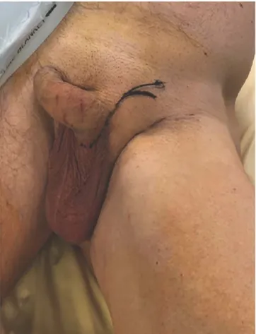
FIGURE 1 Initial skin markings. Arciform incision made on the inguinal ligament continuing on the lateral base of penis
FIGURE 2 Superthin external pudendal artery flap (STEPA) flap's vascular anatomy
cervical lymph node dissection and reconstruction with a left STEPA flap to reconstruct a defect about 8 x 6 cm2• The preoperative international index of erectile function (IIEF-5) survey recorded a mild erectile dys function (score 19 of 25).
The installation was standard for the resection of an oropharyn geal tumor in cervicofacial surgery, with the left lower limb entirely in the operative field so as to be able to position it in slight abduction and release the scrotum to perform the removal of the flap. An arciform incision was performed on the inguinal ligament then the dis section began in the subfascial layer (Figure 1). The first stage con sisted of locating the suprapubic artery at its cross-over with the spermatie cord (Figure 2). lts prudent retrograde dissection was per formed up to the base of its pedicle on the artery and the external
pudendal vein, in the subcremasteric area. The artery and external pudendal vein were dissected, respectively, up to their base, femoral artery and large saphenous vein (Figure 2). The caliber of the artery and vein were, respectively, 2 and 2.2 mm and the length of the pedi cle was 7.2 cm. The second step consisted of incising the scrotum at the median raphe following the preestablished limits of the cutaneous palette necessary for reconstruction with a dimension of 9 cm by 7 cm in our case. The incision began on the lateral portion of the base of the penis and continued along the median raphe. The testis and the testicular cord were separated from the scrotal sac by blunt dis section and carefully removed. The incision was continued inferiorly then parallel to the groin to join the initial exploratory incision. After ligation of the pedicle, the flap was placed on hold at the orapharyngeal receiving site after tunneling of the pedicle at the floor of the mouth
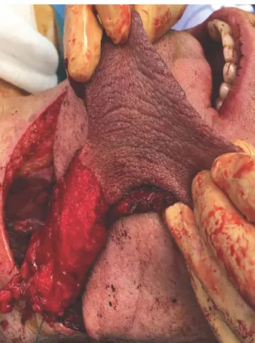
FIGURE 3 Scrotal free flap harvested, sized 9 x 7 cm2• Super thin
extemal pudendal artery flap (STEPA) free flap after vascular ligature and before tunneling of the pedicule at the floor of the mouth
(Rgure 3). Anastomoses of the external pudendal vessels (one artery and one vein) were performed by separate points of Ethilon 8.0, begin ning with anastomosis of the artery with left facial artery, and then with that of the vein with the left facial vein. Then the flap was inset on the defect (Rgure 4i After reintegration of the testis into the scrotum, the donor site was primary closed without tension in two layers. The post operative period was marked on the fourth day by a contraction of the flanks of the flap secondary to a major edema. On the 15th postopera tive day, we proceeded with a surgical revision for disunion exceeding the junction of the soft palate with the flap on an irradiated area. Scar ring was then obtained. Postoperative staging was pT2N0ROM0 with a supervisory guide as part of carcinological follcm-up. No complication was observed at the donor site. Scarring was of good quality with a dis crete scar hidden in the patient's undergarments (Figure 5). After 12 month of follow-up, the patient reports no aesthetic or sexual corn plaints and no erectile function irnpairment was recorded follcming the postoperative IIEF-5.
3 1 DISCUSSION
Oropharyngeal or oral floor reconstruction in a carcinological situation by a STEPA Flap is promising.
FIGURE 4 Postoperative result. Super thin external pudendal artery flap (STE PA) flap inset in the defect after sutures
The surgical technique is simple and quick. The pedicle vessels have a satisfactory diameter, 2.81 mm for the external pudendal artery at its base and 4.44 mm for the external pudendal vein, with a pedicle length up to 11 cm after dissection within the subcutaneous tissue. The skin is thin and supple, 1.1 mm thick, ideal for cervicofacial reconstructions (Phoon et al., 2014). The absence of functional com plication and the aesthetic result conceming the donor site make it a flap with low morbidity in comparison to the radial forearm flap (Benateau et al., 2002).
Regarding the free radial forearm flap, its fineness, flexibility, ver satility, lem hair growth, pedicle length (7-12 cm; Chen et al., 2005) and reliability (>90% success; Chen et al., 2005; Soutar
&
Mc Gregor, 1986) make it a choice element in cervicofacial reconstruction. lts pri mary disadvantage is the morbidity of the donor site, both functionally and aesthetically, with an incidence of complications estimated between 6 and 53% (Bootz&
Biesinger, 1991; Loeffelbein et al., 2012; Timmons, Missotten, Poole,&
Davies, 1986; Toschka et al., 2001). More than 16% of patients have a functional impact on the mobility of their forearm and/or wrist, more than 28% complain of aesthetic result (Bootz&
Biesinger, 1991; Loeffelbein et al., 2012; Timmons et al., 1986; Toschka et al., 2001). Closure of the donor site is carried out most frequently by skin grafting, which may become partially necrotic in 19-53% of cases, with a risk of tendon exposure ranging from 13 to 33% (Lutz, Wei, Chang, Yang,&
Chen, 1999).•
FI GU
RE 5
Scarring of the donor site at 6 month. Donor site with a good quality scar which is hiddenProblems with localized sensitivity at the donor site and in the sensory area of the radial nerve occur in 7.7 (Meland, Core, Hoverman, Dunet,
&
Leyder, 1993)-54% (Lutz et al., 1999) of cases and up to 82% at 3 months postoperative (Chen et al., 2005). Functional compli cations such as stiffness of the wrist with decreased joint range or grip strength may occur, respectively, in more than 26.7% of cases (Lutz et al., 1999) and in 1 1-40% of cases (Huang, Chen, Huang, Mardini,&
Feng, 2003; Timmons et al., 1986). Patients with a radial forearm flap may also have other complications at the donor site such as wrist or hand edema or an intolerance to cold. Regarding cosmetic results, two studies report patient satisfaction in 94.3% (Toschka et al., 2001) and 98% (Lutz et al., 1999) of cases, others are more mixed with 28.4% (Richardson, Fisher, David Vaughan, & Brown, 1997), 24% (Bootz & Biesinger, 1991), and 16.7% (Swanson&
MR, 1990) of patients being somewhat dissatisfied to dissatisfied.ln cervicofacial reconstruction, the execution of a radial forearm flap can be carried out simultaneously with the excision of the operative specimen but the proximity of the two sites may constrain surgeons during the ipsilateral lymph node dissection. Another advantage of the STEPA Flap is allowing for more comfortable dou ble team work.
Various studies were carried out in order to simplify closure of the donor site and decrease its morbidity when radial forearm free flap was the procedure of choice. But these flaps designs may involves extensive scars or, as the radial forearm snake free flap, may limits flap length pedicle or skin paddle size (Garg, Wieland, Poore, Sanchez,
&
Hartig, 2017). The direct closure of the donor site without tension with a hidden scar is one of the main advantages of the STE PA flap.The primary disadvantages of the STEPA flap are the hairiness of the cutaneous palette, which can hinder the patient during hair
growth, as well as the risk of inguinal lymphedernas, estimated at 10% (Phoon et al., 2014).
The scrotal flap was widely used in urology reconstruction includ ing hypospadias reconstruction or reconstruction of penile after injury. The rate of scrotal surgery complications was well-described in the literature with reported rate of hematoma and infections in 5% and 3.6% of case (Swartz, Morgan,
&
Krieger, 2007). As in this case, no impairment of erectile function and no impact on testicular func tion is described at this point in the literature (Phoon et al., 2014; Zhao, Zhang, Yu,&
Long, 2009). Although no psychological impact of moving scrotal skin in the mouth was recorded, it needs to be taken into account. Whether hyper-pigmentation could be a problem in facial reconstruction, it is not a disadvantage to be considered in the context of oral cavity reconstruction.The STEPA flap presents the plastic qualities suitable to cer vicofacial reconstruction. The authors believe it may therefore be an alternative to the radial forearm flap in the consideration of recon struction in a carcinological context, particularly for the oropharynx and oral cavity. However, this is the first STEPA flap used in head and neck surgery. Additional cases are still required to determine the reliability of this flap compared to the radial forearm free flap in particula r.
FINANCIAL DISCLOSURE STATEMENT
None.ORCID
REFERENCES
Benateau, H., l.araba, C., Alix, T., & Compere, J. F. (2002). Radial forearm or Chinese flap. Revue de S tomatologie et de Chirurgie Maxillo-Fadale, 103(1), 35 40.
Bootz, F., & Biesinger, E. (1991). Reduction of complication rate at radial forearm flap donor sites. ORL, 53(3), 160 164.
Chen, C. M., Lin, G. T., Fu, Y. C., Shieh, T. Y., Huang, 1. Y., Shen, Y. S., & Chen, C. H. (2005). Complications of free radial forearm flap transfers
for head and neck reconstruction. Oral Surgery, Oral Medicine, Oral
Pathology, and Oral Radiology, 99(6), 671 676.
Garg, R. K, Wieland, A. M., Poore, S. O., Sanchez, R., & Hartig, G. K. (2017). The radial forearm snake flap: A novel approach to oral cavity and oropharyngeal reconstruction that reduces forearm donor site
morbidity. Microsurgery, 37(1), 6 11.
Huang, C., Chen, H., Huang, Y., Mardini, S., & Feng, G. (2003). Comparison of the radial forearm flap and the thinned anterolateral thigh cutane ous flap for reconstruction of tongue defects: An evaluation of donor site morbidity. Plastic and Reconstructive Surgery, 114, 1704 1710. Kiranantawat, K., Yeo, M. S. W., lmaizumi, A., Sitpahul, N., Ciudad, P.,
Nicoli, F., ... Chatdokmaiprai, C. (2018). The scrotal free flap: First suc cessful clinical application of a free super-th in external Pudendal artery
(STEPA) flap for reconstruction of a foot defect. Journal of Plastic,
Reconstructive & Aesthetic Surgery, 71(2), 262 264.
Loeffelbein, D. J., AI-Benna, S., SteinstraBer, L, Satanovskij, R. M., Rohleder, N. H., Mücke, T., ... Kesting, M. R. (2012). Reduction of donor site morbidity of free radial forearm flaps: What level of evi dence is available? Ep/asty, 12, e9.
Lutz, B. S., Wei, F. C., Chang, S. C. N., Yang, K. H., & Chen, 1. (1999). Donor site morbidity after suprafascial elevation of the radial forearm flap: A
prospective study in 95 consecutive cases. Plastic and Reconstructive
Surgery, 103(1), 132 137.
Meland, N.B., Core, G. B., Hoverman, V. R., Dunet, É., & Leyder, P. (1993).
The radial forearm flap donor site. Plastic and Reconstructive Surgery, 91(Supplement), 865 870.
Orlik, J. R., Horwich, P., Bartlett, C., Trites, J., Hart, R., & Taylor, S. M. (2014). Long-term functional donor site morbidity of the free radial
forearm flap in head and neck cancer survivors. Journal of Otolaryngol
ogy -Head & Neck Surgery, 43, 1 7.
Phoon, A. F., Shah, A. K, Cormack, G. C., & Saint-Cyr, M. (2014). The super thin extemal pudendal artery (STEPA) flap. Journal of Plastic, Recon
structive & Aesthetic Surgery, 67(10), 1397 1406.
Richardson, D., Fisher, S. E., David Vaughan, E., & Brown, J. S. (1997).
Radial forearm flap donor-site complications and morbidity: A pro spective study. Plastic and Reconstructive Surgery, 99(1), 109 115. Soutar, D. S., & Mc Gregor, 1. A. (1986). The radial forearm flap in intraoral
reconstruction: The experience of 60 consecutive cases. Plastic and Reconstructive Surgery, 78 (1), 1 8.
Swanson, E., & MR, B. J. B. (1990). The radial forearm flap: Reconstructive applications and donor-site defects in 35 consecutive patients. Plastic and Reconstructive Surgery, 85(2), 258 266.
Swartz, M. A., Morgan, T. M., & Krieger, J. N. (2007). Complications of scrotal surgery for benign conditions. Urology, 69(4), 616 619.
Timmons, M. J., Missotten, F. E. M., Poole, M. D., & Davies, D. M. (1986).
Complications of radial forearm flap donor sites. British Journal of Plas
tic Surgery, 39(2), 176 178.
Toschka, H., Feifel, H., Erli, H., Minkenberg, R., Paar, O., Riediger, D. (2001). Aesthetic and functional results of harvesting radial forearm flap, especially with regard to hand function. Int ernational Journal of Oral and Maxil/ofadal Surgery, 30, 42 48.
Zhao, Y. Q., Zhang, J., Yu, M. S., & Long, D. C. (2009). Functional restora tion of penis with partial defect by scrotal skin flap. The Journal of Urol ogy, 182(5), 2358 2361.
How to cite this article: Dupret-Bories A, Roumiguie M, De Bonnecaze G, et al. The super thin external pudendal artery (STEP A) free flap for oropharyngeal reconstruction - A case report Microsurgery. 2019;39:758-762. https://doi.org/10.