OATAO is an open access repository that collects the work of Toulouse
researchers and makes it freely available over the web where possible
Any correspondence concerning this service should be sent
to the repository administrator: [email protected]
This is an author’s version published in:
http://oatao.univ-toulouse.fr/27606
To cite this version:
Potet, Pauline and De Bonnecaze, Guillaune and Chabrillac, Emilien and
Dupret Bories, Agnès
and Vergez, Sébastien and Chaput, Benoit Closure of
radial forearm free flap donor site: A comparative study between keystone flap
and skin graft. (2020) Head & Neck, 42 (2). 217-223. ISSN 1043-3074
DOi: 10. 1002/hed.25977
Closure of radial forearm free Oap donor site: A comparative
study between keystone flap and skin graft
Pauline Potet MD
1œ
IGuillaune De Bonnecaze MD, PhD
1 1Emilien Chabrillac MD
1Cl!)
1Agnès Dupret-Bories MD, PhD
2 1Sébastien Vergez MD, PhD
1,2 1Benoit Chaput MD, PhD
31Department ofENT and Head and Neck Surgery, Toulouse University Hospital, Hôpital Larrey, Toulouse, France 1)epartment of surgery, Institut Universitaire du Cancer de Toulouse, Toulouse, France
3Department of Plastic and Aesthetic Surgery, Toulouse University Hospital, Hôpital Rangueil, Toulouse, France Correspondence
Pauline Potet, Service ORLet CCF Hôpital Larrey, 24 chemin de Pouvourville 314-00 Toulouse, France.
Email: pauline [email protected]
1
1INTRODUCTION
Abstract
Background: The aim was to investigate the feasibility of radial foreann free flap (RFFF) donor site closure by keystone flap (KF) and compare its outcomes to those of skin graft (SG) closure.
Methods: One hundred and one patients who underwent RFFF for head and neck reconstruction were included (35 KF closure and 65 SG closure). Duration of wound healing and donor site complications was collected. After a minimal follow-up of
1 year, patients were questioned about functional and esthetic impairment.
Results: Coverage of donor site by KF was successful in ail cases. The duration of wound healing was longer after SG than after KF (32 days vs 18 days, P < .001). Healing complications, esthetic and functional results were not statistically different. Conclusion: Forearm donor site closure by KF is a feasible alternative to the tradi tional SG. lts main advantages are the reduced wound healing time and the avoid ance of a second donor site.
KEYWORDS
esthetic and functional results, donor site rnorbidity, keystone flap, radial forearrn free flap, skin graft
The fasciocutaneous radial forearm free flap (RFFF), increasingly used since Yang's description in a large series in 1981,1 is commonly used in reconstructive surgery for head and neck tumors. Since the survival rate of the flap approaches 95%,2 the attention of the surgeon has gradually focused on postoperative quality of life, most particularly on the donor site morbidity, which is often underestimated.3
We propose to adapt the use of keystone local flap to the closure of RFFF donor site. Keystone perforator island flap is a multiperforator advancement flap based on musculocutaneous or fasciocutaneous perforators, described by Behan in 2003.5
This study presents the Keystone local flap surgical tech nique and the outcomes of the RFFF donor site closure with both techniques.
Our objective was to prove the feasibility of closure by keystone flap (KF) and to compare its outcomes with skin graft (SG) in terms of wound healing, esthetic, and func tional impairment.
Many possibilities of donor site closure have been described, ranging from direct closure to local flaps, includ ing thin or full skin grafting, artificial dermis, expanders, or negative pressure therapy. Although skin grafting is the most frequently used technique, the question of an ideal way to close the donor site remains unanswered.4
2 1 METHODS
We performed a monocentric retrospective study from January 2013 to October 2017 in the ENT and Head and Neck surgery
department of the University Institute of Cancer in Toulouse, France. We included 101 patients who received RFFF for reconstruction after head and neck tumor. Patient's agreement was collected in order to use their data for research and publica tion purposes.
We divided the population into two groups: a group of 36 patients for which a first surgical team systernatically and consecutively perforrned a KF (group A), and a group of 65 patients for which a second surgical team systematically and consecutively perforrned a SG (group B).
Patients' dernographics and characteristics were collected in the medical record: age, sex, co-morbidities (including tobacco use, diabetes, and cardiovascular disease), local complications, wound healing disorders and healing duration (in days).
The area of substance loss was calculated using the length and width of the antebrachial flap harvested (elliptical area for Keystone reconstructions and rect.angu lar area for skin graft coverage ).
After at least 12 months, patients were asked (on the phone or during consultation) about donor site current pain (visual analog scale for pain, out of 10), loss of grip strength (yes/no), local sensitivity (normal/decreased/abolished), esthetic self assessment from 1 to 5 (1: ugly, 3: fair, 5: perfect). If neces sary, the healing duration data were cross-referenced with the patient's staternents. They were also asked a free description of their current global fi.mctional irnpairment. An esthetic hetero evaluation was performed for ail donor sites by an independent examiner (sarne scale from 1 to 5), during consultation or by photograph.
2.1
1Surgical technique
The KF can be used for elliptical defects, with transfer of adjacent tissue for better color and contour match. The flap
(A)
(B)
is designed as two opposite V-Y flaps that are oriented paral lel to the log axis of the defect.6
In the case of RFFF closure, the KF wil l be harvested from the ulnar side, vascularized by a rich network of ulnar artery perforators (Figures 1 and 2).7 Skin laxity is tested preoperatively by pinch test and must be sufficient to al low
F l G URE 2 Surgical technique of coverage by keystone flap. A, Design of keystone flap. B, Blunt dissection. C, Oosure with interrupted suture (nonabsorbable thread)
F l G UR E 1 Design of keystone flap. A, Design of flap on ulnar side of
defect. B, Distribution of tensions on the scar after closure
a direct closure by interrupted suture with nonabsorbable thread, generally at the cost of excessive tension.
An incision at 90° from the tip of the defect is performed to join the curvilinear line of the flap mark out. The flap width is equal to that of the defect. Blunt dissection of the subcutaneous tissue ensures mobilization of the flap and pre serves venous and nervous networks. The closure is obtained with both subcutaneous and cutaneous interrupted suture.
3
1ST A TISTICAL ANAL YSIS
Categorical variables were expressed as proportions and com parecl between groups using Fisher's exact test. Continuous var iables were expressed as means with ranges and comparecl between groups using the Mann-Whitney U test. A two-sided
P value <.05 was considered significant. AU statistical analyses
were performed using BiostaTGV software (Jussieu, France).
4
1RESULTS
Between January 2013 and October 2017, 101 patients underwent a head and neck reconstruction with forearm free
flap. A KF closure was performed for 36 of them (group A), and a SG coverage for 64 patients (group B).
One patient received an upfront SG closure, and a KF 15 days later due to SG loss with tendinous exposure. The latter was evaluated in group B concerning the initial outcomes, and then in group A.
KF postoperative care consisted of Redon drainage for 1 or 2 days, with a dry bandage. The sutures were removed after an average of 10 days.
The wound healing protocol after skin grafting consisted of an applied fixed fatty bandage removed after an average period of 5 days, as well as a wrist splint. Tuen the fat dress ing was reproduced every day until complete healing.
Both groups were comparable in terms of age (P = .696), sex ratio (P = .109), and BMI (P = .912). The skin defect was significantly larger in group B comparecl to group A with 41.1 ± 13.4 and '22,.7 ± 5.1 cm2, respectively (P < .0001) (Table 1).
Coverage of the RFFF donor site was successful for all patients in group A. The median duration of local care was 14 days in group A and 28 days in group B (P < .001) (Table 2).
In group A, 11 patients had at least one wound healing com plication. There were six wounds reopening (16.7%) with one
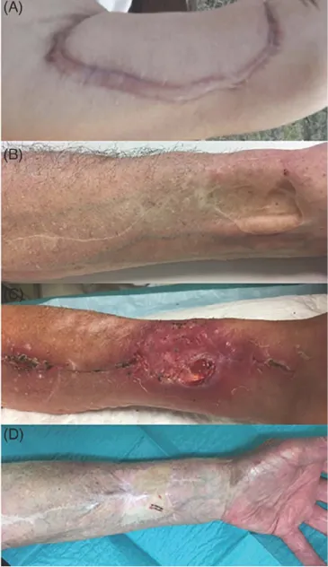
F l G UR E 3 &thetic results of radial foreann free flap donor site. A, Fîve years after closure by keystone flap. Esthetic score by patient two of five. B, One year after closure by keystone flap. Esthetic score by patient three of five. C, One y� after closure by Keystone flap. Esthetic score by patient ail of five. D, Two years after closure by skin graft. Esthetic score by patient one os five. F, R>ur years after closure by skin graft. Esthetic score by patient three of five. F, Two years after closure by skin graft. Esthetic score by patient ail of five
FIGURE 4 Wound complication examples. A, Hypertrophie scar after keystone flap. B, skin trough. C, Tendinous exposure. D, Pigmentation disorder
tendon exposure, three local infections, and four inflammations with significant distal edema (11 %) also occurred, requiring in one case a stitch release to avoid compartment syndrome.
In group B, 21 patients presented at least one wound healing complication. There were 16 partial graft necroses (24.6%) with nine tendon exposures, three local infections, and four hematomas. One of the tendon exposures motivated a secondary coverage at 15 days by KF.
The morbidity was assessed after a minimum follow-up of 12 months (max: 5 years), either by telephone or during a consultation (l'able 3). In group A, the average visual analog scale for pain was 0.61/10. Seven patients complained about hyperesthesia to touch or cold (27%), limited to the scar for three of them. Two patients (7%) deplored a "ca rdboardy" appearance of the reconstruction, and one patient described neuropathie pain (3.8%).
TABLE 1 Patient characteristics GroupA
keystone Group B skin
flap (n
=
36) graft (n=
65) p Age, median 60.5 (34 79) 61 (29 87) (years) Sex•
Male 23 51 .109•
Female 13 14 BMI, median 23.15 23.3 .9 (16.3 31.4) (14.2 31.6) Comorbidities•
Smoking 21 47•
Diabetes 2 6•
Cardiovascular 5 7 disease Tumor site•
Oral cavity 17 29•
Oropharynx 15 22•
Hypopharynx 3 9•
Lip 0 2 Flap surface (cm2) 28.7 41.1 .001TABLE 2 Healing complication and duration
GroupA GroupB
keystone skin graft (n
=
36) (n=
65) pPatients with at least one 11 (30.5%) 21 (32.3%) complication • Partial necrosis of 16(24.6%) skin graft • Wound reopening 6 (16.7%) 0 • Tendinous exposure 1 (2.8%) 9 (13.8%) .091 • Hematoma 1 (2.8%) 4 (6.2%) • Infection 3 (8.3%) 3 (4.6%) • Edema 4 (11.1 %) 0 (± epidermolysis)
Healing lime, median 14 (7 42) 28 (14 168) .001
(days)
In group B, the average visual analog scale for pain was 1.4/10 (Figure 3). Ten patients (21 %) had hyperesthesia, six patients (12%) complained about the cardboard-like appear ance, and one felt neuropathie pain (2.1 %).
Regarding the sensitivity of the reconstmcted skin pad dle, a disorder (hypoesthesia or anesthesia) was reported by nine patients (34%) in group A and 28 patients (59%) in group B (P = .049).
TABLE3 Esthetic and functional evaluation after at least 1 year GroupA GroupB
keystone skin graft
(n = 26) (n = 47) p
Pain, mean (V AS from 0.61 1.4 0 to 10)
Skin paddle sensitivity 9 (34.6%) 28 (59.5%) .049
disorder
Decreased grip strength 5 (19.2%) 11 (23.4%) Esthetic self evaluation, 3.46 3.54
mean (from 1 to 5)
Esthetic hetero evaluation, 2.76 2.70
mean (from 1 to 5) Discomfort
•
Hyperesthesia 7 (26.9%) 10 (21.3%)•
Cardboard like 2 (7.7%) 6 (12.8%) appearance•
Neuropathie pain 1 (3.8%) 1 (2.1 %)The patients' mean esthetic score was 3.46/5 and 3.51/5, respectively, in group A and B. The average score given by the independent examiner was 2.76/5 and 2.70/5, respec tively. The esthetic score differences were not statistically significant.
S I DISCUSSION
This case-control study compares SG with KF for donor site closure after forearm free flap harvesting.
In our study, the overall early wound healing complica tion rate after skin grafting (partial graft necrosis, hematoma, infection, and tendon exposure) was 32%. In a prospective study on 86 patients, Richardson et al. 8 found 16% of partial SG necrosis, and 13% of tendon exposure, comparable to our results: 13% of tendinous exposure and 24% of partial graft necrosis.
Wound healing complications after donor site closure are responsible for an increased length of postoperative care (Figure 4). In our study, the average healing time after skin grafting was 4 weeks, which is consistent with the literature: 4.68 weeks in a study by Karini et al.9
The major late complaints described in the literature are lack of cutaneous thickness, loss of grip strength, decrease in joint amplitude, dysesthesia, hypoesthesia in the tenitory of the super ficial sensory branch of the radial nerve, paresthesia, pruritus, hypersensitivity to cold, neuropathie pain, and unsightly scar.4·10-14 In group B, 59.5% of patients had sensitivity disorders of the reconstructed skin paddle (hypoesthesia or anesthesia). 34.6% patients reported hyperesthesia to touch. Twenty-two
patients reported a tolerable pain at rest (V AS 1.4). 23.8% of patients reported a subjective decrease in strength or amplitude of the wrist.
The sequelae as well as the healing disorders may be related to the RFFF harvesting itself and are therefore not modifiable: consequence of the vascular flow changes after radial artery flow interruption, causing, for example, hyper sensitivity to cold.
These sequelae and complications may also be due to the flap dissection procedure, the patient's comotbidities and, finally, the covering technique. Severa] studies compared various closure techniques and failed to demonstrate the superiority of one of them.4·15·16
Sorne procedures are known to reduce the donor site mor bidity. Supra-fascial dissection can provide a complication rate
of 6%.17 Oosing by a purse string suture can reduce by up to
44% the area to be grafted and thus the risk of healing compli cation.9·18·19 A careful tendinous coverage by peritendinous tis sue ensures both better support for engraftment and a lesser risk of postoperative tendon exposure.17 The use of a wrist splint in order to limit wrist mobility for a short time tends to improve healing, especially after skin grafting.13 The use of artificial der
mis or collagen matrix associated or not with skin grafting, may allow for a reduction of scarring complications?>
The split-thickness skin graft is widely used because it is simple and not limited by the size of the defect. However, it requires an additional donor site (frequently the anteromedial side of the thigh), involves more pain and wound care, and can cause skin depigmentation. This thin coverage provides a cuta neous depression, a risk of pruritus, and a lesser protection of the noble structures (particularly tendinous and nervous ones) favoring hypersensitivity to touch and cold.
Full-thickness skin graft is a comparable type of wound do sure that provides slightly better esthetic outcomes than split thickness skin graft. Its donor site is closed primarily and is thus less painful.21 Sorne authors harvested a full-thickness skin graft at the level of the RFFF pedicle, avoiding an additional donor site.9,22
A primary coverage by local flap, with consistency and color similar to those of defect, theoretically provides greater protec tion against tendon exposure and skin paddle hyperesthesia
Foissac et al. suggest the use of a perforator flap that arises distally to the ulnar artery for tendon exposure cover age after primary closure.23 Elliot et al. in 198824 and then Jaquet et al. in 201212 described the use of ulnar transposi tion flap, based on an ulnar perforator artery, for primary closure of small and medium donor site defects.
Our study brings to light that KF provides a reduction in healing time, with a median duration of 14 days in group A and 28 days in group B (P < .001). Moreover, we observed less ten don exposure after KF closure, at the cost of an increased risk of
compartment syndrome. Barly wound healing disorders were not greater in group A.
Concerning esthetic and functional outcomes, sensitivity disorders were less frequent after KF closure than after skin grafting (34.6% vs 59.5%, P = .049) This can be explained by the preservation of deep sensory pedicles. The skin pad dle hypoesthesia, frequently reported after skin grafting, is limited to the scar with KF closure, thus decreasing the risk of injury. The analysis of other parameters (esthetic self and hetero-evaluation, grip strength, and distal pain) did not show any significant difference between the two groups.
Postoperative pain was not assessed because this criterion is neither reliable nor reproducible in a retrospective study. Nev ertheless, it seems to us that the KF is more painful during the first few days (because of the significant tension of the closure, leading to local edema), while the skin graft preferentially cau ses burning pain on the secondary harvesting site.
The main complication of this closure is the compartment syndrome, requiring close monitoring of edema, tenderness, and distal motricity in the early postoperative days.
The KF is suitable for small to medium size defects. In our study, the calculated area of group A flaps (elliptical) is statistically smaller than the estimated area of group B grafts (rectangular) (28.69 vs 41.05 cm2; P < .001). The average area of group A flaps is comparable to literature data for local perforator flaps: between that of the series of Jaquet et al. (19.8cm2
)12 and Hsieh et al. (47cm2 ).25
In our study, the preoperative assessment of KF coverage possibility is not based on the defect size, but on the ratio of the flap width to the forearm perimeter as well as on skin laxity.
This flap is less favorable on colored skin.26 It is indeed based on tension distribution on scars, which is a risk factor for keloid scars.
The limitations of our study were the bias of retrospec tively collected data and the absence of morbidity analysis of the SG secondary harvesting site. Our study does not establish clear-cut criteria for choosing this type of recon struction, but it demonstrates that this technique is an inter esting alternative to skin grafting.
Future studies should prospectively evaluate postopera tive pain, skin graft donor site morbidity, and objective mea sures of the ratio of the flap width to the forearm perimeter in order to propose precise indications for KF.
6 1 CONCLUSION
While the radial forearm is the ideal choice for closure of many defects, the donor site morbidity remains an issue, with no method of closure conclusively superior to another. This study offers a valuable alternative to the closure of small to medium sized radial forearm flap donor sites. Keystone-type local flap closure is an interesting option to skin graft closure that
provides coverage of the donor site with reduced healing time and acceptable outcomes.
CONFLICT OF INTEREST There are no conflicts of interest. ORCID
Pauline Potet
G
https://orcid.org/0000-0001-6869-6284Emilien Chabrillac <) https://orcid.org/0000-0003-4113-2846
REFERENCES
1. Yang G, Chen B, Gao Y, et al. Foreann free skin flap transplanta tion [in Chinese]. Nat/ Med J China. 1981;61:139.
2. Wood JW, Broussard KC, Burkey B. Preoperative testing for radial foreann free flaps to reduce donor site morbidity. JAMA Otolaryngol Neck Surg. 2013;139(2): 183.
3. Harris BN, Bewley AF. Minimizing free flap donor site morbidity.
Curr Opin Otolaryngol Head Neck Surg. 2016;24(5):447 452. 4. Pabst AM, Werk meister R, Steegmann J, Holzle F, Bartella A. Is
there an ideal way to close the donor site of radial forearm free flaps? Br J Oral Maxillofac Surg. 2018;56(6):444 452.
5. Behan FC. The keystone design perforator Island flap in recontructive surgery. ANZJ Surg. 2003;73(3):112 120.
6. Chaput B, Herlin C, Espié A, Meresse T, Grolleau JL, Ganido I. The
keystone flap alternative in posttraumatic lower extrernity reconstruc tion. J PlastReconstr Aesthet Surg. 2014;67(1):130 132.
7. Abraham JT, Saint Cyr M. Keystone and pedicle perforator flaps in reconstructive surgeiy. Clin Piast Surg Avr. 2017;44(2):
385 402.
8. Richardson D, Fisher SE, Vaughan ED, Brown JS. Radial forearm flap donor site complications and morbidity: a prospective study. Piast Reconstr Surg. 1997 Jan ;99(1):109 115.
9. Karirni A, Mahy P, Reychler H. Oosure of radial forearm free flap donor site defect with a local meshed full thickness skin graft : a retrospective stndy of an original technique. J Cranio Maxillofac Surg. 2007;35 (8):369 373.
10. de Witt CA, de Bree R, Verdonck de Leeuw IM, Quak JJ, Leemans CR. Donor site morbidity of the fasciocutaneous radial foreann flap: what does the patient really bother? Eur Arch
Otorhinolaryngol. 2007;264(8):929 934.
11. Orlik JR, Horwich P, Bartlett C, Trites J, Hart R, Taylor S. Long term functional donor site morbidity of the free radial forearm flap in head and neck cancer survivors. J Otolaryngol Head NeckSurg.
2014;43(1): 1.
12. J aquet Y. Radial forearm free flap don or site morbidity ulnar based transposition flap vs Split thickness skin graft. Arch
Otolaryngol Head NeckSurg. 2012;138(1):38.
13. Emerick KS, Deschler DG. Incidence of donor site skin graft loss requiring surgical intervention with the radial forearm free flap.
Head Neck J. 2007;29(6):5 73 5 76.
14. Fang QG, Shi S, Zhang X, Li Z N, Liu F Y, Sun C F. Upper extrernity morbidity after radial forearm flap harvest : a prospec
15. Ho T, Couch M, Carson K, Schimberg A, Manley K, Byrne PJ. Radial foreann free flap donor site outcomes comparison by closure
methods. Otolaryngol Head Neck Surg. 2006;134(2):309 315. 16. Zuidam JM, Coert JH, Hofer SOP. Closure of the donor site of the
free radial foreann flap: a comparison of full thickness graft and
Split thickness skin graft. AM Piast Surg. 2005;55(6):612 616. 17. Shonka DC, Kohli NV, Milam BM, Jameson MJ. Suprafascial bar
vest of the radial forearm free flap decreases the risk of postopera tive tendon exposure. AM Otol Rhinol I.aryngol. 2017;126(3):
224 228.
18. D'arpa S, Cillino M, Mazzucco W, et al. An algorithm to improve
outcomes of radial forearm flap donor site. Acta Chir Belg. 2018; 118(4):219 226.
19. Winslow CP, Hansen J, Mackenzie D, Cohen n, Wax MK. Pur sestring closure of radial foreann fasciocutaneous donor sites. Laryngoscope. 2000;110(11):1815 1818.
20. Wester JL, Pittman AL, Lindau RH, Wax MK. AlloDerm with Split thickness skin graft for coverage of the foreann free flap
donor site. Otolaryngol Head Neck Surg J. 2014;150(1):47 52.
21. Kim TB, Moe KS, Eisele DW, Orloff LA, Wang SJ. Full thickness skin graft from the groin for coverage of the radial foreann free flap donor site. Am J Otolaryngol. 2007;28(5):325 329.
22. Riecke B, Assaf AT, Heiland M, et al. Local full thickness skin graft of the donor arm a novel technique for the reduction of
donor site morbidity in radial forearm free flap. /nt J Oral Maxi/
l.ofac Surg. 2015;44(8):937 941.
23. Foissac R, Benatar M, Dassonville 0, Bozec A, Poissonnet G, Camuzard O. Coverage of tendon exposure after radial foreann free flap by the Dorsoulnar artery perforator flap. Otolaryngol
Head Neck Surg. 2017;156(5):822 827.
24. Elliot D, Bardsley AF, Batchetor AG, Soutar OS. Direct closure of the radial forearm flap donor defect Br J Piast Surg . 1988;41(4):358 360.
25. Hsieh C H, Kuo Y R, Yao S F, Liang CC, Jeng S F. Primary clo sure of radial forearm flap donor defects with a bilobed flap based
on the fasciocutaneous perforator of the ulnar artery. Piast Reconstr Surg. 2004;113(5): 1355 1360.
26. Bhat S. Keystone flaps in coloured skin: flap technology for the
masses? Jndian J Piast Surg. 2013;46(1):36.
How to cite this article: Potet P, De Bonnecaze G, Chabrillac E, Dupret-Bories A, Vergez S, Chaput B. Closure of radial forearm free flap donor site: A comparative study between keystone flap and skin graft. Head & Neck. 2020;42:217 223. https://doi.