HAL Id: hal-03023558
https://hal.archives-ouvertes.fr/hal-03023558
Submitted on 15 Dec 2020
HAL is a multi-disciplinary open access
archive for the deposit and dissemination of
sci-entific research documents, whether they are
pub-lished or not. The documents may come from
teaching and research institutions in France or
abroad, or from public or private research centers.
L’archive ouverte pluridisciplinaire HAL, est
destinée au dépôt et à la diffusion de documents
scientifiques de niveau recherche, publiés ou non,
émanant des établissements d’enseignement et de
recherche français ou étrangers, des laboratoires
publics ou privés.
Treatment of experimental autoimmune
encephalomyelitis with engineered bi-specific Foxp3+
regulatory CD4+ T cells.
Manish Malviya, Abdelhadi Saoudi, Jan Bauer, Simon Fillatreau, Roland
Liblau
To cite this version:
Manish Malviya, Abdelhadi Saoudi, Jan Bauer, Simon Fillatreau, Roland Liblau. Treatment of
exper-imental autoimmune encephalomyelitis with engineered bi-specific Foxp3+ regulatory CD4+ T cells..
Journal of Autoimmunity, Elsevier, 2020, 108, pp.102401. �10.1016/j.jaut.2020.102401�. �hal-03023558�
Contents lists available atScienceDirect
Journal of Autoimmunity
journal homepage:www.elsevier.com/locate/jautimm
Treatment of experimental autoimmune encephalomyelitis with engineered
bi-speci
fic Foxp3+ regulatory CD4+ T cells
Manish Malviya
a, Abdelhadi Saoudi
a, Jan Bauer
b, Simon Fillatreau
c,d, Roland Liblau
a,∗aCentre de Physiopathologie Toulouse-Purpan (CPTP), Université de Toulouse, Centre National de la Recherche Scientifique (CNRS), Institut National de la Santé et de la
Recherche Médicale (Inserm), Université Paul Sabatier (UPS), Toulouse, France
bDepartment of Neuroimmunology, Center for Brain Research, Medical University of Vienna, A-1090, Austria
cInstitut Necker-Enfants Malades (INEM), INSERM U1151-CNRS UMR 8253, Université Paris Descartes, Sorbonne Paris Cité, Bâtiment Leriche, 75993, Paris, France dAP-HP, Hôpital Necker Enfants Malades, Paris, France
A B S T R A C T
The use of autoantigen-specific regulatory T cells (Tregs) as a cellular therapy for autoimmune diseases is appealing. However, it is challenging to isolate and expand large quantity of Tregs expressing disease-relevant T-cell receptors (TCR). To overcome this problem, we used an approach aiming at redirecting the specificity of polyclonal Tregs through autoreactive TCR gene transfer technology. In this study, we examined whether Tregs engineered through retroviral transduction to express a TCR cross-reactive to two CNS autoantigens, myelin oligodendrocyte glycoprotein (MOG) and neurofilament-medium (NF-M), had a superior protective efficacy compared with Tregs expressing a MOG mono-specific TCR. We observed that engineered Tregs (engTregs) exhibited in vitro regulatory effects related to the antigenic specificity of the introduced TCR, and commensurate in potency with the avidity of the transduced TCR. In experimental autoimmune encephalomyelitis (EAE), adoptively transferred engTregs proliferated, and migrated to the CNS, while retaining FoxP3 expression. EngTregs expressing MOG/NF-M cross-reactive TCR had superior protective properties over engTregs expressing MOG-specific TCR in MOG-induced EAE. Remarkably, MOG/NF-M bi-specific TCR-engTregs also improved recovery from EAE induced by an unrelated CNS autoantigen, proteolipid protein (PLP). This study underlines the benefit of using TCRs cross-reacting towards multiple autoantigens, compared with mono-reactive TCR, for the generation of engTregs affording protection from autoimmune disease in adoptive cell therapy.
1. Introduction
Multiple sclerosis (MS) is a chronic inflammatory disease of the central nervous system (CNS) likely caused by an autoimmune response against self-antigens expressed in the CNS [1–3]. Prevention of auto-immunity relies, at least in part, on a subset of regulatory CD4 T cells (Tregs) characterized by the expression of the transcription factor forkhead-box protein P3 (Foxp3), which is essential for their develop-ment and suppressive functions [4,5]. This population limits tissue damage and inflammation by inhibiting the activation and effector functions of several immune cells, including conventional CD4 and CD8 T cells, B cells, NK cells, NKT cells, and monocytes as well as dendritic cells [6,7].
Autoreactive Tregs express self-reactive T-cell receptor (TCR) with higher affinity than conventional T cells (Tconvs) for the corresponding self-antigen [8]. An imbalance in Treg/Tconv immune homeostasis contributes to autoimmune pathogenesis [9]. In multiple sclerosis (MS) patients [10] and other autoimmune diseases, such as rheumatoid ar-thritis [11], type 1 diabetes [12] and myasthenia gravis, either the frequency of Tregs is reduced or their functional properties are altered [13–15]. One possibility to restore the Treg/Tconv balance would be to
adoptively transfer polyclonal Tregs. However, the adoptive transfer of polyclonal Tregs could have beneficial effects on autoimmunity while bearing deleterious consequences on protective systemic immune re-sponses [16,17]. Thus, the adoptive transfer of disease-relevant an-tigen-redirected functional Tregs might be a possible therapeutic strategy in MS and other autoimmune diseases [18–21].
Importantly, whereas the adoptive transfer of polyclonal Tregs had little effect, myelin-reactive Tregs cured recipient mice from experi-mental autoimmune encephalomyelitis (EAE), an animal model of MS [8,22]. However, autoantigen-specific Tregs are rare and remain diffi-cult to isolate. Furthermore, to obtain the desired number of antigen-specific Tregs for adoptive transfer studies, multiple rounds of ex vivo expansion using antigen-loaded antigen-presenting cells (APC) are needed, which can lead to decreased suppressive function of Tregs and promote their plasticity into Th17 cells [23,24]. Several alternate methods for in vivo expansion of antigen-specific Tregs have been re-ported, such as injection of nanoparticle attached self-peptides [25], and oral or intravenous injection of self-peptides [26,27] or low dose IL-2 [21]. However, the injected self-peptides can also be taken-up, pro-cessed, and presented by APC to Tconvs, which can further enhance pathogenic immune response to self-antigens.
https://doi.org/10.1016/j.jaut.2020.102401
Received 12 December 2019; Accepted 1 January 2020
∗Corresponding author. Centre de Physiopathologie de Toulouse-Purpan Hospital, Place du Docteur Baylac TSA 40031, 31059, Toulouse Cedex 9, France.
E-mail address:roland.liblau@inserm.fr(R. Liblau).
Journal of Autoimmunity 108 (2020) 102401
Available online 13 January 2020
0896-8411/ © 2020 Elsevier Ltd. All rights reserved.
A promising approach for generating self-antigen-specific Tregs is the introduction of chosen TCR genes by retroviral or lentiviral gene transfer in polyclonal Tregs. Several studies have demonstrated the feasibility of this method both in mice and humans [28–30]. For ex-ample, the adoptive transfer of myelin oligodendrocyte glycoprotein (MOG)-specific Tregs generated by TCR gene transfer was more potent at preventing mice from EAE than that of polyclonal Tregs [8]. Simi-larly, auto-antigens redirected engTregs generated with chimeric an-tigen receptors were superior to polyclonal Tregs at preventing ex-perimental models of inflammatory bowel disease, autoimmune disease, and graft-versus host disease [31–35].
It is now well documented that the TCR cross-react with multiple antigens, though with different avidity, so that the limited number of T cells can cope with the vast number of peptide ligands. Autoreactive T cells recognizing multiple self-antigens have been reported in several immune-mediated diseases, including MS [36,37]. Interestingly, ef-fector CD4 T cells targeting both MOG and neurofilament-medium (NF-M) have emerged as highly pathogenic from our studies of EAE in C57BL/6 mice. These dual self-antigens cross-reactive Tconv cells were able to induce EAE when either of the two target auto-antigens were present (MOG−/− or NF-M−/− mice), and were highly pathogenic when both target antigens were expressed in wild type (WT) mice [38–41]. Harnessing the functional properties of high-avidity dual self-antigens cross-reacting TCR for therapeutic application could represent an interesting option to generate bi-specific protective Tregs. In the present study, we investigated the in vivo therapeutic effect of MOG/NF-M bi-specific TCR-engineered Tregs (engTregs) and compared them with MOG-specific TCR-engTregs. To this goal, we engineered poly-clonal Tregs to express one of two MOG/NF-M cross-reactive TCRs, and one of two MOG mono-specific TCRs with varying avidity towards their cognate antigen(s), and tested whether cross-reactivity of TCR, its avidity or both on the engTregs correlated with the level of protection against T cell-mediated immunopathology in EAE. We demonstrate that TCR-engTregs bearing TCR cross-reactive to two self-antigens not only strongly suppress an autoimmune response to these self-antigens, but also inhibit EAE induced by an unrelated autoantigen. Hence these data suggest that generating Tregs with cross-reactive TCR may be a valu-able therapeutic option in MS, and other autoimmune diseases. 2. Experimental procedures
2.1. Mice
Male C57BL/6 mice, 10–15 weeks old, were purchased from Charles River Laboratories, France. MOG-specific TCR-transgenic 2D2 mice on a C57BL/6 background [42] have been backcrossed with CD45.1 con-genic animals. Mice were kept in specific pathogen-free conditions and used in accordance of the European Union guidelines following ap-proval of the local ethics committee.
2.2. Peptides
The peptides MOG (35–55) (MEVGWYRSPFSRVVHLYRNGK), NF-M (15–35) (RRVTETRSSFSRVSGSPSSGF), and (PLP178–191) (NTWTTCQS-IAFPSK) were purchased from Polypeptide Laboratories (San Diego, CA) with a purity > 95%.
2.3. Cloning of the TCR genes into a retroviral vector
MOG-specific T-cell hybridomas were previously described [40,43,44]. The paired TCRα and TCRβ cDNA sequences of chosen TCR clones were amplified using gene specific Vα and Vβ forward primers and a common Cα and Cβ reverse primers as described previously [43], and linked using a 2A element of porcine teschovirus (P2A) by PCR and cloned into the MP71 retrovirus vector [8,45] using NotI and EcoRI restriction sites to obtain TCRβ-P2A-TCRα. In addition, the Thy1.1 gene
was linked to the TCR chains using a 2 A linker element of teschovirus (T2A) to produce TCRβ-P2A-TCRα-T2A-Thy1.1 that provided a surface marker for the identification of virus transduced T cells.
2.4. Production of retrovirus and T-cell transduction
Retrovirus particles were produced using Plat-E cells by transient transfection with the TCR or control retrovirus vector using Lipofectamine®2000 transfection reagent (Invitrogen). Supernatants containing retroviral particles were harvested at 48 h and 72 h after transfection, centrifuged at 350 g for 1 min and carefullyfiltered using 40 μm sterile cell strainer to remove cell debris. Supernatants were mixed with 5μg/ml polybrene infection reagent (Sigma) for 30 min at 37 °C and used for transduction of Tregs or Tconvs.
CD4+TCR+CD25hiTregs or CD4+TCR+CD25− Tconvs were iso-lated via cell sorting using a BD FACSAriaflow cytometer from pooled spleen and lymph nodes of four tofive donor male C57BL/6 mice. These cells were then stimulated in aflat bottom 96-well tissue culture plate (TRP) coated with 1μg/ml anti-CD3 and soluble anti-CD28 (BD) mAbs or with anti-CD3/CD28 dynabeads (Invitrogen). Activated CD4 Tconvs were cultured in complete medium (RPMI 1640 medium with 10% FBS, 100 units/mL penicillin, 100 mg/mL streptomycin, 1 mM non-essential amino acids, 50 mMβ-mercaptoethanol, 1 mM sodium pyruvate, and HEPES). The Treg culture medium also contained 1000 U/ml re-combinant mouse IL-2 (R&D systems), and, where indicated, 100 nM rapamycin (Sigma). Cultured Tregs or Tconv were transduced on day 2 and 3 after isolation, by centrifugation at 32 °C for 2 h in six-well plates coated with 10μg/ml RetroNektin (TaKaRa Biomedicals). The in vitro and in vivo experiments were performed 4–6 days after the transduced Treg cells were generated.
2.5. Adoptive transfer of Tregs and EAE induction
EAE was induced and assessed as described [8,39,41]. In brief, 10 to 12-week-old C57BL/6 male mice were immunized subcutaneously at the base of the tail either with 100μg of MOG (35–55) or 200 μg of PLP (178–191) emulsified in complete Freund's adjuvant (CFA; BD) con-taining 500μg of Mycobacterium tuberculosis (Strain H37 Ra, BD). Per-tussis toxin (200 ng; List Biological Laboratories) was injected in-travenously at days 0 and 2 post-immunization. Retrovirus transduced TCR-engTregs (106
cells), control Tregs (106cells) or PBS were injected intravenously either one day before EAE induction or on day 9 post-immunization. Disease severity was scored daily on a 5 point scale: 0, no neurological sign; 1, tail atony; 2, hind limb weakness; 3, hind limb paralysis; 4, forelimb paralysis; and 5, moribund.
2.6. Peripheral and CNS mononuclear cell isolation
Mice were anaesthetized with ketamine and transcardially perfused with cold PBS. Spleen, lymph nodes (cervical, axillary & inguinal), brain and spinal cord were collected separately. Blood samples were collected in EDTA-coated tubes before perfusion. Cells from spleen, lymph nodes and blood were treated with ACK (Ammonium-Chloride-Potassium) lysing buffer before CD4 T cells were enriched using the dynabeads untouched Mouse CD4 kit (Invitrogen). Mononuclear cells from brain and spinal cord were isolated by homogenization and di-gestion using collagenase D (Roche) and DNaseI (Sigma) followed with a triple percoll gradient (30, 37, 70%) and centrifugation for 20 min at 2000 rpm [41,46].
2.7. Flow cytometry
Cell surface staining was performed with mAbs specific for CD4, CD8, TCR, Thy1.2, Thy1.1, PD1, CTLA-4, CD25, ICOS, GITR, CD103 as well as a viability dye (eFluor 780). Expression of other cell surface markers CD11b, CD11c, CD19, MHC-II, CD45, ICOS and CCR7 was also
assessed by staining of mononuclear cells isolated from the CNS of mice with EAE, using the respective mAbs.
For intracellular cytokine staining, cells were stimulated in culture medium either with MOG or NF-M peptide loaded APC for 72 h and with phorbol 12-myristate 13-acetate (PMA, 50 ng/ml, Sigma), iono-mycin (1 mg/ml, Sigma) for the last 5 h in culture medium containing GolgiStop (1μl/ml, BD Biosciences) at 37 °C in a humidified 5% CO2 atmosphere [46]. After staining for surface markers (CD4, CD8, Thy1.1 and Thy1.2) and viability, cells were fixed and permeabilized for staining of IL-2, IL-10, IL-17, IL-35(IL-12p35), GM-CSF, TNFα, IFNγ, TGFβ(LAP), Granzyme B and FoxP3 using respective cytoplasmic or intra-nuclear fixation/permeabilization buffer kit from eBiosciences according to the manufacturer's instructions.
For the peptide:MHC-II tetramer staining, 106cells were incubated with MOG-PE:I-Ab, NF-M-APC:I-Abor control CLIP-PE/APC:I-Ab tetra-mers overnight at 37 °C with 5 μl tetramer in 500 μl RPMI culture medium.
Data were acquired on cytometer LSRII or Fortessa (BD) and ana-lyzed with FlowJo software (Tree Star Inc., Ashland, OR, USA). 2.8. Regulatory T cell-mediated suppression of proliferation assays
Treg suppression assays were performed following the protocol of [47] with slight modifications. Briefly, Tconvs from 2D2 mice (CD4+TCR+CD25−) expressing a MOG/NF-M bi-specific transgenic TCR were isolated and labeled with CellTrace Violet (CTV) dye. Hun-dred thousand labeled 2D2 Tconvs per well were activated with 50μg/ ml of MOG or NF-M peptide loaded APC at the ratio of 1:3 in a round bottom 96-well tissue culture plate (TRP) and cultured alone (no Tregs) or in the presence of either TCR-engTregs or control engTregs at dif-ferent Tregs:Tconv ratios. After 72 h, the suppressive efficiency of en-gTregs was determined byflow cytometry analysis of CTV dilution in the responder 2D2 Tconv cells.
2.9. Statistical analyses
EAE disease courses were compared by repeated-measure two-way ANOVA. Cumulative disease scores and data from peptide stimulations were analyzed with one-way ANOVA test. Reported significant values were obtained after multiple comparisons performed using two-way and one-way ANOVA by comparing mean of each group with the mean of every other group. Statistical analyses were performed with GraphPad Prism Software. *p < 0.05; **p < 0.01; ***p < 0.001; ****p < 0.0001.
3. Results
3.1. Validation of recombinant exogenous TCR on Tconv cells
In MS, the paucity of Tregs within inflammatory CNS lesions, as confirmed here in Table 1, likely contributes to the disease process [48,49]. Adoptive cell therapy using Tregs in human autoimmune dis-eases is a highly investigated option that has the potential to increase the frequency of Tregs in the blood and in inflamed tissues [20,21].
In the present study, we investigated, in an animal model of MS, the potential of adoptive cell therapy using Tregs redirected to recognize neural autoantigens by TCR gene transfer. We selected TCRs from two clones reacting only to MOG and from two MOG/NF-M cross-reactive T cell clones [40]. We reprogrammed the antigen specificity of polyclonal T cells by introducing either MOG-specific or MOG/NF-M bi-specific TCR through retroviral gene transfer. In order to recognize the two self-antigens, MOG/NF-M cross-reactive TCR likely contact the shared amino acid residues within the core epitopes of MOG (38–50) and NF-M (18–30) (Fig. 1A). The TCRs used in this study have either low or high avidity for the cognate peptide(s), use different V(D)J germline gene segments and have distinct CDR3 sequences (Fig. 1B). We used a
multicistronic retroviral expression vector to produce engineered Tconvs (engTconvs) or Tregs (engTregs) expressing the recombinant TCR together with an identifiable surface marker, Thy1.1. A retroviral vector expressing Thy1.1 but no exogenous TCR was used to generate control engTregs throughout this study (Fig. 1C & D). The engTregs expressing a recombinant TCR are named TCR-engTregs, while those transduced with the control vector are named control-engTregs.
First, we validated this engineering strategy in engTconvs by as-sessing surface expression of the recombinant TCRs and Thy1.1, and by performing functional analysis of transduced cells. For this purpose, we isolated polyclonal CD4+TCR+CD25−Tconvs and activated them with anti-CD3ε and anti-CD28 antibodies 48 h before transducing them with TCR-encoding or control retroviruses. Due to the unavailability of clo-notype-specific antibodies to specifically detect the expression of the recombinant TCR on the transduced cells, we relied on Thy1.1 ex-pression to evaluate transduction efficiency. Transduction efficiency was about 80% (Fig. 1E). The surface expression level of TCRβ on TCR-engTconvs and on non-transduced T cells was similar (Fig. 1E).
We then assessed the pairing of recombinant TCRα and β chains at the cell surface of TCR-engTconvs, using fluorescently-labeled peptide:I-Abtetramers and functional response to cognate antigen(s). Transduced CD4 Tconvs were incubated with MOG:I-Ab, NF:M-I-Aband the non-specific CLIP:I-Abtetramers, and the tetramer staining among the Thy1.1+population was analyzed byflow cytometry. As expected, engTconvs expressing a MOG mono-specific TCR bound to MOG-I-Ab only (Fig. 1F), whereas Tconvs expressing a MOG/NF-M cross-reactive TCR bound to both MOG:I-Aband NF-M:I-Abtetramers (Fig. 1F). The two MOG/NF-M bi-specific TCR have low avidity towards MOG (Fig. 1F). However, one of them exhibited a strong binding to the NF-M:I-Abtetramers, as assessed by frequency and MFI of staining, and was therefore named MOGlow/NF-Mhi TCR, while the other was named MOGlow/NF-MlowTCR (Fig. 1F). Similarly, among the two MOG mono-specific TCRs, MOG:I-Abtetramer binding confirmed that one has high avidity for MOG, thus named MOGhiTCR, and the other one has low avidity, hence named MOGlowTCR (Fig. 1F). These data indicate the efficient pairing of the recombinant TCRα and β chains on the surface of transduced cells.
To test the functionality of the recombinant TCRs, engTconvs were
Table 1
Quantification of FoxP3 Tregs in the CNS of MS patients versus non-neurolo-gical controls.
Gender Age (Years) CD3 (cells/mm2) % FoxP3+ Treg cells
Multiple sclerosis Female 68 52.7 0.0 Female 45 190.4 3.0 Female 45 204.2 0.0 Female 40 39.8 0.0 Female 57 63.7 0.0 Female 20 56.5 1.7 Female 55 126.2 0.9 Female 71 22.6 0.1 Female 83 16.0 0.0 Male 45 190.4 3.0 Male 36 32.5 0.0 Average 52.0 75.3 0.5 Controls Female 45 2.8 0.0 Female 42 13.7 0.0 Female 30 26.3 0.0 Female 47 13.0 0.0 Female 36 5.4 0.0 Female 39 12.2 0.0 Female 71 18.6 0.0 Female 70 9.6 0.0 Male 37 13.3 0.0 Male 65 6.0 0.0 Male 83 6.4 0.0 Average 51.4 11.5 0.0
co-cultured with MOG or NF-M peptide-loaded APC and production of IL-2 was assessed byflow cytometry. MOG (35–55) peptide induced significant IL-2 expression in both MOG/NF-M cross-reactive and MOG mono-specific TCR-engTconvs, confirming that all recombinant TCRs are MOG reactive (Fig. 1G). The proportion of IL-2-producing cells in response to the MOG (35–55) peptide correlated well with the binding of MOG:I-Abtetramers to the exogenous TCR (Fig. 1G, left panel). On the other hand, stimulation with NF-M(15–35) peptide elicited IL-2
production only in MOG/NF-M cross-reactive TCR-engTconv cells, confirming the cross-reactivity of these TCR towards MOG and NF-M (Fig. 1G, right panel). Here again the functional avidity of the TCR-engTconvs correlated with the binding of NF-M:I-Abtetramers to the exogenous TCR. These data indicate that the recombinant TCR induce efficient signaling in the transduced cells, and show that the functional avidity of these TCR correlated with the magnitude of tetramer staining.
3.2. Functional characterization of engineered Tregs expressing recombinant TCR
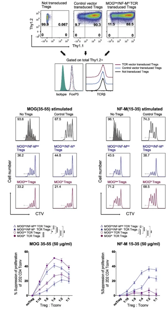
We characterized the expression and function of TCR-engTregs for the four recombinant TCRs. Polyclonal CD4+TCR+CD25hiT cells were isolated by FACS and expanded using anti-CD3ε and CD28 anti-bodies before retrovirus transduction. Expression of FoxP3 by sorted Tregs was assessed before and six days after retrovirus transduction. Transduction efficiency on Tregs was determined by Thy1.1 staining (Fig. 2A). For this study, only experiments in which more than 70% of Tregs were efficiently transduced, and in which more than 97% of cells expressed FoxP3 were used for in vitro and in vivo experiments (Suppl. Fig. 1). We also assessed whether retrovirus transduction altered some key features of Tregs by comparing the expression of cell surface mar-kers and intracellular cytokines in non-transduced vs. transduced en-gTregs on day six post-transduction. We did not observe notable dif-ferences among thefive types of Tregs (Suppl.Fig. 2), and detected no changes in surface TCR expression after transduction (Fig. 2A).
We next tested in vitro the antigen-specific suppressive function of TCR-engTregs, using as responder cells Tconvs from 2D2 mice, which carry a transgenic TCR specific for both MOG and NF-M [38]. Sup-pression of 2D2 Tconv proliferation to MOG or NF-M peptides was evaluated at various ratios of TCR-engTregs to 2D2 Tconvs (Fig. 2B). The control-engTregs modestly suppressed 2D2 Tconv proliferation at a ratio of 1:2 (Fig. 2B; top panels), a likely consequence of activation of the control-engTregs for the retroviral transduction. A stronger antigen-specific suppression of 2D2 cell proliferation was observed for each of the four types of TCR-engTregs even at 1:8 Treg:Tconv ratios (Fig. 2B). The magnitude of this antigen-dependent suppression correlated with the avidity of the recombinant TCR for the tested self-antigens. Thus, engTregs expressing the MOGhiTCR achieved a 47% antigen-specific suppression of 2D2 Tconvs proliferation at a ratio of 1:2, while this value was 29% for MOGlowTCR-engTregs. The two bi-specific TCRs also endowed polyclonal Tregs with antigen-specific suppressive capacities. Indeed, engTregs expressing the MOGlow/NF-Mlowor MOGlow/NF-Mhi TCR suppressed 2D2 Tconvs proliferation to 37% and 21%, respectively at the ratio of 1:2 (Fig. 2B; upper & lower panels). Importantly, the MOG/NF-M bi-specific TCR-engTregs additionally suppressed the re-sponse of 2D2 Tconvs to NF-M, with a stronger suppressive effect ob-served with engTregs carrying the MOGlow/NF-MhiTCR (Fig. 2B; upper & lower panels). In contrast, TCR-engTregs bearing a MOG-mono-spe-cific TCR had no effect on NF-M stimulated cultures (Fig. 2B; upper & lower panels). We conclude that TCR-engTregs suppress autoreactive Tconvs in an antigen-dependent manner, so that bi-specific TCR un-iquely expand the reactivity profile of TCR-engTregs.
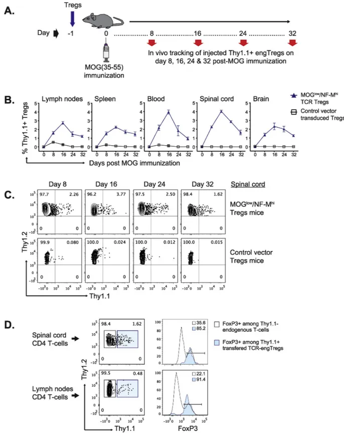
3.3. In vivo persistence and stability of transferred engineered Tregs We next evaluated the activation of adoptively transferred engTregs, and their maintenance of FoxP3 expression, in recipient mice
during EAE. TCR-engTregs or control-engTregs, were administered to Thy1.2 C57BL/6 recipient mice one day before EAE induction by im-munization with MOG (35–55). Lymph nodes, spleen, blood, spinal cord, and brain samples were then analyzed on days 8, 16, 24 and 32 post-immunization (Fig. 3A). TCR-engTregs were detected in all ex-amined tissues on day 8, and at even higher frequencies on day 16, while their abundance then progressively declined at later time points. This response was antigen-specific because, in contrast, control-en-gTregs did not display any expansion (Fig. 3B & C). Although there was not a drastic increase in TCR-transduced Treg frequency in the CNS vs. lymphoid tissues, there seems to be a preferential retention of TCR-transduced Tregs in the CNS at day 32 (Suppl.Fig. 3). Thesefindings suggest that TCR-engTregs were stimulated by the MOG (35–55) pep-tide used for immunization, and subsequently accumulated in the CNS. However, the supra physiological quantity of MOG-specific Tregs used to treat mice may have saturated the local MOG antigen-presentation capacities in the CNS and thereby Treg retention. Importantly, the TCR-engTregs retained high FoxP3 expression in recipient mice for at least 32 days (Fig. 3D).
3.4. TCR cross-reactivity and functional avidity determine the protective function of engineered Tregs during MOG-induced EAE
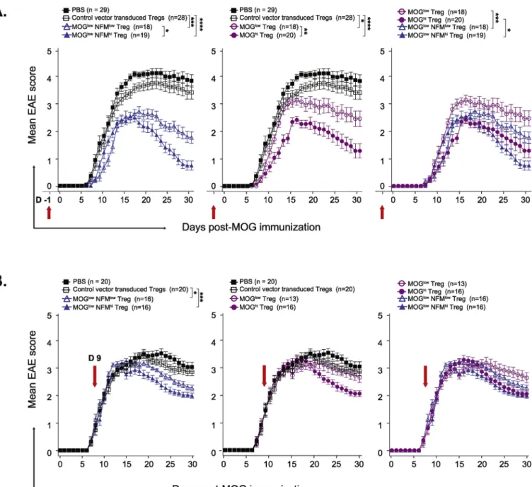
We next tested whether the protective function of TCR-engTregs was influenced by the bi-vs. mono-specificity of their recombinant TCR, or their functional avidity towards their cognate antigen, during EAE. In a preventive disease setting, the adoptive transfer of 106 TCR-engTregs one day before EAE induction resulted in a less severe disease and a faster recovery in recipient mice, compared to mice that received control-engTregs or PBS (Fig. 4A;Table 2). The properties of the re-combinant TCR determined the protective efficacy of the transferred Tregs. High avidity TCR either for NF-M (MOGlow/NF-MhiTregs) or for MOG (MOGhiTregs) conferred better protective function to transduced Tregs compared to low avidity TCRs (Fig. 4A; left & middle panels). Notably, among the two-high avidity TCRs, the bi-specific TCR-en-gTregs were more protective than TCR-enTCR-en-gTregs expressing the mono-specific TCR (Fig. 4A; right panel). These data suggest that both the functional avidity and the cross-reactivity of the self-reactive TCRs impacted the protective function of TCR-engTregs in EAE.
We then assessed the in vivo protective function of TCR-engTregs in a therapeutic setting by injecting these cells on day 9 post-immuniza-tion. The engTregs expressing the TCRs with high avidity to NF-M (bi-specific) or MOG (mono-(bi-specific), reduced disease severity when ad-ministered in recipient mice after onset of EAE signs (Fig. 4B,Table 3). The bi-specific MOGlow/NF-MlowTCR-engTregs also provided benefit, whereas the MOGlowTCR-engTregs did not. These data suggest that the engTregs with high avidity and/or bi-specific TCRs are more effective for the treatment of autoimmune diseases.
Fig. 1. Transfer of recombinant TCR from autoreactive CD4 T-cell hybridomas into polyclonal T cells via a retroviral expression system. (A) Shared amino acid residues between the MOG and NF-M peptides in the context of I-Abare shown in red.(B) CDR3 regions of the two chosen MOG/NF-M cross-reactive TCR (2MOG60
& 3MOGN204) and the two MOG-specific TCR (1MOG26 & 1MOG210) as reported in Lucca et al., 2014. The short names attributed to each TCR for easy reading are shown in the right column.(C) Retroviral vector genome: the paired TCRα and β chain genes of each MOG-reactive T-cell hybridoma were inserted in frame into a MP71 retroviral vector together with a Thy1.1 gene as a congenic surface marker. The P2A and T2A peptide sequences were used to link the TCRβ, α, and Thy1.1 genes. The control retroviral vector carries only the Thy1.1 gene. LTR - long terminal repeat.(D) Experimental scheme: autoreactive TCRs were transferred into polyclonal CD4 Tregs or Tconvs via retroviral transduction to redirect their specificity resembling a monoclonal population. (E) Representative flow cytometry plots showing Thy1.1 staining (upper panels) and TCRβ staining (endogenous + exogenous; lower panel) among not transduced versus control vector or TCR vector (MOGlow/NF-MhiTCR) engTconvs on day 6 after transduction. The lower panel is an overlay histogram showing TCRβ expression on the X-axis and the count of
events on the Y-axis.(F) Representative histograms showing frequency of peptide:MHC-II tetramer binding cells among Thy1.1+
engTconvs. Retroviral transduced Tconvs expressing one of the indicated recombinant TCRs were stained with MOG:I-Ab-PE, NF-M:I-Ab-APC (open histograms) and control CLIP:I-Ab-PE/APC
tet-ramers (filled histograms). Results from one representative of three independent experiments are shown. (G) IL-2 production by recombinant engTconvs. TCR-engTconvs expressing each of the MOG/NFM cross-reactive or MOG-reactive TCRs, or control TCR-engTconvs were stimulated with MOG (35–55) or NF-M(15–35) peptides at the indicated concentrations, and the proportion of IL-2-producing cells were quantified in the Thy1.1+population byflow cytometry analysis. Data are
Fig. 2. TCR-engineered Tregs mediate an-tigen-specific immunosuppression in vitro. (A) Representative flow cytometry plots assessing transduction efficiency of Tregs based on Thy1.1 staining (upper panels) as well as levels of FoxP3 and TCR expression (lower panels) among not transduced versus control vector or TCR vector (MOGlow
/NF-Mhi TCR) transduced FACS sorted CD4+TCR+CD25hi cells on day 6 after
transduction. (B) CD4 Tconvs from 2D2 mice were isolated and labeled with CellTrace Violet (CTV) dye. Labeled 2D2 Tconvs were activated with MOG or NF-M peptide-loaded APC in the absence (no Tregs) or presence of TCR-engTregs or control-engTregs at different ratios. After 72 h, the suppressive property of Tregs was determined by assessing inhibition of CTV dilution byflow cytometry analysis. Top: Representative histograms showing Treg-mediated immunosuppression as measured by CTV dilution. Labeled 2D2 CD4 Tconv cells were activated with MOG (left panels) or NF-M (right panels) and cultured alone or with the indicated Tregs at a Treg:Tconv ratio of 1:2. Representative results from one of three independent experiments. Number indicates the percentage of 2D2 Tconvs that underwent at least one cell division. Bottom: Graph showing the antigen-spe-cific suppression of proliferation of 2D2 Tconvs mediated by TCR-engTregs, ob-tained after subtraction of the antigen-in-dependent suppression induced by control-engTregs. Graphs show mean ± SEM of three pooled independent experiments each performed in triplicates.
Fig. 3. In vivo persistence of adoptively transferred TCR-engineered Tregs. (A) Experimental scheme: TCR-engTregs expressing Thy1.1 and the MOGlow/NF-Mhi cross-reactive TCR, or control-engTregs expressing only Thy1.1, were administered to C57BL/6 mice one day before EAE induction by immunization with MOG (35–55). Lymph nodes, spleen, blood, spinal cord, and brain samples were collected on day 8, 16, 24 and 32 post-immunization. Flow cytometry analyses were performed to quantify the frequency of Thy1.1+transferred Tregs in the harvested tissues.(B) Kinetics of the expansion and persistence of MOGlow/NF-Mhi
cross-reactive TCR-engTregs versus control-engTregs in C57BL/6 mice. Mice were sacrificed at the indicated time points (n = 2/3 per group and per timepoint) and expression of Thy1.1 among total CD4 T cells was analyzed in organs of the recipient animals. Graphs show the percentage of Thy1.1+cells among CD11b−CD45hi
CD4+Thy1.2+viable cells (mean ± SEM).(C) Representativeflow cytometry dot plots showing Thy1.1+staining among total CD4 T cells from the spinal cord of
mice injected with MOGlow/MF-Mhi cross-reactive TCR-engTregs or control-engTregs. Values indicate the percentage of cells falling in each quadrant. (D)
Representativeflow cytometry dot plots showing expression of FoxP3 among transferred Thy1.1+Thy1.2+MOGlow/NF-MhiTCR-engTregs (filled histogram), and
among host Thy1.1-Thy1.2+CD4 T cells (open histogram), on day 32 post-immunization in the spinal cord and lymph nodes of a representative recipient mouse.
3.5. TCR-engineered Tregs regulate autoreactive Tconv in vivo
To investigate how TCR bi-specificity affects the im-munosuppressive function of engTregs in vivo, we compared the auto-immune response in mice treated with engTregs expressing either the bi-specific MOGlowNF-MhiTCR or the mono-specific MOGlowTCR. The engTregs were administered into C57BL/6 mice one day before EAE induction by immunization with MOG (35–55), and the CD4 T cell response was analyzed in recipient mice at disease onset (day 8), peak of disease (day 16) and during recovery (day 32) in lymph nodes, spinal cord, and brain. Remarkably, the administration of MOGlowNF-Mhi TCR-expressing engTregs resulted in a profound reduction of CD4 T cells accumulation in the brain of mice at all time points, while
engTregs carrying the MOGlowTCR had no impact on this parameter (Fig. 5A). In agreement, mice treated with MOGlow/NF-Mhibi-specific TCR-engTregs displayed a reduced frequency of host CD4 T cells pro-ducing IFNγ and IL-17 in lymph nodes, spinal cord, and brain at all time points, while MOGlowTCR engTregs did not significantly reduce this response (Fig. 5B and C). These results correlated with reduced accu-mulation of host MOG tetramer-positive Thy1.2+Thy1.1-CD4+T cells in the brain on day 32 (Fig. 5D). We conclude from these results that the expression of a bi-specific TCR enhances the beneficial effect of en-gTregs against autoimmune disease.
Fig. 4. Protective function of the TCR-engineered Tregs on MOG-induced EAE. C57BL/6 mice received either 106recombinant TCR-engTregs, or 106control
engTregs or PBS one day before immunization with MOG (35–55) in a prophylactic setting (A) or on day 9 post-immunization with MOG (35–55) in a therapeutic setting(B) as shown by arrows. Clinical signs were scored daily until day 30 post-immunization and shown as mean ± SEM. The left graphs show EAE score for PBS and control-engTregs treated groups versus the two MOG/NF-M bi-specific TCR-engTregs groups. The graphs in the middle show EAE score for PBS and control-engTregs treated groups versus the two MOG specific TCR-control-engTregs groups. The right graphs show EAE scores for the two MOG/NF-M cross-reactive TCR-control-engTregs groups versus two MOG mono-reactive TCR-engTregs groups. N represents the total number of mice used in three independent experiments. Statistical analyses were performed comparing TCR-engTregs versus control-engTregs, then comparing the different TCR-engTregs; only significant differences are shown.
3.6. TCR-engineered Tregs protect recipient mice from EAE induced by an unrelated CNS autoantigen
The observation that bi-specific TCRs confer improved protective function to engTregs in comparison to mono-specific TCR led us to address whether bi-specific TCR-engTregs could also regulate a patho-genic autoimmune response directed towards a CNS autoantigen they do not recognize. To test this possibility, we assessed the protective effect of engTregs expressing the MOG/NF-M or MOG mono-specific TCR in EAE induced with the PLP (178–191) peptide. The TCR-engTregs or control-TCR-engTregs were adoptively transferred in recipient mice either one day before EAE induction or on day 9 post-im-munization (Fig. 6;Table 2and 3). In a prophylactic setting, the ad-ministered TCR-engTregs did not affect disease initiation, as predicted by the fact that they were not stimulated by the immunizing antigen, but they significantly improved recovery from paralysis, when a bi-specific TCR (MOGlow/NF-Mlow and MOGlow/NF-Mhi), or the MOGhi mono-specific TCR was used (Fig. 6A). In contrast, only the two bi-specific TCR conferred a protective function to engTregs in a ther-apeutic setting (Fig. 6B). These data further highlight the unique therapeutic value of engTregs expressing bi-specific TCR.
4. Discussion
In this study, we engineered, using retroviral transduction, Tregs to express TCR specific for MOG or for both MOG and NF-M. The engTregs exhibited in vitro regulatory properties related to the antigenic specifi-city of the transduced TCR, with potency commensurate with their TCR's avidity. Following their adoptive transfer in an EAE setting, the TCR-engTregs proliferated, migrated to the CNS and retained elevated FoxP3 expression. The TCR-engTregs reduced the severity of
MOG-induced EAE in preventive and therapeutic settings. Importantly, the magnitude of this beneficial effect was dependent on both the avidity of the transduced autoreactive TCR and its bi-reactivity, as assessed by tetramer staining and IL-2 production in response to graded con-centrations of self-peptides. A similar trend was also observed when the MOG/NF-M bi-specific and MOG mono-specific TCR-engTregs were used to treat EAE induced by PLP. The therapeutic effect of the trans-ferred TCR-engTregs was associated with reduced T infiltration in the CNS of the recipient mice, and lower production of the pro-in-flammatory cytokines IFN-γ and IL-17.
Autoreactive Tregs are more efficient at controlling organ-specific autoimmune diseases than polyclonal Tregs and have therefore at-tracted a lot of attention [20,50]. Strategies have been developed to either induce/amplify in vivo these autoreactive Tregs or to isolate and expand them ex vivo for subsequent adoptive transfer [51]. However, those approaches are technically challenging and may carry risk of disease exacerbation if pathogenic Tconvs are activated. To overcome this problem, we used a strategy to redirect the antigen-specificity of polyclonal Tregs using autoreactive TCR gene transfer [8,28,30–32,52]. The major advantage of this approach is the relative ease to generate large numbers of Tregs carrying the chosen MHC class I or MHC class II:peptide specificity [28,53]. Indeed, in our hands, based on cell sur-face expression of Thy1.1 protein more than 70% of the engTregs have been efficiently transduced. Tetramer staining and functional in vitro and in vivo experiments confirm efficient TCR gene transfer. Of note, we detected similar TCR density on the cell surface of transduced and not-transduced T cells. This is probably due to the competition of trans-ferred TCRα/β with endogenous TCRα/β chains for the limited number of endogenous CD3 chains (γ,δ,ε and ζ) to form a cell surface TCR/CD3 functional complex [54,55]. Nonetheless, during the expression of the recombinant TCR in engTregs, there is the possibility of mispairing of
Table 2
Prevention of MOG or PLP -induced EAE by transfer with recombinant TCR engineered Tregs one day before immunization.
Groups Mice (Number) Day of onset (mean ± SEM) Max Score (mean ± SEM) Cumulative score (mean ± SEM) Disease incidence % MOG (35–55) induced EAE
PBS only (No Tregs) 29 9.10 ± 0.34 4.4 ± 0.09 80.13 ± 3.80 96.66 (29/30) Control Tregs 28 9.03 ± 0.36 4.0 ± 0.15 71.75 ± 3.60 93.33 (28/30) MOGlowTregs 18 9.38 ± 0.25 3.47 ± 0.16 56.86 ± 4.43 90 (18/20)
MOGhiTregs 20 10.6 ± 0.46 2.7 ± 0.16 37.45 ± 4.24 100 (20/20)
MOGlowNF-MlowTregs 18 10.61 ± 0.52 3.08 ± 0.14 44.97 ± 2.19 90 (18/20)
MOGlowNF-MhiTregs 19 9.52 ± 0.28 2.84 ± 0.10 36.89 ± 2.73 95 (19/20)
PLP (178–191) induced EAE
PBS only (No Tregs) 20 10.35 ± 0.37 3.4 ± 0.09 54.3 ± 1.47 100 (20/20) Control Tregs 20 11.35 ± 0.44 3.3 ± 0.13 51.02 ± 1.53 100 (20/20) MOGlowTregs 12 10 ± 0.53 3.2 ± 0.11 48.79 ± 2.46 80 (12/15)
MOGhiTregs 14 9.6 ± 0.40 2.8 ± 0.16 42.32 ± 2.22 93.33 (14/15)
MOGlowNF-MlowTregs 14 9.8 ± 0.44 3.07 ± 0.12 46.42 ± 2.10 93.33 (14/15)
MOGlowNF-MhiTregs 12 10.83 ± 0.75 2.79 ± 0.15 39.45 ± 3.04 80 (12/15)
Table 3
Prevention of MOG or PLP -induced EAE by transfer with recombinant TCR engineered Tregs on day 9 post-immunization.
Groups Mice (Number) Day of onset (mean ± SEM) Max Score (mean ± SEM) Cumulative score (mean ± SEM) Disease incidence % MOG (35–55) induced EAE
PBS only (No Tregs) 20 8.30 ± 0.26 3.9 ± 0.13 69.27 ± 2.60 100 (20/20) Control Tregs 20 8.65 ± 0.31 3.7 ± 0.13 65.00 ± 2.09 100 (20/20) MOGlowTregs 13 8.30 ± 0.30 3.57 ± 0.11 62.50 ± 2.53 86.66 (13/15)
MOGhiTregs 16 8.31 ± 0.31 3.53 ± 0.14 58.62 ± 2.15 100 (16/16)
MOGlowNF-MlowTregs 16 7.87 ± 0.20 3.53 ± 0.10 62.46 ± 0.93 100 (16/16)
MOGlowNF-MhiTregs 16 8.43 ± 0.34 3.43 ± 0.12 54.75 ± 2.03 100 (16/16)
PLP (178–191) induced EAE
PBS only (No Tregs) 16 9.75 ± 0.23 3.71 ± 0.17 61.90 ± 3.22 80 (16/20) Control Tregs 16 10.12 ± 0.32 3.71 ± 0.18 59.59 ± 2.56 80 (16/20) MOGlowTregs 10 10.00 ± 0.29 3.65 ± 0.21 57.65 ± 3.27 83.33 (10/12)
MOGhiTregs 11 10.36 ± 0.27 3.22 ± 0.12 49.95 ± 1.09 91.66 (11/12)
MOGlowNF-MlowTregs 13 10.23 ± 0.25 3.23 ± 0.20 49.50 ± 2.21 86.66 (13/15)
the exogenous TCRα or β chains with the endogenous TCR chains. However, based on MOG/NF-M:MHC-II tetramer binding, and IL-2 production in response to cognate antigen(s), we assume that most of the recombinant exogenous TCRα/β chains were co-expressed, retained
expected functional avidity to the cognate antigen(s), and were able to propagate TCR signaling in the transduced cells. Potential mispairing of exogenous TCR with endogenous TCR chains could be overcome by the addition of a second disulphide bond between the constant regions of
Fig. 5. Effect of TCR-engineered Tregs on host MOG-reactive CD4 Tconv cells in EAE. TCR-engTregs expressing a MOG-specific or a MOG/NF-M bi-specific TCR, control-engTregs, or PBS were administered to C57BL/6 mice one day before EAE induction by immunization with MOG (35–55). CD4 T cells were isolated from the lymph nodes (LN), spinal cord (SC) and brain at the indicated time points after immunization.(A) The numbers of brain-infiltrating CD4 T cells at first signs of EAE (day 8), at EAE peak (day 16) and during recovery (day 32). Graphs show the number (mean ± SEM) of host CD4 T cells (CD45hiCD11b−Thy1.2+Thy1.1-CD4+
cells).(B) Brain mononuclear cells were isolated, activated with MOG (35–55) peptide-loaded APCs for 72 h and with phorbol 12-myristate 13-acetate (PMA)/ ionomycin for the last 5 h, and stained for intracellular cytokines. A representative dot plot showing IFNγ and IL-17 production by brain-infiltrating host CD4 T cells, on day 32 post-immunization, from mice transferred with control-engTreg and MOGlow/NF-MhiTCR-engTregs. Data show cytokine-producing cells gated on CD45hi
CD11b−Thy1.2+Thy1.1-CD4+T cells.(C) The frequencies of IFNγ and IL-17 producing CD4 T cells isolated and activated from LN, SC and brain as described
above. Data represent the percentage of cytokine-producing cells within the host Thy1.2+Thy1.1-CD4+cell population at days 8 and 32 post-immunization. Graphs
show mean ± SEM values obtained from 2 independent experiments, each involving 2–3 mice. (D) Mononuclear cells isolated on day 32 post-immunization were stained with the I-Ab:MOG (35–55)-PE or control I-Ab:CLIP-PE tetramers, followed by a cocktail containingαCD4, αCD8, αThy1.2, αThy1.1 mAbs and a viability dye.
Flow cytometry analyses were performed to quantify the frequency of viable tetramer + cells among the host Thy1.2+Thy1.1-CD4+T cells. The upper panels show
representativeflow cytometry dot plots. The lower panels show mean ± SEM values obtained from 2 independent experiments, each involving 5 mice.
Fig. 6. TCR-engineered Tregs can reduce EAE induced by another myelin antigen, PLP. C57BL/6 mice received 106recombinant TCR-engTregs or control-engTregs,
or PBS one day before immunization with PLP (178–191) in a prophylactic setting (A) or on day 9 post-immunization with PLP (178–191) in a therapeutic setting (B) as shown by arrows. Clinical signs were scored daily until day 30 post-immunization and shown as mean ± SEM. The left graphs show EAE score for PBS and control-engTregs treated groups versus the two MOG/NF-M bi-specific TCR-engTregs groups. The graphs in the middle show EAE score for PBS and control-engTregs treated groups versus the two MOG specific TCR-engTregs groups. The right graphs show EAE scores for the two MOG/NF-M cross-reactive TCR-engTregs groups versus two MOG reactive TCR-engTregs groups. N represents the total number of mice used in three independent experiments. Statistical analyses were performed comparing TCR-engTregs versus control-engTregs, then comparing the different TCR-engTregs; only significant differences are shown.
the recombinant TCRα/β chains [56–58]. Other limitations relate to the suboptimal regulatory potency of Tregs from MS patients [15], and the reduced migration and/or retention of Tregs in the CNS of MS patients. These may be overcome by adapting the ex vivo culture condition and/ or by targeting the transferred Tregs in vivo [59]. Recently, adding metabolites or Th1-favoring cytokines during the ex vivo expansion phase of human Tregs cultures has been shown to modify their ex-pression of homing receptors, thereby promoting migration to in-flammatory sites [60]. The use of anti-IL-2 antibody stabilizing IL-2 or of orthogonal IL-2-IL-2 receptor complexes may provide a way to se-lectively expand the transferred Tregs and to enhance their regulatory functions [61,62].
In agreement with previous work, high avidity MOG-specific TCR-engTregs were more potent at preventing and reversing MOG-induced EAE than TCR-engTregs of lower functional avidity [8]. Similarly, in a mouse model of transplantation, graft survival was increased in re-cipients of Tregs expressing a high avidity TCR as compared to Tregs expressing the lower avidity TCR [52]. In addition to higher TCR avidity, cross-reactivity to multiple antigens is also likely to be an im-portant contributing factor to the suppressive potency of a transgenic Treg population. Indeed, we demonstrate here that engTregs expressing TCR cross-reactive for two CNS self-antigens have superior protective function over those expressing mono-specific TCR. Our study demon-strates that TCR gene transfer can be used to produce bi-specific Tregs, and underlines that this improves their protective function compared to mono-specific TCR.
Some T cells can express a singleβ chain and two α chains and may recognize several antigens. Nonetheless, T cells with a single TCRα/β pair have also been shown to recognize several antigens [37,63]. The cross recognition of MOG and NF-M peptides occurs via a single TCRα/ β pair and is related to shared amino acid contact residues between the two peptides [40]. Based on theoretical and experimental grounds cross-reacting TCRs have been assumed to be common [37,63,64]. This provides TCR the opportunity to face the enormous number of foreign antigens. We show here that it can be of benefit for immune regulation mediated by autoreactive Tregs.
It has been shown that during infection or repeated exposure to pro-inflammatory cytokines, Tregs can lose FoxP3 expression and in some cases even convert into Tconvs [65,66]. However, in our study, the TCR-engTregs retained FoxP3 expression up to 32 days after transfer. Nonetheless, we found that, when the cycles of in vitro expansion were repeated, the TCR-engTregs cultured progressively display a decreasing proportion of FoxP3+cells. We currently do not know whether this is due to overgrowth of Tconvs, or the loss of Treg phenotype. It might be possible to limit this problem by introducing exogenous FoxP3 and Id3 genes together with the TCR genes into the retroviral vector to improve the continuous expression of Foxp3 and Treg stability [67]. It should be noted, however, that human Tregs ex vivo expanded for 2 weeks re-tained their Treg phenotype for several weeks - up to one year - post-transfer in an autologous setting, which is reassuring [59,68].
An important issue in the context of autoimmune disease is whether the Tregs used therapeutically need to recognize the autoantigen(s) targeted by the pathogenic T cells or whether Tregs specific for other tissue-specific antigens would be efficacious. Clinical translation would be simplified if the latter were the case, obviating the need of identi-fying the autoantigens relevant for the disease process. Our data show that the inhibition of EAE was achieved even when the engTregs and the pathogenic T cells recognized different autoantigens within the same tissue, a phenomenon likely related to ‘bystander suppression’. These findings are similar to those in which TCR-transgenic or en-gineered Tregs recognizing one myelin antigen could suppress EAE induced by immunization with a different antigen [22,30,69]. This is particularly relevant because the antigenic specificity of the pathogenic T cells in MS, and the characterization of their TCR, is being progres-sively understood [1,2]. Bystander suppression likely involves soluble mediators and does not require the Tregs and Tconvs to interact with
the same APC [30]. In some instances [46], but not always [30], Tconvs have been shown to be at least partially resistant to the suppressive effects of Tregs in highly inflammatory settings. Therefore, cellular therapy using eng-Tregs may be more effective if combined with im-munomodulatory/immunosuppressive therapies.
As clinical translation would require curing rather than preventing disease, we also tested the impact of transferred engTregs on ongoing EAE. In this therapeutic setting, the significant improvement achieved may be related to the local effect of Tregs within the CNS as demon-strated in other settings [70,71]. Arguing in favor of their effect within the CNS, rather than in secondary lymphoid organs, the high avidity TCR engTregs had no effect on the initial phase of PLP-induced EAE, but significantly improved recovery. Detection of the engTregs at high frequency within the tissue in our study provides credence to this hy-pothesis. CNS-infiltrating Tregs, most likely upon recognition of auto-antigens released from injured tissue, may both regulate glial activation and neurotoxicity [70] and promote remyelination [71] through the local release of amphiregulin and CCN3, respectively. By contrast, in the prophylactic setting, the beneficial effect of the cellular therapy on EAE severity occurs early during disease development and is marked. It is likely that the effect of TCR-engineered Tregs takes place, at least in part, in secondary lymphoid organs where MOG peptide can reside for prolonged period of time following immunization in complete Freund adjuvant. We, therefore, postulate that the mechanisms and even site of action of the Tregs differ in the therapeutic vs. prophylactic schedule but also when disease is induced by MOG vs. PLP. This is supported by the fact that, in the prophylactic setting, EAE onset is delayed in MOG-induced EAE (with reduced peak EAE score) whereas in PLP-MOG-induced EAE disease regulation only occurs after the peak of disease (see Figs. 4A & 6A).
In this context, a salient point of this study is the added value to use engTregs whose TCR recognizes two tissue-relevant autoantigens, which have a unique capacity to promote recovery from disease, even in a therapeutic setting, and for a pathogenic T cell response driven by a distinct autoantigen. This is consistent with the local bystander sup-pression that can be afforded by Tregs with specificity distinct to that of the pathogenic T cells [30,72]. In conclusion, our study highlights the unique advantages of bi-specific TCR for the development of ther-apeutic engineered Treg as a platform to treat autoimmune diseases. Author contributions
M.M. conducted all the experiments, prepared the figures and drafted the manuscript; A.S contributed to the development of the project, analyzed the data, and edited the manuscript; S·F provided the retrovirus vector backbone, and protocol to engineer Tregs, contributed in experiment planning, and edited the manuscript; R.L supervised the project, contributed to the development of the project, analyzed the data, interpreted the results and edited the manuscript.
Declaration of competing interest
The authors have declared that no conflict of interest exists. Acknowledgments
We thank Dr. Sylvie Guerder (Centre de Physiopathologie Toulouse-Purpan, Toulouse, France) for generously providing Plat-E retroviral packaging cell line. We thank NIH Tetramer Core Facility for providing the peptide:MHC-II tetramers. We thank the Cytometry platform (Ms. Anne Laure Iscache, and Drs Fatima L'faqihi and Valérie Duplan-Eche) for performing the cell sorting, and Animal facility for taking care of the mice, at the Centre de Physiopathologie Toulouse-Purpan. We thank to Clemence Queriault for assisting in CNS mononuclear cell isolation. M.M. was supported by“ITN-NeuroKine” and “ECTRIMS” postdoctoral fellowships. This work was supported by grants from the French MS
society (ARSEP), The Foundation pour la Recherche Médicale (FRM), the French Research Agency (ANR TCRinMS), the ITN-Neurokine, ERA-NET NEURON (Meltra-BBB), and the Institut Universitaire de France. Appendix A. Supplementary data
Supplementary data to this article can be found online athttps:// doi.org/10.1016/j.jaut.2020.102401.
References
[1] I. Jelcic, et al., Memory B cells activate brain-homing, autoreactive CD4(+) T cells in multiple sclerosis, Cell 17 (2018) 85–100,https://doi.org/10.1016/j.cell.2018. 08.011e123.
[2] R. Planas, et al., GDP-l-fucose synthase is a CD4(+) T cell-specific autoantigen in DRB3*02:02 patients with multiple sclerosis, Sci. Transl. Med. 12 (2018),https:// doi.org/10.1126/scitranslmed.aat4301462:pii.eaau8826.
[3] D. Lodygin, et al., Beta-Synuclein-reactive T cells induce autoimmune CNS grey matter degeneration, Nature 567 (2019) 7749, https://doi.org/10.1038/s41586-019-0964-2e15.
[4] Z. Li, D. Li, A. Tsun, B. Li, FOXP3+ regulatory T cells and their functional reg-ulation, Cell. Mol. Immunol. 12 (2015) 558–565,https://doi.org/10.1038/cmi. 2015.10.
[5] A.Y. Rudensky, Regulatory T cells and Foxp3, Immunol. Rev. 241 (2011) 260–268,
https://doi.org/10.1111/j.1600-065X.2011.01018.x.
[6] M. Romano, G. Fanelli, C.J. Albany, G. Giganti, G. Lombardi, Past, present, and future of regulatory T cell therapy in transplantation and autoimmunit, Front. Immunol. 10 (2019) 43,https://doi.org/10.3389/fimmu.2019.00043. [7] E.M. Shevach, Foxp3(+) T regulatory cells: still many unanswered questions-A
perspective after 20 Years of study, Front. Immunol. 9 (2018) 1048,https://doi. org/10.3389/fimmu.2018.01048.
[8] E. Kieback, et al., Thymus-derived regulatory T cells are positively selected on natural self-antigen through cognate interactions of high functional avidity, Immunity 44 (2016) 1114–1126,https://doi.org/10.1016/j.immuni.2016.04.018. [9] J.B. Wing, A. Tanaka, S. Sakaguchi, Human FOXP3+Regulatory T cell
hetero-geneity and function in autoimmunity and cancer, Immunity 50 (2019) 302–316,
https://doi.org/10.1016/j.immuni.2019.01.020.
[10] V. Viglietta, C. Baecher-Allan, H.L. Weiner, D.A. Hafler, Loss of functional sup-pression by CD4+CD25+ regulatory T cells in patients with multiple sclerosis, J. Exp. Med. 199 (2004) 971–979,https://doi.org/10.1084/jem.20031579. [11] F.A. Cooles, J.D. Isaacs, A.E. Anderson, Treg cells in rheumatoid arthritis: an
up-date. Curr Rheumatol Rep. 15 (2013) 352, https://doi.org/10.1007/s11926-013-0352-0.
[12] A. Visperas, D.A. Vignali, Are regulatory T cells defective in type 1 diabetes and can wefix them? J. Immunol. 197 (2016) 3762–3770,https://doi.org/10.4049/ jimmunol.1601118.
[13] A. Balandina, S. Lécart, P. Dartevelle, A. Saoudi, S. Berrih-Aknin, Functional defect of regulatory CD4(+)CD25+ T cells in the thymus of patients with autoimmune myasthenia gravis, Blood 105 (2005) 735–741, https://doi.org/10.1182/blood-2003-11-3900.
[14] P. Putheti, A. Pettersson, M. Soderstrom, H. Link, Y.M. Huang, Circulating CD4+CD25+ T regulatory cells are not altered in multiple sclerosis and unaffected by disease-modulating drugs, J. Clin. Immunol. 24 (2004) 155–161,https://doi. org/10.1023/B:JOCI.0000019780.93817.82.
[15] K. Venken, N. Hellings, R. Liblau, P. Stinissen, Disturbed regulatory T cell home-ostasis in multiple sclerosis, Trends Mol. Med. 16 (2010) 58–68,https://doi.org/10. 1016/j.molmed.2009.12.003.
[16] B. Arellano, D.J. Graber, C.L. Sentman, Regulatory T cell-based therapies for au-toimmunity, Discov. Med. 22 (2016) 73–80.
[17] T.J. Curiel, Tregs and rethinking cancer immunotherapy, J. Clin. Investig. 117 (2007) 1167–1174,https://doi.org/10.1172/JCI31202.
[18] P.R. Adair, Y.C. Kim, A.H. Zhang, J. Yoon, D.W. Scott, Human Tregs made antigen specific by gene modification: the power to treat autoimmunity and antidrug an-tibodies with precision, Front. Immunol. 8 (2017) 1117,https://doi.org/10.3389/ fimmu.2017.01117.
[19] W.J. Chae, A.L.M. Bothwell, Therapeutic potential of gene-modified regulatory T cells: from bench to bedside, Front. Immunol. 9 (2018) 303,https://doi.org/10. 3389/fimmu.2018.00303.
[20] J.H. Esensten, Y.D. Muller, J.A. Bluestone, Q. Tang, Regulatory therapy for auto-immune and autoinflammatory diseases: the next frontier, J. Allergy Clin. Immunol. 142 (2018) 1710–1718,https://doi.org/10.1016/j.jaci.2018.10.015.
[21] A. Sharabi, M.G. Tsokos, Y. Ding, T.R. Malek, D. Klatzmann, G.C. Tsokos, Regulatory T cells in the treatment of disease, Nat. Rev. Drug Discov. 17 (2018) 823–844,https://doi.org/10.1038/nrd.2018.148.
[22] L.A. Stephens, K.H. Malpass, S.M. Anderton, Curing CNS autoimmune disease with myelin-reactive Foxp3+ Treg, Eur. J. Immunol. 39 (2009) 1108–1117,https://doi. org/10.1002/eji.200839073.
[23] P. Hoffmann, et al., Loss of FOXP3 expression in natural human CD4+CD25+ regulatory T cells upon repetitive in vitro stimulation, Eur. J. Immunol. 39 (2009) 1088–1097,https://doi.org/10.1002/eji.200838904.
[24] Z. Zhang, W. Zhang, J. Guo, Q. Gu, X. Zhu, X. Zhou, Activation and functional specialization of regulatory T cells lead to the generation of Foxp3 instability, J.
Immunol. 198 (2017) 2612–2625,https://doi.org/10.4049/jimmunol.1601409. [25] P. Serra, P. Santamaria, Antigen-specific therapeutic approaches for autoimmunity,
Nat. Biotechnol. 37 (2019) 238–251,https://doi.org/10.1038/s41587-019-0015-4. [26] E.G. Schmitt, C.B. Williams, Generation and function of induced regulatory T cells,
Front. Immunol. 4 (2013) 152,https://doi.org/10.3389/fimmu.2013.00152. [27] S. Vigouroux, E. Yvon, E. Biagi, M.K. Brenner, Antigen-induced regulatory T cells,
Blood 104 (2004) 26–33,https://doi.org/10.1182/blood-2004-01-0182. [28] C.M. Hull, et al., Generation of human islet-specific regulatory T cells by TCR gene
transfer, J. Autoimmun. 79 (2017) 63–73,https://doi.org/10.1016/j.jaut.2017.01. 001.
[29] Y.C. Kim, et al., Engineered antigen-specific human regulatory T cells: im-munosuppression of FVIII-specific T- and B-cell responses, Blood 125 (2015) 1107–1115,https://doi.org/10.1182/blood-2014-04-566786.
[30] Y.C. Kim, et al., Engineered MBP-specific human Tregs ameliorate MOG-induced EAE through IL-2-triggered inhibition of effector T cells, J. Autoimmun. 92 (2018) 77–86,https://doi.org/10.1016/j.jaut.2018.05.003.
[31] E. Elinav, et al., Redirection of regulatory T cells with predetermined specificity for the treatment of experimental colitis in mice, Gastroenterology 134 (2008) 2014–2024,https://doi.org/10.1053/j.gastro.2008.02.060.
[32] E. Elinav, et al., Amelioration of colitis by genetically engineered murine regulatory T cells redirected by antigen-specific chimeric receptor, Gastroenterology 136 (2009) 1721–1731,https://doi.org/10.1053/j.gastro.2009.01.049.
[33] D. Blat, et al., Suppression of murine colitis and its associated cancer by carci-noembryonic antigen-specific regulatory T cells, Mol. Ther. 22 (2014) 1018–1028,
https://doi.org/10.1038/mt.2014.41.
[34] M. Fransson, et al., CAR/FoxP3-engineered T regulatory cells target the CNS and suppress EAE upon intranasal delivery, J. Neuroinflammation 9 (2012) 112,
https://doi.org/10.1186/1742-2094-9-112.
[35] K.G. MacDonald, et al., Alloantigen-specific regulatory T cells generated with a chimeric antigen receptor, J. Clin. Investig. 126 (2016) 1413–1424,https://doi. org/10.1172/JCI82771.
[36] G. Cai, D.A. Hafler, Multispecific responses by T cells expanded by endogenous self-peptide/MHC complexes, Eur. J. Immunol. 37 (2007) 602–612,https://doi.org/10. 1002/eji.200636787.
[37] M.E. Birnbaum, et al., Deconstructing the peptide-MHC specificity of T cell re-cognition, Cell 157 (2014) 1073–1087,https://doi.org/10.1016/j.cell.2014.03. 047.
[38] G. Krishnamoorthy, et al., Myelin-specific T cells also recognize neuronal auto-antigen in a transgenic mouse model of multiple sclerosis, Nat. Med. 15 (2009) 626–632,https://doi.org/10.1038/nm.1975.
[39] L.E. Lucca, et al., Myelin oligodendrocyte glycoprotein induces incomplete toler-ance of CD4(+) T cells specific for both a myelin and a neuronal self-antigen in mice, Eur. J. Immunol. 46 (2016) 2247–2259,https://doi.org/10.1002/eji. 201646416.
[40] L.E. Lucca, et al., Bispecificity for myelin and neuronal self-antigens is a common feature of CD4 T cells in C57BL/6 mice, J. Immunol. 193 (2014) 3267–3277,
https://doi.org/10.4049/jimmunol.1400523.
[41] A. Ramadan, et al., In situ expansion of T cells that recognize distinct self-antigens sustains autoimmunity in the CNS, Brain 139 (2016) 1433–1446,https://doi.org/ 10.1093/brain/aww032.
[42] E. Bettelli, et al., Myelin oligodendrocyte glycoprotein-specific T cell receptor transgenic mice develop spontaneous autoimmune optic neuritis, J. Exp. Med. 197 (2003) 1073–1081,https://doi.org/10.1084/jem.20021603.
[43] R. Alli, P. Nguyen, T.L. Geiger, Retrogenic modeling of experimental allergic en-cephalomyelitis associates T cell frequency but not TCR functional affinity with pathogenicity, J. Immunol. 181 (2008) 136–145,https://doi.org/10.4049/ jimmunol.181.1.136.
[44] J.C. Guery, A. Sette, E. Appella, L. Adorini, Constitutive presentation of dominant epitopes from endogenous naturally processed self-beta 2-microglobulin to class II-restricted T cells leads to self-tolerance, J. Immunol. 154 (1995) 545–554https:// www.jimmunol.org/content/154/2/545/tab-article-info.
[45] B. Engels, et al., Retroviral vectors for high-level transgene expression in T lym-phocytes, Hum. Gene Ther. 14 (2003) 1155–1168,https://doi.org/10.1089/ 104303403322167993.
[46] T. Korn, et al., Myelin-specific regulatory T cells accumulate in the CNS but fail to control autoimmune inflammation, Nat. Med. 13 (2007) 423–431,https://doi.org/ 10.1038/nm1564.
[47] L.W. Collison, D.A.A. Vignali, In vitro Treg suppression assays, Methods Mol. Biol. 707 (2011) 21–37,https://doi.org/10.1007/978-1-61737-979-6_2.
[48] J.S. Tzartos, et al., Interleukin-17 production in central nervous system-infiltrating T cells and glial cells is associated with active disease in multiple sclerosis, Am. J. Pathol. 172 (2008) 146–155,https://doi.org/10.2353/ajpath.2008.070690. [49] B. Fritzsching, et al., Intracerebral human regulatory T cells: analysis of CD4+
CD25+ FOXP3+ T cells in brain lesions and cerebrospinalfluid of multiple sclerosis patients, PLoS One 6 (2011) e17988, ,https://doi.org/10.1371/journal. pone.0017988.
[50] Q. Tang, et al., In vitro-expanded antigen-specific regulatory T cells suppress au-toimmune diabetes, J. Exp. Med. 199 (2004) 1455–1465,https://doi.org/10.1084/ jem.20040139.
[51] K.V. Tarbell, et al., Dendritic cell-expanded, islet-specific CD4+ CD25+ CD62L+ regulatory T cells restore normoglycemia in diabetic NOD mice, J. Exp. Med. 204 (2007) 191–201,https://doi.org/10.1084/jem.20061631.
[52] J.Y. Tsang, et al., Conferring indirect allospecificity on CD4+CD25+ Tregs by TCR gene transfer favors transplantation tolerance in mice, J. Clin. Investig. 118 (2008) 3619–3628,https://doi.org/10.1172/JCI33185.
receptor gene transfer, PLoS One 5 (2010) e11726, ,https://doi.org/10.1371/ journal.pone.0011726.
[54] A. Alcover, B. Alarcón, V. Di Bartolo, Cell biology of T cell receptor expression and regulation, Annu. Rev. Immunol. 36 (2018) 103–125,https://doi.org/10.1146/ annurev-immunol-042617-053429.
[55] K.N. Brazin, et al., The T cell antigen receptor alpha transmembrane domain co-ordinates triggering through regulation of bilayer immersion and CD3 subunit as-sociations, Immunity 49 (2018) 829–841,https://doi.org/10.1016/j.immuni.2018. 09.007e6.
[56] C.J. Cohen, Y.F. Li, M. El-Gamil, P.F. Robbins, S.A. Rosenberg, R.A. Morgan, Enhanced antitumor activity of T cells engineered to express T-cell receptors with a second disulfide bond, Cancer Res. 67 (2007) 3898–3903,https://doi.org/10.1158/ 0008-5472.can-06-3986.
[57] J. Kuball, et al., Facilitating matched pairing and expression of TCR chains in-troduced into human T cells, Blood 109 (2007) 2331–2338,https://doi.org/10. 1182/blood-2006-05-023069.
[58] S. Thomas, et al., Targeting the Wilms tumor antigen 1 by TCR gene transfer: TCR variants improve tetramer binding but not the function of gene modified human T cells, J. Immunol. 179 (2007) 5803–5810,https://doi.org/10.4049/jimmunol.179. 9.5803.
[59] J.A. Bluestone, et al., Type 1 diabetes immunotherapy using polyclonal regulatory T cells, Sci. Transl. Med. 7 (2015) 315ra189,https://doi.org/10.1126/scitranslmed. aad4134.
[60] R.E. Hoeppli, et al., Tailoring the homing capacity of human Tregs for directed migration to sites of Th1-inflammation or intestinal regions, Am. J. Transplant. 19 (2019) 62–76,https://doi.org/10.1111/ajt.14936.
[61] E. Trotta, et al., A human anti-IL-2 antibody that potentiates regulatory T cells by a structure-based mechanism, Nat. Med. 24 (2018) 1005–1014,https://doi.org/10. 1038/s41591-018-0070-2.
[62] J.T. Sockolosky, et al., Selective targeting of engineered T cells using orthogonal IL-2 cytokine-receptor complexes, Science 359 (IL-2018) 1037–104IL-2,https://doi.org/10. 1126/science.aar3246.
[63] L. Wooldridge, et al., A single autoimmune T cell receptor recognizes more than a million different peptides, J. Biol. Chem. 287 (2012) 1168–1177,https://doi.org/ 10.1074/jbc.M111.289488.
[64] D. Mason, A very high level of crossreactivity is an essential feature of the T-cell receptor, Immunol. Today 19 (1998) 395–404, https://doi.org/10.1016/S0167-5699(98)01299-7.
[65] G. Oldenhove, et al., Decrease of Foxp3+ Treg cell number and acquisition of ef-fector cell phenotype during lethal infection, Immunity 31 (2009) 772–786,
https://doi.org/10.1016/j.immuni.2009.10.001.
[66] X.O. Yang, et al., Molecular antagonism and plasticity of regulatory and in-flammatory T cell programs, Immunity 29 (2008) 44–56,https://doi.org/10.1016/ j.immuni.2008.05.007.
[67] K.S. Rauch, et al., Id3 maintains Foxp3 expression in regulatory T cells by con-trolling a transcriptional network of E47, spi-B, and SOCS3, Cell Rep. 17 (2016) 2827–2836,https://doi.org/10.1016/j.celrep.2016.11.045.
[68] M. Dall'Era, et al., Autoimmunity Centers of Excellence, Adoptive Treg cell therapy in a patient with systemic lupus erythematosus, Arthritis Rheum. 71 (2019) 431–440,https://doi.org/10.1002/art.40737.
[69] P. Yu, et al., Specific T regulatory cells display broad suppressive functions against experimental allergic encephalomyelitis upon activation with cognate antigen, J. Immunol. 174 (2005) 6772–6780,https://doi.org/10.4049/jimmunol.174.11. 6772.
[70] M. Ito, et al., Brain regulatory T cells suppress astrogliosis and potentiate neuro-logical recovery, Nature 565 (2019) 246–250, https://doi.org/10.1038/s41586-018-0824-5.
[71] Y. Dombrowski, et al., Regulatory T cells promote myelin regeneration in the central nervous system, Nat. Neurosci. 20 (2017) 674–680,https://doi.org/10. 1038/nn.4528.
[72] G.P. Wright, et al., Adoptive therapy with redirected primary regulatory T cells results in antigen-specific suppression of arthritis, Proc. Natl. Acad. Sci. U. S. A. 106 (2009) 19078–19083,https://doi.org/10.1073/pnas.0907396106.